Antibacterial Improvement of
Leather by Surface Modification
using Corona Discharge and Silver
Nanoparticles Application
Ani Idris1, Zohreh Majidnia1, Peiman Valipour2
1
Department of Bioprocess Engineering, Faculty of Chemical Engineering, Universiti Teknologi Malaysia,81300 UTM, Skudai, Johor,Malaysia
2
Department of Textile Engineering, Faculty of Textile Engineering, Azad Universiti, Ghaemshahr, Iran.
*Corresponding email: [email protected]
Abstract
One of the significant problems of used natural leather shoes is the growth of bacteria which contributes to the unpleasant smell during use. In this study, an attempt was made to improve the antibacterial property of cow and goat leather by surface modification using corona discharge. The corona discharge surface modification was performed at different stages of the oiling process. The effectiveness of the different process was
evaluated using two representative bacteria named S. aureus and E. coli. The atom
absorption and FTIR results indicated that the silver nanoparticles adhere better on the modified corona discharged leather compared to unworked leather. The microbial experiments also revealed that the antibacterial property in the modified corona discharged leather is better than the unworked leather which indicates the essentiality of the corona discharged process. In addition the goat leather which was initially treated with corona discharge process showed better antibacterial property compared to the cow leather. The reduction in bacterial growth indirectly reflects the ability of the corona discharge process to reduce the unpleasant smell of leather.
1. INTRODUCTION
One of the problems in leather is the unpleasant odor in leather shoes [1] which is due to the bacteria growth on the leather surface after a certain period of time. As a result of the bacterial growth, foot odor developed due ovaleric acid, which is produced when Staphylococcus epidermidis, a resident species of the normal cutaneous microbial flora, which degrades leucine present in sweat. In addition, Bacillus subtilis was detected in the skin of subject with strong foot odor, and this species is shown to be closely associated with increased foot odor [2]. Successful treatment of foot odor very much depends on removing of these microorganisms. In microbial evaluation for materials and antibacterial polymers standard method such as odor of index bacteria were used. These bacteria are: E. coli and S. Aureus [3].
Many researchers have studied about the property of silver as photocatalysts [4], antibacterial properties of silver in polymers and nanocomposites fibers with silver nanoparticles exhibiting good antibacterial properties [5]. There are significant advantages of leather shoes that are made with leather containing silver nanoparticles; legs and foot remain dry without any allergy, problematic bacteria producing odor on the shoes are removed, biological balance and preservation of skin in athlete’s activities and fungal disease healed between fingers [6-7]. Recently, it was reported that the application of corona discharge has improved the moisture content of artificial and natural polymer surface. The occurrence of such phenomenon is important in composite materials [8-10]. Other effects of this process to wool textiles include increase in resistance against condensation and change in antibacterial properties [11], increase in humidity and increase in rubbing resistance [12]. One of the most important steps in leather production in the leather shoes is the oil impregnation step. The oil impregnation step includes the doping process which includes covering the surface of leather with a thin layer of oil [5].
Thus in this study the corona discharge technology is introduced to the leather industry where the leather surface is first exposed to corona discharge process before the oil impregnation step. The oil impregnation step includes the doping process which includes covering the surface of leather with a thin layer of oil. If leather is not be lubricated at the time of drying, it will become hard and austere and its fibers cannot stumble over each other, thus it would not have consistency against tension force and against friction and ablation [19]. In addition, a combination of silver nanoparticles with oil is used in the impregnation step so as to increase the antibacterial properties in the leather. The amount of silver nanoparticles left on the leather is measured using X-RD, SEM and TEM. Atomic absorption and antibacterial test with 21196 standard tests were also performed.
2. MATERIALS AND METHODS
2.1 Materials
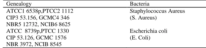
In this study two kinds of leather samples were used; cow and goat leather samples with 1.5 mm thickness, without oiling and without color of Vatan leather factory. Nano particles having 10 nm in size and concentration 0.1 mg/ml is obtained from Nano star company, emulsifier from Zamzam Company. The bacteria strain having features as depicted in Table 1 is collected from BoAli research center of Mashhad, Iran.
Table 1: Bacteria used for antibacterial test
Genealogy Bacteria
ATCC1 6538p,PTCC2 1112 CIP3 53.156, GCMC4 346 NBR5 12732, NCIB6 8625
Staphylococcus Aureus (S. Aureus)
ATCC 8739p,PTCC 1330 CIP 53.126, GCMC 1576 NBR 3972, NCIB 8545
Escherichia coli (E. Coli)
2.2 Method
Figure 1: Surface modification using corona discharge at different stages of the oiling process defined by Standard (no corona discharge applied), first and second method.
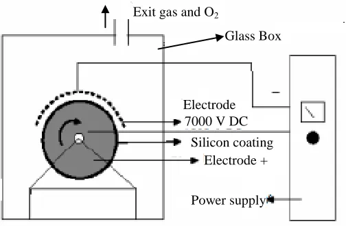
In this study, the Corona machine as shown in Figure 2 is obtained from Azad industry electric factory in Iran and it is equipped with atomic absorption with AA-20model from Australia. It is used for the surface modification of both the cow and goat leather.
Figure 2: Schematic diagram of the corona motion machine on the film, textile and leather
Standard Method
Impregnation with silver nanoparticle and oil
Drying in room temperature
First Method Method
Impregnation with silver nanoparticle and oil
Drying in room temperature Corona Process (30 cycles)
Second Method Method
Impregnation with silver nanoparticle and oil
Drying in room temperature Corona Process (30 cycles)
Exit gas and O2
Glass Box
Electrode 7000 V DC
Silicon coating Electrode +
The cow and goat leather samples were placed on the silicon roller which was placed at a distance of 0.3 cm from the negative electrode. The voltage applied in corona discharge is 7000 volts and liner speed is 2 m/min. In this method, the samples were exposed to the corona discharge for 30 cycles (because if more than 30 cycles were applied, the leather will burn and less than 30 cycles is applied the pores created will be insufficient).
2.3 Characterization of the Samples
2.3.1 Atomic Absorption and TEM
First of all, a certain weight of sample was heated in the container. Heating was continued until the leather begun to burn. Then the solvent was added to the sample and amount of dismissed silver in water was measured. The size and distribution of silver nanoparticles detected using TEM (Philips CM120 BioTWIN).
2.3.2 X-RD analysis and SEM
X-RD analysis was performed on the XR-D machine from Phillips Co. made in Holland (cathode made from copper and Anode from tungsten). X-RD test was done on produced leather samples containing silver nanoparticles and also on raw leather in order to confirm presence of silver nanoparticles. The leather surface that was treated under the corona process was analyzed using the SEM. The EVO Series Environmental Scanning Electron Microscopes were used.
2.4 Evaluation of Microbial Properties
In order to evaluate the degree of anti bacterial property of produced leather the number of colonies was counted according to the international standard method of Tran, no 21196. Based on this method, leather samples along with marker samples in size of 5 x 5
cm2 were treated using 70% ethanol for 20 min, so as to ensure they are free of bacteria.
Suspension of tested bacteria with standard opacity of 0.5 Mc Farland was provided and
a working suspension (with a number of bacteria to 1.3 x 10³ bacteria in each ml of
Muller Hintons slant agar) was also prepared. Samples were separately put in sterile plates and subsequently a suitable volume of provided bacteria suspension was added. After the required time, 10 ml of suspension of leather surface was picked, and the numbers of survived bacteria were counted. They were then cultivated on the Hepton
agar Muller cultivation and placed in the incubator for 24 h at 37 oC. Counting the
3. RESULTS AND DISCUSSION
3.1 Characterization of the Silver Nanoparticles
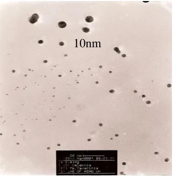
The TEM results in Figure 3 illustrates that the size of the silver nanoparticles used in this study is in the range of 10 nm and they are spherical in shape.
Figure 3: TEM spectrum of silver nanoparticles
3.2 Surface Analysis of the Leather Samples
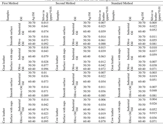
In general, the atomic absorption spectroscopy results in Table 2 reveals that the first method is better than second method and standard method, because the content of silver nanoparticles in the leather processed using method (1) is much higher compared to (2) and standard method. The application of corona discharge before the oiling process helps in modifying the leather surface through the creation of more pores on the leather surface thus allow better penetration or adhesion of the silver nanoparticles to the surface. However, in method (2) corona discharge was performed only after impregnation with silver nanoparticles. The presence of the nanoparticles and oil layer blocks the corona discharge and thus do not allow surface modification of the leather. The formation of pores cannot occur and thus the applied nanoparticles do not adhere to the leather surface, thus resulting in a loss of nanoparticles. In the standard method corona discharge was not performed thus no sufficient pores were created for easy adhesion of silver nanoparticles in them. A similar trend is observed for the goat’s leather where the olive oil seemed to assist the nanoparticles to adhere to the leather better. The results also revealed that the silver nanoparticles tend to adapt better with goat’s leather compared to the cow’s leather. As can be observed in Table 2, the goat
leather compared to cow leather for the same condition has much more silver nanoparticles content, because goat’s leather has much more pores compared to cow’s leather, thus the silver nanoparticles in oil (olive oil or industrial oil) can permeate better into the pores and stabilize there. Moreover goat’s leather has longer naps thus providing larger area when compared to cow’s leather and this allows more silver nanoparticles to adhere better to the leather. The silver nanoparticles tend to be more stable in these naps, because when leather has so many naps, the cross section of leather can attract silver nanoparticles more effectively. Also the highest ratio of silver nanoparticles to oil (60:40) is the best ratio, because higher content of silver nanoparticles in the leather will contribute to improved antibacterial properties. It is observed that the olive oil seems to be a better choice because it is able to retain higher nanoparticles compared to industrial oil for the same ratio of silver nanoparticles to oil.
The results in Table 2 also indicates that the olive oil can dissolve silver nanoparticles better than industrial oil, thus allowing more silver particles to be retained on the leather. The presence of higher content of fatty acids (C3-C18), in olive oil probably tends to dissolve the silver nanoparticles better. Also, the results in Table 2 reveals that the content of silver nanoparticles in the surface with naps is more than the smooth surface, because silver nanoparticles tend to get absorbed in the naps more than the smooth surface. Leather with naps has a larger surface area compared to the leather with smooth surface, thus the more silver nanoparticles tend to be place in these naps.
As can be observed from Figure 4, the existence of the peak at 38.12 confirms the presence of Ag and the peak is observed in the leather samples treated with nanoparticles. No peaks at 38.12 were observed for the raw leather sample. The results also show that the silver nanoparticles were well retained in the leather sample thus depicting the oil impregnating steps were well performed.
Table 2: Content of silver nanoparticles after corona process in cow and goat leather with different ratios of oil and silver nanoparticles
First Method Second Method Standard Method
Sam p les Oil Ag :Oil Sil v er in leath er (p p m )* 0 .0 1 Sam p les Oil Ag :Oil Sil v er in leath er (p p m )* 0 .0 1 Sam p les Oil Ag :Oil Sil v er in leath er (p p m )* 0 .0 1 Go at leath er Sm o o th s u rf ac e In d u str ial Oil
03:03 0.310
Go at leath er Sm o o th s u rf ac e In d u str ial Oil
03:03 3.300
Go at leath er Sm o o th s u rf ac e In d u str ial Oil
03:03 0.005
03:03 3.3.1 03:03 3.3.0 03:03 0.022
03:.3 3.30. 03:.3 3.300 03:.3 0.052
Oliv
e
Oil
03:03 3.310
Oliv
e
Oil
03:03 3.314
Oliv
e
Oil
03:03 0.011
03:03 3.300 03:03 3.301 03:03 0.055
03:.3 3.30. 03:.3 3.388 03:.3 0.081
Su rf ac e with n ap s In d u str ial Oil
03:03 3.310
Su rf ac e with n ap s In d u str ial Oil
03:03 3.310
Su rf ac e with n ap s In d u str ial Oil
03:03 0.010
03:03 3.3.0 03:03 3.300 03:03 0.037
03:.3 3.300 03:.3 3.300 03:.3 0.059
Oliv
e
Oil
03:03 3.3.0
Oliv
e
Oil
03:03 3.31.
Oliv
e
Oil
03:03 0.007
03:03 3.300 03:03 3.3.. 03:03 0.038
03:.3 3.300 03:.3 3.30. 03:.3 0.071
C o w leath er Sm o o th s u rf ac e In d u str ial Oil
03:03 3.31
C o w leath er Sm o o th s u rf ac e In d u str ial Oil
03:03 3.307
C o w leath er Sm o o th s u rf ac e In d u str ial Oil
03:03 0.003
03:03 3.300 03:03 3.3.. 03:03 0.019
03:.3 3.300 03:.3 3.30. 03:.3 0.051
Oliv
e
Oil 03:03 3.31.
Oliv
e
Oil 03:03 3.311
Oliv
e
Oil 03:03 0.007
03:03 3.301 03:03 3.300 03:03 0.048
03:.3 3.300 03:.3 3.385 03:.3 0.081
Su rf ac e with n ap s In d u str ial Oil
03:03 3.31.
Su rf ac e with n ap s In d u str ial Oil
03:03 3.300
Su rf ac e with n ap s In d u str ial Oil
03:03 0.004
03:03 3.3.. 03:03 3.30. 03:03 0.026
03:.3 3.30. 03:.3 3.301 03:.3 0.052
Oliv
e
Oil 03:03 3.3.0
Oliv
e
Oil 03:03 3.319
Oliv
e
Oil 03:03 0.015
03:03 3.30. 03:03 3.3.1 03:03 0.036
03:.3 3.300 03:.3 3.300 03:.3 0.071
3.3 Antibacterial Properties
Based on the 21196 standard, it was stated that every polymer that can remove S. aureus
Figure 4: X-RD absorption from leather sample a) without silver nanoparticles and b) with silver nanoparticles
.
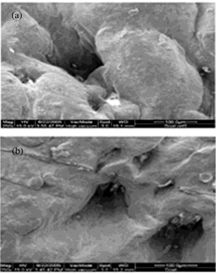
Figure 5: SEM analysis of the leather (a) before corona (raw leather), (b) after corona
L
in
(
C
o
u
n
ts
)
20 30 60 70
2-theta Scale 0
200 300
100
50 40
(a)
Microbial tests results revealed increased antibacterial property for the leather treated using the corona discharge process. The corona operations on the leather caused corrosion and porosity on the leather surfaces. The porosity created by the corona discharge process enhanced the moisture content and improve the breathability of leather. The surface modification on the leather allows better adhesion of the nanometer-sized silver nanoparticles to leather thus improve the antibacterial properties in exhibited in Table 3. The samples that were subjected to the corona discharge showed improvement in antibacterial properties because the silver nano particles tend to adhere better to the leather surface which subsequently provides the antibacterial effect. The corona discharge applied to the leather makes the surface more porous and thus increases the level of humidity and breathing on the leather
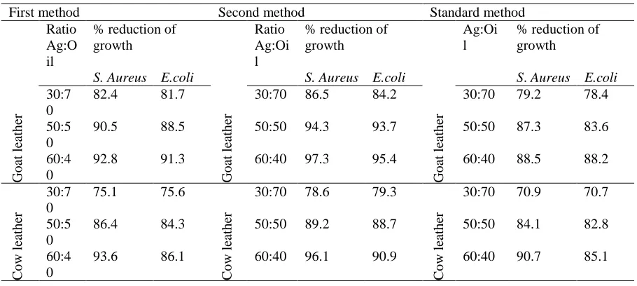
Table 3: The effect of corona discharge and silver nanoparticles for antibacterial properties in the goat and cow leather
First method Second method Standard method
Go at leath er Ratio Ag:O il
% reduction of growth
S. Aureus E.coli
Go at leath er Ratio Ag:Oi l
% reduction of growth
S. Aureus E.coli
Go at leath er Ag:Oi l
% reduction of growth
S. Aureus E.coli
30:7 0
82.4 81.7 30:70 86.5 84.2 30:70 79.2 78.4
50:5 0
90.5 88.5 50:50 94.3 93.7 50:50 87.3 83.6
60:4 0
92.8 91.3 60:40 97.3 95.4 60:40 88.5 88.2
C o w leath er 30:7 0
75.1 75.6
C
o
w
leath
er
30:70 78.6 79.3
C
o
w
leath
er
30:70 70.9 70.7
50:5 0
86.4 84.3 50:50 89.2 88.7 50:50 84.1 82.8
60:4 0
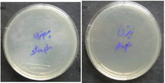
Figure 6: Microbial cultivation of evidence sample without silver nanoparticles in the
standard method in the presence of a) E. Coli b) S. Aureus
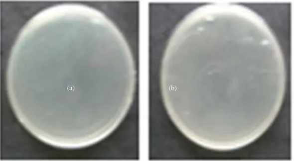
Figure 7: Microbial cultivation in first method of (a) goat leather sample with silver
nanoparticles and (b) cow leather with silver nanoparticles in presence of S. Aureus
bacteria.
(a)
(b) (b) (a)
Figure 8: Microbial cultivation in second method of a) goat leather sample with silver
nanoparticles and b) cow leather with silver nanoparticles in presence of S. Aureus
bacteria.
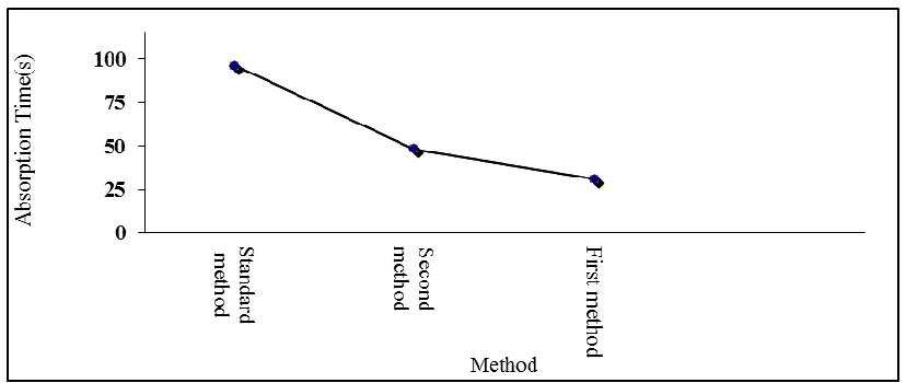
surface and this is proven by the absorption test results as shown in Fig. 9. The improvement in the antibacterial property will solve the problem of unpleasant odor
[8-10] caused by bacteria such as E. Coli and S. Aureus. Corona effect of processing on the
samples is clearly visible in Figure 9. The corona discharge process performed before impregnating of silver nanoparticles in samples (first method) was observed to improve the surface hydrophilicity of the leather tremendously and this is proven by the shortest absorption time amongst the 3 methods. The second method also improved the surface hydrophilicity of the leather but as not as good as the first method. No improvement of surface hydrophilicity can be observed when using the standard method as illustrated by the long absorption time [14]. The occurrence of such phenomenon is due to the fragmentation of linkages between macromolecules, caused by the bombardment of ionized gas particles which osculate together [12-14]. Moreover the free polar compounds on the surface can react with the hydrophilic groups in the material such as the carboxyl and hydroxyl groups and this consequently cause an increase in the level of humidity as observed in many reported polymeric material [13-15].
Figure 9: Content of humidity of goat leather with corona process and without corona process (standard method)
4. CONCLUSIONS
REFERENCES
[1] Bhavan S , Raghava J. (2008)A potential new commercial method for processing
leather to reduce environmental impact. Environ Sci Pollut Res, 15: 293-295
[2] Widrow C A, Kellie S M, Saltzman B R, et al. (1991)Pyomyositis in patients
with the human immu-nodeficiency virus: an unusual form of disseminated bacterial infection, American Journal of Medicine ,91:129-136
[3] Technical Committee ISO/TC 61.( 2007) Plastics, Subcommittee SC 6, Ageing,
chemical and environmental resistance,International standard ISO 22196.first edition. 10-15
[4] Zhu M S, Chen P L, M H. (2012)Visible-light-driven Ag/Ag3PO4-based
plasmonic photocatalysts: Enhanced photocatalytic performance by hybridization with graphene oxide, Chin. Sci. Bull, doi:10.1007/s 11434-01-5367-9
[5] Rajamaran R, Poorneswari S, Bangaruswamy S, et al.( 1978) Influence of
different tanning systems on the characteristics of leather, Leather Sci, 25:394– 399
[6] Crawford F, Hart R, Torgerson D. et al.( 2001) Athlete’s foot and fungally
infected toenails, BMJ, 11: 548-56
[7] Misner ,B D. (2007)A novel aromatic oil compound inhibits microbial
overgrowth on feet: a case study. J Int Soc Sports Nutr.13: 3-4
[8] Valipour P.( 2006) Improvement of Polypropylene Fabric Dye Ability With
Disperse Dye by Corona Plasma Discharge Treatment. In: Korea, Valipour P, Bagheri H A. International Fiber Conference
[9] Aal A A. (2008)Effect of Corona Discharge on Surface of Leather. In:
Dubrovnik ,Aal A A , Maleknia L, alebian A T. 4th International Textile, Clothing & Design Conference. 5-8
[10] Pavlath A E, Slater R F .( 1971) Low-temperature plasma chemistry. Polym.
Symp, 18: 1317- 1324
[11] Riccobono PX, Rolden L.( 1973) Plasma treatment of textiles:A novel approach
[12] Stone R B, Barrett J R. (1962)Study reveals interesting effects of gas plasma radiations on cotton yarn. Textile Bull, 88: 65-68
[13] Jung H Z, Ward T L, Benerito R R.( 1977) The effect of argon cold plasma on
water absorption of cotton. Textile Res. J, 47: 217-222
[14] Ward T L, Jung H Z, Hinojosa O, et al. (1978)Effect of RF cold plasmas on
polysaccharides, J. Surf. Sci, 76: 257-273
[15] Shishoo L R.( 1996) Plasma Treatment-Industrial Applications and its Impact on
the C&L Industry. In:Dubrovnik, Shishoo LR. The 6th. Int. Conf. on Tex. Coat. 35-47
[16] Lieberman M A, Lichtenberg A J.( 1994) Principle of Plasma Discharges and
Materials Processing. John Wiley,2:1-53
[17] Rakowski W, Okoniewski M, Bartos K, et al.( 1982) Plasma treatment of textiles
- potential applications and future prospects. Melliand Textilber, 63: 301-313
[18] Marsh D E. (1978)Plasma torch cutting of textiles. Melliand Textilber, 68:
558-560
[19] Benerito R R, Ward T L, Soignet D M, et al(1981). Modifications of cotton