Detecting the COVID-19
Pravin Pokhrel, Changpeng Hu, Hanbin Mao*
Department of Chemistry and Biochemistry, Kent State University, Kent, OH, USA
*Author of correspondence: Hanbin Mao, PhD
Department of Chemistry and Biochemistry, Kent State University, OH, USA [email protected]; (+1) 330-672 9380
850 University Esplanade, Kent, OH, 44242-0001, P.O. Box 5190
Conflict of Interest: The authors declare no conflict of interest.
Acknowledgement: HM thanks NIH (R01 CA236350) and NSF (CBET-1904921) for financial support.
Abstract
The COVID-19 pandemic has created huge damage to society and brought panics around the world. Such panics can be ascribed to the seemingly deceptive features of the COVID-19: compared to other deadly viral outspreads, it has medium transmission and mortality rates. As a result, the severity of this virus was deeply underestimated by the society at the beginning of the outbreak. Based on this, in this review, we define the viruses with features similar to those of COVID-19 as the Panic Zone viruses. To contain those viruses, accurate and fast diagnosis followed by effective isolation and treatment of patients are pivotal at the early stage of virus breakouts. This is especially true when there is no cure or vaccine available for a transmissible disease, which is the case for current COVID-19 pandemic. As of April 2020, more than one hundred kits for the COVID-19 diagnosis on the market are surveyed in this review. It is of critical importance to rationally use these kits for the efficient management and control of the Panic Zone viruses. Therefore, we discuss guidelines to select diagnostic kits at different outbreak stages of the Panic Zone viruses, COVID-19 in particular. While it is of utmost importance to use detection kits with low false negativity at the early stage of an outbreak, the low false positivity gains its importance at later stages of the outbreak. Finally, since a massive attack from a viral pandemic requires a massive defense from the whole society, we urge both government and private sectors to research and develop affordable point-of-care (POC) detection kits, which can be used massively by the general public (and therefore called as massive POC) to contain Panic Zone viruses in future.
Keywords: Diagnosis, Detection Kits, RT-PCR, Immunoassay, False Negative, False Positive,
1. Background
Since the beginning of the 21st century, our world has been facing unprecedented crises of deadly viruses like Zika, Ebola, SARS, MERS, and so forth. The epidemics of these viral diseases were sparked either by the evolution of pre-existing viruses or by the emergence of new viral species. Such diseases have already caused colossal damage to the society. Loss of lives struck the most, but the consequences aftermath were equally dreadful: the psychological wellbeing of survivors and socio-economic fallout were rather distressing. Now, in December 2019, the world was hit by yet another virus known as SARS-CoV-2 (or COVID-19).
Figure 1. Viruses with high transmission rates (R0) are less fatal. R0 is the reproduction rate of a
virus, which measures its transmissibility1. Solid curve represents an inverse fitting between the
mortality rate and the R0, which has been proposed as the trade-off principle between the
virulence and transmissibility of virus2. The inverse function fits well except for the two viruses in
the Death Zone (blue), which is defined to have a rather high mortality rate. The Panic Zone contains viruses with medium levels of transmission and mortality rates. The data used here are taken from references3–6.
1.1. The Panic Zone viruses
supplies is profit driven. Decisions regarding the management of disease can no longer be made based solely on scientific grounds. Unless a disease poses a specific risk to a wide population, its mere presence in a localized area or population may not be significant from a business perspective. As a result, necessary resources such as PPE (Personal Protective Equipment) are in short supply to fight pandemic diseases promptly. Due to these reasons, the diseases in the Panic Zone often wreck huge collateral damages due to its paralyzing role for the whole society.
In the Panic Zone, SARS was most recently contained by means of massive syndromic surveillance, prompt isolation of patients, and strict quarantine of all contacts. By interrupting all human-to-human transmissions, SARS was effectively eradicated in 20038. Although there are striking similarities between SARS and COVID-19, the difference in the virus characteristics will ultimately determine whether the same measures for SARS will also be successful for the current COVID-19 outbreak. COVID-19 differs from the SARS in terms of infectious period, transmissibility, clinical severity, and extent of the community spread. Although COVID-19 has lower transmissibility than SARS9, many more COVID-19 patients have mild symptoms that contribute to the rapid spread of the virus as these patients are often missed and not isolated.
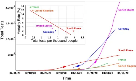
1.2. The early detections
It is generally true that for a rapidly transmitting disease with no cure or vaccine available, the most effective way to curb its spread is to isolate patients. The first step to achieve this is to identify these patients using detection kits. Never before is a virus detection system so critical to contain a viral outbreak as dangerous as COVID-19. As shown in Figure 2, for the five countries with similar age distribution and hospital resources, the more extensive the early tests on the COVID-19, the lower the overall mortality rates in a country. Indeed, Korean and Germany did a substantial number of the tests right at the beginning of the COVID-19 outbreak. Correspondingly, their death rates are among the lowest so far (Figure 2, inset). This confirmed the importance of the early testing to curb the spreading of the COVID-19.
In this survey, we first describe the COVID-19 outbreak briefly. Given the importance of the diagnosis for this deadly pandemic disease, we then survey the detection kits used for the COVID-19. Next, we propose and discuss guidelines to use various kits during different stages of the COVID-19 outbreak. We then wrap up by extending these guidelines to potential
Figure 2. Critical importance of the early detection in the COVID-19 outbreak. Total COVID-19
daily tests are shown for 5 countries with similar medical resources and age distributions. Inset shows the death rates as of 04/08/2020 vs the number of the early detections per thousand population performed during 03/04/2020 - 03/26/2020. The early detection data for each country10 are taken from different periods (marked by stretches) to reflect the timing of the
outbreak in Asian, Europe, and North America (~2 weeks apart). The inset data are linearly fit (r=-0.914), which indicates a decent negative correlation between the early detection and mortality rate.
2. The COVID-19 outbreak 2.1. Covid-19 Timeline
In December 2019, a cluster of pneumonia cases were reported in Wuhan, China11. The causative agent of that disease was unknown until January 7, 2020, when the Chinese
authorities confirmed that they have identified a new virus and named it 2019-nCoV (later called as COVID-19, CoronaVirus Disease 2019, by the WHO) since the virus shared ~80% genome from the SARS-CoronaVirus12. On January 11, the first death caused by this virus was reported in China. This disease was highly contagious. The WHO declared a Public Health Emergency of International Concern (PHEIC) within a month after the first case. On March 11, WHO declared COVID-19 as a pandemic disease.
2.2. Clinical characteristics of COVID-19
cases, by the end of the first week, the disease can develop into dyspnea and/or hypoxia. In deadly cases, the disease can quickly progress to acute respiratory distress syndrome, septic shock, coagulation disorders, and multiple organ failure13. It’s noteworthy that patients with high viral loads may have low or insignificant fever during the infection. Some children and neonates did not have typical symptoms, but they presented with gastrointestinal symptoms such as vomiting and diarrhea or presented with depression or shortness of breath14. The elderly and patients with chronic underlying diseases had poor prognosis15.
2.3. Epidemiology of COVID-19
People are generally susceptible to COVID-19 at all ages. The infection is transmitted by droplet (direct inhalation of droplets from the sneeze, cough, or talking of an infected person) or contact (contacting the virus deposited on the object surface, which then enters the body via the mouth, nose, eyes, or other mucous membrane16). Study showed a higher viral load in the nasal cavity than the throat, suggesting the nasal sampling is a more effective approach to detect the virus. There was no difference in the viral load between symptomatic and asymptomatic
patients17, the latter of which can also transmit the disease18. Guan et al. reported that some patients were tested positive for COVID-19 in stool and urine samples13.
3. Diagnosis of the COVID-19 infection
As discussed in the Introduction, in the absence of effective therapeutic drugs or vaccines for COVID-19, it is essential to detect the disease at an early stage and immediately isolate infected patients.Currently, there are three methods in clinical practice to diagnose COVID-19, which are summarized below.
3.1. Chest CT Imaging
Studies showed that chest CT images contained characteristic features for COVID-19 patients. The hallmarks of these CT images include ground glass opacities, crazy-paving
pattern, consolidative opacities, septal thickening, and the reverse-halo sign19–22. These features demonstrate a highly organized pattern of pneumonia20. Unlike these features, nodules, cystic changes, bronchiectasis, pleural diffusion, and lymphadenopathy are less common22.
Despite such features, the Centers for Disease Control (CDC) in the US does not currently recommend CT to diagnose COVID-19. Laboratory testing of the virus remains the reference standard, even if the CT findings are suggestive of COVID-19 infections23. This is because features of the chest imaging from COVID-19 patients may overlap with other infections caused by influenza, H1N1, or SARS-CoV24,25.
were RT-PCR negative showed initial positive CT images consistent with COVID-19 infections. From the patients in the recovery stage, 42% showed improvement in CT features before their RT-PCR results turned negative.
According to these diagnostic studies, RT-PCR assays were not as sensitive and reliable as CT images. The false negative results from RT-PCR assays can be detrimental to the control of the COVID-19, especially at the beginning of the outbreak. The caveat for the CT scans is that at an early stage of infection, the lungs of a patient may not develop damaging features that can be picked up by CT scans, increasing its false negative rate. In addition, the COVID-19 CT features share similarities with other viral pneumonia, resulting in false positive detections. Nevertheless, given the rapidly spreading of the COVID-19, the priority is to identify any suspicious case for isolation and proper treatment. In the context of emergency disease control, some false-positive cases may be acceptable. It is the false negative cases, due to the poor sensitivity of methods, that present a threat to public health at the beginning of an
outbreak. In some cases, chest CT imaging showed positive COVID-19 infection while RT-PCR testing was negative26. A combination of clinical symptoms, epidemiologic history, and CT imaging of a patient is instrumental to identify COVID-19 infections at the time when chemical detection kits are in short supply.
3.2. Nucleic acid based methods
After identification of the COVID-19 virus for this pandemic outbreak, the COVID-19 genome was quickly sequenced28, from which unique sequences have been identified for COVID-19 diagnosis. Reverse transcriptase-polymerase chain reaction (RT-PCR) is a nucleic acid amplification assay that has been used routinely for the detection of RNA viruses in clinical settings29. In RT-PCR, reverse transcriptase is first used to convert RNA to its complementary DNA, which is amplified by PCR (polymerase chain reaction). There are variants of RT-PCR method that share the same mechanism while differing in the detection strategy. For example, real time RT-PCR reads fluorescent signals in real time during PCR30 to quantify the target, whereas nested RT-PCR uses two sets of primers to avoid non-specific PCR amplification31.
The COVID-19 genes targeted for detection so far include the RdRP gene, Nucleocapsid (N) gene, E gene, Spike protein (S gene), and ORF1ab gene. Chu et al. used two different one-step real-time RT-PCR approaches to detect ORF1ab and N genes of the viral genome32. This assay showed a high dynamic range of 0.0002-20 TCID50 (50% tissue culture infective dose) per reaction and the detection limit below 10 RNA copies per reaction. Later, WHO developed a technical guidance including the protocols from different countries to aid COVID-19 diagnosis33. In the US, CDC developed a real time RT-PCR diagnostic kit with detection limits as low as 4-10 RNA copies per µl. Scientists from Germany used the E gene for the first-line screening and the RdRP gene for confirmatory testing. This method further increased sensitivity to detect as low as 5.6 RNA copies per reaction for the E gene and 3.8 RNA copies per reaction for the RdRP gene. In Hongkong, the N gene was used as the first-line screening while the ORF1b as the confirmatory testing. In France, two RdRP genes were used for initial screening followed by the confirmatory E gene testing. In Japan, nested RT-PCR was used, which significantly reduced the non-specific target amplification, leading to decreased false-positive results. In general, the sensitivity of these assays ranges from 3.8 to 10 RNA copies per reaction, with high
High-throughput sequencing presents another confirmatory method to detect COVID-19. However, the application of high-throughput sequencing in clinical diagnosis is limited because of its equipment dependency and high cost. Zhang et al. demonstrated a point-of-care (POC) testing based on CRISPR-based SHERLOCK34to detect synthetic COVID-19 RNA fragments35. They utilized isothermal DNA amplification method and targeted the ORF1ab and S genes with a detection limit as low as 10 copies/µl.
In the public health emergency, highly sensitive methods are desirable. Although studies have shown that RT-PCR may be less sensitive than CT imaging, its specificity makes it
superior to other methods to detect COVID-19 or other viruses. It is of critical importance to rationally choose specific diagnostic methods to battle outbreaks of viral diseases. Any
negligence or compromise in the diagnosis may lead to devastating consequences. Wang et al. suggested combining RT-PCR with other methods as well as epidemiological history of patients to diagnose COVID-19 infection more credibly36. Indeed, the Chinese authority has adopted this approach to diagnose COVID-19 in Wuhan by combining RT-PCR with CT scans27. Studies also showed that the sensitivity of RT-PCR varies with the specimen types. To et al.revealed that the saliva samples were more promising to be used in RT-PCR37 while Yam et al. concluded that testing more than one specimen could significantly maximize the sensitivity of the RT-PCR testing38. These findings suggest it is rather important to apply nucleic acid based kits with optimized conditions to maximize their diagnosis potency.
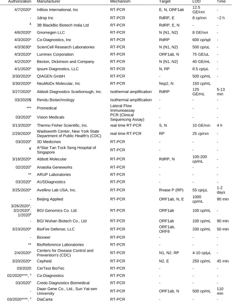
Table 1 lists the nucleic acid based kits used for the diagnosis of COVID-19. The sensitivity of those kits ranges from 100-1000 copies/mL.
3.3. Immunoassays
Immunoassay is another established method to diagnose diseases. This method detects serum antibodies generated in patients who have been exposed to the COVID-19. These antibody tests are important in detecting infections with few or no symptoms.
In the COVID-19 infection, studies have shown that the seroconversion in the patient-generally starts after a week of the first symptom39. In a study of post symptomatic patients, Amanat et al.detected high IgA and IgM immune responses40. Using recombinant viral proteins, this immunoassay could detect antibodies as early as 3 days after the development of the first symptom. Liu et al. reported that the accuracy of the ELISA for IgG and IgM antibodies was more than 80%41. The efficacy of the immunoassay also depends on the specificity of the antigens used to capture the antibodies from the patients. Between the spike (S) proteins and nucleocapsid (N) proteins, the sensitivity of the S proteins is higher for the antibody capture. Among various spike proteins, the S1 protein has shown more capabilities to bind to COVID-19 antibodies42. In a comparative study, both ELISA and colloidal gold immunochromatographic kits showed equal sensitivity with 100% specificity for the COVID-19 detection43.
4. Ideal characteristics of diagnostic methods
Diagnostic testing has become indispensable for diagnosis, prognoses, and monitoring the progress of different diseases. Efficient diagnostic testing is an important intervention for outbreak management and control. WHO has developed the ASSURED criteria as a benchmark to decide if a test efficiently addresses the needs for disease control: Affordable, Sensitive,
Specific, User-friendly, Rapid and robust, Equipment-free and Deliverable to end-users45. It’s ideal to have all the criteria fulfilled in a single test. In practice, however, testing methods can rarely fit all the ASSURED criteria. In pandemic outbreaks for example, rapid and sensitive methods are dearly needed at the beginning of the outbreak. But many kits available require qualified laboratories and personnel for testing. In such a case, accommodation of the ASSURED principles must be taken to facilitate the testing.
In a pandemic outbreak, it is always important to understand the nature of the pathogen before developing efficient diagnostic tests. Translating the tests into the point-of-care (POC)46 mode can help decision-making and improve the efficiency of the treatment. POC provides rapid and actionable information for patient management and care at the time when it is most needed. Many affordable POC kits such as lateral flow immunoassays47 are also appropriate for
resource-limited settings in middle- or low-income countries where laboratory infrastructure is weak. One example for affordable POC testing is the pregnancy strip test48 in which pregnancy can be quickly determined at home using paper strips on specimens such as noninvasive urine samples. Due to the requirements of easy usage and cheap price, they often use colloidal gold based immunoassay mechanisms. Such POC testing kits perhaps represent the best solution to fight fast transmitting pandemics.
5. Rationales in choosing diagnostic methods in the COVID-19 outbreak
Figure 3. Intervention of the COVID-19 outbreak. The intervention at an early stage (before the
inflection point, which is the point where the half width of a Gaussian peak is equivalent to the sigma of the Gaussian) of a virus breakout is the key to slow down the transmission of the virus. It not only decreases the peak value of newly confirmed daily cases, but also saves the time to increase the hospital capacity, each of which reduces the overall mortality rate.
Among all current methods, nucleic acid based kits are considered the most reliable because of their high specificity and accuracy. This is not surprising since these methods target unique sequences in the viral genome for identification. Due to these advantages, it becomes a detection of choice at the beginning of a virus outbreak. At this stage, it is critical to identify and isolate all possible patients before the virus enters an exponential growth stage (around the inflection point, see Figure 3). Therefore, it is important to reduce the false negative results of the diagnosis. To achieve this, high sensitivity is a necessity. The PCR amplification used in various RT-PCR kits can detect as low as 100 copies/mL reaction (see Table 1), which is equivalent to 0.167 attoMolar (for a reaction volume of 100 microliters). It is noteworthy that high sensitivity is accompanied with increased false positive results. But at the beginning of a virus outbreak, some false positive level may be tolerated. Since there are not so many infected patients at the initial stage of the outbreak, the chance of cross contamination from COVID-19 patients to these false positive cases is small, even if they are isolated together (but well protected by PPE) in spacious locations such as convention centers. When the viral outbreak becomes stronger, false positive cases should be reduced as much as possible due to the increasing cross contamination concerns.
Another way to reduce the false negativity in nucleic acid based testing is to perform CT scans. As discussed in Section 3.1, it can be more sensitive to diagnose COVID-19 using CT scans. The caveat for the CT scan is its relatively low specificity (false positive results), which may be tolerated at the initial stage of an outbreak. However, positive CT scans only show for patients at the later stage of their COVID-19 infections, which limits its use for early stage screening. The method is still valuable to quickly screen serious cases from mild ones. Due to limited testing kits and over-burdened clinical resources, many patients with mild symptoms have been self-isolated first. When their conditions deteriorate, it becomes important to
streamline life-threatening cases as soon as the patients are sent to the hospital. Due to the fast performance and interpretation of CT scans within tens of minutes as demonstrated in China for example, these patients can be quickly identified, followed by appropriate treatment to save lives.
Immunoassays work well only after the human body develops antibodies to fight viruses. Therefore, these kits are not appropriate to detect infection cases at the early stage of an
infection at which patients may be asymptomatic. Given that asymptomatic patients also transmit COVID-1918, it is not recommended to use immunoassays at the beginning of the pandemic. In the current COVID-19 breakout, we have often seen that during the exponential increase stage of the disease (around the inflection point, seeFigure 3), there have been insufficient number of nucleic acid based kits to test all suspicious cases. Current strategy to solve this issue is rather impassive. The precious testing kits are reserved only for more serious cases. For the patients with light symptoms, they were sent home for self-isolation. The
immunoassay can be used to test those patients after their symptoms lasted about one week. Since these tests are cheaper, faster, and easier to perform47 with respect to nucleic acid based methods, they can be quickly and massively conducted by staff at drive-through stations.
In the future, the high-throughput, microarray-based testing may be able to address the bottleneck diagnosis problem caused by shortage of testing kits. In such a method, thousands of tests can be run simultaneously on a chip49. For this method, the time limiting step becomes the sample collection, which must be performed one-at-a-time. The other direction to resolve the bottleneck testing problem is to develop affordable POC kits as discussed in section 4. These kits can be performed at home for self-isolated people with mild symptoms. If they are tested positively by the POC kits, their conditions will be closely monitored for further medical treatments or other interventions. The inherent properties of these POC kits (cheap, fast, and easy-to-use) afford their massive usage by the general public to fight with pandemic outbreaks. We therefore name such an approach as massive POC strategy. Given there is no such
massive POC product on the market for the COVID-19 yet, research and development on the affordable POC kits are dearly needed at this stage for virus detections.
6. Conclusions and Perspectives
available detection kits, it is crucial to reduce false negative results at the expense of some false positive level during the early stage of the outbreak. It becomes important to reduce the false positivity in later stages of the outbreak. Although nucleic acid based detection kits, RT-PCR in particular, offer best solutions so far to these requirements because of their high sensitivity and specificity, immunoassays can well supplement the detection armory due to their cheaper price, simpler operation, and faster detection time. The use of immunoassays is especially useful at the later stages of the virus outbreak when patients already develop symptoms for a while or when nucleic acid based testing kits are unavailable or not enough due to mass inflow of
Table 1: Kits based on nucleic acid detection
Authorization Manufacturer Mechanism Target LOD Time
4/7/2020* InBios International, Inc RT-PCR E, N, ORF1ab 12.5
GE/rxn -
- 1drop Inc RT-PCR RdRP, E 8 cp/rxn ~2 h
ƛ 3B BlackBio Biotech India Ltd RT-PCR RdRP, E, N - -
4/6/2020* Gnomegen LLC RT-PCR N (N1, N2) 8 GE/rxn -
4/3/2020* Co-Diagnostics, Inc RT-PCR RdRP 600 cp/spl -
4/3/3030* ScienCell Research Laboratories RT-PCR N (N1, N2) 500 cp/uL -
4/3/2020* Luminex Corporation RT-PCR ORF1ab, N 75 GE/uL -
4/2/2020* Becton, Dickinson and Company RT-PCR N (N1, N2) 40 GE/mL -
4/1/2020* Ipsum Diagnostics, LLC RT-PCR N, RP 8.5 cp/µL -
3/30/2020* QIAGEN GmbH RT-PCR - 500 cp/mL -
3/30/2020* NeuMoDx Molecular, Inc RT-PCR Nsp2, N 150 cp/mL
3/27/2020* Abbott Diagnostics Scarborough, Inc. Isothermal amplification RdRP 125 GE/mL
5-13 min
03/2020$ Rendu Biotechnology Isothermal amplification - - -
*** Promedical Lateral Flow
Immunoassay - - -
03/2020# Vision Medicals PCR (Clinical
Sequencing Assay) - - -
3/13/2020* Thermo Fisher Scientific, Inc. real time RT-PCR S, N 10 GE/rxn 4 h
2/29/2020* Wadsworth Center, New York State Department of Public Health's (CDC) real time RT-PCR RP 25 cp/rxn -
03/2020# 3D Medicines RT-PCR - - -
€ A*Star Tan Tock Seng Hospital of
Singapore RT-PCR - - -
3/18/2020* Abbott Molecular RT-PCR RdRP, N 100-200
cp/mL -
02/2020# Anatolia Geneworks RT-PCR - - -
** ARUP Laboratories RT-PCR - - -
03/2020# AUSDiagnostics RT-PCR - - -
3/25/2020* Avellino Lab USA, Inc. RT-PCR Rnase P (RP) 55 cp/µL 1-2
days
- Beijing Applied RT-PCR ORF1ab, N, E 1000 cp/mL 90 min
3/26/2020*, 3/2/2020#,
1/2020$
BGI Genomics Co. Ltd. RT-PCR ORF1ab 100 cp/mL -
- BGI Wuhan Biotech Co., Ltd RT-PCR ORF1ab 100 cp/mL 90 min
3/23/2020* BioFire Defense, LLC RT-PCR ORF1ab,
ORF8 330 cp/mL 50 min
- Bioneer RT-PCR - - -
** BioReference Laboratories RT-PCR - - -
2/4/2020* Centers for Disease Control and
Prevention's (CDC) RT-PCR N1, N2. RP 4-10 cp/µL -
3/20/2020* Cepheid RT-PCR N2, E 250 cp/mL 45 min
03/2020 CerTest BioTec RT-PCR - - -
02/2020****, # Co-Diagnostics RT-PCR - - -
03/2020# Credo Diagnostics Biomedical RT-PCR - - -
- Daan Gene Co., Ltd., Sun Yat-sen
University RT-PCR ORF1ab, N 500 cp/mL
110 min
**** Diagnostic Solutions Laboratory RT-PCR - - -
3/19/2020* DiaSorin Molecular LLC RT-PCR ORF1ab, S 500 cp/mL 1-1.5
h
- Diatherix Eurofins RT-PCR - - -
03/2020# Genetic Signatures RT-PCR - - -
03/2020# Genomica/PharmMar Group RT-PCR - - -
3/16/2020* Hologic, Inc. RT-PCR -
10-2
TCID50/m L
-
* Integrated DNA technologies/Danaher RT-PCR - - -
€ JN Medsys RT-PCR - - -
#, ψ Kogene Biotech RT-PCR - - -
3/16/2020* Laboratory Corporation of America
(LabCorp) RT-PCR
Rnase P (RP),
N 6.25 cp/µL -
* LGC, Biosearch Technologies RT-PCR - - -
3/27/2020* Luminex Molecular Diagnostics, Inc. RT-PCR ORF1ab, N
gene, E 1.5 cp/µL 4 h
- Maccura Bio-tech Co., RT-PCR ORF1ab, N, E - 2 h
3/30/2020* NeuMoDx Molecular RT-PCR - - -
3/20/2020*, 2/2020#,
3/26/2020ƛ
Novacyt/Primerdesign RT-PCR - - -
02/2020# OsangHealthCare RT-PCR - - -
3/24/2020* PerkinElmer, Inc. RT-PCR ORF1ab, N 20 cp/mL -
3/20/2020* Primerdesign Ltd. RT-PCR - 0.33 cp/µL -
3/17/2020* Quest Diagnostics Infectious Disease,
Inc. RT-PCR N1 and N3 136 cp/mL -
3/23/2020*,
3/2020# Quidel Corporation RT-PCR pp1ab 0.8 cp/µL 75 min
3/12/2020* Roche Molecular Systems, Inc. RT-PCR E - 3 hrs
- SANSURE Bio-tech Co., Ltd RT-PCR ORF1ab, N 200 cp/mL 90 min
02/2020#, $$ See Gene RT-PCR - - -
- Shanghai Bio Germ RT-PCR ORF1ab, N 1000 cp/mL 90 min
- Shanghai GeneoDx Biotech Co., Ltd RT-PCR ORF1ab, N 500 cp/mL 90 min
- Shanghai ZJ Bio-tech Co., Ltd. RT-PCR ORF1ab, N, E 1000
cp/mL 90 min
2/2020#, $$, $$$ SolGent RT-PCR - - -
03/2020# Systaaq Diagnostic Products RT-PCR - - -
03/2020# TIB MolBiol Synthesalabor RT-PCR E - -
- Ustar RT-PCR ORF1ab, N - 90 min
- Wuhan Easydiagnosis RT-PCR ORF1ab, N - 75 min
3/23/2020* Mesa Biotech Inc. RT-PCR and colorimetry N 100 cp/rxn 30 min
3/19/2020* GenMark Diagnostics, Inc. RT-PCR, electrowetting
and sensing -
10^5
cp/mL 2 h
** Fulgent Genetics/MedScan laboratory Sequencing - - -
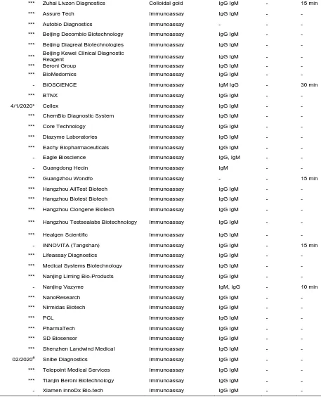
Table 2: Kits based on immunoassay
Authorization Manufacturer Mechanism Target LOD Time
*** Beijing O&D Biotech Colloidal gold - - -
*** Nantong Diagnos Biotechnology Colloidal gold - - -
*** Zuhai Livzon Diagnostics Colloidal gold IgG IgM - 15 min
*** Assure Tech Immunoassay IgG IgM - -
*** Autobio Diagnostics Immunoassay - - -
*** Beijing Decombio Biotechnology Immunoassay IgG IgM - -
*** Beijing Diagreat Biotechnologies Immunoassay IgG IgM - -
*** Beijing Kewei Clinical Diagnostic
Reagent Immunoassay IgG IgM - -
*** Beroni Group Immunoassay IgG IgM - -
*** BioMedomics Immunoassay IgG IgM - -
- BiOSCiENCE Immunoassay IgM IgG - 30 min
*** BTNX Immunoassay IgG IgM - -
4/1/2020* Cellex Immunoassay IgG IgM - -
*** ChemBio Diagnostic System Immunoassay IgG IgM - -
*** Core Technology Immunoassay IgG IgM - -
*** Diazyme Laboratories Immunoassay IgG IgM - -
*** Eachy Biopharmaceuticals Immunoassay IgG IgM - -
- Eagle Bioscience Immunoassay IgG, IgM - -
- Guangdong Hecin Immunoassay IgM - -
*** Guangzhou Wondfo Immunoassay - - 15 min
*** Hangzhou AllTest Biotech Immunoassay IgG IgM - -
*** Hangzhou Biotest Biotech Immunoassay IgG IgM - -
*** Hangzhou Clongene Biotech Immunoassay IgG IgM - -
*** Hangzhou Testsealabs Biotechnology Immunoassay IgG IgM - -
*** Healgen Scientific Immunoassay IgG IgM - -
- INNOVITA (Tangshan) Immunoassay IgG IgM - 15 min
*** Lifeassay Diagnostics Immunoassay IgG IgM - -
*** Medical Systems Biotechnology Immunoassay IgG IgM - -
*** Nanjing Liming Bio-Products Immunoassay IgG IgM - -
- Nanjing Vazyme Immunoassay IgM, IgG - 10 min
*** NanoResearch Immunoassay IgG IgM - -
*** Nirmidas Biotech Immunoassay IgG IgM - -
*** PCL Immunoassay IgG IgM - -
*** PharmaTech Immunoassay IgG IgM - -
*** SD Biosensor Immunoassay IgG IgM - -
*** Shenzhen Landwind Medical Immunoassay IgG IgM - -
02/2020# Snibe Diagnostics Immunoassay IgG IgM - -
*** Telepoint Medical Services Immunoassay IgG IgM - -
*** Tianjin Beroni Biotechnology Immunoassay IgG IgM - -
- Xiamen innoDx Bio-tech Immunoassay IgG IgM - -
Table 3: Kits based on 'not identified' mechanism
- Biological Technologies Co., LTD - - - -
- (Chongqing) Bio-tech, Co., Ltd - - - -
- Health Technology Co., Ltd - - - -
- Bio-tech Co., Ltd - - - -
- Medical Technology Co., Ltd - - - -
- Biotechnologies (Hangzhou) Ltd - - - -
- Biomedicine Co., Ltd. - - - -
*US EUA Authorized, **US EUA Planne,***US Notified FDA under section IV.D, ****US EUA Submitted, #European Union
Conformity Marked, $The National Medical Product Administration Authorized China, $$Korea Ministry of Food and Drug Safety, $$$Philippines Food and Drug Administration, €Singapore Health Sciences Authority, personal authorization for clinical use, ƛEUA
India, ψKorea Centers for Disease Control and the Korea Food and Drug Administration -Data Not Available
References for kits in table 1-3: https://www.fda.gov/medical-devices/emergency-situations-medical-devices/emergency-use-authorizations#coronavirus2019;
http://ph.china-embassy.org/eng/sgdt/t1760281.htm;
References:
1. Dietz, K. The estimation of the basic reproduction number for infectious diseases. Stat. Methods Med. Res.2, 23–41 (1993).
2. Lipsitch, M. & Moxon, E. R. Virulence and transmissibility of pathogens: what is the relationship? Trends Microbiol.5, 31–37 (1997).
3. Basic reproduction number. Wikipedia (2020).
https://en.wikipedia.org/wiki/Basic_reproduction_number 4. List of human disease case fatality rates. Wikipedia (2020).
https://en.wikipedia.org/wiki/List_of_human_disease_case_fatality_rates
5. Harding, A & Lanese, N. The 12 Deadliest Viruses on Earth. livescience.com (March 4, 2020).https://www.livescience.com/56598-deadliest-viruses-on-earth.html.
6. Chen, J. Pathogenicity and transmissibility of 2019-nCoV—a quick overview and comparison with other emerging viruses. Microbes Infect. (2020).
7. Baud, D. et al. Real estimates of mortality following COVID-19 infection. Lancet Infect. Dis. (2020).
8. Wilder-Smith, A., Chiew, C. J. & Lee, V. J. Can we contain the COVID-19 outbreak with the same measures as for SARS? Lancet Infect. Dis. (2020).
9. Du, Z. et al. The serial interval of COVID-19 from publicly reported confirmed cases. medRxiv (2020).
10. Roser, M., Ritchie, H. & Ortiz-Ospina, E. Coronavirus Disease (COVID-19) – Statistics and Research. Our World Data (2020).
11. WHO. Novel Coronavirus (2019-nCoV) situation report - 1. (January 21, 2020). https://www.who.int/emergencies/diseases/novel-coronavirus-2019/situation-reports
12. Guo, Y.-R. et al. The origin, transmission and clinical therapies on coronavirus disease 2019 (COVID-19) outbreak–an update on the status. Mil. Med. Res.7, 1–10 (2020).
13. Guan, W. et al. Clinical characteristics of coronavirus disease 2019 in China. N. Engl. J. Med. (2020).
14. Zeng, L. K. et al. First case of neonate infected with novel coronavirus pneumonia in China. Zhonghua Er Ke Za Zhi Chin. J. Pediatr.58, E009 (2020).
15. Chen, N. et al. Epidemiological and clinical characteristics of 99 cases of 2019 novel coronavirus pneumonia in Wuhan, China: a descriptive study. The Lancet395, 507–513
(2020).
16. Lu, C., Liu, X. & Jia, Z. 2019-nCoV transmission through the ocular surface must not be ignored. The Lancet395, e39 (2020).
17. Zou, L. et al. SARS-CoV-2 viral load in upper respiratory specimens of infected patients. N. Engl. J. Med.382, 1177–1179 (2020).
18. Rothe, C. et al. Transmission of 2019-nCoV infection from an asymptomatic contact in Germany. N. Engl. J. Med.382, 970–971 (2020).
19. Bernheim, A. et al. Chest CT findings in coronavirus disease-19 (COVID-19): relationship to duration of infection. Radiology 200463 (2020).
20. Kong, W. & Agarwal, P. P. Chest imaging appearance of COVID-19 infection. Radiol. Cardiothorac. Imaging2, e200028 (2020).
21. Pan, F. et al. Time course of lung changes on chest CT during recovery from 2019 novel coronavirus (COVID-19) pneumonia. Radiology 200370 (2020).
22. Shi, H. et al. Radiological findings from 81 patients with COVID-19 pneumonia in Wuhan, China: a descriptive study. Lancet Infect. Dis. (2020).
24. Hosseiny, M., Kooraki, S., Gholamrezanezhad, A., Reddy, S. & Myers, L. Radiology
perspective of coronavirus disease 2019 (COVID-19): lessons from severe acute respiratory syndrome and Middle East respiratory syndrome. Am. J. Roentgenol. 1–5 (2020).
25. American Roentgen Ray Society. Novel coronavirus (COVID-19) imaging features overlap with SARS and MERS: COVID-19’s imaging features are variable and nonspecific, but the imaging findings reported thus far do show. ScienceDaily
https://www.sciencedaily.com/releases/2020/02/200228142018.htm
26. Fang, Y. et al. Sensitivity of chest CT for COVID-19: comparison to RT-PCR. Radiology 200432 (2020).
27. Ai, T. et al. Correlation of chest CT and RT-PCR testing in coronavirus disease 2019 (COVID-19) in China: a report of 1014 cases. Radiology 200642 (2020).
28. Heymann, D. L. & Shindo, N. COVID-19: what is next for public health? The Lancet395, 542–545 (2020).
29. Bustin, S. A. & Mueller, R. Real-time reverse transcription PCR (qRT-PCR) and its potential use in clinical diagnosis. Clin. Sci.109, 365–379 (2005).
30. Bustin, S. A. Quantification of mRNA using real-time reverse transcription PCR (RT-PCR): trends and problems. J. Mol. Endocrinol.29, 23–39 (2002).
31. Chen, H. et al. Clinical characteristics and intrauterine vertical transmission potential of COVID-19 infection in nine pregnant women: a retrospective review of medical records. The Lancet395, 809–815 (2020).
32. Chu, D. K. et al. Molecular diagnosis of a novel coronavirus (2019-nCoV) causing an outbreak of pneumonia. Clin. Chem.66, 549–555 (2020).
33. WHO. Summary table of available protocols. (2020) https://www.who.int/docs/default-source/coronaviruse/whoinhouseassays.pdf?sfvrsn=de3a76aa_2
34. Kellner, M. J., Koob, J. G., Gootenberg, J. S., Abudayyeh, O. O. & Zhang, F. SHERLOCK: nucleic acid detection with CRISPR nucleases. Nat. Protoc.14, 2986–3012 (2019).
35. Zhang, F., Abudayyeh, O. O. & Jonathan, S. G. A protocol for detection of COVID-19 using CRISPR diagnostics. 2020-02-14. https://broad.io/sherlockprotocol
36. Wang, C., Horby, P. W., Hayden, F. G. & Gao, G. F. A novel coronavirus outbreak of global health concern. The Lancet395, 470–473 (2020).
37. To, K. K.-W. et al. Consistent detection of 2019 novel coronavirus in saliva. Clin. Infect. Dis. Off. Publ. Infect. Dis. Soc. Am. (2020).
38. Yam, W. C. et al. Evaluation of reverse transcription-PCR assays for rapid diagnosis of severe acute respiratory syndrome associated with a novel coronavirus. J. Clin. Microbiol.
41, 4521–4524 (2003).
39. Gao, Y. et al. Evaluation the auxiliary diagnosis value of antibodies assays for detection of novel coronavirus (SARS-Cov-2) causing an outbreak of pneumonia (COVID-19). medRxiv (2020).
40. Amanat, F. et al. A serological assay to detect SARS-CoV-2 seroconversion in humans. medRxiv (2020).
41. Liu, W. et al. Evaluation of Nucleocapsid and Spike Protein-based ELISAs for detecting antibodies against SARS-CoV-2. J. Clin. Microbiol. (2020).
42. Okba, N. M. et al. SARS-CoV-2 specific antibody responses in COVID-19 patients. medRxiv (2020).
43. Xiang, J. et al. Evaluation of Enzyme-Linked Immunoassay and Colloidal
Gold-Immunochromatographic Assay Kit for Detection of Novel Coronavirus (SARS-Cov-2) Causing an Outbreak of Pneumonia (COVID-19). medRxiv (2020).
44. Sheridan, C. Fast, portable tests come online to curb coronavirus pandemic. Nat. Biotechnol. (2020) doi:10.1038/d41587-020-00010-2.
46. US FDA. Coronavirus (COVID-19) Update: FDA Issues New Policy to Help Expedite Availability of Diagnostics. FDA
https://www.fda.gov/news-events/press- announcements/coronavirus-covid-19-update-fda-issues-new-policy-help-expedite-availability-diagnostics
47. Hesterberg, L. K. & Crosby, M. A. An overview of rapid immunoassays. Lab. Med.27, 41–
46 (1996).
48. Butler, S. A., Khanlian, S. A. & Cole, L. A. Detection of early pregnancy forms of human chorionic gonadotropin by home pregnancy test devices. Clin. Chem.47, 2131–2136
(2001).