TITLE
SARS-CoV-2 related deaths in routine forensic autopsy practice: histopathological patterns
AUTHORS
Antonio Tombolini Roberto Scendoni
UOC Medicina legale - ASUR AV3 62100 Macerata, Italy
Via Annibali 31L - Piediripa
Corresponding authors: Antonio Tombolini - UOC Medicina legale - ASUR AV3- 62100 Macerata, Italy -Via Annibali 31L – Piediripa - [email protected]
ABSTRACT
“Severe acute respiratory syndrome” (SARS) due to Coronavirus (SARS-CoV) infection is a known cause of death. Sometimes demise can occur unexpectedly in apparently previous healthy individual after a brief period of trivial flue-like symptoms. In this dobtfull cases the forensic pathologist could be requested to define cause of death occurred outside hospital. In this report the authors describe two thorough autopsied cases of SARS-CoV-2 related deaths occurred suddenly at home and not preceded by hospitalization, highlighting associated histopathologic patterns and correlating them to pathophysiology of viral infection.
Keywords SARS-CoV-2 – Forensic autopsy – Histopathology
INTRODUCTION
2020 pandemic outbreak of SARS-CoV-2 infection constitutes actually one of the leading health risk worldwide.
In Italy, during the first four months of 2020, especially among erderly and/or immunocompromised patients, SARS has been responsible of a peak of morbidity and mortality, still developing today [1].
It also constitutes not only a cause of work overload but also a peculiar health risk for all healthcare staff.
On april 2020 the italian Health Ministry had advised to avoid autopsy in case of comproved COVID-19 related death occurred in hospital or at home [2].
Nevertheless, the forensic pathologist is not infrequently requested to autoptically define cause of death occurred outside hospital in dobtfull cases, not immediately suspected to be SARS-CoV related.
In the present report the authors briefly describe two autoptic cases of SARS-CoV-2 related deaths. Autopsies were requested by family physician in order to define the cause of unexpected demise of two “young” erderly” woman, preceded only by a brief period of trivial flue-like symptoms, occurred suddenly at home without previous hospitalization or esecution of nasopharyngeal swabs.
The aim of this report is to highlight histopathologic pattern detected at light microscopy (H&E) plus IHC survey and to correlate them with conceivable pathophysiology of viral attack.
CASES REPORT
Thorough forensic autopsies were performed on two caucasian woman, respectively 67- and 61-years old, who suddenly died after one week of persistent low-grade fever that not required hospitalization.
Deaths, occurred at home, were preceded by sudden onset of a rapidly worsening dyspnoea and anticipated the emergency staff intervention.
The 61-years old woman was affected by diabetes mellitus non insulin-dependent and Hashimoto’s thyroiditis cronically treated with steroids.
Apparently no relevant disease affected the other.
Before beginning autopsy, nasopharyngeal swabs were sampled and transferred to local pathoclinical laboratory. RT PCR search for SARS-coV-2 RNA gave positive results.
At autopsy, in both cases lungs appeared not particularly congested but heavy and hepatizated, without macroscopic evidence of thromboembolism.
The 67-year old woman showed splenomegaly (spleen weight 290 gramms).
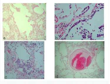
At light microscopy (H&E) survey the lung parenchima showed classic histopathologic triad of diffuse alveolar damage (DAD) [3]: hyaline membranes plastered along alveolar walls, type II pneumocytes hyperplasia and arterial thrombi (Fig.1).
Hyperplastic type II pneumocytes appeared marked atypical showing high nuclear:cytoplasmic ratio, bizzarre eosinophilic nuclear inclusions, cytoplasmic accumulation of viral particles, syncytial fusion or formation of cellular aggregates (Fig.2).
Not infrequently type II pneumocytes gain an excepionally abnormal (sometimes monstrous) appearance (Fig.3).
They also contain Cowdry type A nuclear inclusions (Fig.4).
No increment of CD68+ alveolar macrophage was reported, present as single elements, the AE1/AE3 hyperplastic type II pneumocytes constituting overtly the great mayority of cells inside alveolar space (Fig.5).
Mild to moderate interstitial mononuclear infiltration, intralveolar fine fibrin meshwork or serous/hemorrhagic oedema were seldom focally evident.
Organizing mixoid fibroblast plugs and bronchial/bronchiolar inflammation were absent.
Naked megakaryocytes were overrepresented in lung small vessel, sometimes coated by thin rim of cytoplasm (Fig.6).
Naked megakaryocytes were also clearly visible in myocardial, glomerular and adrenal cortex capillaries (Fig.7).
Stress-related microscopic hemorrhagic foci were found in adrenal cortex.
Expansion of red pulp and a severe reduction of white pulp were evident in spleen. No germinal centers were detected.
DISCUSSION
SARS is a potentially lethal lung acute disease, requiring intensive care especially mechanical ventilation.
SARS was reported for the first time during Coronavirus (SARS-CoV-1) pandemia occurred in 2002/2003 [4].
In that occasion was also defined the histopathologic pattern associated with the clinical syndrome, DAD, featured by a triad: hyaline membranes plastered along alveolar walls, type II pneumocytes hyperplasia, and thrombi present in distal arteries [5].
In peculiar circumstances, unknown fatal cases of SARS could request the intervention of the forensic pathologist to ascertain the cause of death: this is the case of unexpected demise, preceded only by a brief period of trivial flue-like syntoms, occurred at home and not preceded by hospitalization or esecution of nasopharyngeal swabs.
Aim of authors is to share histopatologic findings detected in two autopsy cases of unsuspected acute fatal SARS due to SARS-CoV-2 infection, not altered by effects of mechanical ventilation or intensive pharmacological therapy, at the same time attempting to infer some insights in pathophysiology of viral attack.
It is immediately evident from our two autoptic cases that DAD still represents the main histopathologic background of SARS.
Microscopic topography of lung damage shows that SARS-CoV-2 retains a selective tropism for type I pneumocytes: ubiquitous hyaline membranes and florid type II pneumocytes hyperplasia represent response to extensive type I pneumocytes death. Also type II pneumocytes are electively assaulted by virus as pointed out by unusual highly atypical reactive hyperplasia (enlarged cell with high nuclear:cytosplamic ratio, bizzarre eosinophilic nuclear inclusions seldom band-like, tendency to form syncytial aggregates, monstrous or vacuolate form) and nuclear viral cytopathic effect (Cowdry type A inclusions).
No relevant inflammation in lung tissue was detected: only a mild to moderate focal mononuclear interstitial infiltrate.
No bronchitis/bronchiolitis nor pneumonia were detected. No other lung compartments were affected by virus.
Collateraly SARS-CoV-2 infection elicites a release from bone marrow of megakaryocytes that embolize to lung capillaries.
Single naked megakaryocytes are usual finding within pulmonary capillaries of patients with febrile illnesses but, in our knowledge, they are never been noted in other organs [6].
Megakaryocytes release induced by SARS-CoV-2 infection is striking because very conspicuous: released megakaryocytes are so many that they are able to filter through lung capillaries and embolize further in other vital organs like hearth, kidney and adrenal glands.
Since naked megakaryocytes in bone marrow are tipically associated to an increase of peripheral demand of thrombocytes, this naked megakaryocytes storm means that SARS-CoV-2 could be able also to alter profoundly human coagulation system, like fibrin thrombi formation in pulmonary distal arteries suggests.
Compliance with ethical standards
No ethical approval for this work was requested. The authors declare the report not include reasearch involving Human Participants and/or Animals. No informed consent was considered necessary.
Conflict of interest The authors declare that they have no conflict of interest.
Funding No funding was obtained.
REFERENCES
1.Istituto nazionale di Statistica, Impatto dell’epidemia COVID-19 sulla mortalita’ totale della popolazione residente – primo trimestre 2020, 4-5-2020
2. Ministero della Salute, “Indicazioni emergenziali connesse ad epidemia COVID-19 riguardanti il settore funebre, cimiteriale e di cremazione, 01/04/2020.
3. Anna-Luise A. Katzenstein Diagnostic Atlas of Non-Neoplastic Lung Disease, demos MEDICAL, 2016, pp.115-126
4. CDC (2003), Severe acute respiratory syndrome—Singapore, MMWR, 52:405– 411
5. Chong PY, Chui P, Ling AE, Franks TJ, Tai DY, Leo YS, Kaw GJ, Wansaicheong G, Chan KP, Ean Oon LL, Teo ES, Tan KB, Nakajima N, Sata T, Travis WD (2004), Analysis of deaths during the severe acute respiratory syndrome (SARS) epidemic in Singapore: challenges in determining a SARS diagnosis, Arch Pat Lab Med, 128(2):195-204