The Spatial Evolution of Tau Pathology
in Alzheimer’s Disease:
Influence of Functional Connectivity and Education
Inaugural Dissertation
zur
Erlangung des Doktorgrades
Dr. nat. med.
der Medizinischen Fakultät und
der Mathematisch-Naturwissenschaftlichen Fakultät der Universität zu Köln
vorgelegt von
Merle Christine Hönig
aus Frankfurt am Main
buchdruck.de, Berlin
The Spatial Evolution of Tau Pathology in Alzheimer’s Disease:
Influence of Functional Connectivity and Education
Inaugural Dissertation
zur
Erlangung des Doktorgrades
Dr. nat. med.
der Medizinischen Fakultät und
der Mathematisch-Naturwissenschaftlichen Fakultät der Universität zu Köln
vorgelegt von
Merle Christine Hönig
aus Frankfurt am Main
buchdruck.de, Berlin
Betreuer: Prof. Dr. Alexander Drzezga Referent*innen: Prof. Dr. Gereon R. Fink
Prof. Dr. Silvia Daun
Prof. Dr. Kathrin Reetz
A B S T R A C T
Alzheimer’s disease is neuropathologically characterized by extracellular accumulation of amyloid plaques and intracellular aggregation of misfolded tau proteins, which eventually lead to neurodegeneration and cognitive impairment. With the recent advances in neuroimaging, these two proteinopathies can now be studied in vivo using positron emission
tomography (PET). Combining this imaging technique with functional magnetic resonance imaging has consistently revealed a spatial overlap between amyloid accumulates and functional connectivity networks (Buckner et al., 2009; Grothe et al., 2016), indicating
functional connectivity as mechanistic pathway in the distribution of neuropathologies. While
the infiltration of these neuronal networks by amyloid deposits seems uniform across
individuals with Alzheimer’s disease, there nevertheless exists inter-individual differences in the clinical expression of the disease despite similar pathological burden (Stern, 2012). This observation has fuelled the concept of existing resilience mechanisms, which are supported by lifetime and –style factors and, which magnitude varies between individuals, contributing to
the clinical heterogeneity seen in Alzheimer’s disease.
Even though the spreading and resilience mechanisms in the phase of amyloid accumulation
are now better understood, no information on tau pathology in vivo were available in this regard
until recently. Given the recent introduction of tau PET compounds, this thesis therefore aimed to address two questions: 1) whether functional connectivity contributes to the distribution of tau pathology across brain networks, and 2) whether the consequence of tau pathology on cognitive and neuronal function is mitigated by a resilience proxy, namely education. Using [18F]-AV-1451 PET imaging to quantify tau pathology in a group of Alzheimer’s disease patients, we observed that tau pathology arises synchronously in independent components of the brain, which in turn moderately overlap with known functional connectivity networks. This suggest that functional connectivity may act as contributing factor in the stereotypical distribution of tau pathology. Moreover, the results of this thesis demonstrate that the consequence of regional tau pathology on cognition differs depending on the level of education. Despite equal clinical presentation, higher educated patients can tolerate more tau pathology, already in regions related to advanced disease stage, than lower educated patients. Furthermore, tau pathology is less paralleled by neuronal dysfunction at higher levels of education. Thus, higher educated individuals show a relative preservation of neuronal function despite the aggregation of misfolded tau proteins. This maintenance of neuronal function may in turn explain the relative preservation of cognitive function albeit progressive tau pathology aggregation.
Taken together, the results of this thesis provide novel insights into the spreading mechanisms and the role of resilience factors towards tau pathology aggregation, which may not only be
ii
tauopathies. Better understanding of the spreading mechanisms in these diseases will permit a more precise prediction of disease progression and will thus be valuable for disease monitoring. Concomitantly, the development of sensitive biomarkers for disease monitoring is crucial for the evaluation of anti-tau-based therapies. Regarding the development of pharmacological strategies, the current results also indicate that proxy measures of resilience, such as education, need to be considered when allocating patients to treatment groups. Biased allocation may otherwise lead to a misinterpretation of observed effects that are not due to the drug but the group characteristics. Aside from this, sensitive tools for the early identification of at-risk individuals with high resilience need to be established to allow for a timely intervention. Current hypothesis is that an early intervention has the highest chance of success in modifying the disease course. However, as demonstrated by this work, individuals with high resilience remain undiagnosed until late in the disease course. Further research into resilience mechanisms may thus support the development of sensitive diagnostic tools and additionally offer potential targets that can be harnessed for novel treatment strategies. Hopefully, one day supporting the development of effective disease-modifying treatments.
Z U S A M M E N F A S S U N G
Die Alzheimer-Erkrankung ist neuropathologisch gekennzeichnet durch die extrazelluläre
Ansammlung von Amyloid Plaques und die intrazelluläre Aggregation von fehlgefalteten
Tau-Proteinen, die schließlich zu Neurodegeneration und charakteristischen kognitiven Beeinträchtigungen führen. Mit den neuesten Fortschritten in der Hirnbildgebung können diese Proteinopathien in vivo mittels der Positronen-Emissions-Tomographie (PET) untersucht
werden. Die Kombination dieser Bildgebungstechnik mit der funktionellen Magnetresonanztomographie hat dabei konsistent eine räumliche Überlappung zwischen
Amyloid Akkumulationen und funktionellen Konnektivitätsnetzwerken bei Patienten/Innen
mit einer Alzheimer-Erkrankung ergeben (Buckner et al., 2009; Grothe et al., 2016). Diese
räumliche Überlappung weist darauf hin, dass die funktionelle Konnektivität zur Verteilung von Neuropathologien beiträgt. Während die Infiltration dieser neuronalen Netzwerke durch
Amyloid Plaques bei Individuen mit einer Alzheimer-Krankheit konsistent erscheint, gibt es dennoch interindividuelle Unterschiede zwischen der pathologischen Belastung und dem klinischen Ausdruck der Erkrankung (Stern, 2012). Diese Beobachtung deutet auf Resilienzmechanismen hin, die durch Lebensstilfaktoren unterstützt werden, und deren Ausmaß individuell variiert und dadurch zur klinischen Heterogenität der Alzheimer-Erkrankung beiträgt.
Auch wenn die Ausbreitungs- und Resilienzmechanismen der Amyloid Pathologie inzwischen besser erforscht sind, lagen diesbezüglich bis vor Kurzem keine Informationen über
die in vivo Tau-Pathologie vor. Angesichts der jüngsten Verfügbarkeit von
Tau-PET-Substanzen in klinischen und Forschungseinrichtungen war daher das Ziel dieser Dissertation
folgende Fragestellungen zu untersuchen: 1. ‚Trägt die funktionelle Konnektivität zur
Verteilung der Tau-Pathologie entlang bestimmter Gehirnnetzwerke bei?‘; β. ‚Wird die Konsequenz der Tau-Pathologie in Bezug auf die kognitive und neuronale Funktion durch ein Resilienzmaß, nämlich Bildungsniveau, gemildert?‘. Die Analyse von [18F]-AV-1451 PET-Bildgebungsdaten zur Quantifizierung der Tau-Pathologie bei einer Gruppe von Alzheimer-Patienten/Innen ergab, dass die Tau-Pathologie synchron in unabhängigen Komponenten des Gehirns auftritt, die wiederum moderat räumlich mit bekannten funktionellen Konnektivitätsnetzwerken überlappen. Dies deutet darauf hin, dass die funktionelle Konnektivität als beitragender Faktor für die stereotype Verteilung der Tau-Pathologie fungiert. Darüber hinaus zeigen die Ergebnisse dieser Arbeit, dass die Konsequenzen der regionalen Tau-Pathologie für die Kognition abhängig vom Bildungsniveau sind. Trotz gleicher klinischer Schwere können hoch gebildete Patienten/Innen eine schwerwiegendere Tau-Pathologie tolerieren als niedrig gebildete Patienten/Innen. Zusätzlich scheint der neurotoxische Effekt der Tau-Pathologie bei einem höheren Bildungsniveau weniger stark ausgeprägt zu sein. So zeigen hochgebildete Patienten/Innen eine relative Erhaltung der neuronalen Funktion trotz der Aggregation der Tau-Pathologie. Diese Präservation der neuronalen Funktion ermöglicht
iv
vermutlich wiederum die relative Erhaltung der kognitiven Funktion trotz erhöhter Tau-Pathologie-Last.
Insgesamt dienen die Ergebnisse dieser Arbeit einem besseren Verständnis des Ausbreitungsmechanismus der Tau Pathologie und dem Beitrag von Resilienzfaktoren zur Aggregation der Tau-Pathologie, die nicht nur für die Alzheimer-Erkrankung, sondern auch für andere neurodegenerative Erkrankungen, insbesondere Tauopathien, relevant sein können. Ein erweitertes Verständnis der Ausbreitungsmechanismen dieser Krankheiten wird eine genauere Vorhersage des Krankheitsverlaufs ermöglichen und somit für das Krankheitsmonitoring wertvoll sein. Gleichzeitig ist die Entwicklung sensitiver Biomarker für das Krankheitsmonitoring entscheidend für die Auswertung von anti-Tau-basierten Therapien. Im Hinblick auf die Entwicklung von pharmakologischen Strategien deuten die aktuellen Ergebnisse darauf hin, dass bei der Zuordnung von Patienten/Innen zu Behandlungsgruppen Annäherungsmaße der Resilienz, wie z.B. Bildung, berücksichtigt werden müssen. Eine einseitig vorgenommene Zuordnung kann ansonsten zu einer Fehlinterpretation der beobachteten Effekte führen, die nicht auf das Medikament, sondern auf die Gruppeneigenschaften zurückzuführen sind. Darüber hinaus müssen sensitive Instrumente für die frühzeitige Identifizierung von Risikopersonen mit hoher Resilienz entwickelt werden, um eine rechtzeitige Intervention zu ermöglichen. Die aktuelle Hypothese ist, dass eine frühzeitige Intervention die höchste Erfolgschance in der Modifikation des Krankheitsverlaufes besitzt. Wie diese Arbeit jedoch zeigt, werden Personen mit einer hohen Resilienz erst zu einem späten Zeitpunkt im Krankheitsverlauf diagnostiziert. Insgesamt kann die Erforschung von Resilienzmechanismen zur Entwicklung von sensitiven Diagnoseinstrumenten beitragen. Zusätzlich bieten identifizierte Resilienzmechanismen potenzielle Ansätze, die für neue Behandlungsstrategien genutzt werden können. Die Ergebnisse werden hoffentlich eines Tages auch die Entwicklung effektiver krankheits-modifzierender Behandlungsstrategien unterstützen.
A C K N O W L E D G E M E N T S
It has been an honour working together with so many talented people, without whom this work would not have been possible.
In particular, I would like to thank my supervisor Prof. Dr. Alexander Drzezga, who has always had time to discuss research projects despite his busy schedule. I admire his passion for science and highly appreciate that he has given me the freedom to work on my own. I am also truly thankful that he has provided me the opportunity to attend international meetings to present our work. It has been a privilege working for him.
Furthermore, I am very grateful for being able to work in the multimodal neuroimaging group of Prof. Dr. Thilo van Eimeren, who has been an out-of-the-boxing-thinking, creative and patient supervisor throughout this time. He has always motivated me and challenged my way of thinking, thereby greatly contributing to the work of this thesis. I am impressed by his tremendous curiosity in neurodegenerative diseases.
Another important person is Dr. Gérard Bischof, who has supported me even at the busiest
times, who was always there to discuss my “final_final_final” results and to critically review
my work. He has been a valuable teacher throughout my PhD. His profound knowledge on
Alzheimer’s disease and his passion for research is inspiring. But most importantly, without Dr. Bischof I would not have been able to play a piano duet for a Nobel laureate.
I would also like to thank Kathrin Giehl for being my PhD-companion in the multimodal neuroimaging group and sharing the experiences and struggles of doing a PhD. It was certainly fun to learn how to let our solar plexus shine.
Furthermore, I wish to thank Dr. Jochen Hammes, who has been a patient teacher in all kinds of computer- and programming-related issues.
Moreover, I would like to thank the RTG-NCA for granting me a 3-year fellowship. Especially, I would like to thank my RTG tutors, Prof. Dr. Silvia Daun and Prof. Dr. Gereon R. Fink, for their valuable feedback at the thesis committee meetings, Dr. Katerina Vlantis, Dr. Isabell Witt and Kathy Jörgens for helping with administrative issues and Dr. Katharina Kastenholz and Manish Tomar for the good times at the retreats.
I would like to thank my parents, Elke and Jürgen, and my partner, Alex, for always believing in me and trying to calm me down before a talk. I know it was not easy at times and I am very grateful for your patience and support.
Finally, yet importantly, I would like to thank the clinical staff involved in the acquisition of the data and the patients and caregivers for willing to participate in this research.
T A B L E O F C O N T E N T S
Abstract ... i Zusammenfassung ... iii Acknowledgements ... v Abbreviations ... 1 List of Publications ... 2 Introduction ... 4Clinical presentation and diagnosis of Alzheimer’s disease ... 6
Neuropathological hallmarks of Alzheimer’s disease ... 7
Amyloid ... 8
Neurofibrillary tangles ... 9
Positron emission tomography in Alzheimer’s disease ... 11
Amyloid PET ... 11
Tau PET ... 12
FDG PET ... 12
Temporal evolution of Alzheimer’s disease biomarkers ... 14
Spatial distribution of Alzheimer’s disease biomarkers ... 15
The network degeneration hypothesis ... 17
The heterogeneity in clinical expressions of Alzheimer’s disease - The role of resilience ... 19
Cognitive reserve... 19
Brain reserve ... 20
Brain maintenance ... 21
Rationales and aims of the dissertation ... 22
Rationale & Aim I ... 22
Rationale & Aim II ... 23
Rationale & Aim III ... 24
Publication I ... 25
Networks of tau distribution in Alzheimer's disease ... 25
Contribution to this work ... 26
Additional information ... 26
Publication II ... 27
Tau pathology and cognitive reserve in Alzheimer's disease ... 27
Contribution to this work ... 28
Publication III ... 29
Level of education mitigates the impact of tau pathology on neuronal function ... 29
Contribution to this work ... 30
Additional information ... 30
Discussion ... 31
Spreading mechanisms of tau pathology ... 31
Prion-like spreading mechanisms of tau pathology ... 32
The role of functional connectivity in the spread of tau pathology ... 33
Factors beyond structural and functional connectivity ... 34
Susceptibility of the default mode network to tau pathology ... 35
Resilience against the evolution of tau pathology ... 38
An interpretation based on cognitive reserve and brain reserve ... 38
Animal models for the investigation of resilience and maintenance mechanisms ... 39
The role of early lifetime intervention ... 40
Implications of resilience for the diagnosis, prognosis and drug evaluation ... 40
Resilience mechanisms prompt refinement of current model of Alzheimer’s disease biomarkers ... 42
From education towards a more direct measure of resilience ... 44
Limitations ... 46
Conclusion & Outlook ... 48
References ... 49
Appendix ... 63
A1.) Abstract I –Regional suceptibility of the default mode network in Alzheimer’s disease ... 64
A2.) Abstract II – Functional connectivity in the hippocampus associated with cognitive reserve ... 65
B1.) Publication I –Networks of tau distribution in Alzheimer’s disease ... 66
B2.) Publication II – Tau pathology and cognitive reserve in Alzheimer’s disease ... 67
B3.) Publication III – Level of education mitigates the impact of tau pathology on neuronal function ... 68
T A B L E O F F I G U R E S
Figure 1
Proteolytic pathways of the transmembrane amyloid precursor protein (APP). ... 8 Figure 2
The human microtubule-associated tau protein (MAPT) gene and its isoforms. ... 10 Figure 3
Illustration of PET tracers and their binding sites. ... 13 Figure 4
Update on hypothetical model of Alzheimer’s disease biomarkers (Jack Jr et al., β01γ). ... 14 Figure 5
The stereotypical distribution pattern of amyloid and tau pathology in Alzheimer’s disease. ... 16 Figure 6
Pathophysiological topographies and their overlap with functional connectivity networks in the
clinical phenotypes of Alzheimer’s disease. ... 18 Figure 7
The framework of resistance and resilience in Alzheimer’s disease. ... 20 Figure 8
Regional susceptibility of the posterior default mode network in phenotypes of Alzheimer’s disease... 36 Figure 9
Hypothetical model on the evolution of Alzheimer’s disease biomarkers based on the level of resilience. ... 43 Figure 10
A B B R E V I A T I O N S
AICD amyloid precursor protein intracellular domain
FDG fluorodeoxyglucose
ApoE 4 apolipoprotein E4 (f)MRI (functional) magnetic resonance imaging
APP amyloid precursor protein ICA independent component
analysis BDNF brain derived neurotrophic
factor MAO monoamine oxidase
BM brain maintenance MAPT microtubule-associated tau
protein
BR brain reserve MCI mild cognitive impairment
CBD corticobasal degeneration NFT neurofibrillary tangles
PiB Pittsburgh Compound B NIA-AA National Institute on Aging
and Alzheimer's Association
CR cognitive reserve PCA posterior cortical atrophy
CSF cerebrospinal fluid PET positron emission tomography
CTF C-terminal fragment PSP progressive supranuclear palsy
DMN default mode network TPN tau pathology network
2
L I S T O F P U B L I C A T I O N S
This dissertation is based on the following publications:
1) Hoenig MC, Bischof GN, Seemiller J, Hammes J, Kukolja J, Onur ÖA, Jessen F, Fliessbach K, Neumaier B, Fink GR, van Eimeren T, Drzezga A. Networks of tau
distribution in Alzheimer's disease. Brain, 2018 Feb 1;141(2):568-581. doi:
10.1093/brain/awx353. PubMed PMID: 29315361.
2) Hoenig MC, Bischof GN, Hammes J, Faber J, Fliessbach K, van Eimeren T, Drzezga
A. Tau pathology and cognitive reserve in Alzheimer's disease. Neurobiology of Aging,
2017 Sep;57:1-7. doi: 10.1016/j.neurobiolaging.2017.05.004. PubMed PMID: 28577411.
3) Hoenig MC, Bischof GN, Onur ÖA, Kukolja J, Jessen F, Fliessbach K, Neumaier B, Fink GR, Kalbe E, Drzezga A, van Eimeren T. Level of education mitigates the impact of tau pathology on neuronal function. European Journal of Nuclear Medicine and
Molecular Imaging, 2019 Aug;46(9):1787-1795: 1787. doi: 10.1007/s00259-019-04342-3. PubMed PMID: 31183635.
In addition, this dissertation contains data in form of two abstracts:
a. Hoenig MC, Bischof GN, Onur ÖA, Jessen F, Fliessbach K, Neumaier B, van Eimeren T, Drzezga A. Regional susceptibility of the default mode network is associated with
clinical phenotypes of Alzheimer’s disease. Presented at: Deutsche Gesellschaft für Nuklearmedizin, 2019, Bremen, Germany.
b. Hoenig MC, Bischof GN, Lopes Alves I, Ahlswede M, Sakagiannis P, Jessen F, Schmidt S, Barkhof F, van Eimeren T, Drzezga A for the AMYPAD Consortium.
Hippocampal intrinsic connectivity supports cognitive reserve in amyloid-positive
cognitively normal subjects and Alzheimer’s disease patients. Presented at: AAIC, 2019, L.A., USA.
Co-authorships:
Hammes J, Theis H, Giehl K, Hoenig MC, Greuel A, Tittgemeyer M, Fink GR, Drzezga A, van Eimeren T. Dopamine metabolism of the nucleus accumbens and fronto-striatal connectivity modulate impulse control. Brain, 2019 Mar 1;142(3):733-743. doi: 10.1093/brain/awz007.
PubMed PMID:30753324.
Bischof GN, Ewers M, Franzmeier N, Grothe M, Hoenig MC, Kocagoncu E, Neitzel J, Rowe JB, Strafella A, Drzezga A, van Eimeren T on behalf of the MINC faculty. Connectomics and
molecular imaging in neurodegeneration. European Journal of Nuclear Medicine and
Molecular Imaging, 2019 Jul 11. Epub ahead of print. doi: 10.1007/s00259-019-04394-5. PubMed PMID: 31292699.
Seemiller J, Bischof GN, Hoenig MC, Tahmasian M, van Eimeren T, Drzezga A. Indication of retrograde tau spreading along Braak stages and functional connectivity pathways. Acta
4
I N T R O D U C T I O N
“Where are you right now?”
„Here and everywhere. Here and now. You must not think badly of me."
Alois Alzheimer & Auguste D. It has been more than a century since Alois Alzheimer first reported on the case of Auguste D., who showed a progressive loss of memory and speech as well as agitation. The brain autopsy of Auguste D. revealed an accumulation of senile plaques, neurofibrils, and general atrophy, which Alois Alzheimer concluded to cause the clinical symptoms. Several years later, Emil Kraepelin introduced the term “Alzheimer’s disease”, which was initially considered a rare disease, but has become the most common neurodegenerative disease world-wide, currently affecting around 50 million people above the age of 65 (Gaugler et al., 2019). With the
demographic change in Western societies, it is anticipated that the prevalence will triple by 2050, increasing the societal and economic burden tremendously. Despite all efforts, there is still no disease-modifying treatment available. Consensus is that only an early diagnosis and intervention has the highest chance of success in modifying this devastating disease.
The incremental growth in scientific knowledge about Alzheimer’s disease over the past decades has revealed that the neuropathological hallmarks of Alzheimer’s disease, namely
amyloid plaques and neurofibrillary tangles (composed of misfolded tau proteins), begin to accumulate more than 20 years before initial clinical symptoms occur. Importantly, these neuropathological characteristics can nowadays be visualized and studied in vivo using positron
emission tomography (PET). Especially the recent introduction of tau PET compounds has opened new possibilities in research and clinical settings. By means of tau PET imaging, information on the molecular mechanisms and distribution patterns of tau pathology in
Alzheimer’s disease and its interaction with other pathophysiological processes can now be gathered in vivo. This is further important for the identification of underlying resilience
mechanisms against the spatial evolution of tau pathology and its neurotoxic effects given the
heterogeneity in clinical expressions seen in Alzheimer’s disease. Some individuals can tolerate
more neuropathology albeit similar clinical severity than others and some never even develop clinical symptoms despite showing the neuropathological hallmarks of Alzheimer’s disease (Stern, 2012). Understanding the mechanisms driving this resilience against brain pathology is crucial for the development of sensitive diagnostic and prognostic biomarkers as well as novel treatment strategies.
This dissertation is intended to examine the role of functional connectivity as potential spreading mechanism of tau pathology employing tau PET imaging ([18F]-AV-1451) in combination with resting-state functional magnetic resonance imaging (MRI). Moreover, this thesis work elucidates the contribution of a resilience surrogate, namely education, concerning the impact of tau pathology aggregation on cognition and neuronal function. This investigation
is important on the one hand regarding the understanding of the mechanistic pathways of the pathophysiological processes in Alzheimer’s disease, and on the other hand concerning the interpretation of potential resilience mechanisms modifying the clinical expression of
Alzheimer’s disease.
In the introduction of this dissertation, a brief overview on the current knowledge about the
neuropathological hallmarks of Alzheimer’s disease, their temporo-spatial evolution and a description of potential mechanisms supporting resilience against these pathologies is provided, with an emphasis on PET imaging studies. The introduction is followed by three publications, which focus on the role of functional connectivity and education regarding the spread and neuronal consequences of tau pathology. Finally, the papers will be discussed in a larger context.
6
C L I N I C A L P R E S E N T A T I O N A N D D I A G N O S I S
OF ALZHEIMER’S DISEA
S E
Sporadic late-onset Alzheimer’s disease usually occurs in individuals older than 65 years and accounts for more than 95% of all cases with Alzheimer’s disease (Gaugler et al., 2019). The
remaining cases relate to familial and early-onset forms of Alzheimer’s disease with an age of onset ranging from 30 to 65 years. Within the realm of this dissertation, the term Alzheimer’s disease will refer to sporadic late-onset Alzheimer’s disease.
The cardinal symptom of Alzheimer’s disease is a progressive loss of memory, with subsequent appearance of executive, visual-spatial, language and neuropsychiatric impairments. At advanced stages of the disease, motor symptoms such as difficulties with swallowing and walking may also occur. Despite the typical presentation of Alzheimer’s disease, also atypical forms exist. These atypical forms present the same neuropathological characteristics, namely
amyloid plaques and misfolded tau proteins, but are characterized by different clinical phenotypes. These variants have been associated with an earlier disease onset (Snowden et al.,
2007; Koedam et al., 2010) and encompass posterior cortical atrophy (PCA), the logopenic
variant and the dysexecutive/ behavioural variant of Alzheimer’s disease. PCA is characterized by predominant visual-spatial deficits, the logopenic variant by language difficulties and the behavioural variant by deficits in executive function. Independent of the typical or atypical phenotype of Alzheimer’s disease, the clinical symptoms eventually interfere with activities of daily living, rendering the individual with Alzheimer’s disease dependent on others. The average life-expectancy after the diagnosis of Alzheimer’s disease is about 8-10 years, but significantly depends on how impaired the individual already is at the point of diagnosis (Larson
et al., 2004; Helzner et al., 2008).
At present, the clinical diagnosis of Alzheimer’s disease is based on the recommended diagnostic National Institute on Aging and Alzheimer's Association (NIA-AA) guidelines from 2011 (Albert et al., 2011; Jack Jr et al., 2011; McKhann et al., 2011; Sperling et al., 2011) and
the international working group criteria on Alzheimer’s disease (Cummings et al., 2013; Dubois
et al., 2014). According to the NIA-AA guidelines, Alzheimer’s disease is a continuum, which
can be categorized into three stages: preclinical, mild cognitive impairment (MCI), and
Alzheimer’s dementia. In the preclinical stage, molecular changes are detected including amyloid build-up, but no significant cognitive deficits are yet observable (Sperling et al.,
2011). In the MCI stage, initial memory impairments occur that are greater than of an age-matched person, but which do not yet interfere with daily living (Albert et al., 2011). In addition
to the accumulation of amyloid , a marker of neuronal injury including tau becomes abnormal, which can be assessed using PET or cerebrospinal fluid (CSF) markers (Albert et al., 2011). In
the final stage, Alzheimer’s dementia, cognitive deficits interfere with the person’s ability to function independently and both biomarkers are abnormal (McKhann et al., 2011). Importantly,
the guidelines for the symptomatic stages were intended to support the clinical diagnosis, whereas the preclinical definition was meant to offer researchers a common definition for the identification of participants, who show abnormal Alzheimer’s disease biomarkers in absence of clinical symptoms. Moreover, the integration of Alzheimer’s disease biomarkers in form of PET imaging or CSF analysis is considered as aid for the clinical setting, but the recommended guidelines are primarily based on clinical criteria.
The current diagnostic guidelines have therefore shifted from a syndromal to a biological construct, which have recently been outlined in a new research framework (Jack Jr et al.,
2018a). In this framework, the presence or absence of abnormal Alzheimer’s disease biomarker profiles are the building blocks used to describe the Alzheimer’s disease spectrum, while considering cognitive impairment as the result of the disease rather than its definition. The workgroup suggested the so-called A-T-N classification scheme, which is based on brain imaging (i.e., PET or MRI) and CSF biomarkers. “A” refers to amyloid pathology, “T” to tau
pathology, and “N” to neurodegeneration. In addition to the presence or absence of these three markers, a fourth factor namely the cognitive continuum can be included. Importantly, in contrast to the NIA-AA guidelines, a clear distinction is set between tau pathology and neurodegeneration/neuronal injury. The A-T-N classification states that abnormal amyloid and tau levels are a necessary condition for Alzheimer’s disease, whereas neurodegeneration is not specific to Alzheimer’s disease. As it is currently only a research framework, it still needs to be validated in clinical settings. However, so far, it appears to be a well-suited tool for the
biological diagnosis of Alzheimer’s disease by taking advantage of the currently available biomarkers for the quantification of the neuropathological hallmarks of Alzheimer’s disease.
N E U R O P A T H O L O G I C A L H A L L M A R K S
OF ALZHEIMER’S DISEA
S E
Alzheimer’s disease is a dual proteinopathy characterized by the extracellular deposition of amyloid in senile plaques and the intracellular aggregation of neurofibrillary tangles (NFTs). According to the prominent amyloid cascade hypothesis (Hardy and Higgins, 1992), amyloid deposition is the initial event, which triggers the formation of senile plaques and NFTs. This in turn leads to neuronal death and ultimately to dementia. Much support has been provided for this hypothesis. Nonetheless, there remain objections against it – one being that no phase 3 clinical trial targeting amyloid plaques has yet been successful in modifying the disease course (Gilman et al., 2005; Doody et al., 2014; Salloway et al., 2014; Siemers et al., 2016; Honig et al., 2018) and another one being that these two proteinopathies may actually act independently
with tau pathology preceding amyloid deposition, as recently suggested by autopsy data (Jack Jr et al., 2013). Despite the uncertainties regarding the temporal trajectory of the pathogenesis
of Alzheimer’s disease, there is no doubt that the two neuropathological hallmarks take on a
8
Amyloid
The extracellular accumulation of amyloid peptides was first discovered as main constituents
of senile plaques in Alzheimer’s disease and Down’s Syndrome (Glenner and Wong, 1984; Masters et al., 1985a; Masters et al., 1985b). A while later, genetic studies on familial
Alzheimer’s disease found that mutations in genes encoding the amyloid precursor protein (APP) (Goate et al., 1991), preselin-1 and preselin-2 (Levy-Lahad et al., 1995; Sherrington et al., 1995) were associated with elevated levels of amyloid . It is now known that preselin-1
and -2 are involved in the APP processing pathway. Although the functional role of APP itself remains unresolved, much knowledge has been gathered about the formation of amyloid deposits:
The amyloid peptide is derived from APP, a transmembrane protein which can undergo two proteolytic pathways, the α-pathway or -pathway. The former represents the non-amyloidogenic and the latter the non-amyloidogenic pathway (see Figure 1). In the
non-amyloidogenic process, APP is sequentially cleaved close to the membrane by α-secretase, liberating sAPPα and generating an alpha-C-terminal fragment (CTFα). Cleavage of CTFα by -secretase generates a small p3 fragment and a cytosolic element, the APP intracellular domain (AICD), which remains bound to the membrane and carries a role in signal transduction (Chen
et al., 2017). The catalytic subunit of -secretase is encoded by either presilin-1 or presilin-2.
In the amyloidogenic pathway, APP is cleaved by -secretase, which results in a large
derivative, sAPP , and a beta-C-terminal fragment (CTF ) that remains bound to the membrane (Selkoe, 1994; Olsson et al., 2014). CTF is then cleaved by -secretase, which results in the
soluble amyloid peptide and the membrane-bound AICD.
Figure 1– Proteolytic pathways of the transmembrane amyloid precursor protein (APP). In the non-amyloidogenic pathway, APP is cleaved by α-secretase generating sAPPα and CTFα followed by cleavage of -secretase, liberating pγ and producing AICD. In the amyloidogenic pathway, -secretase cleavage results in sAPP and a larger CTF fragment followed by -secretase producing amyloid and AICD. CTF = C-terminal fragment; AICD= APP intracellular domain. Adapted from Cheignon et al., 2018.
As the cleavage process in the amyloidogenic pathway is somewhat imprecise, numerous
species of amyloid exist (Takami et al., 2009). The most common species are amyloid 40
and amyloid 42, which are more hydrophobic and fibrillogenic than shorter forms of amyloid (Takami et al., 2009). It is assumed that the deposition of amyloid is likely a consequence
of impaired clearance and degradation mechanisms, which promote the assembly of amyloid monomers to oligomers, protofibrils and eventually to amyloid plaques in the brain parenchyma (Mawuenyega et al., 2010). Importantly, it was recently reported that the soluble forms of
amyloid are more toxic to neuronal cells than senile plaques (Bao et al., 2012; Esparza et al.,
2013). Given their solubility, these forms can spread throughout the brain (Chen et al., 2017),
disrupt synaptic function and trigger downstream toxic pathways (Mucke and Selkoe, 2012). Moreover, in accordance with the amyloid cascade hypothesis, transgenic experiments demonstrated that soluble amyloid forms increase tau phosphorylation (Seino et al., 2010)
and induce tau aggregation and seeding (De Felice et al., 2008; Seino et al., 2010; Vergara et al., 2019), which represents the second neuropathological hallmark of Alzheimer’s disease.
Neurofibrillary tangles
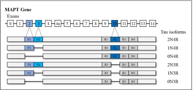
Neurofibrillary tangles (NFTs) are intracellular aggregates, which are composed of the hyperphosphorylated tau protein. The tau protein in its native form is abundant in the axons of neurons and carries an important role in the stabilization of the microtubules and axonal transport (Morris et al., 2011). It is encoded by the microtubule-associated tau protein (MAPT)
gene (Weingarten et al., 1975; Grundke-Iqbal et al., 1986) and contains four areas: an
N-terminal region, a proline-rich domain, a microtubule-binding domain, and a C-N-terminal projection region (Mandelkow et al., 1996). In the adult human brain six isoforms of tau are
present, which are produced by alternative splicing of exon 2, 3, and 10 (see Figure 2). These
isoforms can further be separated based on the length of their repeat binding domain, namely three or four carboxy repeat domains (3R and 4R). In the adult human brain, the 3R and 4R forms of tau are equally expressed, but this ratio changes in neurodegenerative diseases (Gao
et al., 2018). Tau aggregation is not only a characteristic hallmark of Alzheimer’s disease, but
also other neurodegenerative diseases known as tauopathies (e.g., progressive supranuclear palsy (PSP) or corticobasal degeneration (CBD)). These tauopathies can be classified based on the overexpression of the tau isoforms, namely: 3R-tauopathies (e.g., Pick’s disease), 4R-tauopathies (e.g., PSP or CBD), and 3R/4R tauopathies, to which Alzheimer’s disease belongs. Albeit the differences in tau isoform expression, the pathological tau lesions are highly phosphorylated across tauopathies, whereby in Alzheimer’s disease both forms of tau (γR and
4R) undergo hyperphosphorylation (Buée et al., 2000). The exact mechanism leading to this
hyperphosphorylation remains elusive. But given that various kinases and phosphatases regulate tau phosphorylation in the normal state, it has been suggested that an imbalance between these enzymes causes hyperphosphorylation of tau (Noble et al., 2013).
Importantly, the hyperphosphorylation of tau appears to precede the aggregation of misfolded tau proteins into paired helical filaments and then into insoluble NFTs (Alonso , 2001;
10
Chohan et al., 2005). The aggregation process, in turn, is facilitated through mechanisms such
as impaired degradation, truncation, or missorting of tau (Guillozet‐Bongaarts et al., 2006;
Dickey et al., 2007). Regarding the truncation of tau, it was shown that it suppresses the
formation of the so called paperclip structure, which usually hinders tau to aggregate (Jeganathan et al., 2006). Notably, tau in its native form is unfolded and does not tend to
aggregate (Mukrasch et al., 2009). However, through the truncation process, tau loses the
paperclip formation, which in turn promotes the aggregation of tau proteins. Moreover, recent studies showed that the hyperphosphorylation of tau can lead to missorting of tau from the axon to the somato-dendritic compartment (Thies and Mandelkow, 2007; Hoover et al., 2010;
Zempel et al., 2010; Zempel et al., 2017), which can cause synaptic dysfunction (Thies and
Mandelkow, 2007; Hoover et al., 2010).
Overall, a multitude of processes involved in the hyperphosphorylation and aggregation of misfolded tau proteins have been identified so far (for a detailed review see Morris et al., 2011;
Wang and Mandelkow, 2016). Nevertheless, the exact trigger of tau pathology in Alzheimer’s disease remains unknown as the MAPT gene is not genetically linked to Alzheimer’s disease.
This lack of genetic association to Alzheimer’s disease suggests that tau pathology is a downstream process of the amyloid - induced neurodegenerative cascade – a cascade, which is further characterized by pathophysiological alterations including the loss of synapses, initial hippocampal and later general atrophy, neuroinflammation in form of reactive astrocytes and activated microglia, and depletion of distinct neurotransmitter systems (Luca et al., 2018).
Importantly, with the developments in PET imaging, several processes of this pathophysiological cascade can nowadays be visualized and studied in vivo. The principle of
PET imaging and the main PET compounds used in Alzheimer’s disease research and diagnosis
will therefore be discussed in the next section.
Figure 2 - The human microtubule-associated tau protein (MAPT) gene and its isoforms. Representation of the six isoforms of the tau protein produced by alternative splicing of exon 2 ,γ, and 10 is illustrated. In Alzheimer’s disease, 3R and 4R isoforms are equally hyperphosphorylated. N= N-terminal; R = microtubule-binding repeat domain. Adapted from Wang and Mandelkow, 2016.
P O S I T R O N E M I S S I O N T O M O G R A P H Y
I N
ALZHEIMER’S DISEASE
Positron emission tomography (PET) is an imaging technique permitting the visualization of molecular changes and protein aggregations in vivo by injection of radioactively labelled tracers
into the blood, which bind to the biomolecules of interest. Briefly, PET imaging is based on the following technique: The radioactively labelled tracers contain short-lived positron-emitting radionuclides such as fluorine-18 or carbon-11. The beta decay of these radionuclides (e.g., 18F) attached to the target biomolecule (e.g., glucose) results in the emission of a positron that annihilates with an electron after travelling less than 1mm in the tissue. The annihilation process results in two gamma-photons being emitted in opposite directions. These gamma-rays are then detected by scintillation detectors, which register the annihilation photons in coincidence and store the events. Finally, using computer analysis, the PET activity distributions are reconstructed as three-dimensional images based on the coincidence events. The final images are then used for diagnostic or research purposes.
Up to now, several PET tracers have been developed, which can cross the blood brain barrier
and visualize Alzheimer’s disease-related pathophysiological changes. The most commonly used tracers, which will be discussed in the following, can visualize amyloid pathology, tau pathology and changes in glucose metabolism (see Figure 3).
Amyloid PET
The most widely studied amyloid tracer is [11C]-Pittsburgh Compound B ([11C]-PiB), which was developed by Chet Mathis and William Klunk in 2002 (Mathis et al., 2002). This tracer
shows high affinity and selectivity to fibrillar amyloid in senile plaques (Mathis et al., 2002;
Cohen et al., 2012). However, given the short half-life of [11C]-PiB of only 20 minutes, its use
is limited to centres that have a cyclotron and a department of radiochemistry on-site. Due to this limitation, F18-labelled tracers with similar affinity profiles, but a half-life of around 120 minutes, were developed, among them: [18F]Florbetapir (Wong et al., 2010), [18F]Florbetaben (Rowe et al., 2008), and [18F]Flutemetamol (Rinne et al., 2012). Ever since these amyloid
tracers have been available, a large body of evidence has been gathered supporting the utility of these tracers as diagnostic tool for dementia due to Alzheimer’s disease. In addition, these tracers have been useful for patient selection and the evaluation of drug efficacy in clinical trials (Fleisher et al., 2011). However, the drawback of the currently available amyloid tracers is that
they only bind to insoluble plaques and not to the more toxic and soluble forms of amyloid (Haass and Selkoe, 2007). Moreover, albeit its major role in defining Alzheimer’s disease,
amyloid PET imaging appears not to be suitable for the short-term prediction of individuals converting from prodromal stages or MCI to Alzheimer’s dementia (Iaccarino et al., 2017).
Furthermore, amyloid PET shows relatively low correlation with clinical and cognitive parameters (Brier , 2016) indicating that this PET modality is less well-suited for staging
12
of the disease. Thus, for better information regarding the progression and staging of the disease, [18F]-Fluorodeoxyglucose ([18F]-FDG) PET and more recently tau PET compounds have been considered.
Tau PET
The complexity of the tau protein given its heterogenous isoforms and its intracellular location have been major challenges in the development of selective tau PET tracers. Overcoming these challenges, several radioactive substances have recently been developed. The most widely studied are: [18F]-THK5117 (Harada
et al., 2015), [18F]-THK5351 (Harada et al., 2016), [18
F]-AV-1451 (Chien et al., 2014), and [11C]-PBB3 (Maruyama et al., 2013). Before that time, solely
CSF measures could provide information on abnormalities in tau phosphorylation in the central nervous system, but no information on the regional distribution of tau pathology could be obtained. The introduction of tau PET tracers has therefore led to new possibilities for diagnostic and research-oriented considerations (van Eimeren et al., 2017). A progressively
accumulating body of evidence suggests that tau PET is a suitable progression and staging marker, because it is more closely associated with neurodegeneration and cognitive decline than amyloid PET (Bischof et al., 2016; Brier et al., 2016; Ossenkoppele et al., 2016; Schöll et al.,
2016; Schwarz et al., 2016). Additionally, recent longitudinal tau PET studies have provided
first insights into the pathogenic cascade of Alzheimer’s disease (Chiotis et al., 2018a; Jack Jr et al., 2018b; Southekal et al., 2018). Importantly, in contrast to amyloid PET, tau PET allows
differentiation between typical and atypical phenotypes of Alzheimer’s disease, and primary tauopathies from secondary tauopathies1 (Kikuchi et al., 2016; Ossenkoppele et al., 2016; Dronse et al., 2017; Hammes et al., 2017; Passamonti et al., 2017; Whitwell et al., 2017;
Whitwell et al., 2018b). It is thus a meaningful biomarker for differential diagnosis. However,
despite the advances of tau PET imaging, an unresolved issue of the first-generation tau PET tracers remains the off-target binding to subcortical structures (Marquié et al., 2015; Lowe et al., 2016; Ng et al., 2017) and the lower affinity to different tau isoforms (Smith et al., 2017).
Therefore, second-generation tracers have been developed with improved binding properties and lower off-target signal, among them [18F]-PI-2620 (Mueller et al., 2017) and [18 F]-MK-6240 (Walji et al., 2016). These tracers are currently under investigation for their clinical and
research utility (Hostetler et al., 2016; Villemagne et al., 2018). Aside from this, tau PET
imaging provides unique information on underlying disease mechanisms, which are not only relevant for Alzheimer’s disease but also for other tauopathies.
FDG PET
The PET compound with the longest history in the investigation and diagnosis of neurodegenerative diseases is FDG PET, which is sensitive to changes in glucose metabolism.
1Primary tauopathies are considered diseases with misfolded tau proteins being the predominant pathological signature such as PSP or CBD. Secondary tauopathies account for diseases such as Alzheimer’s disease, which
It has been postulated that FDG PET measures synaptic function rather than overall neuronal function (Harris et al., 2012). Thus, a decrease in FDG PET signal (i.e., hypometabolism)
reflects an index of synaptic failure (Iaccarino et al., 2017). Over the past two decades, FDG
PET has been demonstrated to provide high diagnostic accuracy. Like tau PET, FDG-PET can aid in the differential diagnosis given that it offers information on the underlying pattern of neuronal dysfunction (Foster et al., 2007). Accordingly, distinct regional patterns of
hypometabolism have been observed for the clinical phenotypes of Alzheimer’s disease (Ossenkoppele et al., 2016; Dronse et al., 2017). Moreover, a close spatial relationship between
the tau PET signal and the FDG PET signal has been reported by several studies, whereas this is not the case for amyloid PET (Bischof et al., 2016; Brier et al., 2016; Ossenkoppele et al.,
2016). However, in contrast to amyloid and tau PET, FDG imaging does not provide any information on the underlying neuropathology. Therefore, an additional marker such as liquor
measurements or PET imaging is necessary to confirm the diagnosis of Alzheimer’s disease or
another neurodegenerative disease. Despite this, FDG PET may nevertheless be preferred to novel and more expensive PET compounds in the clinical setting given that it comes at relatively low costs in comparison to other PET tracers (van Eimeren et al., 2017).
Collectively, the use of these three PET modalities provide unique information on the evolution of the molecular characteristics in Alzheimer’s disease. The additional use of other PET compounds in research settings, for example for the visualization of neuroinflammation (e.g., [11C]-PBR28) and changes in synaptic density (e.g., [11C]-UCB-J), permits the investigation of the temporo-spatial relationship between these markers in the pathogenic cascade of
Alzheimer’s disease. In addition, combining these PET modalities with neuroimaging techniques such as diffusion tensor imaging (DTI) or functional MRI (fMRI) allows the investigation of spreading mechanisms across structural and functional pathways, which will support better understanding of this complex neurodegenerative disease (Bischof et al., 2019).
Figure 3– Illustration of PET tracers and their binding sites. AV-1451 is used to visualize paired helical filaments and neurofibrillary tangles in the neuron. C-PiB binds to amyloid plaques in the extracellular space and FDG is a marker of metabolic consumption mainly at the synapse. The pathological processes of Alzheimer’s disease are accompanied by the accumulation of reactive astrocytes and activated microglia, which can also be visualized using different PET compounds. C-PiB = Pittsburgh compound B; FDG = fluorodeoxyglucose.
14
T E M P O R A L E V O L U T I O N
OF ALZHEIMER’S DISEA
S E B I O M A R K E R S
An ever-increasing body of evidence suggests that the two neuropathological hallmarks of Alzheimer’s disease appear to evolve in temporal order with a long preclinical phase (Braak et al., 2011). In 2010, Jack and colleagues introduced a hypothetical model of Alzheimer’s disease
biomarker evolution for the in vivostaging of Alzheimer’s disease. This model was based on
evidence from cross-sectional, longitudinal and autopsy studies (Jack Jr et al., 2010). Three
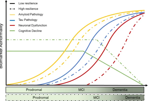
years later the model was revised to incorporate the observed variability in clinical expressions despite similar pathological burden and to specify the temporal ordering of certain biomarkers (Jack Jr et al., 2013). According to this well-established model (see Figure 4), abnormalities in
amyloid 42 in the CSF followed by abnormalities in amyloid PET imaging are detected up to two decades before clinical symptom onset. Following amyloid accumulation, abnormalities in tau CSF become apparent closely followed by biomarker abnormalities of neuronal function, such as measured with FDG PET, or of atrophy, as quantified by structural MRI. The trajectories of tau pathology, neuronal dysfunction and atrophy patterns are thereby more closely associated in time with the onset of clinical symptoms. By the time cognitive symptoms occur, amyloid pathology begins to plateau, whereas tau pathology and neurodegeneration continue to disperse. The onset of clinical symptoms can differ between individuals as represented by two trajectories relating to cognitive decline over time for individuals at low and high risk of developing mild cognitive impairment and Alzheimer’s dementia. Importantly, all trajectories are considered to be sigmoidal implying an initial phase of acceleration followed by a deceleration and plateau.
Figure 4 –Update on hypothetical model of Alzheimer’s disease biomarkers (Jack Jr et al., 2013). Temporal evolution of currently available Alzheimer’s disease biomarkers, which are colour- coded as depicted in the upper left corner. CSF = cerebrospinal fluid; A = amyloid ; FDG = fluorodeoxyglucose; MRI = magnetic resonance imaging; PET = positron emission tomography;MCI = mild cognitive impairment.
Notably, the model is based on currently available and detectable abnormalities in Alzheimer’s
disease biomarkers based on CSF, PET or structural MRI, which may not be sensitive enough
to detect the earliest changes in the neurodegenerative cascade of Alzheimer’s disease. This
may explain the discrepancy to recent autopsy studies, which suggested that tau pathology already appears in young people below 30 years of age, whereas amyloid plaques start accumulating in the fourth age-decade (Braak et al., 2011). Based on this evidence, it was
proposed that tau and amyloid may represent independent pathophysiological processes sharing an upstream causative factor, as also proposed by the dual pathway hypothesis (Small and Duff, 2008). Further investigations are required to proof or refute this assumption. Yet, the
current body of evidence is unambiguous regarding the fact that Alzheimer’s disease has a long
preclinical phase and that by the time initial clinical symptoms occur, amyloid pathology is already widely distributed across many brain regions, whereas tau pathology and neurodegeneration continue to spatially disperse in a stereotypical manner.
S P A T I A L D I S T R I B U T I O N
OF ALZHEIMER’S DISEA
S E B I O M A R K E R S
One of the most puzzling features of the amyloid cascade hypothesis remains the spatial disconnection between amyloid and tau pathology. Autopsy studies and recent PET imaging
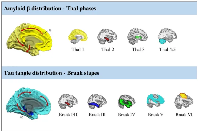
studies indicate that both proteinopathies follow distinct topographies, which initially occur in distal regions of each other. The stereotypical distribution patterns have been summarized for amyloid pathology by the Thal phases (Thal et al., 2002) and for tau pathology by the Braak
stages (Braak and Braak, 1991; Braak et al., 2006), which are both based on autopsy data.
According to the Thal phases, amyloid initially occurs in the neocortex, in particular in frontal areas (Phase 1), followed by accumulation in the hippocampus and entorhinal cortex (Phase 2), the basal ganglia (Phase 3), the brainstem (Phase 4), and finally the cerebellum (Phase 5). In contrast, tau pathology first gradually deposits in the transentorhinal cortex (Braak I) from where it spreads to the entorhinal region and hippocampal formation (Braak II), to temporal areas (Braak III), the precuneus (Braak IV), the parietal, occipital, and frontal regions (Braak V) and finally to the somatosensory cortex (Braak VI). Braak stages I-II represent the prodromal phase, Braak sages III-IV the early-moderate phase, and Braak V-VI the advanced and final stage of the disease. More recently, it was suggested that tau pathology may already start in the locus coeruleus and spread from there to subcortical and neocortical regions in a stereotypical manner (Braak et al., 2011).
Both staging systems suggest a well-defined neuroanatomical propagation pattern of the two proteinopathies (see Figure 5). However, interestingly, studies comparing autopsy data and in vivo imaging revealed that the earliest Thal phases cannot be visualized using amyloid PET
(Murray et al., 2015; Thal et al., 2015). In contrast, tau PET imaging studies consistently
16
et al., 2016; Schwarz et al., 2016; Hoenig et al., 2017). Moreover, the spread of tau pathology
is spatially more closely related with measures of neuronal dysfunction and neurodegeneration than the amyloid distribution patterns. This is the case for typical and atypical forms of
Alzheimer’s disease such as PCA, the logopenic and the dysexecutive variant as illustrated in Figure 6 (Bischof et al., 2016; Ossenkoppele et al., 2016; Dronse et al., 2017; Whitwell et al.,
2018).
Up to date, several factors and hypotheses have been considered concerning the stereotypical spread of these neuropathologies, such as susceptibility of distinct neuron groups (Shen et al.,
2016), gene expression patterns (Grothe et al., 2018; Sepulcre et al., 2018), and cell-to-cell
transmission processes (Clavaguera et al., 2009; De Calignon et al., 2012). Furthermore,
multimodal imaging studies consistently reported that the topographies of neurodegenerative disease pathologies overlap with large-scale neuronal networks (Drzezga, 2018). This suggests that functional and structural connectivity between regions promotes the distribution of these pathologies across neuronal networks, an observation that is summarized by the network degeneration hypothesis (Palop et al., 2006; Seeley et al., 2009), which will be elaborated on
in the following.
Amyloid β distribution - Thal phases
Tau tangle distribution - Braak stages
Figure 5 – The stereotypical distribution pattern of amyloid and tau pathology in Alzheimer’s disease. Brain regions of initial amyloid accumulation contain the frontal cortex (dark yellow) from where it spreads throughout the neocortex to subcortical regions and cerebellar regions. Tau pathology initially occurs in the entorhinal cortex (dark blue) from where it spreads to limbic and neocortical regions. The spatial distribution of amyloid plaques is defined by the Thal phases (top row) and the tau pathology distribution pattern is defined by the Braak stages (bottom row). The highlighted regions represent the newly affected region. FC= frontal cortex; EC=entorhinal cortex.
T H E N E T W O R K D E G E N E R A T I O N H Y P O T H E S I S
An accumulating body of evidence indicates that neurodegenerative diseases do not randomly spread across the brain, but coincide with specific functional brain networks (Tahmasian et al.,
2016). Based on these observations, the network degeneration hypothesis has been formulated, which postulates that neurodegenerative disease pathologies expand along functional networks eventually leading to failure of them (Palop et al., 2006; Seeley et al., 2009). Network failure
ultimately results in clinical symptomatology, which corresponds to the functional network being affected. This hypothesis suggests that functional connectivity between cortical nodes of neuronal networks acts as driver in the spread of pathology.
Over the past decade, several functional connectivity networks have been identified such as the executive control, language, visual-spatial, and the default mode network (DMN) (Yeo et al.,
2011), each carrying the name of the supporting cognitive domain. Compelling evidence has been gathered by studies combining fMRI and structural MRI or FDG PET, which reported a susceptibility of the aforementioned networks to neurodegeneration (Desgranges et al., 2002;
Seeley et al., 2009; Drzezga et al., 2011). In particular, the DMN has consistently been found
to be disrupted in Alzheimer’s disease (Greicius et al., 2004; Buckner et al., 2005; Buckner et al., 2009; Jones et al., 2015). The DMN is a highly active network during rest and is deactivated
during externally oriented tasks. Interestingly, amyloid PET imaging studies demonstrated predominant amyloid accumulation in hubs of the DMN (Buckner et al., 2005), which
eventually leads to disconnection of this network (Jones et al., 2015). Importantly, although
some studies have pointed at a susceptibility of amyloid in hub regions of functional networks, network degeneration appears to rather depend on global levels of amyloid than local levels (Drzezga et al., 2011; Iaccarino et al., 2018). This may be due to the location of amyloid ,
which distributes diffusely in the extracellular space throughout the brain. Therefore, it was recently suggested that tau pathology due to its intracellular location, its trans-synaptic spreading potential, and its close relationship to neurodegeneration, better relates to network
dysfunction and degeneration in Alzheimer’s disease. With the recent development of tau PET compounds in combination with fMRI, investigation of functional network infiltration by tau pathology is now feasible. Indeed, first in vivo studies have documented a close relationship
between functional networks and tau pathology distribution patterns in typical Alzheimer’s
disease (Hansson et al., 2017; Jones et al., 2017; Hoenig et al., 2018), as will be elaborated on
in publication I and the discussion. Whether similar mechanisms also account for atypical forms
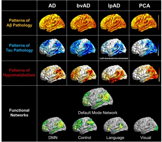
of Alzheimer’s disease still needs to be investigated. However, these atypical forms represent characteristic topographic patterns of hypometabolism and tau pathology, which resemble known functional connectivity networks (see Figure 6). A PET imaging study combing amyloid
and FDG PET revealed that these variants are indeed associated with degeneration of phenotype-specific networks (Lehmann et al., 2013). In contrast, amyloid deposition only
18
et al., 2013). This points towards a role of tau pathology in specific network degeneration in
atypical Alzheimer’s disease, as will be discussed later.
Generally, it is not yet clear why brain pathologies affect distinct brain networks. It could be explained by a combination of cell-to-cell transmission processes, inherent genetical, developmental susceptibility or by the type of underlying pathological aggregation (Drzezga, 2018). Notably, the consequences of the observed network degeneration differ between individuals. For example, it was recently shown that modulation in efficacy of affected networks supports maintenance of cognitive performance in the phase of Alzheimer’s disease- related brain pathology (Weiler et al., 2018). Some individuals are thus capable of coping with
network dysfunction or degeneration, thereby contributing to the clinical heterogeneity seen in
Alzheimer’s disease.
Figure 6 – Pathophysiological topographies and their overlap with functional connectivity networks in the clinical phenotypes of Alzheimer’s disease. Illustrated are the distribution patterns of amyloid pathology (yellow), tau pathology (blue), neuronal dysfunction (red), and resembling functional connectivity networks (green) for typical and atypical phenotypes of Alzheimer’s disease. Tau pathology and neuronal dysfunction patterns appear closely associated with distinct functional networks, but with predominant susceptibility of the posterior DMN across phenotypes. AD = Alzheimer’s disease; bvAD = behavioural variant of Alzheimer’s disease; lpAD = logopenic variant of Alzheimer’s disease; PCA = posterior cortical atrophy; DMN = default mode network. This figure was adapted from the originally published figure (courtesy of Merle Hönig) in the Journal of Nuclear Medicine. Drzezga A. The Network Degeneration Hypothesis: Spread of Neurodegenerative Patterns Along Neuronal Brain Networks. J Nucl Med. 2018; 59(11): 1645-8. © SNMMI.
T H E H E T E R O G E N E I T Y I N C L I N I C A L E X P R E S S I O N S O F
ALZHEIMER’S DISEASE
- T H E R O L E O F R E S I L I E N C E
Already in the late 80s, Katzman and colleagues reported a disparity between the extent of brain pathology and the individual clinical expression in patients with dementia (Katzman et al.,
1988). Over the past decades, it turned out that the rate of cognitive decline given a certain level of pathological burden is highly variable between individuals. This notion has been acknowledged, among other things, by the revision of the Jack model integrating two trajectories for low and high risk of cognitive decline (Jack Jr et al., 2013). These trajectories
are based on coping mechanisms, which are associated with factors such as greater educational attainment, higher IQ and grey matter volume. As several terms have been used across studies to define the disparity between brain pathology and clinical symptoms, a framework was recently published to harmonize terminologies (Arenaza-Urquijo and Vemuri, 2018). According to this framework, resistance is defined as a mechanism to avoid brain pathology, thus preventing Alzheimer’s disease-related pathology aggregation. In contrast, resilience is referred to coping with Alzheimer’s disease-related pathology. This framework further incorporates the concepts of cognitive reserve, brain reserve and brain maintenance, while simultaneously distinguishing between the presence and relative absence of brain pathology (Arenaza-Urquijo and Vemuri, 2018). An overview of the framework on resistance and resilience is provided in Figure 7. Importantly, within the realm of this dissertation, the focus will be laid on resilience mechanisms as the studies conducted as part of this dissertation are based on data of individuals, which already present Alzheimer’s disease pathology.
In the following, the potential coping mechanisms in form of cognitive reserve, brain reserve and maintenance will be elucidated in more detail:
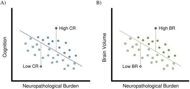
Cognitive reserve
Cognitive reserve (CR) refers to the adaption of cognitive processes in the presence of brain pathology (Stern, 2002, 2009; Stern et al., 2018a). Most commonly used surrogate measures of
CR are education, occupation, lifetime experience, verbal and general IQ (Yoo et al., 2015). It
is believed that CR acts through the more efficient use and modulation of distinct brain networks or recruitment of additional brain areas in the phase of pathology aggregation. Recent imaging studies support this assumption (Morbelli et al., 2013; Yoo et al., 2015; Franzmeier et al., 2017a; Franzmeier et al., 2017b; Stern et al., 2018b; Weiler et al., 2018). In particular, these
studies showed that network adaptations and compensation are associated with higher levels of educational attainment (Morbelli et al., 2013; Yoo et al., 2015; Franzmeier et al., 2017a;
Franzmeier et al., 2017b; Stern et al., 2018b; Weiler et al., 2018). The adaptation of network
efficacy and recruitment of additional brain areas may in turn provide a coping mechanism for increased pathological burden as reported by recent PET studies. These studies demonstrated that individuals with higher levels of education can maintain higher cognitive function at similar
20
levels of amyloid burden than lower educated individuals (Roe et al., 2008). Likewise,
individuals with higher CR can tolerate more amyloid pathology (Kemppainen et al., 2008),
hippocampal atrophy (Vuoksimaa et al., 2013) and hypometabolism in temporo-parietal areas
than lower educated individuals with similar clinical impairment (Kemppainen et al., 2008;
Ewers et al., 2013; Morbelli et al., 2013). Information regarding tau pathology load in vivo and
CR is still relatively limited due to the only recent availability of tau PET compounds (Hoenig
et al., 2017; Rentz et al., 2017; Shimada et al., 2017). Publication II of this dissertation is one
study investigating the association between level of education, as proxy measure of CR, and
tau pathology in Alzheimer’s disease.
Brain reserve
Although the observations relating to CR may be explained by network adaptations, it needs to be noted that these adaptations would not be possible without a biological foundation such as neuroplasticity, which in turn relates to the concept of brain reserve (BR). BR thereby refers to the neurobiological capital, hence brain integrity, of an individual (Stern et al., 2018a). The
concept of BR is based on a threshold model, which states that an individual with high BR can tolerate greater amounts of brain damage or brain pathology than individuals with low BR before showing clinical symptoms (Stern, 2002). This is because an individual with high BR obtains enough neuronal substrate to compensate the brain damage or pathology. Most commonly, grey matter volume, intracranial volume, but also head circumference have been used as surrogate measures of BR. Several structural MRI studies have provided support for the concept of BR using these proxies and further implicated higher education with better brain integrity (Schofield et al., 1997; Perneczky et al., 2010; Chang et al., 2016; Groot et al., 2018).
Thus, highly educated individuals presenting greater pathology burden may not only possess higher CR, but also better brain integrity to cope with the harmful effects of neuropathology. In publication III, this assumption is elaborated on by assessing the effects of tau pathology on neuronal function at different levels of education (Hoenig et al., 2019).
Figure 7 –The framework of resistance and resilience in Alzheimer’s disease. Contributing factors that support resilience and resistance mechanisms are for example lifestyle factors or distinct genes. Resistance is associated with brain maintenance, whereas resilience is closely associated with brain reserve and cognitive reserve. These three concepts in turn modulate the relationship between pathology build-up, neuronal dysfunction and cognition. Adapted from Arenaza-Urquijo and Vemuri, 2018.