ECG data compression using a neural network
model based on multi-objective optimization
Bo Zhang1*, Jiasheng Zhao2, Xiao Chen3, Jianhuang Wu2*
1 Department of Ultrasound in Medicine, Shanghai East Hospital, Tongji University School of Medicine, Shanghai, China, 2 Shenzhen Institutes of Advanced Technology, Chinese Academy of Sciences, Shenzhen, China, 3 School of Computing and Communications, Faculty of Engineering and Information Technology, University of Technology Sydney, Sydney, Australia
*[email protected](JHW);[email protected](BZ)
Abstract
Electrocardiogram (ECG) data analysis is of great significance to the diagnosis of cardiovas-cular disease. ECG compression should be processed in real time, and the data should be based on lossless compression and have high predictability. In terms of the real time aspect, short-time Fourier transformation is applied to the processing of signal wave for reducing computational time. For the lossless compression requirement, wavelet-transformation that is a coding algorithm can be used to avoid loss of data. In practice, compression is required to avoid storing redundant recording data that are not useful in the diagnosis platform. The obtained data can be preprocessed to remove noise by using wavelet transform, and then a multi-objective optimize neural network model is used to extract feature information. Com-pared with the existing traditional methods such as direct data processing method and trans-form method, our proposed compression model has self-learning ability to achieve high data compression ratio at 1:19 without losing important ECG information and compromising qual-ity. Upon testing, we demonstrated that the proposed ECG data compression method based on multi-objective optimization neural network is effective and efficient in clinical practice.
1. Introduction
Electrocardiogram (ECG) is widely used in modern medicine as a diagnostic parameter. How-ever, medical experts has to record huge chunks of such clinical data, and if these data cannot be compressed, it will increase storage cost due to large hard-disk space required. From the technical aspect, ECG data compression has these characteristics: 1) real time, lossless com-pression and high comcom-pression rate, and 2) the comcom-pression data can be used directly without full decompression. At the same time, electrocardiogram (ECG) that is recorded by automatic monitoring has significance to the diagnosis of cardiovascular disease. However, it usually takes a long time to record ECG data. On the other side, a large amount of electrocardiogram data is required to be analyzed and stored, while some of the meaningful feature information in these data is useful to diagnose. Therefore, it is necessary to adopt data compression a1111111111 a1111111111 a1111111111 a1111111111 a1111111111 OPEN ACCESS
Citation: Zhang B, Zhao J, Chen X, Wu J (2017)
ECG data compression using a neural network model based on multi-objective optimization. PLoS ONE 12(10): e0182500.https://doi.org/10.1371/ journal.pone.0182500
Editor: Quan Zou, Tianjin University, CHINA Received: April 5, 2017
Accepted: July 19, 2017 Published: October 3, 2017
Copyright:©2017 Zhang et al. This is an open access article distributed under the terms of the
Creative Commons Attribution License, which permits unrestricted use, distribution, and reproduction in any medium, provided the original author and source are credited.
Data Availability Statement: All ECG files are
available from the PhysioBank database (doi:10. 13026/C2F305),https://physionet.org/physiobank/.
Funding: This work was supported in part by
National Natural Science Foundation of China (No.61672510, 81571693, 81401428), Guangdong Natural Science Foundation (No.
2014A030313690), Shenzhen Science and Technology Program (No.
SGG20150602143414338, No.
JCYJ20160331191401141), Guangdong Science and Technology Program (No.2016A020220016), Pudong New Area Committee of Science and
algorithm to conduct compression on electrocardiogram data, in order to improve the storage and analysis efficiency of electrocardiogram.
The current ECG data compression algorithms [1,2] can be divided into three classes: 1) direct data processing; 2) transform; and 3) neural network approaches. Direct ECG data pro-cessing method usually conducts data compression by eliminating redundant information in ECG, by using methods such as Evolutionary Computation, Turning Point Scan-Along Polyg-onal Approximation, and Differential-Pulse Coding Modulation (EC, TP, SAPA and DPCM) algorithms [3–5]. Transform method usually conducts data compression by mathematical function, such as Kanade Lucas Tomasi, Discrete Cosine Transform, Fast Fourier Transform (KLT, DCT and FFT) algorithms. Based on other school of thoughts, the method that is based on neural network [6–9] usually conducts data compression by extracting the feature informa-tion implied in ECG through self-learning.
ECG data compression method that based on neural networks has gained growing atten-tion for its characteristics, which pertains to strong adaptability, parallel processing, good qual-ity of configurable waveform, and anti-interference. On one hand, the ECG data compression should achieve a data compression ratio as high as possible; on the other hand, it is required not to lose valid information or minimize losing electrocardiogram information. Hence, a suit-ably designed multi-objective function can optimize ECG data compression. If the current neural network based on one objective function is applied to achieve compression, we can only get a local optimal solution due to the focus on the optimization of one objective in data quality improvement. It is worthwhile noting that the neural network can easily fall into local minimum and lose ECG data.
Therefore, this paper proposes a theory model of multi-objective optimization neural net-work based on multi-objective constrained optimization theory [10–13], and then it studies the ECG data compression method that based on the multi-objective optimization neural work. Generally, this method is based on the changes of ECG characteristics so that neural net-work can learn under the guidance of the multi-objective function and adjust its structural parameters (i.e. coupling weight and offset value). With the purpose of extracting the feature information that implied in the ECG, it can realize effective ECG data compression [14–16]. In our paper, we study the theoretical model and learning algorithm of multi-objective optimi-zation neural networks, and then discusses ECG compression based on an optimizing neural network. Finally, we confirm the feasibility and advancement of this method through various carefully designed computational experiments in this paper.
2. Methodology
In this section, we present the mathematical formulations of the Discrete Wavelet Transform approach and neural network approach, which are implemented in this paper.
2.1 Wavelet transform used in ECG data compression
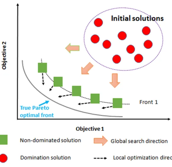
ECG feature extraction [4,17,18] is required to remove noise signal before feature extract pro-cessing due to vulnerability from noise in the environment. Then, a Pareto-optimal solution is required to achieve the best data compression versus compromising high quality (Fig 1). Wavelet transform is greatly effective for the instantaneous and time variant signal, which can help to eliminate baseline drift noise. A wavelet transform module has an input signal, which is defined as an integration of smaller version of the mother wavelet signal. Here, we present
Technology (No.PKJ2015-Y17), and the Academic Leaders Training Program of Pudong Health Bureau of Shanghai (grant No. PWRd2013-02). The funders had no role in study design, data collection and analysis, decision to publish, or preparation of the manuscript.
Competing interests: The authors have declared
that no competing interests exist.
Abbreviations: BP, Back Propagation; CC,
Correlation Coefficient; DCT, Discrete Cosine Transform; DPCM, Differential-Pulse Coding Modulation; DTW, Dynamic Time Warping; DWT, Discrete Wavelet Transform; EC, Evolutionary Computation; ECG, Electrocardiogram; EZW, Embedded Zerotree Wavelet; FFT, Fast Fourier Transform; KLT, Kanade Lucas Tomasi; PRD, Percentage Root-mean-squared Difference; SAPA, Scan-Along Polygonal Approximation; SPECK, Set Partitioning Embedded Block Coder; SPIHT, Set Partitioning Hierarchical Trees; TP, Turning Point.
the integral equations of our wavelet functions: WsfðtÞ ¼ 1 s Z 1 1 fðtÞ s t t s dt: ð1Þ
Wavelet scale parameters generally select a value of 2 as the form of exponent that results in the expressions= 2j, wherej= 1,2,. . .,m. Note thatφis the parameter of the mother wave func-tion.Eq (1)can hence be expressed as:
φsðtÞ ¼1
sφ t
s : ð2Þ
Following the previous formulation, the wavelet transform can be expressed inEq (3)as:
W2jfðtÞ ¼ 1 ffiffiffiffi 2j p Z þ1 1 fðtÞ pffiffiffiffi2j tffiffiffiffit 2j p dt: ð3Þ
Discrete signal requires the use of Discrete Wavelet Transform (DWT). Now, binarization of digital signals based on the DWT algorithm can be performed to give Eqs(4)and(5)as Fig 1. Multi-objective optimization leading to s Pareto-front of all solutions as the main objective.
follows: W2jfðnÞ ¼ X k2Z gkS2j1fðn 2j 1kÞ; ð4Þ S2jfðnÞ ¼ X k2Z hkS2j 1fðn 2 j 1 kÞ; ð5Þ
whereS2jis a smooth function,S2jfðnÞis the original signal (low frequency coefficients) that
serves as the approximation function, andW2jfðnÞis the original signal (high frequency
coefficients). It is worthwhile noting thathkandgkpertain to a low-pass and high-pass filter
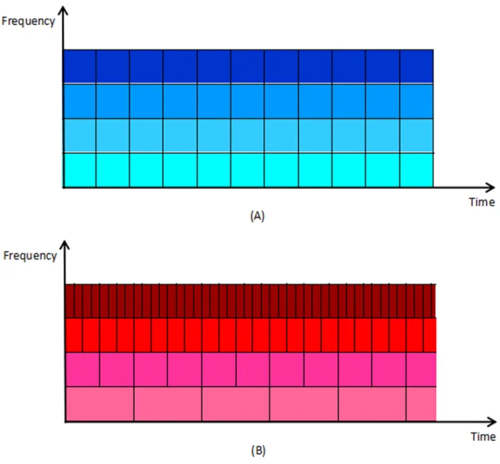
coefficients respectively. Details of ECG signal can be observed after the DWT process. Selection of wavelet functions in the decomposition process is the key to analysis ECG sig-nals, and hence a scale through short-time Fourier transformation and wavelet-transforma-tion is to be used (Fig 2).
In a baseline breathing exercise, the frequency ranges from 0.15Hz to 0.3Hz. Wavelet trans-formation can eliminate baseline drift of signals noise disturbance, because there is no latency and reduced distortion. Wavelet ECG signal degradation for approximate signal (high ampli-tude and low frequency signals) and the detail signal (low ampliampli-tude of high frequency signals) can help to distinguish the desired signal and noise signal.
2.2 ECG testing and selection of feature
Electrocardiographic signal, which is based on the electrical activity from the heart, is made up of a series of waves including the R wave, QRS-wave, P wave, T wave, and U wave. The QRS wave represents ventricular depolarization process two potential changes and the first down-ward wave of QRS wave is the Q waves. Due to the R-wave arrived amplitude maximum, it is easy to detect the QRS wave after locating the position of R. QRS complex detection algorithm based on wavelet transform, the core is in a scale or search within a certain scale wavelet form modulus maxima-minima between zero R-wave locations. [19–21] Scale wavelet trans-form can be achieved following these steps:
1. Thef(n) of ECG can transform toW2jfðnÞ;ðj2zþÞ, which is based on small wavelets coeffi-cients. This process utilizes the secondary wavelets and multi-scale decomposition of samples.
2. Whenj = 3, the positive thresholds1and negative thresholds2can detect the maximal and
minimal wavelets.
3. Locate the value that is over zero point, between the maximum value and minimum value. 4. The modified point of R-wave location can be acquired by23 1
2 ¼4.
After locating R, we can be certain that every beat will contain the P-QRS-T waves. Note that our ECG database is based on 251 points in a heartbeat cluster, such that we have R before 90 points, and R behind 160 points as two groups. The QRS-wave signal frequency content concentrate on details with a scale of 3, 4, and 5. Next, the T and P waves mainly concentrate details with a scale of 3, while other levels that do not contain noise are discarded. Notably, the time domain characteristics in ECG and RR intervals constitute a feature vector, which forms the foundation of signal classification.
2.3 Model of multi-objective optimization neural network
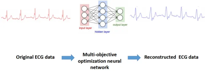
Neural network has appeared in increasing applications in the field of optimal computation, pattern recognition, intelligent control, and signal processing. However, multi-objective func-tion [22,23] is an index in a large number of engineering applications. Generally speaking, the feature of ECG can be reserved in this pattern, and it has high access ability without losing any useful information through the NN hidden layers (Fig 3). Therefore, the simultaneous optimi-zation of multi-objective function shall be described through the following mathematical Fig 2. Determination of sinusoidal frequency and phase content of local sections of ECG signal versus time based on (A) short-time-Fourier-transformation; and (B) wavelet-transformation.
problem, and the integral inEq (6)can be expressed as: min~y¼~fð~xÞ ~ x2D ; min~y¼ ðf1ð~xÞ;f2ð~xÞ;. . .;fmð~xÞÞ; ð6Þ Wherefðx*Þ ¼ ðf1ðx * Þ;f2ðx * Þ; ;fmðx *
ÞÞis the multi-objective vector criterion function,x*is an Euclidean space vector ofn-dimensional,Xis a set of constraints,~yis the objective vector, and
Dis search space.
In a multi-objective optimization problem, the non-inferior solution concept is usually adopted to describe the solution of vector function optimization. This means that a feasible decision vectorx’2Xis non-inferior solution, andx’2Xdoes not exist, and thereforeEq (7) becomes:
fðxÞ fðx0
Þ: ð7Þ
The non-inferior solution of multi-objective optimization problem can be obtained by the following, and thenEq (8)can be expressed as:
min x *2X Xm i¼1 oifiðxÞ; ð8Þ whereωi>0 and Xm i¼1 oi¼1.
In the case of a convex objective function and convex constraint,xis completely deter-mined by the changes ofo~i¼ ðoi1;oi2; oimÞ, so that a multi-objective convex optimization can be solved by weighting and secularization optimization. If a feed forward neural network is used to solve the multi-objective optimization problem, then this neural network can mini-mize the energy function in the following form, and the integral inEq (9)can be expressed as:
E¼X
m
i¼1
oifiðxÞ: ð9Þ
Fig 3. Flowchart of multi-objective optimization neural network for reconstruction of ECG data.
According toEq (9), the learning equation for multi-objective optimization is derived as follows, and thenEq (10)becomes:
doij dt ¼ a Xm k¼1 ok@fkðxÞ @oij ; ð10Þ
whereωijis a weight between neuronsiand neuronsj,αis the neural network learning rate,
andfk(x), (k= 1,2, m) is the objective function that is to be determined by the existing
prob-lem. Finally,Eq (11)becomes:
oi>0 and X
m
i¼1
o¼1: ð11Þ
2.4 ECG data compression based on multi-objective optimization
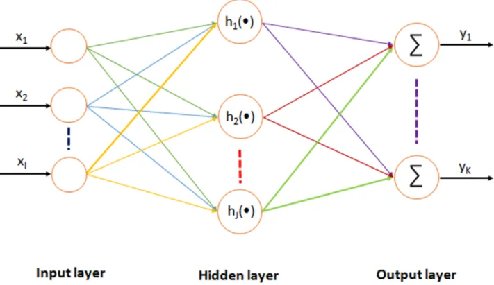
The structure of the multi-objective optimization neural network of ECG data compression is shown inFig 4. It is a three-layer feed forward neural network, including input layer, implica-tion layer and output layer. The input of neurons in input layer is the sampling point data of ECG. The neurons in hidden layer change according to the characteristics of ECG by learning to adjust the weight and bias value between it and input layer neurons. It is possible to extract the feature information implied in ECG (expressed as the output information of implied neu-ron). [15,24,25] ECG waveform after data compression can be reconstructed by output layer neurons based on the ECG feature information that is extracted by hidden neurons, based on its weight and offset value with hidden layer neurons. If the general back propagation (BP)
Fig 4. Neural network connections with the input, hidden and output layers of nodes representing a connection from a neural output to the input of a neuron.
algorithm is applied to train the above-mentioned neural network, then the following prob-lems exist: 1) there is a long processing time during network training; 2) the solution easily falls into a local minimum; and 3) the weights of the hidden layer neurons is difficult to determine.
The hidden layer decides the compression ratio. Note that if there are too many hidden layer neurons, the data compression ratio will decrease. On the other hand, if the hidden layer neurons are too few, the data compression performance will reduce, and resulting in significant distortion of reconstructed ECG. Nevertheless, an effective and practical ECG data compression algorithm requires not only a high data compression ratio but also, the reconstructed ECG shall retain or minimize loss of the effective ECG information as much as possible. Meanwhile, the real-time performance of algorithm is also required in practice [10,26–29]. Therefore, ECG data compression can be expressed as a multi-objective optimi-zation problem mathematically, which means to seek for the optimal data compression effect under the constraints of following multi-objective functions: 1) data compression ratio; 2) valid information loss after data compression; and 3) the real-time performance of data compression.
The model of multi-objective neural network that is discussed in the previous section is pre-sented here to solve multi-objective optimization problem. Following that, it can be used to achieve multi-objective compression of ECG data. Currently, the key question is how to sum-marize the multi-objective optimization function of ECG data compression. [30,31]
At present, the percentage root-mean-squared difference (PRD) and correlation coefficient (CC) are widely adopted as indicators to evaluate the loss of effective information after data compression, then Eqs(12)and(13)becomes:
PRD¼ ffiffiffiffiffiffiffiffiffiffiffiffiffiffiffiffiffiffiffiffiffiffiffiffiffiffiffiffiffiffiffiffiffiffiffiffiffiffiffiffiffiffiffiffiffiffiffiffiffiffiffiffiffi 1 N XN i¼1 ððri rÞ ðoi oÞ 2 Þ s ffiffiffiffiffiffiffiffiffiffiffiffiffiffiffiffiffiffiffiffiffiffiffiffiffiffiffiffiffiffi 1 N XN i¼1 ðoi oÞ 2 s ; ð12Þ CC¼ 1 N XN i¼1 ððoi oÞ ðri rÞÞ ffiffiffiffiffiffiffiffiffiffiffiffiffiffiffiffiffiffiffiffiffiffiffiffiffiffiffi 1 N XN i¼1 ðoi oÞ s ffiffiffiffiffiffiffiffiffiffiffiffiffiffiffiffiffiffiffiffiffiffiffiffiffiffiffiffiffi 1 N XN i¼1 ðri rÞ 2 s ; ð13Þ
Whereoiindicates the value of sampling pointiin the original waveform,riis the value of
sam-pling pointiin the restored waveform, to restore the value of the sampling point of the wave-form ofi,ois the average value of all sampling points in the original waveform, andris the average value of all sampling points in the restored waveform. [32] From Eqs(12)and(13), it can be seen that PRD represents the error magnitude contained in the waveform; and CC rep-resents the correctness of restored waveform. Therefore, multi-objective optimization function of ECG data compression can be summarized as follows inEq (14):
E¼W1NdþW2PRDþW3 ð1 CCÞ; ð14Þ
whereNdrepresents the number of neurons in hidden layer, PRD represents normalized RMS
error, and CC represents the correlation coefficient. In addition, theW1,W2andW3represent
the following indicators: 1) the weight of compression ratio, 2) normalized RMS error, and 3) correlation coefficient in the multi-objective ECG data compression, respectively.
The implication is as follows: solving the minimum value ofEq (14)requires seeking the optimal compromise solution between the effective ECG information and the data compres-sion ratio to be as high as possible. The main purpose of the first term inEq (14)is to improve the data compression ratio while the second term mainly reflects the error magnitude con-tained in ECG waveform, with the purpose to reduce the total amount of error in the restored electrocardiogram [1,5,20,33]. The third term mainly reflects the correctness of ECG wave-form restoration, with the purpose to reduce the recovery error of all sampling points in ECG waveform. According to [32–34], we can derive the learning equation of neural network for ECG data compression inEq (15)as:
doij
dt ¼ a
@E
@oij; ð15Þ
which then leads toEq (16)as
doij dt ¼a o1 @Nd @oijþo2 @PRD @oij þo3 @CC @oij !! ; ð16Þ
Whereαis the neural network learning rate. Note thatω1,ω2andω3are positive numbers less
than 1, and that their sum of weights is such thatω1+ω2+ω3= 1.
3. Results and discussion
3.1 Experimental verification
In order to verify the effectiveness and advancement of theoretical model and learning algo-rithm of multi-objective optimization neural network in the applications of ECG data com-pression, we conduct ECG data compression study based on neural network with partial ECG waveform T100, T105, T106, T108, T111, T112, T217, T219, T220 and T221 from MIT / BIH ECG database [2,3,14].
In our experiment, all parameters are set as follows. For the ECG waveform of data com-pression, each heart beat consists of 105 points before the R-wave peak, and165 points after the R-wave peak. Then, we conduct samples in the 270 points data such that all 15 points are used around the R point. For the other sections, samples are carried out every 6 points, so that each heart beat has 70 data points.
3.2 Reconstructed waveform based on other ECG data compression
algorithms
The neurons number in both input layer and output layer is 70, and the neurons number in hidden layer can be obtained by a multi-objective optimization function. The inputs neurons in the neural network correspond to the sampling data of ECG waveform. The outputs of hid-den neurons correspond to the implicit feature information of each ECG waveform. Next, after compression, the ECG waveform data is acquired through the weight between input neu-rons and hidden neuneu-rons. The sampling data of input ECG waveform and output neuneu-rons cor-responds to the data of reconstructed ECG waveform, which is acquired using the weight between hidden neurons, output neurons, and the output of hidden neuron. At this time, the weight between input neurons and hidden neurons, the weight between hidden neurons and output neurons, and the offset value between hidden neurons and output neurons are obtained by neural network through learning in regards of ECG data compression. (Fig 4) First of all, 14 neurons are selected as hidden neurons, and we select 40 waveforms from the T100 ~ T221 series in order to train the neural network for 10,000 cycles. Here, the E value in
multi-objective compression function of ECG data is 3.557, (parameters in the learning algorithm of multi-objective optimization neural network are set as:ω1= 0.25,ω2= 0.45,ω3= 0.3, andα=
0.4). Then, we implemented 16 neurons as hidden neurons, where parameters of neural net-work training data and training time are the same as above. Here, the E value in multi-objec-tive compression function of ECG data is 3.975. Finally, 12 neurons are selected as hidden neurons, whose training data, training time and learning algorithm parameters are the same as above. Then, theEvalue in multi-objective compression function of ECG data is 3.764.
As can be seen from the above results, selecting 14 neurons as hidden neuron is appropriate. Moreover, in order to check the learning outcomes of neural network, the studied and non-studied ECG waveforms (based on selection of 40 waveforms from T100 ~ T221 ECG wave-form, from which 67% are studied) are regarded as the input of neural network, then the hidden layer neurons record the compressed data of each ECG waveform. In the following modules, the output layer neuron can reconstruct the ECG waveform based on the output information of hidden neurons, and the weight between hidden neurons and output neurons that obtained through neural network learning. At this time, the evaluation indexes values of ECG compres-sion are: data comprescompres-sion ratio is 1:19, PRD = 12%, and CC = 99%, as shown inFig 5.
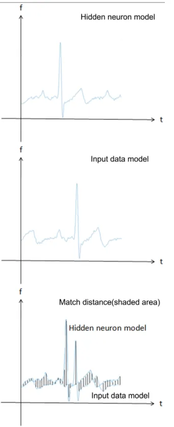
The hidden neurons match the output neurons based on Dynamic Time Warping (DTW). Notably, the DTW can recognize the all ECG waves, and then classify different ECG waves into output layers. The frequency of the wave can be detected by trained waves, and the trained wave can predict the income waves according to an Euclidean metric. There areMframes in hidden neuron andNframes in input neuron, wheredrepresents the distance between the hidden neuron and input neuron. Each frames based onMandNhas a certain distance (Fig 6). Therefore, the output data can be screened by this distance. The defined distance is set as 0.3 in order to raise the accurate of output data.
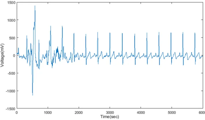
Fig 5. Reconstructed signal output after ECG compression for a longer period of 6000 seconds.
Fig 6. Comparison of waveform by the hidden neuron and input data models using a match distance approach.
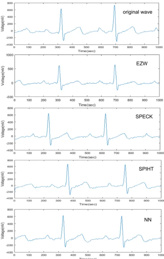
Fig 7. Reconstructed ECG signal waveform based on the wavelet compression and neural network methods in comparison with the original signal.
Fig 8. Average percentage root-mean-squared difference (PRD) results based on different ECG data compression ratios using transform and neural network approaches (A); Average encoding time versus ECG compression ratio using transform and neural network approaches (B).
Using the above analysis, it can be seen that multi-objective optimization neural network that used for ECG data compression is significantly better than several other data compression algorithms. Neural networks can adjust the parameters of network structure by learning the ECG data compression that comes with different characteristics. In addition, it can carry out learning under the guidance of multi-objective function in order to achieve the best result of data compression. This means that we are able to achieve a data compression ratio to be as high as possible without losing useful ECG information or losing as few information as much as possible.
Fig 7demonstrates the reconstructed ECG signals after undergoing the data compression process. Our method based on neural network can achieve fewer probe average values in dif-ferent compression ratio in comparison to other methods such as Embedded Zerotree Wavelet (EZW), Set Partitioning Embedded Block Coder (SPECK), and Set Partitioning Hierarchical Trees (SPIHT) as demonstrated byFig 8(A). At same time, our method requires less computa-tional time as compared to these methods based on the same compression ratio. (Fig 8(B))
4. Conclusion
In this paper, we put forward our mathematical model and learning algorithm for a neural net-work that is based on multi-objective optimization. This approach is then successfully applied onto ECG data compression. In our computational experiments, a satisfactory ECG data com-pression result is achieved, and we compared our neural network approach with the wavelet transform approaches to demonstrate its superiority. For future implementation, it may be of interest to compare this type of technique with direct data processing methods. Our model can process the useful data adaptively and efficiently, which comes at a lower cost in comparison with the traditional ECG compression techniques already in practice. Furthermore, the effec-tiveness and advancement of ECG data compression method that based on multi-objective optimization neural network are confirmed through comparison with these existing techniques.
Author Contributions
Conceptualization: Jianhuang Wu. Data curation: Jiasheng Zhao. Formal analysis: Bo Zhang.
Funding acquisition: Bo Zhang, Jianhuang Wu. Investigation: Bo Zhang.
Methodology: Jianhuang Wu.
Project administration: Jianhuang Wu. Resources: Jianhuang Wu.
Software: Xiao Chen. Supervision: Jianhuang Wu.
Validation: Jiasheng Zhao, Xiao Chen. Visualization: Jiasheng Zhao.
Writing – original draft: Bo Zhang, Jianhuang Wu. Writing – review & editing: Bo Zhang, Jianhuang Wu.
References
1. Ahmeda SM, Abo-Zahhad M (2001) A new hybrid algorithm for ECG signal compression based on the wavelet transformation of the linearly predicted error. Med Eng Phys 23: 117–126. PMID:11413064 2. Clifford GD, Behar J, Li Q, Rezek I (2012) Signal quality indices and data fusion for determining clinical
acceptability of electrocardiograms. Physiol Meas 33: 1419–1433.https://doi.org/10.1088/0967-3334/
33/9/1419PMID:22902749
3. Kors JA, van Herpen G, van Bemmel JH (1992) Variability in ECG computer interpretation. Analysis of individual complexes vs analysis of a representative complex. J Electrocardiol 25: 263–271. PMID: 1402511
4. Marcus M, Cohen A, Hammerman H, Inbar G (1987) Ordinal discrimination of ECG orthogonal features. J Electrocardiol 20 Suppl: 91–96. PMID:3320260
5. Kors JA, van Herpen G, Willems JL, van Bemmel JH (1992) Improvement of automated electrocar-diographic diagnosis by combination of computer interpretations of the electrocardiogram and vector-cardiogram. Am J Cardiol 70: 96–99. PMID:1615877
6. Ertosun MG, Rubin DL (2015) Automated Grading of Gliomas using Deep Learning in Digital Pathology Images: A modular approach with ensemble of convolutional neural networks. AMIA Annu Symp Proc 2015: 1899–1908. PMID:26958289
7. Dranca L, Goni A, Illarramendi A (2009) Real-time detection of transient cardiac ischemic episodes from ECG signals. Physiol Meas 30: 983–998.https://doi.org/10.1088/0967-3334/30/9/009PMID: 19696464
8. Xue Y, Jiang J, Zhao B, Ma T (2017) A self-adaptive artificial bee colony algorithm based on global best for global optimization. Soft Computing: 1–18.
9. Gu B, Sheng VS (2016) A Robust Regularization Path Algorithm forν-Support Vector Classification. IEEE Transactions on Neural Networks & Learning Systems PP: 1–8.
10. Li X, Cheung WK, Liu J (2010) Improving POMDP tractability via belief compression and clustering. IEEE Trans Syst Man Cybern B Cybern 40: 125–136.https://doi.org/10.1109/TSMCB.2009.2021573 PMID:19651557
11. Braojos R, Beretta I, Ansaloni G, Atienza D (2014) Early classification of pathological heartbeats on wireless body sensor nodes. Sensors (Basel) 14: 22532–22551.
12. Rautaharju PM, MacInnis PJ, Warren JW, Wolf HK, Rykers PM, Calhoun HP. (1990) Methodology of ECG interpretation in the Dalhousie program; NOVACODE ECG classification procedures for clinical tri-als and population health surveys. Methods Inf Med 29: 362–374. PMID:2233384
13. Sharma LN, Dandapat S, Mahanta A (2012) Multichannel ECG data compression based on multiscale principal component analysis. IEEE Trans Inf Technol Biomed 16: 730–736.https://doi.org/10.1109/
TITB.2012.2195322PMID:22542694
14. Dassen WR, Mulleneers RG, den Dulk K, Talmon JL (1993) Artificial neural networks and ECG interpre-tation. Use and abuse. J Electrocardiol 26 Suppl: 61–65.
15. Cadieu CF, Hong H, Yamins DL, Pinto N, Ardila D, Solomon EA, et al. (2014) Deep neural networks rival the representation of primate IT cortex for core visual object recognition. PLoS Comput Biol 10: e1003963.https://doi.org/10.1371/journal.pcbi.1003963PMID:25521294
16. Fira CM, Goras L (2008) An ECG signals compression method and its validation using NNs. IEEE Trans Biomed Eng 55: 1319–1326.https://doi.org/10.1109/TBME.2008.918465PMID:18390322 17. Brohet CR, Derwael C, Robert A, Fesler R (1990) Methodology of ECG interpretation in the Louvain
program. Methods Inf Med 29: 403–409. PMID:2233388
18. Devoe LD (2011) Fetal ECG analysis for intrapartum electronic fetal monitoring: a review. Clin Obstet Gynecol 54: 56–65.https://doi.org/10.1097/GRF.0b013e31820a0ee7PMID:21278502
19. Dubra A, Paterson C, Dainty C (2004) Wave-front reconstruction from shear phase maps by use of the discrete Fourier transform. Appl Opt 43: 1108–1113. PMID:15008490
20. Li R, Zhang W, Suk HI, Wang L, Li J, Shen D, et al. (2014) Deep learning based imaging data comple-tion for improved brain disease diagnosis. Med Image Comput Comput Assist Interv 17: 305–312. PMID:25320813
21. Park J, Lee S, Kang K (2015) Arrhythmia detection using amplitude difference features based on ran-dom forest. Conf Proc IEEE Eng Med Biol Soc 2015: 5191–5194.https://doi.org/10.1109/EMBC.2015.
7319561PMID:26737461
22. Belciug S, Gorunescu F (2015) Improving hospital bed occupancy and resource utilization through queuing modeling and evolutionary computation. J Biomed Inform 53: 261–269.https://doi.org/10.
23. Fearon WF, Lee DP, Froelicher VF (2000) The effect of resting ST segment depression on the diagnos-tic characterisdiagnos-tics of the exercise treadmill test. J Am Coll Cardiol 35: 1206–1211. PMID:10758962 24. Cohen AR, Gomes FL, Roysam B, Cayouette M (2010) Computational prediction of neural progenitor
cell fates. Nat Methods 7: 213–218.https://doi.org/10.1038/nmeth.1424PMID:20139969
25. Manor R, Geva AB (2015) Convolutional Neural Network for Multi-Category Rapid Serial Visual Presen-tation BCI. Front Comput Neurosci 9: 146.https://doi.org/10.3389/fncom.2015.00146PMID:26696875 26. Heaton LL, Lopez E, Maini PK, Fricker MD, Jones NS (2012) Advection, diffusion, and delivery over a
network. Phys Rev E Stat Nonlin Soft Matter Phys 86: 021905.https://doi.org/10.1103/PhysRevE.86.
021905PMID:23005783
27. Chaturvedi A, Lujan JL, McIntyre CC (2013) Artificial neural network based characterization of the vol-ume of tissue activated during deep brain stimulation. J Neural Eng 10: 056023.https://doi.org/10.
1088/1741-2560/10/5/056023PMID:24060691
28. Iwata A, Nagasaka Y, Suzumura N (1990) Data compression of the ECG using neural network for digital Holter monitor. IEEE Eng Med Biol Mag 9: 53–57.
29. Gu B, Sun X, Sheng VS (2016) Structural Minimax Probability Machine. IEEE Transactions on Neural Networks & Learning Systems PP: 1–11.
30. Ledezma CA, Severeyn E, Perpinan G, Altuve M, Wong S (2014) A new on-line electrocardiographic records database and computer routines for data analysis. Conf Proc IEEE Eng Med Biol Soc 2014: 2738–2741.https://doi.org/10.1109/EMBC.2014.6944189PMID:25570557
31. Cichy RM, Khosla A, Pantazis D, Torralba A, Oliva A (2016) Comparison of deep neural networks to spatio-temporal cortical dynamics of human visual object recognition reveals hierarchical correspon-dence. Sci Rep 6: 27755.https://doi.org/10.1038/srep27755PMID:27282108
32. van Bemmel JH, Zywietz C, Kors JA (1990) Signal analysis for ECG interpretation. Methods Inf Med 29: 317–329. PMID:2233378
33. Ditzler G, Polikar R, Rosen G (2015) Multi-Layer and Recursive Neural Networks for Metagenomic Classification. IEEE Trans Nanobioscience 14: 608–616.https://doi.org/10.1109/TNB.2015.2461219 PMID:26316190
34. Zhang W, Li R, Deng H, Wang L, Lin W, Ji S, et al. (2015) Deep convolutional neural networks for multi-modality isointense infant brain image segmentation. Neuroimage 108: 214–224.https://doi.org/10.