VISUAL WORKING MEMORY REPRESENTATIONS ACROSS EYE MOVEMENTS
by
BRITTANY J. DUNGAN
A DISSERTATION
Presented to the Department of Psychology and the Graduate School of the University of Oregon
in partial fulfillment of the requirements for the degree of
Doctor of Philosophy June 2015
DISSERTATION APPROVAL PAGE Student: Brittany J. Dungan
Title: Visual Working Memory Representations Across Eye Movements This dissertation has been accepted and approved in partial fulfillment of the
requirements for the Doctor of Philosophy degree in the Department of Psychology by:
Edward K. Vogel Chairperson
Edward Awh Core Member
Paul Dassonville Core Member
Cristopher Niell Institutional Representative
and
Scott L. Pratt Dean of the Graduate School
Original approval signatures are on file with the University of Oregon Graduate School. Degree awarded June 2015
© 2015 Brittany J. Dungan
DISSERTATION ABSTRACT Brittany J. Dungan
Doctor of Philosophy Department of Psychology June 2015
Title: Visual Working Memory Representations Across Eye Movements
We live in a rich visual world that we experience as a seamless and detailed stream of continuous information. However, we can only attend to and remember a small portion of our visual environment. The visual system is tasked with stitching together snapshots of the world through near constant eye movements, with around three saccades per second. The situation is further complicated with the visual system being
contralaterally organized. Each eye movement can bring items in our environment into a different visual hemifield. Despite the many challenges and limitations of attention and the visual system, how does the brain stitch together our experience of our visual environment?
One potential mechanism that could contribute to our conscious perception of a continuous visual experience could be visual working memory (VWM) working to maintain representations of items across saccades. Electrophysiological activity using event-related potentials has revealed the contralateral delay activity (CDA), which is a sustained negativity contralateral to the side of the visual field where subjects are attending. However, how does this work if we are constantly moving our eyes? How do we form a stable representation of items across eye movements? Does the representation transfer over to the other side of the brain, constantly shuffling the items between the
hemispheres, or does it stay in the hemisphere contralateral to the visual field where the items were located when we originally created the representation? The consequences of eye movements need to be examined at multiple levels and time points throughout the process.
The goal of my doctoral dissertation is to investigate VWM representations throughout the dynamic peri-saccadic window. In Experiment 1, I will first compare VWM representations across shifts of attention and eye position. With the focus on the effect of maintaining attention on items across eye movements, Experiment 2 will also explore eye movements both towards and away from attended visual hemifields. Finally, Experiment 3 is designed to substantiate our use of the CDA as a tool for examining VWM representations across eye movements by confirming that the CDA is indeed established in retinotopic coordinates.
CURRICULUM VITAE NAME OF AUTHOR: Brittany J. Dungan
GRADUATE AND UNDERGRADUATE SCHOOLS ATTENDED: University of Oregon, Eugene
DEGREES AWARDED:
Doctor of Philosophy, Psychology, 2015, University of Oregon Master of Science, Psychology, 2011, University of Oregon Bachelor of Arts, Psychology, 2010, University of Oregon AREAS OF SPECIAL INTEREST:
Cognitive Psychology Cognitive Neuroscience
PROFESSIONAL EXPERIENCE:
Graduate Research Fellow, Department of Psychology, University of Oregon Eugene, OR 2010-2015
Graduate Teaching Fellow, Department of Psychology, University of Oregon Eugene, OR 2012, 2013
Independent Contractor, SplitSage, Inc., 2014-2015
GRANTS, AWARDS, AND HONORS:
UO Promising Scholar Award, University of Oregon, 2010-2011
William & Molly Morgan Scholarship, University of Oregon, 2010-2011 Cognitive and Systems Neuroscience Training Program, Institute of
PUBLICATIONS:
Dungan, B. J., & Vogel, E. K. (2015) Short-term memory. In: Arthur W. Toga, editor. Brain Mapping: An Encyclopedic Reference. (Vol.3, pp. 481-485). Academic Press: Elsevier. In Press.
ACKNOWLEDGMENTS
I would like to express my gratitude to my advisor, Ed Vogel, for providing me with countless opportunities to pursue research both in depth and in a wide variety of areas. From my time as an undergraduate research assistant through my graduate career, I have valued his guidance and support. I would like to thank my other dissertation
committee members Ed Awh, Paul Dassonville, and Cris Niell for their feedback on my research projects over the years. To those Vogel and Awh lab members who came before me and helped shaped the researcher I have become, I thank Keisuke Fukuda, Andrew McCollough, Roy Luria, Hiroyuki Tsubomi, and Edward Ester. And to all those current lab mates throughout the Vogel, Awh, and Mayr labs who have stood around the white board with me as we try to understand the complexities of human cognition, I thank David Anderson and Irida Mance (we made it!), Kirsten Adam, David Sutterer, Joshua Foster, Richard Matullo, Jason Hubbard, Pablo Morales, and Atsushi Kikumoto. Thank you to my research assistant Joshua Zhilong Wu for his indispensible assistance in the data collection process and Zhan Xu for our many discussions and collaborations. And finally, to my parents who encouraged me to always do my best, I couldn’t have done this without you.
TABLE OF CONTENTS
Chapter Page
I. THE IMPACT OF EYE MOVEMENTS ON ATTENTION ... 1
Introduction ... 1
Eye Movements ... 1
Neuroanatomy and Physiology of the Visual and Ocular Motor System ... 2
Eye Movements: Motor Planning and Mechanics ... 4
Saccades ... 4
Motor Level ... 4
Tracking Where the Eyes Need to Move ... 6
Determining When or If the Eyes Need to Move ... 7
Corollary Discharge ... 9
What Eye Movements Can Tell Us About Underlying and Related Processes ... 9
Saccadic Suppression ... 9
Attention ... 12
Attention and Visual Working Memory ... 14
Contralateral-Delay Activity ... 15
Attentional Tracking ... 17
Perceptual and Cognitive Processes in a Spatial Reference Frame ... 18
Spatial Perception ... 18
Spatial Attention ... 20
Spatial Working Memory ... 21
Chapter Page
Eye Movements and Memory/Trans-Saccadic Memory ... 23
Eye Movements and Attention ... 26
Retinotopic Attentional Trace ... 27
Attention Across Saccades ... 29
Perceptual/Visual Stability ... 32
Neural Evidence Related to Perceptual/Visual Stability ... 33
Remapping ... 34
Predictive Remapping ... 36
Remapping in Other Contexts ... 37
Remapping Across Hemispheres ... 38
Conclusions and Future Directions ... 39
II. VISUAL WORKING MEMORY REPRESENTATIONS ACROSS EYE MOVEMENTS ... 41
Introduction ... 41
Materials and Methods ... 43
Overview ... 43
Participants ... 44
Experiment 1 ... 44
Behavioral Procedure ... 44
Eye Tracking Recordings and Analysis ... 46
Electrophysiological Recordings and Analysis ... 46
Experiment 2 ... 48
Behavioral Procedure ... 49
Eye Tracking and Electrophysiological Recordings and Analysis ... 50
Experiment 3 ... 50
Behavioral Procedure ... 50
Eye Tracking and Electrophysiological Recordings and Analysis ... 51
Results ... 52
Experiment 1 ... 52
Behavioral Results ... 52
Eye Tracking Results ... 52
Electrophysiology Results ... 52
Experiment 2 ... 59
Behavioral Results ... 59
Eye Tracking Results ... 59
Electrophysiology Results ... 60
Experiment 3 ... 67
Behavioral Results ... 68
Eye Tracking Results ... 68
Electrophysiology Results ... 69
Discussion ... 70
Implications and Future Directions ... 72
LIST OF FIGURES
Figure Page
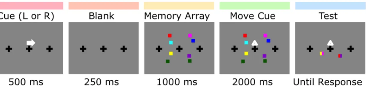
1. Behavioral procedure for Experiment 1 ... 45
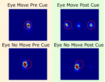
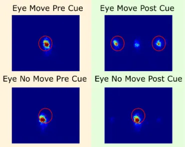
2. Fixations from Experiment 1. ... 48
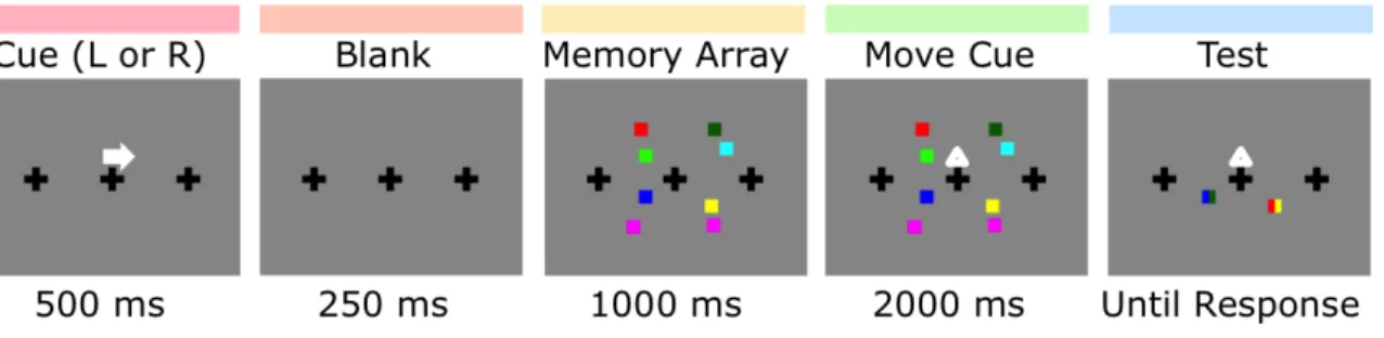
3. Behavioral procedure for Experiment 2 ... 49
4. Behavioral procedure for Experiment 3 ... 51
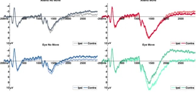
5. Contralateral – ipsilateral difference waves for Experiment 1 ... 53
6. Contralateral and ipsilateral activity for Experiment 1 ... 54
7. Subtraction of attend no move – attend move for Experiment 1 ... 55
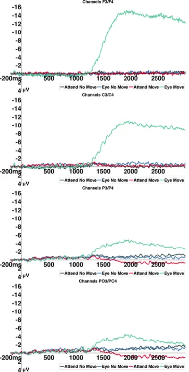
8. Topography of contralateral-ipsilateral difference waves for Experiment 1 ... 57
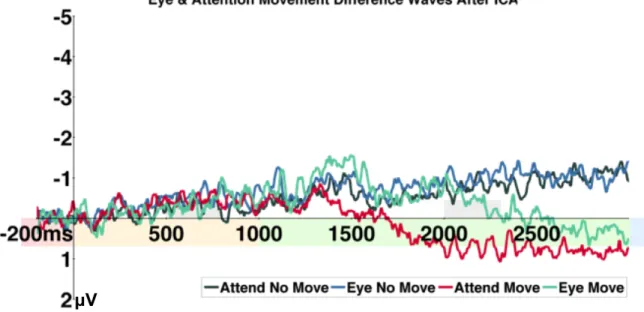
9. Contralateral – ipsilateral difference waves after ICA for Experiment 1 ... 58
10. Fixations from Experiment 2 ... 60
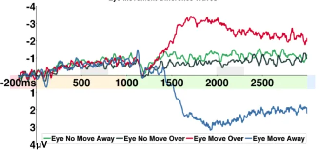
11. Contralateral – ipsilateral difference waves for Experiment 2 ... 61
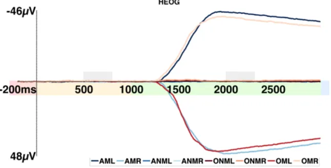
12. HEOG activity for Experiment 2 ... 62
13. Average of difference waves for eye no move and move for Experiment 2 ... 63
14. Contralateral and ipsilateral activity for average eye move for Experiment 2 ... 64
15. Subtraction of average eye no move – average eye move for Experiment 2 ... 65
16. Contralateral – ipsilateral difference waves after ICA for Experiment 2 ... 66
17. Contralateral and ipsilateral activity for Experiment 2 ... 67
18. Contralateral – ipsilateral difference waves for Experiment 3 ... 69
CHAPTER I
THE IMPACT OF EYE MOVEMENTS ON ATTENTION
INTRODUCTION
We live in a rich visual world that we experience as a seamless and detailed stream of continuous information. However, we can only attend to and remember a small portion of our visual environment. The visual system is tasked with stitching together snapshots of the world through near constant eye movements, with around three saccades per second. The situation is further complicated by the contralateral organization of the visual system, whereby information from one visual hemifield is primarily processed in the opposite cortical hemisphere of the brain. Each eye movement can bring items in our environment into a different visual hemifield and research has explored how the brain keeps track of these changes. Despite the many challenges and limitations of attention and the visual system, how does the brain stitch together our experience of our visual environment?
EYE MOVEMENTS
Eye movements are a complex behavior that is a result of, leads to, and interacts with many cognitive functions, and quite literally have the ability to change our point of view with respect to the world around us. Some researchers have approached this topic by examining visual perception and the eye movements themselves (Sommer & Wurtz, 2008), while others have extended this to the effects of saccades on spatial attention, visual short-term memory, and other cognitive mechanisms (Irwin, Zacks, & Brown,
1990; Hollingworth, Richard, & Luck, 2008; Golomb, Nguyen-Phuc, Mazer, McCarthy, & Chun, 2010). Past single-unit recording literature examined the remapping of receptive fields (Colby, Duhamel, & Goldberg, 1992), while more recent evidence supports
converging receptive fields at saccade target locations with implications for attentional facilitation (Zirnsak, Steinmetz, Noudoost, Xu, & Moore, 2014). Spatial updating research continues to investigate pre-saccadic effects (Parks & Corballis, 2008), while other research also suggests lingering memory representations following eye movements (Merriam, Genovese, & Colby, 2003). The field continues to investigate the many peri-saccadic phenomena at the intersection between attention and eye movements.
A separate but related area of research studies the impact of attention on eye movements, including what drives changes of gaze and eye movements in scene perception. However, the present review will focus primarily on the impact of eye movements on attention and visual working memory to help understand how the visual system overcomes various challenges to create our conscious experience of our visual world. In particular, activity in humans will be covered, as well as activity in non-human primates, and other animal, human homologs. First, eye movements themselves will be examined: how are saccades initiated, how does the brain tell the eyes when and where to move, and how is this information organized? Second, the attention and visual working memory literature will be addressed before specifically examining the interaction between eye movements and attention.
Neuroanatomy and physiology of the visual and ocular motor system
into signals that are interpreted as our visual experience. However, our eyes are not always fixed in space. The human visual system has evolved to perform visually guided eye movements to locate objects in space, which requires a complex interplay of many brain structures to coordinate the areas that process the incoming information and
produce the eye movements (Schall, 2009). Visual input from the retina goes through the lateral geniculate nucleus (LGN) and superficial layers of superior colliculus to transmit the information to visual cortex and then on to parietal, including lateral intraparietal area (LIP), and temporal cortices before reaching the fontal eye fields (FEF) (Schall, 2009). FEF plays an important role in saccade production and oculomotor systems. It has projections to various relevant areas and is innervated by many others, including strong interconnectivity with extrastriate visual cortex. FEF is further connected with
dorsolateral prefrontal cortex (DLPFC) and supplementary eye field (SEF), and feeds into the saccade generator through intermediate layers of the superior colliculus, basal ganglia through ipsilateral striatum, and the cerebellum through the pontine nuclei of the brain stem (Schall, 2009). The role of FEF in producing saccades was originally suspected when electrical stimulation in one FEF hemisphere in macaques caused contralaterally directed saccadic eye movements (Schall, 2009). Such stimulation across FEF evokes saccades of gradually varying direction and amplitude, providing early insight into how saccades are generated (Schall, 2009). At any given moment each of these specialized areas of the visual system must process information projected from previous parts of the processing stream and provide inputs to further processing, while interacting with near and far-reaching corticies. This is an impressive process when the eyes are not rapidly scanning the visual world; however, matters are further complicated when eye
movements are taken into account.
Eye movements: motor planning and mechanics
Saccades
Processing a complex visual scene may require several eye movements to bring potential visual targets over the high-resolution of the fovea for inspection (Pertzov, Avidan, & Zohary, 2011). In fact, such eye movements are necessary to our visual experience as demonstrated when perfect fixation is maintained, the visual system
quickly adapts to the present visual stimulation and everything fades away from our view (Martinez-Conde, Macknik, & Hubel, 2004). Saccades, where the eyes rapidly move between two resting positions, can cause dramatic shifts to the position of the images projected on the retina, even when the items represented remain at fixed positions in space (Ross, Morrone, & Burr, 1997). Although this will be addressed in more detail later, this is a key issue that the brain must resolve to give us a stable perception of the world. The brain must keep track of the current state of the eyes, plan the next eye
movement, determine when or if the eye movement plan should execute, and predict how this eye movement plan will impact the placement of the retinal image.
Motor level
At the motor level, saccades are produced by burst and motor neurons that innervate extraocular motorneurons that provide the force to rapidly rotate the eyes, which is counteracted by a second amount of force from tonic neurons to maintain
eccentric gaze (Schall, 2004a). The saccade generator, located in the brainstem,
ultimately receives the command when it is time to move the eyes and where they should then re-fixate (Schall, 2004a). Past research has shown that visually guided saccades can be made reflexively, while saccades toward targets (pro-saccades) tend to be generated faster than saccades away from targets (anti-saccades) when a cue must be evaluated (Clementz, Brahmbhatt, McDowell, Brown, & Sweeney, 2007). Anti-saccades also differ from pro-saccades by recruiting greater prefrontal cortex (PFC) activity (Clementz, Brahmbhatt, McDowell, Brown, & Sweeney, 2007), as well as activity in other areas of the oculomotor circuit (O’Driscoll, Alpert, Matthysse, Levy, Rauch, & Holzman, 1995). Areas involved in voluntary saccades include FEF, SEF, the parietal eye field (PEF), and precuneus in superior parietal cortex, while striate and extrastriate cortices help guide visuomotor responses related to stimulus processing (Clementz, Brahmbhatt, McDowell, Brown, & Sweeney, 2007).
The pre-saccadic time period involves changes of activity in many brain areas. In particular, single unit recordings in area LIP in rhesus monkeys showed enhanced
sensory responses to the spatial locus of a given receptive field for a covertly attended stimulus as well as for one that will be representing the target of a future saccade (Colby, Duhamel, & Goldberg, 1996). Specifically, the researchers saw increased visual
responses to stimuli presented, regardless of behavioral relevance, during a memory guided saccade task (Colby, Duhamel, & Goldberg, 1996). The responses of LIP neurons were related to sensory processing but were also subject to cognitive factors, such as attentional modulation, and were related to motor planning and saccade production (Colby, Duhamel, & Goldberg, 1996). The various changes that the visual system
undergoes in preparation for an eye movement suggests its high priority in visual processing.
Tracking where the eyes need to move
In regards to where the eyes need to be moved, research has identified multiple reference frames for saccades. Oculomotor space and saccade amplitude have been mapped out across the lateral bank of the intraparietal sulcus (IPS) contralateral to the direction of the eye movement (Savaki, Gregoriou, Bakola, Raos, & Moschovakis, 2010). This further enforces the contralateral organization of the visual system as a whole. Functional magnetic resonance imaging (fMRI) has additionally revealed that saccades of equal amplitude are represented within a retinotopic reference frame in FEF and IPS in human cortex (Pertzov, Avidan, & Zohary, 2011). However, saccades to a common target, despite having different movement vectors, also revealed spatiotopic
representations in the middle aspect of IPS (Pertzov, Avidan, & Zohary, 2011). The study further suggested that eye movements are made both in egocentric and allocentric
representations of space and implicated parietal cortex in maintaining our representations of space across these various reference frames (Pertzov, Avidan, & Zohary, 2011). Areas in multiple corticies are recruited to keep track of upcoming saccade targets.
In non-human primates LIP neurons fire after a visual target is presented and just before a saccade is initiated to that target, as well as sustained activity between the time points (Schluppeck, Glimcher, & Heeger, 2005). In a typical memory-guided or delayed saccade task, the subject is cued to fixate centrally before a target is presented in the periphery for a brief period of time. The subject must maintain central fixation until cued to then make a saccade to the remembered location of the peripheral target (Schluppeck,
Glimcher, & Heeger, 2005). Similar to activity seen in LIP and the parietal reach region (PRR) in macaques, memory-guided saccade tasks with humans using fMRI have shown characteristic sustained activity during the delay-period in visual area 7 (V7), and
intraparietal sulcus 1 (IPS1) and 2 (IPS2) (Schluppeck, Curtis, Glimcher, & Heeger, 2006). The delay period activity was also strongly lateralized with greater activity contralateral to the target’s visual field providing evidence for similar topographic
mapping in human parietal cortex (Schluppeck, Curtis, Glimcher, & Heeger, 2006). Other research has plotted the responses to remembered target angles and found that these positions are held in retinotopic reference frames (Sereno, Pitzalis, & Martinez, 2001). These topographic maps in human parietal cortex can help keep track of where the eyes intend to move to next.
Determining when or if the eyes need to move
In regards to when a saccade must be performed, a plan must first be created, and visual information that helps guide the saccades to their next target must be processed. Posterior parietal cortex (PPC) is implicated in sensorimotor integration between visual and motor areas to help us bring in and act on visual information (Van Der Werf, Buchholz, Jensen, & Medendorp, 2009). Evidence has shown high-frequency neuronal synchronization in PPC in humans during saccade end point planning, which may suggest temporal coding as a method for maintaining the goal of a saccade in mind until it can be executed (Van Der Werf, Buchholz, Jensen, & Medendorp, 2009). Such synchrony may also facilitate the interactions between disparate areas of cortex in planning saccades and warrants further research.
At the lowest level, saccades are generated by pulse-step activity in motor
neurons, with the various features of the stimulation determining the ultimate features of the eye movement itself (Crawford & Muller, 1992). The velocity of the eye movement is determined by the neural burst frequency of the pulse generator, while the amplitude of the saccade, meaning the distance that the eyes move, is determined by the burst duration (Crawford & Muller, 1992). Some degree of variability exists in this system that
encompasses potential errors, which leads to the need for corrections in saccades (Crawford & Muller, 1992).
Once a saccade is planned up it can take a latency of 50ms to cancel the plan if a ‘stop’ cue is presented (Schall & Thompson, 1999). Related to the cancellation or withholding of saccades, single-unit recording investigated the different populations of neurons in supplementary motor area (SMA) and FEF in Rhesus monkeys (Schall, 1991a; Schall 1991b). No-go-specific cells in SMA showed preferential activity when a withheld saccade would have targeted an item in the contralateral visual hemifield (Schall, 1991a). Even though the saccade was not performed, the fact that a saccade had been planned allows the visual system to still catalog information about the location of the potential saccade target (Schall, 1991a). During the pre-saccadic period when the saccade plan will eventually be executed, some cells in FEF and SEF begin to fire, but after the saccade is initiated activity in SEF ceases, while activity in FEF continues until the saccade is completed (Schall, 1991b). A further difference in activity between these areas is that neurons in SEF more strongly code for current eye position than those in FEF (Schall, 1991b). Having these processes dedicated to what happens when a saccade plan is cancelled or withheld from execution suggests that the visual system has developed to
tackle all of the changes it must make when saccades are carried out.
Corollary discharge
With initiating saccades and setting eyes into motion, the brain needs to compensate for and anticipate the eventual change in visual input. Many theories of spatial remapping, discussed in more depth later, require the transfer of visual
information from neurons, which code a target before the saccade, to those neurons that will encode the same area of space following the eye movement. Research has shown that passive displacement of the eye results in a shift of the visual world, while voluntary movement of the eye does not (Bridgeman, Van der Heijden, & Velichkovsky, 1994). This observation suggests a key role of keeping track of pending eye movements in our perception of a stable visual world (Colby, Berman, Heiser, & Saunders, 2005). One method for this may be corollary discharge, which is an internal copy of the eye movement signal sent in parallel to the oculomotor command for the motor neurons (Colby, Berman, Heiser, & Saunders, 2005). One potential pathway for the corollary discharge signals between visual hemifields and cortical hemispheres involves the
forebrain commissures (Colby, Berman, Heiser, & Saunders, 2005). More recent research traces the pathway through superior colliculous and the medial dorsal nucleus of the thalamus to FEF to allow the frontal cortical neurons to alter their activity to match up with the displaced visual input (Sommer & Wurtz, 2008).
What eye movements can tell us about underlying and related processes
Saccadic suppression
Various changes in visual information processing and cognition, including latency and topographical differences, occur with saccades as revealed by
electroencephalography (EEG) in humans (Skrandies & Laschke, 1997). During a saccadic eye movement the stream of visual input is briefly interrupted, with reduced visual sensitivity, while items are rapidly shifted to new retinal locations; however, we are largely unaware of these near constant shifts (Collins, Rolfs, Deubel, & Cavanagh, 2009). The brain suppresses conscious awareness of the movement itself, while masking this potential gap in perception (Collins, Rolfs, Deubel, & Cavanagh, 2009).
Psychophysical evidence points to saccadic suppression influencing magnocellular thalamic neurons, while fMRI in humans shows neural correlates of saccadic suppression in both dorsal and ventral streams (Kleiser, Seitz, & Krekelberg, 2004).
Further insights
Consistent with the reafference principle, research suggests that a copy of the oculomotor signal that indicates self-generated motion of the eyes, along with sensory input, help compensate for the shifts of the retinal images so that we only perceive
motion in our environment when the items themselves move (Kleiser & Skrandies, 2000). Supporting evidence comes from the differences observed in brain activity during a saccade versus when the eyes remain fixated. The visual system performs computations taking into account the relative velocities of the stimulus and eye (Klesier & Skrandies,
2000). The brain must therefore reconcile where items are located in space with their projections onto the retina, as well as take into account the current state of the eyes (Klesier & Skrandies, 2000).
Systematic mislocalization errors observed when an item is briefly presented before or during a saccadic eye movement suggest changes in how space is processed around the target of the saccade (Kaiser & Lappe, 2004). These mislocalizations suggest that space around the saccade target is compressed whereby mislocalizations closer to fixation shows uniform shifts consistent with the saccade direction, while stimuli presented in the distant periphery are often perceptually mislocalized orthogonal to the direction of the saccade (Kaiser & Lappe, 2004). The shifts of the reference points and the eventual compression of perceptual space must be evaluated in the peri-saccadic time period (Kaiser & Lappe, 2004). If these processes take different amounts of time, this may help explain why mislocalizations occur (Kaiser & Lappe, 2004). The potential issues with such variability in the system are likely reduced at relevant points in space by attention shifting from the current point of fixation to the saccade target before the saccade is initiated, facilitating visual processing at these target locations (Kaiser & Lappe, 2004).
Using tasks where a saccade target is shifted, between the time when the saccade is initiated and when the eyes land, offers an opportunity to examine how the visual system codes location across saccades (Collins, Rolfs, Deubel, & Cavanagh, 2009). If the computations necessary for performing the eye movement were exact, we would expect to see the saccade land at the pre-saccadic target location. However, some degree of error in saccade landing points suggests errors in the system. These errors likely reflect
variability in the efference copy vector compared to the saccade vector. Judgments of the displacement between the pre- and post-saccadic targets can be used to investigate how the visual system can track the original pre-saccadic target location (Collins, Rolfs, Deubel, & Cavanagh, 2009). Such discrimination tasks about the direction of displacement provide evidence for egocentric spatial location processing where the efference copy is important to remapping the location of the pre-saccadic target in post-saccadic retinotopic coordinates (Collins, Rolfs, Deubel, & Cavanagh, 2009).
Going back to the role that attention can play in easing the transition of visual processing at the original fixation point and at the location of the saccade target, some studies have investigated differential shifts of the eyes as well as attention. Event-related potentials (ERP) in humans, time-locked to the onset of the saccade, reveal separable components for shifting attention to the stimulus, for further enhancement when this stimulus is also the target of the impending saccade, and for the signal related to saccade initiation (Wauschkuhn, Verleger, Wascher, Klostermann, Burk, Heide, & Kompf, 1998). However, it is clear from the eye movement literature that it is difficult to disentangle eye movements and attention with their interactions in facilitating visual processing and that some form of transient visual memory is necessary to perform simple perception and orienting across eye movements (Hollingworth & Ramussen, 2010).
ATTENTION
With countless objects in our visual environment, not even taking into
consideration our near limitless internal thoughts, our brain is tasked with identifying a smaller subset of items that we can actually process at any given time. The mechanism by
which we filter, focus, and prioritize these items is attention. However, attention is a broad field in and of itself. Therefore, I will be focusing on visual attention, and its complimentary and overlapping cognitive process, visual working memory (VWM).
Attention, meaning the locus of attention and the preferential processing at that location, can be described by attentional facilitation. Behaviorally, attentional facilitation can be operationally defined as increased accuracy or reduced response times when an item in an attended area is probed. At the neuronal level, attentional facilitation can then be quantified as the increased firing rate for neurons whose receptive fields contain the attended item (Gregoriou, Gotts, Zhou, & Desimone, 2009). Research has shown these enhanced firing rates in FEF and visual area 4 (V4) as well as oscillatory coupling
between the regions in the gamma frequency band (Gregoriou, Gotts, Zhou, & Desimone, 2009). This coupling suggests top-down effects of FEF on V4 in tasks that require spatial attention and bottom-up effects of V4 on FEF when sustained attention is required
(Gregoriou, Gotts, Zhou, & Desimone, 2009). More generally, FEF in monkeys and humans has been shown to be involved in covert attention, where attention is directed to a location other than where the eyes are fixated (Gregoriou, Gotts, & Desimone, 2012). This provides a simple demonstration that although attention and gaze are intrinsically related, it is possible to focus attention away from where the eyes themselves are focused.
Zooming out to scalp level, ERP recordings have allowed researchers to investigate the mechanisms of attention in humans. Such studies have shown that attention can have early effects in the visual processing stream, both in voluntary attention and automatic attentional capture contexts (Luck, Woodman, & Vogel, 2000). ERPs have helped address the hotly debated topic of whether attention operates at an
early sensory or later stage once a stimulus has already been perceived (Luck, Woodman, & Vogel, 2000). Research shows evidence for attentional modulation as early as 60ms following the onset of the stimulus, when visual information is just entering extrastriate visual areas (Luck, Woodman, & Vogel, 2000). This evidence points to the impact attention can have on our visual experience, operating at such an early stage of visual processing. Depending on task demands, attention can shift between objects or locations in as quickly as 100ms or take several hundred milliseconds (Luck, Woodman, & Vogel, 2000).
Attention and visual working memory
When attention is sustained or when the attended stimuli are no longer in view, the research field boundaries begin to blur with those of memory. This will become more important when considering where mental representations of visual items are held during a saccade. Memory can be divided into long-term and term memory, where term memory can then be further divided into verbal and visual storage. This visual short-term, or working, memory has a limited capacity of only three to four items and holds items in an easily accessible format (Luck & Vogel, 1997). These items can have as many as four features each, including color and orientation (Vogel, Woodman, & Luck, 2001) and participants can control which features they selectively store in VWM
(Woodman & Vogel, 2008). Much research has examined the binding of these features to a single object representation (Hollingworth & Ramussen, 2010).
A standard task used to measure VWM capacity is the change detection paradigm, which draws on the findings from change blindness studies that illustrated humans’
limited capacity to detect changes between serially presented complex visual displays (Luck & Vogel, 2013). In a standard change detection task the participants are presented with an array of colored squares for a brief period of time, followed by a blank retention interval, and then a test where the colored squares are presented either all identical to the original memory array, or with one item changed in color at one of the previous locations (Luck & Vogel, 2013). Participants then indicate whether the two arrays are the same or different. By manipulating the number of items in the arrays and taking into account any inflation to accuracy from guessing it is possible to calculate an individual’s VWM capacity with the equation K = S(H – F) (Vogel, McCollough, & Machizawa, 2005). In this equation, K is an individual’s VWM capacity estimate, S is the number of items presented in the memory array, H is the hit rate where participants correctly identified a change between the displays, and F is the false alarm rate where participants incorrectly responded that a change occurred when one did not (Vogel, McCollough, & Machizawa, 2005).
Contralateral-delay activity
Turning once again to ERPs, researchers found lateralized activity in humans (Vogel & Machizawa, 2004), and later a homologous component in macaques (Reinhart, Heitz, Purcell, Weigand, Schall, & Woodman, 2012), that tracks visual working memory maintenance during the delay period between the onset of the memory array and the test. In humans, this is seen as a sustained negativity at posterior electrode sites, contralateral to the visual hemifield that is being attended (Vogel & Machizawa, 2004). The amplitude of the component increases as a function of the number of items being held in memory up
to an individual’s VWM capacity (Vogel & Machizawa, 2004). This component is referred to as the contralateral delay activity (CDA) and is sensitive to individual differences in VWM capacity, filtering efficiency when targets and distractors are present, and when adding or excluding items from memory (Vogel & Machizawa, 2004; Vogel, McCollough, & Machizawa, 2005). This activity cannot be explained by
increased illumination by more items at higher set sizes being presented on the screen, nor contamination from microsaccades or load dependent eye movements, but is instead directly tied to an individual’s behaviorally measured VWM capacity (Vogel &
Machizawa, 2004; Kang & Woodman, 2014).
Individuals with higher visual working memory capacity estimates are not only capable of holding more items in memory, but are also more efficient at maintaining only target stimuli in memory while ignoring distractors (Vogel, McCollough, & Machizawa, 2005). This is clearly seen in the CDA, as the ERP difference waves between the
contralateral and ipsilateral activity, where the amplitude for maintaining two items is the same as when maintaining two items when two distractors are present, while a substantial increase in amplitude occurs when four items are being maintained (Vogel, McCollough, & Machizawa, 2005). In contrast, low VWM capacity individuals showed a similar CDA in the two target with two distractor condition as was seen in the four item condition, suggesting that the participants were not able to efficiently filter out the distractors (Vogel, McCollough, & Machizawa, 2005).
Interestingly enough, highlighting the overlap in sustained attention and visual working memory, the CDA, as a measure of cognitive load, can be obtained in a task where the stimuli do not disappear during the retention interval. Instead the items can
remain on the screen while the participants actively monitor the items until the test when they must make a two-alternative forced choice response to a test probe in the attended visual hemifield (Tsubomi, Fukuda, Watanabe, & Vogel, 2013). The results show the same behavioral estimates of VWM capacity, K, as well as a comparable CDA, including the asymptote correlated with individual differences, seen in a standard change detection task (Tsubomi, Fukuda, Watanabe, & Vogel, 2013).
Attentional tracking
Further investigation of sustained attention led to studies in attentional tracking. After allocating attention to a stationary object in our visual environment, how then do we keep track of the item once it starts to move while our eyes remain fixed? Attention can be divided to track up to four simultaneously moving target items, even amongst distractors (Alvarez & Cavanagh, 2005). In our constantly changing visual environment, the visual system must keep track of relevant items amongst irrelevant items, which will only become further complicated once the eyes also start to move. However, when the eyes remain fixated, research has examined how we are capable of overcoming such challenges. Tasks where participants were instructed to attend to a bar of a spinning pinwheel showed separable tracking resources in each of the visual hemifields (Alvarez & Cavanagh, 2005).
The versatile and robust CDA was also used to examine individual differences in attentional tracking ability to moving objects in a multiple object-tracking (MOT) task (Drew & Vogel, 2008). In a modification of a change detection paradigm, participants were presented with an initial cue array of squares where 1, 2, or 3 targets were
highlighted with a color before all of the items became black (Drew & Vogel, 2008). The items moved around in the attended visual hemifield on the screen during the retention period before stopping and one of the items was probed in the test array where the participant had to indicate if the item was one of the originally highlighted items in the cue array (Drew & Vogel, 2008). To perform this task while remaining fixated,
participants would have to covertly attend to the locations of the target items to be able to correctly identify the target items amongst the distractors following the motion phase (Drew & Vogel, 2008). Looking at the contralateral, negative going waves the N2pc component occurred around 200ms following the onset of the cue array and showed graded amplitude increases with the number of targets that were selected by attention (Drew & Vogel, 2008). The CDA showed sustained activity with amplitude sensitive to the number of targets being tracked and reflected individual differences in attentional tracking capacity (Drew & Vogel, 2008).
Perceptual and cognitive processes in a spatial reference frame
Attentional tracking underscores the importance of the spatial reference frames in perception and cognition in the visual modality. When items are briefly hidden from view or move throughout distractors in the environment, how does our brain match up the mental representation of the item in its current position with that where it was first encoded? What kind of coordinate system does the visual system use to maintain an item’s identity, binding its features together despite appearing in different locations?
Spatial perception
Starting at a more basic level, how do we perceive the location of stationary items? fMRI studies in humans have shown the use of egocentric reference frames, meaning with respect to where the observer is located, for both visual and somatosensory stimuli in PPC and associated frontal regions (Galati, Pelle, Berthoz, Committeri, 2010). Egocentric reference frames are particularly useful when the observer is acting on the objects in the environment or is receiving proprioceptive feedback or motor efference copies (Galati, Pelle, Berthoz, Committeri, 2010). Other brain areas, including
parahippocampal and retrosplenial regions, and the precuneus, use persistent cues within the environment to orient objects within allocentric reference frames, meaning with respect to the environment (Galati, Pelle, Berthoz, Committeri, 2010).
It was previously assumed that a spatiotopic reference frame would be necessary to stitch together our conscious perception of a seamless environment, rather than purely perceiving our environment through retinotopic coordinates that would change with every eye movement. At the earliest levels of the visual system it makes sense that stimuli are encoded in retinotopic space where each area of light that falls on the retina corresponds to a specific part of the incoming image. However, the mechanisms by which we
experience contiguous space remain more illusive. At which point could higher cognitive areas interpret these lower level signals in the context of the present scene? fMRI in humans has found stimuli representations in retinotopic coordinates in areas as high in the visual processing stream as cortical area MT (middle temporal), as was found in the homologous area in monkeys (Gardner, Merriam, Movshon, & Heeger, 2008). The researchers further suggested that this was also the case for all of occipital cortex that is
visuotopically organized, although noting a few exceptions in the literature (Gardner, Merriam, Movshon, & Heeger, 2008).
One such study found evidence for head-based receptive fields in lateral occipital complex (LOC) that would be more stable across eye movements (McKyton & Zohary, 2007). The location of items were shifted either in space on the screen or by changing the subject’s eye position, which resulted in adaptation effects when the position of the items on the screen remained constant even if the position on the subject’s retina changed (McKyton & Zohary, 2007). LOC in the hemisphere contralateral to the presented item also showed greater activation (McKyton & Zohary, 2007).
Further evidence suggests a gradient from strictly retinotopic coordinates in primary visual area (V1) to integrating more spatiotopic coordinates along the ventral stream into area V4 and inferior temporal (IT) cortex, whereby the position of the eyes works to modulate receptive field activity (McKyton & Zohary, 2007). Research in monkeys has also investigated this interaction between reference frames in the dorsal stream (Duhamel, Bremmer, BenHamed, & Graf, 1997), while further evidence in ventral intraparietal (VIP) area in head-fixed monkeys has identified head-centered coordinates (as cited in Gardner, Merriam, Movshon, & Heeger, 2008; Colby & Goldberg, 1999).
Spatial attention
Briefly considering the role of attention in driving eye movements, it is important to mention the premotor theory of attention that suggests biases in covert spatial attention by pending oculomotor plans (Thompson, Biscoe, & Sato, 2005). Although we can passively perceive visual input, we often examine the space around us with particular
goals in mind, including the intention to act and involve the motor system, or of primary relevance here, to bring something into clear focus by directing our eyes to a relevant target. Parietal cortex is important in encoding representations of objects in our environment in egocentric reference frames that would allow for future action by the observer (Colby & Goldberg, 1999). To assist with transformations from sensory to motor signals, neurons in parietal cortex are modulated by both top-down and bottom-up attention, which can help the observer select the target (Colby & Goldberg, 1999).
The single-unit recording literature has frequently used tasks that include fixation, peripheral attention, and delayed saccades in an attempt to disentangle the enhanced activity seen in the peri-saccade period as either attentional, meaning related to attention, or intentional, meaning related to the intention to make an eye movement or otherwise act on the target (Colby & Goldberg, 1999). The saliency of the stimulus appears to
ultimately modulate the responses of neurons in LIP, without a further motor plan, eye movement, or other behavioral response being necessary (Colby & Goldberg, 1999; Thompson, Biscoe, & Sato, 2005).
Spatial working memory
Moving into spatial working memory, it can be difficult to disentangle visual working memory for visual items that are perhaps inherently tied to their locations in space where they were encoded, and spatial working memory, which focuses more on the locations themselves. However, studies in macaques and humans have found recruitment of a vast number of areas in oculomotor and mnemonic processing, including activity in extrastriate cortex (Berman & Colby, 2002). Single-unit recording in macaques found
delay period activity in memory-guided saccade tasks in extrastriate visual cortex (Berman & Colby, 2002). On top of higher-order association areas (e.g. PPC, FEF, SEF, and PFC) fMRI studies using memory-guided saccade tasks also found significant activation in extrastriate cortex in humans, specifically in the posterior superior temporal sulcus (PST) and lateral occipitotemporal cortex (LOT) (Berman & Colby, 2002).
Spatial updating
Now that we have reviewed eye movements, visual processing, attention, and working memory occurring within spatial reference frames, we can start to identify how each of these components interacts to create our conscious perception of our visual world. For now, how do we keep track of where objects are located even when we, the observer, are moving? The spatial updating literature has investigated how our brains compensate for observer movements including saccades, smooth pursuit eye movements, as well as whole-body rotations and translations (Klier & Angelaki, 2008). Research has focused on the integration of oculomotor, motor, vestibular, and somatosensory signals with visual processing to keep track of the voluntary motion produced by the observer and how this impacts the visual input (Klier & Angelaki, 2008). This information is then used to impact responses at the neural level in how we process further visual inputs (Klier & Angelaki, 2008). Magnetoencephalography (MEG) in humans has revealed rhythmic neural synchronization in PPC to help emphasize which reference frames are task-relevant for current spatial processing (Van Der Werf, Buchholz, Jensen, & Medendorp, 2013). Research has shown the importance of gaze direction, which includes eye and head angle, in spatial updating that is solidified in early development including the
relevance of gaze direction in individuals who developed blindness later in life
(Reuschel, Rösler, Henriques, & Fiehler, 2012). However, spatial updating at the neural level shows that changes in activity in LIP in macaques are potentially independent of the direction of the saccade (Heiser & Colby, 2006).
Studies of spatial updating with regards to saccades have used the double-step task to investigate the particular timing of potential updating between eye movements (Bellebaum, Hoffmann, & Daum, 2005). Such studies of spatial updating across saccades in humans using ERPs examine the activity time-locked to the onset or offset of the saccade (Bellebaum & Daum, 2006; Bellebaum, Hoffmann, Daum, 2005; Peterburs, Gajda, Hoffmann, Daum, & Bellebaum, 2011). Such research has implicated PPC in spatial updating and suggests that updating does not occur until 150ms after the first saccade is initiated, giving the visual system time to integrate information about the first eye movement with motor information about the second pending saccade (Bellebaum Hoffmann, & Daum, 2005). The authors suggest that the differences in timing they find are related to their task reflecting the updating of motor coordinates rather than
maintaining stimulus locations in memory across saccades (Bellebaum, Hoffmann, & Daum, 2005).
EYE MOVEMENTS AND MEMORY/TRANS-SACCADIC MEMORY Having previously addressed how we move our eyes and then how we can attend to and remember items in our environment when our eyes are stable, even if the items are moving, what happens at the intersection between eye movements, attention, and visual working memory, at a more in-depth level? Going back to the potential challenges posed
by saccades, including saccadic suppression, how do we hold onto relevant items across eye movements? When saccadic suppression was addressed above, it was suggested that some form of attention or memory might be necessary to fill in the gap between one fixation and the next (Luck & Vogel, 2013). This is the territory of trans-saccadic
memory, where attention and working memory play a vital role in maintaining our visual experience during the dynamic peri-saccadic period.
The intention to make an eye movement impacts many other cognitive processes, alerting various cortical areas to hold onto whatever they are currently doing so that it can then be compared to and matched up with new inputs of the same items following the saccade. These effects can be seen both when the saccade is fully executed and, to a lesser extent, when the saccade is planned but ultimately withheld, such as in a go/no-go task (Geng, Ruff, & Driver, 2009). Early theories of ‘trans-saccadic integration’
suggested that information across eye movements was essentially overlapped so that the brain somehow combined the pre- and post-saccadic input to create one conscious percept (Irwin, Brown, & Sun, 1988; Irwin, Zacks, & Brown, 1990; Melcher & Colby, 2008). However, this proved not to be the case. Instead, visual short-term memory (VSTM), or VWM, has been proposed as an ideal mechanism to hold onto visual
information while the retinal coordinates of a visual target are displaced across a saccade (Melcher & Colby, 2008). Although sharing many of the defining characteristics of VSTM and VWM, this storage space is sometimes alternately referred to as a separate cognitive phenomenon, such as trans-saccadic perception (TSP) or simply trans-saccadic memory, as it is memory only with relation to saccades (Prime, Vesia, & Crawford, 2011). Others have also described trans-saccadic memory as VSTM plus an additional
visual analog that holds onto attended and non-attended items across the saccade (Germeys, Da Graef, Van Eccelpoel, & Verfaillie, 2010).
VSTM has also been shown to play a reciprocal role in influencing gaze
correction following an errant saccade trajectory by helping the visual system reacquire the original goal-relevant target (Hollingworth, Richard, & Luck, 2008). Participants were asked to make a saccade from a central fixation cross to a target on an outer wheel of potential stimuli that sometimes rotated and required the participant to make
corrections in their eye movement to eventually land on the correct target (Hollingworth, Richard, & Luck, 2008). VSTM helped create object correspondence across the eye movement, which led to fast, efficient, and accurate saccade correction (Hollingworth, Richard, & Luck, 2008). A concurrent visual working memory task that would load VSTM had detrimental effects on saccade correction, providing further evidence of VSTM’s role (Hollingworth, Richard, & Luck, 2008).
This trans-saccadic memory for items can be disrupted using single pulse transcranial magnetic stimulation (TMS) applied over PPC (Prime, Vesia, & Crawford, 2008). TMS proved most disruptive when participants were asked to remember the orientation of tilted Gabor patches following a saccade, especially when the pulse was applied over right PPC in time with saccade initiation (Prime, Vesia, & Crawford, 2008). While the participants had previously been able to hold three to four Gabor patches in memory, the researchers found a temporary TMS-induced effect whereby participants’ capacity reduced to reporting the features for only one Gabor patch (Prime, Vesia, & Crawford, 2008).
visually guided behaviors, research has found that memory is far more accurate and accumulates fewer errors across saccades when held in the original retinotopic
coordinates of the visual system (Golomb & Kanwisher, 2012). In a VWM continuous color report task, participants have also shown biased errors towards distractor stimuli that appeared at the retinotopic location of a previously displayed cue following a saccade (Golomb & Kanwisher, 2014). The biases in the response distributions showed smaller errors, consistent with mixing of the correct target color and the distractor color, as well as larger errors, consistent with swapping of the memory representation for the distractor color (Golomb & Kanwisher, 2014). This result suggests errors in fully shifting attention to the new retinotopic location of the attended target.
Eye movements and attention
Attention plays an important role before and after the eye movement. Research shows that before a saccade is even initiated, attention is covertly shifted to the target of the pending saccade (Hollingworth, Richard, & Luck, 2008). This attended target then is preferentially processed and consolidated in VSTM, with attention helping to determine what is encoded in this trans-saccadic memory (Hollingworth, Richard, & Luck, 2008; Irwin & Gordon, 1998). While it was the task of VSTM to hold onto the representation of the target in memory across the eye movement, to compare the perceptual information, and if necessary to make corrective saccades (Hollingworth, Richard, & Luck, 2008), attention also continues to linger after the eyes have reached their new landing position (Golomb, Chun, & Mazer, 2008).
fixation; however, shortly before the saccade is initiated attention is shifted to the future location of fixation (Golomb, Marino, Chun, & Mazer, 2011; Harrison, Mattingley, & Remington, 2012). Spatial attention is held within a retinotopic reference frame that must be updated with each eye movement, while our spatiotopic representation of space remains stable (Golomb, Marino, Chun, & Mazer, 2011). Using a novel gaze-contingent behavioral paradigm, studies employing visual probes at various times during a saccade investigated the mapping of spatial attention (Golomb, Marino, Chun, & Mazer, 2011). Even when the demands of the task were set up to emphasize a spatiotopic reference frame and the retinotopic location was no longer behaviorally relevant, there was always evidence for spatial attention lingering in retinotopic coordinates up to 200ms after the eye movement, with spatiotopic representations taking over at later delays (Golomb, Marino, Chun, & Mazer, 2011). However, in a task where a retinotopic reference frame was emphasized there was limited evidence for spatial attention at spatiotopic coordinates (Golomb, Marino, Chun, & Mazer, 2011).
This pattern of results suggests that spatial attention is maintained in retinotopic coordinates, with updating to spatiotopic coordinates occurring over time and only when it is behaviorally necessary to do so (Golomb, Marino, Chun, & Mazer, 2011). The researchers further argued that when attention is maintained across a saccade then attention only needs to be updated when the representations must be maintained in spatiotopic coordinates (Golomb, Marino, Chun, & Mazer, 2011). Within a spatiotopic reference frame, a new population of neurons must become active with the previous population tapering off after the eye movement, while the facilitation seen in retinotopic coordinates would not need to shift (Golomb, Marino, Chun, & Mazer, 2011).
Retinotopic attentional trace
There has been further investigation into this lingering attentional facilitation at retinotopic locations that lasts up to around 100ms after the saccade, termed the
retinotopic attentional trace (Golomb, Pulido, Albrecht, Chun & Mazer, 2010; Golomb, Marino, Chun, & Mazer, 2011; Jonikaitis, Szinte, Rolfs, & Cavanagh, 2013). The
literature on the retinotopic attentional trace hints at the slower dynamics of remapping of attention across saccades. The robust nature of the retinotopic attentional trace has been demonstrated in tasks requiring actively sustained attention, regardless of salient
spatiotopic cues such as a background grid presented behind the stimuli, where attention continues to linger in retinotopic coordinates (Golomb, Pulido, Albrecht, Chun & Mazer, 2010). These representations were only updated to spatiotopic coordinates when it became behaviorally necessary (Golomb, Pulido, Albrecht, Chun & Mazer, 2010).
In particular, studies with fMRI and ERPs provide complimentary evidence that attentional facilitation persists at previous retinotopic locations when attention shifts to a new retinotopic location following a saccade (Golomb, Nguyen-Phuc, Mazer, McCarthy, & Chun, 2010). fMRI showed blood oxygen level-dependent (BOLD) response activity consistent with retinotopic facilitation in V4 following the saccade, even when behavior was biased toward holding representations in spatiotopic coordinates, with a slow shift to spatiotopic representations over the time course of several seconds (Golomb, Nguyen-Phuc, Mazer, McCarthy, & Chun, 2010). The superior temporal resolution of ERPs provides supporting evidence that the retinotopic attentional trace is maximal 50-100ms following the saccade (Golomb, Nguyen-Phuc, Mazer, McCarthy, & Chun, 2010). The retinotopic attentional trace shows attentional modulation of the anterior N1 component
and seems to be strongest for sustained spatial working memory that rehearsed spatiotopic locations (Golomb, Nguyen-Phuc, Mazer, McCarthy, & Chun, 2010).
Tasks with behaviorally irrelevant probes looking at visual elicited ERPs have shown enhanced P1 components between 80 and 100ms at occipital and medial parietal sites following contralaterally presented probes at the retinotopic location (Talsma, White, Mathôt, Munoz, & Theeuwes, 2013). In contrast, a reduced P1 component was found when the probe was presented at the spatiotopic location (Talsma, White, Mathôt, Munoz, & Theeuwes, 2013). Although these results differ slightly from those found in previous papers (Golomb, Nguyen-Phuc, Mazer, McCarthy, & Chun, 2010), the results provide complimentary evidence for the retinotopic attentional trace and the employment of a retinotopic reference frame for eye movements and the differences in the locations and components of interest was likely due to the particular task requirements used (Talsma, White, Mathôt, Munoz, & Theeuwes, 2013).
ATTENTION ACROSS SACCADES
With attention shifting to the target of the saccade and lingering at the previous point of fixation, is there a region of facilitation between the two fixation points?
Following the spotlight metaphor, one might assume that for a single locus of attention to sweep between two points of space it must at some point highlight the intermediate space between the two resting points. However, studies probing intermediate locations between the starting and ending points of saccades showed no evidence for attentional facilitation sliding continuously between the two points following the eye movement (Golomb, Marino, Chun, & Mazer, 2011). Instead they found evidence for lingering facilitation at
the previous retinotopic location that slowly decays over time, while a secondary representation builds at the new spatiotopic location (Golomb, Marino, Chun, & Mazer, 2011).
However, investigating for such continuous shifts of visual attention before an eye movement has revealed some attentional facilitation at intermediate locations along the trajectory between the retinotopic and future-field locations (Harrison, Mattingley, & Remington, 2012). Probes presented in the intermediate locations showed significant cueing effects, revealed as benefits in response times (Harrison, Mattingley, &
Remington, 2012). Importantly, this region of attentional facilitation was in the direction of the upcoming saccade, rather than translationally shifted equidistant but in the opposite direction that would be suggested by shifts in neural receptive fields (Harrison,
Mattingley, & Remington, 2012). The results, therefore, suggest more about how
attention is allocated during the pre-saccade period (Harrison, Mattingley, & Remington, 2012). These results are not directly comparable to those of Golomb, Marino, Chun, & Mazer (2011) as locations investigated in the studies were within different reference frames. While Golomb and colleagues (2011) defined the locations based on the spatiotopic locations before the saccade, which would place them in the opposite
direction as the eye movement, Harrison, Mattingley, & Remington (2012) selected their probed locations in the same direction as the eye movement.
Further behavioral research showed that when a task irrelevant color cue was flashed before an eye movement, attention shifted to the future retinotopic location of the cue although the eyes have not yet begun to move (Jonikaitis, Szinte, Rolfs, & Cavanagh, 2013). Attention was sustained at the location in spatiotopic coordinates, regardless of the
changes in retinotopic coordinates, with attentional facilitation decaying quickly at the retinotopic location after the eye movement was completed (Jonikaitis, Szinte, Rolfs, & Cavanagh, 2013). The allocation of attention must have occurred predictively because the removal of the cue during the saccade itself had no effect (Jonikaitis, Szinte, Rolfs, & Cavanagh, 2013).
Pre-saccadic shifts of attention then have further consequences on vision that can help the observer search through a visual environment (Zhao, Gersch, Schnitzer, Dosher, & Kowler, 2012). In this way, attention can act to scout out the next visual environment before the eyes land during a string of saccades. When performing a perceptual search task, performance was better at eventual goals of saccades than at other locations regardless of memory load, contrasts of the stimulus, and presence of superimposed visual noise (Zhao, Gersch, Schnitzer, Dosher, & Kowler, 2012; Kowler, Anderson, Dosher, & Blaser, 1995). These findings suggest that these shifts of attention that occur before eye movements help to modulate the quality of the visual representations, perhaps acting on the relative levels of signal to noise, as seen with enhanced neural responses at target locations (Zhao, Gersch, Schnitzer, Dosher, & Kowler, 2012). Directly testing the impact of a pending eye movement on enhancement of neural activity related to
behavioral performance, microstimulation in FEF in monkeys showed improved
performance only for the attended item within the range of the stimulated area of cortex, during a spatial attention task at sub-threshold for initiating the saccade (Moore & Fallah, 2001).
Related to the premotor theory of spatial attention, there is growing evidence that oculomotor planning and covert shifts of visuospatial attention may use overlapping
cortical areas (Casarotti, Lisi, Umiltà, & Zorzi, 2012). Attentional allocation itself can also be defined at the neuronal level when the collective activity of certain neurons comes to represent one location instead of another (Schall, 2004b). Neurons in FEF have been implicated in covert and overt shifts of attention as well as in various stages of pro-, anti-, and no saccade singleton search tasks (Schall, 2004b). Studies showed that activity in FEF could be related to guiding attention even in the absence of saccades, though it is most commonly associated with saccade production (Schall, 2004b). Single-unit recordings have also shown that attentional selection, for example of singleton targets, and selection of the endpoint of a saccade appear to be supported by separable types of neurons in FEF, which suggests that selection of these two types of targets are distinct processes (Sato & Schall, 2003). Some of the overlapping cortical areas in visual attention and saccades can also show differential contributions to one process over the other (Powell & Goldberg, 2000).
Perceptual/visual stability
Now that we have all the puzzle pieces for eye movements, attention, and visual working memory, it is time to put them together to investigate how we maintain a stable visual world despite all of the challenges and complications mentioned above. There is a rich history of theories in the field of perceptual and visual stability that has tried to understand how disparate snapshots can be turned into conscious perception of a continuous visual world. Any plausible theory must take into account why we do not experience movement despite our eyes moving around three times every second, as well as how differences in visual input before and after an eye movement are reconciled. It is
also important to note that stability is most likely not for our entire visual world, but instead for the select items that we are able to attend to across the eye movement. Finally, theories must be able to explain the systematic errors that can be observed in the peri-saccadic time period such as mislocalization of objects briefly presented around the time of saccades while objects that remain visible are not vulnerable to spatial instability despite much larger trans-saccadic displacements (Cicchini, Binda, Burr, & Morrone, 2013; McConkie & Currie, 1996).
Proposed solutions have included: elimination, whereby the efference copy subtracts out the effect of the eye movement, translation, which would rely on an as of yet undiscovered overall spatiotopic map, and evaluation, where a stable map is held in mind and changes are only made when updating is required (Bridgeman, Van der Heijden, & Velichkovsky, 1994). More recent theories build off of this third type of mechanism, where the world is assumed to be stable unless there are salient changes, particularly those that fall within the focus of attention (Mathôt & Theeuwes, 2011). The pre-saccadic shifts of attention could come into play by helping to find any blatant differences between pre- and post-saccade images before the eyes land (Mathôt &
Theeuwes, 2011). Then, representations of attended items can somehow be ‘remapped’ to stay consistent with their new retinotopic location following the eye movement (Mathôt & Theeuwes, 2011).
Neural evidence related to perceptual/visual stability
Much of the recent literature on visual stability takes neural evidence into consideration, and thus tries to explain behavioral observations that have detectable
neural correlates. Deficits seen in patients with PPC lesions and single-unit recordings in LIP in primates informed research using TMS over PPC to see if disruption of that area of cortex can impact visual stability (Chang & Ro, 2007). The study did find effects on sensitivity to detecting displacement of stimuli following an eye movement, particularly when TMS was applied to the contralateral hemisphere (Chang & Ro, 2007). Parietal cortex and frontal cortex continue to be areas of interest in visual stability as they receive anticipatory information before the eye movement and are related to the corollary
discharge pathway that carries information about the eye movement itself (Wurtz, Joiner, & Berman, 2011; Wurtz, 2008).
Targeting right human frontal cortex with continuous theta-burst stimulation (cTBS) systematically impacted trans-saccadic perceptions of displacement (Ostendorf, Kilias, & Ploner, 2012). This disruption of visual space across eye movements may have been due to reduced internal estimates of the amplitude of the saccade that was made (Ostendorf, Kilias, & Ploner, 2012). Frontal cortex monitors oculomotor actions to help integrate our perception of space across eye movements and likely contributes one of the variables for the visual stability calculation (Ostendorf, Kilias, & Ploner, 2012).
More recent theories have abandoned explicit spatiotopic maps of the world as a way to organize trans-saccadic representations, while proposing other mechanisms such as remapping of attentional pointers to explain perceptual stability (Cavanagh, Hunt, Afraz, & Rolfs, 2010). With this system it would be possible to transfer neural activation for attended items in anticipation of the saccade without having to shift the receptive fields of the neurons themselves (Cavanagh, Hunt, Afraz, & Rolfs, 2010). This would also preclude the transfer of feature detectors that would ordinarily adapt to constant