Short Communication
Anger and fear responses to stress have different biological profiles
Wesley G. Moons
*, Naomi I. Eisenberger, Shelley E. Taylor
University of California, Los Angeles, United States
a r t i c l e
i n f o
Article history:
Received 6 May 2009
Received in revised form 28 August 2009 Accepted 28 August 2009
Available online 2 September 2009
Keywords: Emotion, Stress Anger Fear Cortisol Proinflammatory cytokine
a b s t r a c t
In contrast to a general model of stress, a functional model suggests that emotions may regulate stress responses in specific adaptive ways. The current study examined whether anger and fear during a chal-lenging stress task (Trier Social Stress Task) were differentially associated with cortisol and proinflamma-tory cytokine responses to an acute stressor. Baseline anger and fear were related to greater cortisol and proinflammatory cytokines. However, anger reactions to the stressor were associated with greater stress-related increases in cortisol over time but not proinflammatory cytokines. In contrast, fear reactions to the stressor were associated with increases in stress-related proinflammatory cytokines over time and a decrease in cortisol. Results are consistent with the functional perspective that distinct emotional experiences appear to trigger temporally-patterned adaptive biological processes to mobilize energy in response to anger and to promote withdrawal in response to fear. Discussion focuses on the role of the HPA axis to increase available metabolic fuel and proinflammatory cytokines to prompt behavioral withdrawal.
Ó2009 Elsevier Inc. All rights reserved.
1. Introduction
A colleague aggressively berates you. Are you angry? Are you afraid? Experiences of stress can engage any of several negative emotions, such as anger or fear. A functional perspective suggests that different emotions trigger distinct cognitive, behavioral, and biological processes that facilitate dealing with the stressor at hand (Frijda, 1986; Lazarus, 1991; Tooby and Cosmides, 1990). However, such flexibility in dealing with specific stressors is difficult to ex-plain with a general model of stress that posits a set of fixed biolog-ical responses to all stressors (e.g.,Selye, 1956).
An overarching assumption in much of the research on stress is that physiological responses to threats or stressful events are gen-eralized and share very similar, if not identical, profiles in response to all stressors. However, a functional model of stress is emerging which maintains that stress responses are adaptively tailored to deal with particular stressors (see Kemeny and Shestyuk, 2008; Weiner, 1992). Emotions are one possible means by which stress responses may be tailored to specific stressors. Distinct emotions may differentially affect stress responses by eliciting biological processes that facilitate responding to different types of stressors, such as anger-eliciting or fear-eliciting stressors. The current research examined whether two negative but distinct emotions, anger and fear, are associated with different biological responses to an acute stressor.
Stress initiates several biological adaptations, including activa-tion of the hypothalamic–pituitary–adrenocortical (HPA) axis. When released into the system, cortisol increases available glu-cose, boosting the metabolic fuel expended in energy-consuming activities. In addition to such neuroendocrine responses, acute stress can also activate immune system responses. The immune system not only repairs bodily damage and fights off infection, but it can also trigger behavioral changes that minimize physical damage from injury or infection. Thus, in addition to coordinating peripheral inflammatory responses to contaminants such as bacte-ria and viruses, proinflammatory cytokines also signal the brain to induce ‘‘sickness behaviors”, which can include reduced eating and drinking, reduced exploratory behavior, and general social withdrawal to promote recovery and recuperation from illness or infection (Hart, 1988; Kent et al., 1992; Maier and Watkins, 1998). Because these same behavioral changes (e.g., social withdrawal) can be useful for dealing with certain psychological stressors for which withdrawal rather than confrontation may be adaptive, the immune system may have been co-opted over time to promote withdrawal in response to certain psychological stressors (see Kemeny, 2007; Maier and Watkins, 1998). Thus, emotions may coordinate neuroendocrine and immune responses to stressors in functional ways.
The idea of specific biological consequences of distinct emotions is consistent with a functional view of emotions (Kemeny, 2007; Kemeny and Shestyuk, 2008), in that cortisol and proinflammatory cytokines appear to be related to only some negative emotions. For example, variation in anger throughout the day has been linked to variation in cortisol levels throughout the day, but cortisol shows
0889-1591/$ - see front matterÓ2009 Elsevier Inc. All rights reserved. doi:10.1016/j.bbi.2009.08.009
* Corresponding author. Address: Department of Psychology, University of California, Los Angeles, 1285 Franz Hall, Los Angeles, CA 90095, United States.
E-mail address:[email protected](W.G. Moons).
Contents lists available atScienceDirect
Brain, Behavior, and Immunity
j o u r n a l h o m e p a g e : w w w . e l s e v i e r . c o m / l o c a t e / y b r b ino such association with sadness (Adam et al., 2006). Anger is a negative emotion associated with appraisals of certainty, low risk, and relative strength (Lerner and Keltner, 2001; Mackie et al., 2000; Smith and Ellsworth, 1985), as well as a motivation to ap-proach with a tendency to aggress (Harmon-Jones and Sigelman, 2001; Lazarus, 1991). In short, anger can be characterized as moti-vating confrontational behavior. Angry individuals who initiate confrontations, whether verbal or physical, would likely benefit from the metabolic fuel that the HPA axis makes available (Kemeny and Shestyuk, 2008).
Fear, in contrast, is associated with appraisals of uncertainty, risk, and relative weakness (Lerner and Keltner, 2001; Mackie et al., 2000; Smith and Ellsworth, 1985). Fear can motivate avoid-ance and withdrawal (Blanchard and Blanchard, 1984; Lazarus, 1991), because fearful individuals may believe they cannot over-come stressful events. In response to fear-inducing stressors, peo-ple may benefit from increases in proinflammatory cytokines that are tied to submissive withdrawal. For example, the avoidance-ori-ented emotion of shame has been linked to increases in proinflam-matory cytokines (Dickerson et al., 2004). Similarly, a fear-driven increase in proinflammatory cytokine levels would likely increase, over time, adaptive withdrawal that can often follow fear.
Participants’ baseline levels of anger, fear, cortisol, the proin-flammatory cytokine interleukin-6 (IL-6), and markers of the pro-inflammatory cytokine tumor necrosis factor-
a
(TNF-a
) were assessed at the beginning of a laboratory stress session. Partici-pants then participated in the Trier Social Stress Test which reli-ably produces neuroendocrine stress responses (Kirschbaum et al., 1993) and can produce either anger or fear. Participants’ post-stressor anger and fear were assessed and used to predict cor-tisol, IL-6, and sTNFa
RII responses to the stressor. We predicted a differentiated effect of emotions in response to the stressor, reflect-ing the functional role of emotions, such that anger would be asso-ciated with increased cortisol, but only fear would be assoasso-ciated with increased IL-6 and sTNFa
RII.2. Method
2.1. Participants
One hundred eighty three students and employees (71 men, 112 women) at a large university participated in exchange for $120. Participants with the following conditions were excluded: mental or physical health problems, use of medications affecting cardiovascular or endocrine function, current treatment from a mental health professional, diagnosis of PTSD, current use of men-tal health medications or oral contraceptives. Women who were pregnant or lactating were also excluded.
2.2. Procedure
Participants reported to the university’s General Clinical Re-search Center between 1:30 and 4:30 in the afternoon to control for diurnal variation in cortisol (Van Cauter et al., 1996). Partici-pants completed health questionnaires that assessed their general health and health-related behaviors. Specifically, participants de-scribed their health using a 5-point scale ranging from 1 (excellent) to 5 (poor). Participants also reported the number of servings of caffeinated beverages they consumed in the past hour, on the day of the experiment, and for the prior 7 days on average as well as the number of cigarettes they smoked and the number of alco-holic beverages they drank the day of the experiment and for the prior 7 days on average.
Ten minutes after arrival, participants provided baseline sam-ples of oral mucosal transudate (OMT), to assess markers of
im-mune system activation reliably (Nishanian et al., 1998). An Orasure collective device (Epitope, Beaverton, OR) was placed be-tween the lower cheek and gum to attain the OMT sample. Using a passive drool method, saliva was collected in 2.0 ml CorningÒ
cryovials (Corning, Inc., Coning, NY) to assess cortisol. Approxi-mately 30 min later, participants supplied a second saliva sample. Cortisol levels from the first and second saliva sample were aver-aged to create a measure of baseline cortisol. Using five point scales from 1 (not at all) to 5 (extremely) participants reported how angry and hostile they felt, averaged into a measure of base-line anger (r= .58), and afraid and scared, averaged into a measure of baseline fear (r= .78).
Each participant then completed the Trier Social Stress Task (TSST), a commonly used stress challenge that reliably elicits bio-logical stress responses (Kirschbaum et al., 1993). Participants pre-pared and delivered a speech on why they would be a good administrative assistant to either no visible audience, a disapprov-ing audience, or an approvdisapprov-ing audience. Because this audience manipulation was not relevant to the current research question, it was statistically controlled in analyses.1 After delivering the
speech, participants completed a mental arithmetic task in which they counted backward by 7s and by 13s from 2395 aloud while the experimenter urged them to go faster.
Approximately 30 min after beginning the TSST, participants provided a second OMT sample from which post-stressor IL-6 and sTNF
a
RII were assessed and a third saliva sample. Participants then completed post-stressor questionnaires including how angry and hostile they felt, averaged to create post-stressor anger (r= .59), and afraid and scared, averaged to create post-stressor fear (r= .77). Approximately 10 min later, participants provided a fourth saliva sample. Cortisol levels from the third and fourth sal-iva samples were averaged as a measure of post-stressor cortisol.2Participants were then debriefed and dismissed.
Saliva samples were shipped for overnight delivery on dry ice to Salimetrics (State College, PA) where cortisol assays were con-ducted. Salivary cortisol levels were determined from a 25-
l
l sam-ple, which was assayed in duplicate by radioimmunoassay using the HS-cortisol High Sensitivity Salivary Cortisol Enzyme Immuno-assay Kit (Salimetrics LLC, State College, PA). The HS-cortisol Immuno-assay allows for robust results when saliva samples have a pH within the range of 3.5–9.0. All samples were within this pH range.The proinflammatory cytokine assays were conducted at the Center for Interdisciplinary Research in Immunology and Disease (CIRID) at the University of California, Los Angeles. IL-6 was mea-sured using the IMx automated microparticle enzyme immunoas-say system (Abbot, Abbott Park, IL). We assessed the soluble receptor for TNF-
a
(sTNFa
RII) because it is more reliably measured than TNF-a
itself (Diez-Ruiz et al., 1995). sTNFa
RII was measured with Quantikine Human sTNF-RII enzyme immunoassay kit manu-factured by R&D Systems (Minneapolis, MN). Protein in oral fluids was quantified by the Bradford method using the Bio-Rad protein assay kit with bovine plasma albumin as the standard. All IL-6 re-sults are reported using analyte-to-protein ratios, because this measure controls for individual differences in salivary flow rate, and is more reliable than the analyte values alone (Dickerson et al., 2004).1
The audience condition did not differentially affect fear or anger reactions to the stressor. Following assumptions for the use of covariates, we verified that the audience manipulation did not interact with any predictor variables in predicting any dependent variables.
2
Cortisol levels of saliva samples 3 and 4 were combined because, based on the established time course of cortisol (Kirschbaum et al., 1993), both samples reflect responses to the stressor. The same pattern of results emerges if samples 3 and 4 are analyzed separately.
3. Results
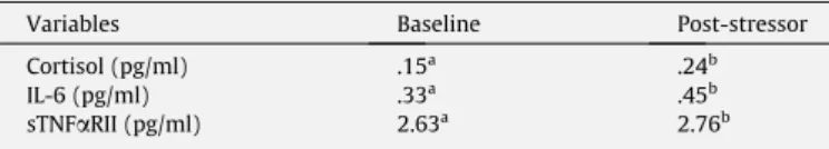
Table 1presents the mean values for baseline and post-stressor levels of cortisol, IL-6, and sTNF
a
RII. In all cases, baseline mean val-ues are significantly lower than post-stressor levels of cortisol, IL-6, and sTNFa
RII.3.1. Simple correlations
Table 2 presents the correlations among baseline measures. Baseline anger and fear were positively associated with baseline cortisol. Baseline fear was positively associated with IL-6, whereas baseline anger was only marginally positively associated with IL-6. A similar, but weaker, pattern emerged for sTNF
a
RII such that baseline fear was marginally positively associated with sTNFa
RII, whereas anger showed no association with sTNFa
RII. Baseline cor-tisol was not associated with baseline levels of IL-6 or sTNFa
RII.Table 3 presents the correlations among post-stressor mea-sures. Post-stressor anger, but not fear, was marginally associated with post-stressor cortisol. In contrast, post-stressor fear, but not anger, was associated with IL-6. Neither post-stressor anger nor post-stressor fear were associated with sTNF
a
RII. Post-stressor cortisol was negatively associated with post-stressor IL-6, but not associated with sTNFa
RII.3.2. Regression analyses
To examine the associations between emotions and cortisol in response to the stressor, a regression analysis predicting post-stressor cortisol was conducted that controlled for the audience manipulation, health questionnaire items (i.e., general health, caf-feine intake, alcohol consumption, and smoking), baseline emo-tions, and baseline cortisol levels.3 As displayed in Table 4 and Fig. 1, when post-stressor anger and post-stressor fear were entered as simultaneous predictors of post-stressor cortisol, anger was sig-nificantly positively associated with cortisol,p= .018, whereas fear was significantly negatively associated with cortisol,p= .042.4
To examine the hypothesized association between emotions and IL-6 and sTNF
a
RII, identical regression models that controlled for baseline IL-6 and sTNFa
RII, respectively, were conducted.5Asdisplayed inTable 4, fear was significantly positively associated with IL-6,p= .003, as expected, whereas anger was not associated with levels of IL-6, p= .93. A similar pattern emerged for sTNF
a
RII, although the positive relationship between fear and sTNFa
RII was marginally significant,p= .076; there was no association betweenanger and sTNF
a
RII,p= .68. None of the reported effects were mod-erated by participant gender.4. Discussion
We explored whether anger and fear are associated with differ-ent, theoretically adaptive patterns of biological responses to stressors. Greater anger in response to the stressor was associated with higher post-stressor cortisol levels, as predicted. Greater fear in response to the stressor was associated with lower post-stressor cortisol levels. As hypothesized, greater fear was associated with higher levels of the proinflammatory cytokine IL-6, whereas anger was not. Similarly, greater fear was marginally associated with higher levels of sTNF
a
RII, whereas anger was not. These findings are consistent with the idea that distinct emotions tailor stress re-sponses to an acute stressor.Post-stressor fear was negatively correlated with post-stressor cortisol levels. Because cortisol suppresses the production of proin-flammatory cytokines (Robles et al., 2005), a reduction in cortisol levels facilitates the proliferation of proinflammatory cytokines. Consistent with this point, the stressor cortisol and post-stressor IL-6 were negatively correlated. Because fear-driven reduction of cortisol would presumably enhance the biological and behavioral consequences of fear-driven IL-6 production, both the increase in IL-6 and the decrease in cortisol are consistent with the idea that fear coordinates a functional response to a fear-elic-iting stressor.
The negative correlation between fear and cortisol emerged be-cause the regression analysis controlled for anger. When anger was not controlled, fear was not correlated with cortisol. Controlling for the shared negative valence between the two emotions leaves only characteristics of fear that distinguish it from anger (e.g., uncer-tainty) to correlate with dependent variables like cortisol. Such an approach may reveal effects of distinct emotions that are attrib-utable to distinctive properties of emotions other than their nega-tive valence.
The current findings are correlational and preclude inferences about causation. However, the observed endocrine and immune ef-fects were most likely triggered by central nervous system pro-cesses, such as the rapid emotional responses to the stressor. Thus, we speculate that the specific appraisals unique to fear and anger precede the endocrine and immune system responses ob-served here.
The present study examined only two emotional responses to stressors. Sadness is a reasonable response to some stressors and may similarly promote withdrawal from the situation, perhaps mediated by changes in proinflammatory cytokines. Although sad-ness is an unlikely emotional response to the stressor in the cur-rent study, our findings suggest that the association between distinct emotions and biological processes might potentially be enlightening with regard to other emotional responses to stress.
Table 1
Baseline and post-stressor mean values of cortisol, IL-6, and sTNFaRII.
Variables Baseline Post-stressor Cortisol (pg/ml) .15a
.24b
IL-6 (pg/ml) .33a .45b
sTNFaRII (pg/ml) 2.63a 2.76b
Note:Values within rows not sharing similar superscripts are significantly different from each other atp< .05.
Table 2
Correlations among baseline anger, fear, cortisol, IL-6, and sTNFaRII. Baseline anger Baseline fear Baseline cortisol Baseline IL-6 Baseline sTNFaRII Baseline Anger – .326*** .166* .143 .066 Baseline Fear – .202** .195** .120 Baseline Cortisol – .073 .061 Baseline IL-6 – .548*** Baseline sTNFaRII – p< .10. * p< .05. ** p< .01. *** p< .001. 3
No results interacted with the audience manipulation. Two dummy coded variables were created to control for the audience manipulation in all regression analyses.
4
The correlations of stressor cortisol with stressor anger and post-stressor fear remained significant when baseline IL-6 and baseline sTNFaRII were included as covariates.
5
IL-6 and sTNFaRII data were log-transformed to normalize distribution of residuals. Three outliers were excluded for producing post-stressor IL-6 scores more than three standard deviations from the mean. The correlation of post-stressor IL-6 with post-stressor fear remained significant when baseline cortisol was included as a covariate.
These results may be revealing as to the time course and coor-dination of psychological and biological stress responses. Because the production of cortisol and proinflammatory cytokines is slow, relative, for example, to sympathetic activation, it is unlikely that these processes play a central role in the immediate fight or flight response to acute stressors. Other faster processes such as changes in cardiovascular functioning are more likely to mobilize the energy expended in immediate fight or flight responses. In-stead, emotion-driven increases in cortisol or proinflammatory cytokines may supplement, over time, the initial generalized reac-tion to stressors, in more specific, emoreac-tion-consistent, and adap-tive ways. For example, anger-driven increases in cortisol can supplement available energy for confrontation. In contrast, fear-driven increases in proinflammatory cytokines may motivate withdrawal when retreat may be advantageous without preclud-ing individuals from an initial flight response driven by cardiovas-cular activity. Thus, emotions may have relatively undifferentiated early fight or flight responses to acute stressors; however, over time more differentiated effects of distinct emotions may emerge and may shape endocrine and immune processes in functional manners. Stress has been found to initiate processes that lead to deferred benefits in other contexts (e.g., production and distribu-tion of leukocytes that prepare the body to mend potential future injury (Dhabhar and McEwen, 1999; Viswanathan et al., 2002); the present findings suggest a potential role for emotions in shap-ing delayed benefits.
Although energy is required for both the fight response linked to anger and the flight response linked to fear, based on the current findings, the motivation to withdraw from a fear-inducing threat may be more important and adaptive for people feeling afraid than the availability of additional energy through the release of cortisol. Because increased cortisol would inhibit increases in withdrawal-linked proinflammatory cytokine production, it may be that a fear-ful individual sacrifices the additional energy that would be made available by cortisol in exchange for increases in the motivation to withdraw from fear-inducing situations.
In summary, the present findings provide beginning support for a functional model of responses to stress in which biological pro-cesses are associated with specific emotional responses to
stress-ors. As such, emotions appear to be likely mechanisms by which stress responses are modified in adaptive ways. Anger, a confronta-tive emotion, demonstrated an association with increased HPA axis activity in response to the stressors, consistent with the idea that energy resources are needed following confrontative responses to stress. Fear, by contrast, was associated with enhanced proinflam-matory cytokine activity, especially IL-6, effects that are consistent with promoting withdrawal which can often follow states of heightened fear.
Table 3
Correlations among post-stressor anger, fear, cortisol, IL-6, and sTNFaRII.
Post-stressor anger Post-stressor fear Post-stressor cortisol Post-stressor IL-6 Post-stressor sTNFaRII Post-stressor Anger – .432*** .131 .096 .057 Post-stressor Fear – .010 .156* .086 Post-stressor Cortisol – .191* .099 Post-stressor IL-6 – .609*** Post-stressor sTNFaRII – p< .10. *p< .05. *** p< .001. Table 4
Post-stressor anger and fear predicting post-stressor cortisol, IL-6, and sTNFaRII. Predictor variables Post-stressor cortisolb Post-stressor IL-6b Post-stressor sTNFaRIIb Post-stressor anger .293* .007 .036 Post-stressor fear .253* .237** .159 p< .10. *p< .05. **p< .01.
Note:Analyses of post-stressor cortisol, IL-6, and sTNFaRII controlled for baseline levels of cortisol, IL-6, and sTNFaRII, respectively. All three regressions controlled for the audience manipulation, health questionnaire items, and baseline emotions.
Fig. 1.Post-stressor anger predicting post-stressor cortisol, IL-6, and sTNFaRII (top panel). Post-stressor fear predicting post-stressor cortisol, IL-6, and sTNFaRII (bottom panel). Analyses of post-stressor cortisol, IL-6, and sTNFaRII controlled for baseline levels of cortisol, IL-6, and sTNFaRII, respectively. All three regressions controlled for the audience manipulation, health questionnaire items, and baseline emotions.
Acknowledgments
Wesley G. Moons, Naomi I. Eisenberger, and Shelley E. Taylor, Department of Psychology, University of California, Los Angeles. This research was supported by a grant from the National Insti-tutes on Aging (AG030309). We thank Ted Robles for his helpful comments.
References
Adam, E.K., Hawkley, L.C., Kudielka, B.M., Cacioppo, J.T., 2006. Day-to-day dynamics of experience-cortisol associations in a population-based sample of older adults. Proc. Natl. Acad. Sci. USA 103, 17058–17063.
Blanchard, D.C., Blanchard, R.J., 1984. Affect and aggression: an animal model applied to human behavior. Adv. Stud. Aggress. 1, 1–62.
Dhabhar, F.S., McEwen, B.S., 1999. Enhancement of the immunization/sensitization phase of cell-mediated immunity: the role of acute stress and adrenal stress hormones. Neuroimmunomodulation 6, 213.
Dickerson, S.S., Kemeny, M.E., Aziz, N., Kim, K.H., Fahey, J.L., 2004. Immunological effects of induced shame and guilt. Psychosom. Med. 66, 124–131.
Diez-Ruiz, A., Tilz, G.P., Zangerle, R., Baier-Bitterlich, G., Wachter, H., Fuchs, D., 1995. Soluble receptors for tumor necrosis factor in clinical laboratory diagnosis. Eur. J. Haematol. 54, 1–8.
Frijda, N.H., 1986. The Emotions. Cambridge University Press, Cambridge, England. Harmon-Jones, E., Sigelman, J., 2001. State anger and prefrontal brain activity: evidence that insult-related relative left-prefrontal activation is associated with experienced anger and aggression. J. Pers. Soc. Psychol. 80, 797–803. Hart, B.L., 1988. Biological basis of the behavior of sick animals. Neurosci. Biobehav.
Rev. 12, 123–137.
Kemeny, M.E., 2007. Emotions and the immune system. In: Ader, R. (Ed.), Psychoneuroimmunology. Academic Press, New York, pp. 619–630.
Kemeny, M.E., Shestyuk, A., 2008. Emotions, the neuroendocrine and immune systems, and health. In: Lewis, M., Haviland-Jones, J.M., Barrett, L.F. (Eds.), Handbook of Emotions. Guilford Press, New York, pp. 661–675.
Kent, S., Bluthé, R.M., Kelley, K.W., Dantzer, R., 1992. Sickness behavior as a new target for drug development. Trends Pharmacol. Sci. 13, 24–28.
Kirschbaum, C., Pirke, K., Hellhammer, D.H., 1993. The ‘Trier Social Stress Test’ – a tool for investigating psychobiological stress responses in a laboratory setting. Neuropsychobiology 28, 76–81.
Lazarus, R.S., 1991. Emotion and Adaptation. Oxford University Press, New York. Lerner, J.S., Keltner, D., 2001. Fear, anger, and risk. J. Pers. Soc. Psychol. 81, 146–159. Mackie, D.M., Devos, T., Smith, E.R., 2000. Intergroup emotions: explaining offensive action tendencies in an intergroup context. J. Pers. Soc. Psychol. 79, 602–616. Maier, S.F., Watkins, L.R., 1998. Cytokine for psychologists: implications of
bidirectional immune-to-brain communication for understanding behavior, mood, and cognition. Psychol. Rev. 10, 83–107.
Nishanian, P., Aziz, N., Chung, J., Detels, R., Fahey, J.L., 1998. Oral fluids as an alternative to serum for measurement of markers of immune activation. Clin. Diagn. Lab. Immunol. 5, 507–512.
Robles, T.F., Glaser, R., Kiecolt-Glaser, J.K., 2005. A new look at chronic stress, depression, and immunity. Curr. Dir. Psychol. Sci. 14, 111–115.
Selye, H., 1956. The Stress of Life. McGraw-Hill, New York.
Smith, C.A., Ellsworth, P.C., 1985. Patterns of cognitive appraisal in emotion. J. Pers. Soc. Psychol. 48, 813–838.
Tooby, J., Cosmides, L., 1990. The pas explains the present: emotional adaptations and the structure of ancestral environments. Ethol. Sociobiol. 11, 375–424. Van Cauter, E., Leproult, R., Kupfer, D.J., 1996. Effects of gender and age on the levels
and circadian rhythmicity of plasma cortisol. J. Clin. Endocrinol. Metab. 81, 2468–2473.
Viswanathan, K., Bilbo, S.D., Dhabhar, F.S., 2002. Acute stress administered before primary immunization or before challenge enhances skin cell mediate immunity to keyhole limpet hemocyanin (KLH). Brain Behav. Immun. 16, 222. Weiner, H., 1992. Perturbing The Organism: The Biology of Stressful Experiences.