JOURNAL OF THE AMERICAN COLLEGE OF TOXICOLOGY Volume 3, Number 6, 1984
Mary Ann Liebert, Inc., Publishers
A
Survey
of
Metal-induced Mutagenicity
in Vitro and in Vivo
K. HANSEN' and R.M. STERN'
ABSTRACT
A survey of the literature shows that organic and inorganic compounds of
53
metals have
been assayed for genotoxic effects in vitro and in vivo. It is falund that there are great
variations in the response obtained with different test systems and that a wide range of
compounds of the different metals is positive in at least one of the short-term tests. Some
of the variation observed could be due to differences in uptake mechanisms. This effect
plus the wide variation in the quantity and quality of the data ]prevents any direct com-
parison of in vitro activity with in vivo potency of the various imetallic species.
INTRODUCTION
HE INTERNATIONAL literature has been surveyed in an attempt to correlate the results of genotox-
T
icity experiments performed in vitro and in vivo using organic and inorganic metal compounds, with known carcinogenic effects of metals in man. Compounds of53
metallic elements are reported to have been tested for genotoxic effects (Table l), but the number of experiments for which the in- dividual metals have been tested is found to vary widely, e.g., chromium has been assayed in more than300
individual experiments, whereas
beryllium has been tested in only24
cases.For
those 5 to10
metals with significant data bases, e.g., nickel and chromium, the prevalence of positive data agrees with the epidemiological evidence, although the wide range of in vivo and in vitro potency still prevents resolution of questions of compound specificity. For the many remaining metals with relatively little available data, it is impossible to determine if there is a preponderance of positiveor
negative results
or
to make comparisons between them. The main purpose of this survey is to review the present status of the short-term screening data base for metals and to identify neglected or con- troversial areas deserving of further study. It is also intended for use as a resource in the process of estimating potential human risk.SURVEY OF THE METALS
A large number of test systems have been used for the study of metals. These systems have been grouped into the following
16
categories:1 . Infidelity of DNA synthesis in vitro
2. DNA damage in prokaryotes 3 . DNA repair in prokaryotes 4. Ames Salmonella-microsome test
'Danish National Institute of Occupational Health, 2900 Hellerup, Denmark, *The Danish Welding Institute, 2600 Glostrup, Denmark.
w CQ h,
Lanthanides1
;;
I I
I 1
f'
Actinides
90*
Th
Pa
Np
Pu
la
2a
36
4b
5b
6b
7b
8
Ib
2b
3a
4a
5a6a
7a
0
1
2
H
He
3*
4*
56
7
8
9
10
Li
Be
BCNOFNe
11*
12*
13*
14*
IS
16
17
18
Na
MgA1
Si
P
S
C1
Ar
-
z
19*20*
21
22*
23*
24*
25*
26*
27*
28*
29*
30*
31*
32
33*
34*
35
36
?i
3
3
87
88
89++
104
105
106
2
K
Ca
Sc
Ti
V
Cr
Mn
Fe
Co
Ni
Cu
Zn
Ga
Ge
As
Se
Br
Kr
%
z
37*
38*
39*
40*
41*
42*
43
44*
45*
46*
47*
48*
49*
SO*
51*
52*
53
54
Rb
Sr
Y
Zr
Nb
Mo
Tc
Ru
Rh
Pd
Ag
Cd
In
Sn
Sb
Te
I
Xe
55*56*
57*'
72
73*
74*
75
76*
77*
78*
79*
80*
81*
82*
83*
84
85
86
Cs
Ba
La
Hf
Ta
W
Re
0sIr
Pt
Au
Hg
T1
Pb
Bi
Po
At
Rn
Fr
Ra
Ac
*At least one compound of the metal has been tested.63*
Eu
95
Am
64
65*
66
67
68*
69
70
71Gd
Tb
Dy
Ho
Er
Tm
Yb
Lu
96
97
98
99
100101
102
103
Cm
Bk
Cf
Es
Fm
Md
No
Lr
METAL-INDUCED MUTAGENICITY
5.
Mutation tests using
Escherichia coli
6.
Eukaryotic microorganisms
7 .
DNA damage and inhibition of DNA synthesis in mammalian cells
8.
DNA repair tests in mammalian cells
9.
Chromosome aberrations (CA) in mammalian cells in vitro
10.
Sister chromatid exchanges (SCE) in mammalian cells in vitro
1 1.Mutation in mammalian cells in vitro
12.
Mammalian celI transformation
13.Drosophila
14.
In vivo mammalian mutations and cytogenetics
15.
Higher plants
16.Others
The principles of the systems and some examples of the type of cells and methods used in these
categories are briefly described in Appendix
1 . The metals and metalloids that have been tested arelisted alphabetically according to their English names in separate sections for each of the elements in
Appendix
2.The literature survey has disclosed that a wide range of salts and other compounds has been
studied, including organic and inorganic compounds of metal ions in various oxidation states. Usu-
ally the chemical formulas have been given, and these are used to identify the compounds listed. In
some cases, especially for complex substances, the chemical formulas have not been disclosed or
defined, and the substances assayed are listed by name only.
Inorganic compounds are listed in subgroups in the order of the oxidation state of the appropriate
metal ion, followed by groups of inorganic salts, and finally complex compounds. The results from
mutagenicity testing of some nonmetallic inorganic compounds are included as well.
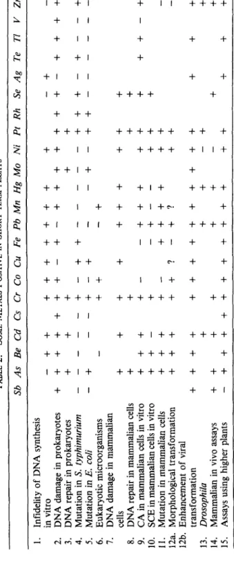
Dividing the many test systems into the 16 groups permits a rapid comparison of the extent of the
database for both metallic ions and individual compounds, a limited overview of which can be seen
in Table
2,for those metals for which there might be interest in ascertaining human risk. A positive
sign indicates at least one positive result regardless of the number
of negatives, and a negative sign
indicates the absence
ofpositive results.
In order to increase the utility
ofthis survey, an attempt has been made to evaluate the data in
each of the original articles. Positive results are classified into three categories according to the
degree to which the information is quantitative:
(1)data that merely indicate positive response at
some dose level, (2) data that permit a comparison of the positive response with a negative control,
(3)
data that indicate a dose-response relationship. Negative test results regardless of quality are
separately indicated. Some results are felt to be equivocal and are so indicated. No attempt has been
made to evaluate the laboratory practice used in the test procedures
aisreported.
In the absence of comments to the contrary, the original papers have been referred to in preparing
the listing, and review articles have been used only as a source of additional references. Limited use
has been made of the available data bases such as BLAISE, TOX-LINE, and the abstracting jour-
nals. The literature has been examined through to the end of
1982.
In
order to faciliate identification of references, entries in Appendix
2consist of a number that
corresponds to an entry in the Reference list.
DISCUSSION
An impression of the current state of genotoxic screening of the metals can be obtained from ex-
amination of Tables 1 and
2.Table
1 shows that inorganic complexes of a relatively large fraction ofthe common elements up to atomic number 83 in the Periodic Table have been examined in at least
one assay. From Table
2it can be seen that only a relatively few elements have been examined in a
significant number
ofthe 16 groups of test systems. It cannot be determined however, to what extent
the metallic compounds have been investigated for their specific activity without detailed examina-
tions of Table
3(Appendix 2).
TABLE
2.
SOME
METALS
POSITIVE
INSHORT-TERM
ASSAYS
Sb
As
Be
Cd
Cs
Cr
Co
Cu
Fe
Pb
Mn
Hg
Mo
Ni
Pt
Rh
Se
Ag
Te
TI
VZn
w 00 P ~~1.
Infidelity
of
DNA
synthesis
-++
+++++++
+
-+
-
2.
DNA
damage
in
prokaryotes
+
+
+
+
+
+
+
-+
-
+
+
+
+
+
+
+
-
+
+
+
+
in
vitro
X
3.
DNA
repair
in
prokaryotes
+
+
+
+
+
4.
Mutation
in
S.
typhimurium
- --
--
+-++----
-++
+-+--+
9
-
-+
-++
---
2:
!2
5.Mutation
in
E.
coli
6.
Eukaryotic
microorganisms
-
++
-+
7.
DNA
damage
in
mammalian
8.
DNA
repair
in
mammalian
cells
+
+
+
+
U9.
CA
in
mammalian
cells
in
vitro
+++
+-
-+++
++
+
++-+
10.
+-
++++
++
-
11.
Mutation
in
mammalian
cells
+++
12b.
Enhancement
of
viral
+-+
--
-+
z
+
$
++
+
3
z
cells
+
+
+
+
+++
++
++-
+
-+--
SCE
in
mammalian
cells
in
vitro
12a.
Morphological
transformation
+++
++?
-+?
++
-
transformation
++++
++++++++++
+
+
+
13.
Drosophila
+
+
+
-+
+
14.Mammalian
in
vivo
assays
++
+
+
+
+
+
+
15.
Assays
using
higher
plants
-+++++++++++
++
3+
+
-METAL-INDUCED MUTAGENICITY
Since a few of the metals (chromium, nickel, arsenic, cadmium, and beryllium) are strongly
suspected human carcinogens based on standard criteria, it might be expected that they have re-
ceived a significant amount of attention. This is, in fact, true with the notable exception
of
beryllium, which for unknown reasons has been little studied. For chromium, nickel, and arsenic,
the preponderance of positive test results as found in many systems is as imight be expected if there is
a connection between mutagenicity, genotoxicity, and carcinogenicity. 'The question of the use of in
vitro studies to predict in vivo risk cannot be resolved for cadmium because of a mixture of positive
and negative results across compounds and systems for this metallic ion.
For some of the metals, e.g., chromium, selenium, and arsenic, the geiiotoxic potency depends on
the oxidation state of the metal ion. Inorganic Cr
111is inactive in test systems using intact cells,
whereas Cr VI is positive in
all
these assays. Compounds of trivalent arsenic are more active than the
pentavalent compounds in chromosome aberrations tests, and Se(1V) compounds seem to be more
potent than the Se(V1) compounds. The results obtained with selenium are especially difficult to in-
terpret, as some of them show antigenotoxic effects whereby selenium diminishes the mutagenicity
induced by other agents. Similar conflicting results are seen in animal cancer studies.
Some of the Cr
111complexes that are carrying stable aromatic amines as ligands show a
mutagenic effect in prokaryotes. Others, such as the complexes of platinum, show a mutagenic re-
sponse depending on the configuration of the complex, e.g., cisplatin tliaminedichloride is a more
potent mutagen than the transconfiguration.
Very few attempts have been made to elucidate the question of synergistic or antagonistic effects
of two or more metals in short-term bioassay. It has been shown that manganese dust reduces cell
transformation induced by nickel subsulfide, that arsenite and
UV
light act synergistically in
mutagenicity and DNA repair assays, and that NiSO, and benzo(a)pyrene show a synergistic effect
in a cell transformation assay using primary Syrian hamster embryo cells.
Some of the variation observed in the results
of the bioassays of different metal complexes could
be due to differences in bioavailability (i.e., uptake mechanisms) rather than reflecting (molar)
genotoxic potency. The extent to which a compound at a given dose is taken up by the biological
system and is able to reach the target molecules depends, among other things, on the nature of the
uptake mechanism and on the (intracellular) reactivity of the compound in the actual surroundings,
e.g., the cell cytoplasm. Insoluble particles are incorporated into cells by different mechanisms than
are the soluble salts, whereas the complex organic compounds may reach the target organelles by yet
other pathways. Test systems using prokaryotes, yeast cells, mammalian cells or entire animals will
each exhibit a wide variation in the extent and rate at which they pick up those various metal com-
pounds that demand different uptake mechanisms.
Thus it may be misleading to compare results of different compounds administered at the same
dose in a given system, or the same compound as administered in different systems. Although the
data base is seen to still be very incomplete, the above arguments indicate that when attempting to
complete the survey
of metals, care should be taken in choosing both in vitro and in vivo short-term
test systems that are relevant to the materials used.
Short-term bioassay might be used to determine the presence or extent of risk of human exposure
to
the metals and their inorganic and organic complexes. Based on examination of the available
data, the tentative conclusion reached is that a summing up of positive and negative test results ob-
tained for each of the metallic compounds cannot be used to predict the potential human risk in-
volved with, for example, occupational exposure to metals. What can be done is to identify which
particular substances, e.g., found in specific occupational exposures, behave in vitro in ways that are
consistent with those for other, similar substances for which there exists a knowledge of eventual
human risk, thereby permitting a transfer of risk assessments from one substance to another or from
exposures in one industry to another.
An effort should be made to extend the use of the data base for the assessment of human risk. It is
undoubtably worthwhile to fill in certain gaps through the systematic use of additional test systems
for specific compounds and additional compounds with specific systems so as to develop further
understanding
ofthe mechanisms controlling biological activity. Without this understanding, it may
in many cases be necessary to treat each metallic complex as a separate entity, since there is such a
wide range of the physical and chemical properties of otherwise related compounds (such as the
ox-
ides). The short-term bioassay data base of inorganic substances does not provide the same tools for
risk estimation as are available for organic chemicals.
APPENDIX 1: TEST SYSTEMS
The test systems have been divided into 16 groups consisting of in vivo and in vitro assays using
prokaryotes and eukaryotes and with different genetic endpoints.
A brief description of each ofthe
groups is presented, with references on the principles and methods of the test systems. Most of the
references are found in a few well-known books and in the reports
of the US EPA Gene Tox Pro-
gram as listed in the Bibliogrophy. However, for those of the test systems not described therein, the
reader is referred to the actual papers used in the metals survey. Apart from the references cited
below a description and an evaluation
of screening assays can be found in IARC, 1980.
Group I . Infidelity
of
DNA Synthesis
In
Vitro
The test systems are based on cell-free DNA synthesis in which the incorporation of noncomple-
ment nucleotides is measured by radioactive labeling. In one test system an isolated viral DNA
polymerase incorporates radioactive-labeled nucleotides in a synthetic DNA template
of restricted
base composition. The test substances are added together with the labeled nucleotides, and the fre-
quency
oferrors is measured.(’0) In another procedure, the reversion frequency from mutant to
wild-type viral DNA is measured.(56)
Group 2. DNA Damage in Prokaryotes
1. Differential killing of
Bacillus subfilis
(rec-assay)
2.
Differential killing of
Escherichia
coli
(Pol A-/Pol
A’)A nonspecific DNA damaging effect in prokaryotic cells is detected by differential killing. The
prokaryotes deficient in DNA repair capacity are more sensitive to DNA-damaging agents than are
the wild-type prokaryotes, (De Serres and Ashby, 1981, Chap. 12, 13, and
14;Leifer et al. 1981,
Gene Tox Program.)
Group 3. DNA Repair in Prokaryotes
I .Inhibition of excision repair in
E. coli
2.Inhibition of SOS repair in
E. coli
Inhibition of the error-free excision repair of pyrimidine dimers after radiation exposure results in
an enhancement of the mutation frequency of
E. coli.
( 4 6 )Inhibition of the postreplication repair
pathway, the error-prone
SOS
repair causing a decrease in, e.g., UV-induced mutations in certain
E.
coli
strains (lacking excision repair capacity).‘”)
Group 4. Ames Salmonella-Microsome Test
In the Ames Salmonella-microsome assay the number
ofreversed mutations in
Salmonella
typhimurium
from histidine requirement to histidine independence expresses the mutagenic activity
of the test compound. The
Salmonella
strains most commonly used are TA 1535, TA 1537, TA 1538,
TA 98, and TA 100 in different test procedures with and without metabolic activation (S-9 mix), see
group 5 (Brusick, 1981, Chap. 9, prot. 1; Stich and San, 1981, Chap. 11, 12).
Group
5.
Mutation Tests Using E. coli
E. coli
is used for detection of mutagenic activity in the same way as
S.
fyphimurium.
Different
strains of
E.
coli
detect forward as well as reversed mutations. Three essentially different test pro-
METAL-INDUCED MUTAGENICITY
cedures are used- the qualitative spot test, in which the test compound iis placed in the middle of the
surface of the agar, the quantitative plate incorporation assay in which the test compound is mixed
with the bacteria in the agar, and the sensitive fluctuation test in whichi the entire procedure is per-
formed in liquid medium. (De Serres and Ashby,
1981,
Chap.
36, 37;
Brusick,
1980,
Gene Tox Pro-
gram).
Group 6. Eukaryotic Mircoorganisms
1 .
Saccharomyces cerevisiae:
mutation, gene conversion, and mitotic crossing over, inhibition of
2.
Schizosaccharomyces pompe:
forward mutation and gene conversion
S.
cerevisiae
is
the most frequently used eukaryotic microorganism. Point mutations as well as
chromosome aberrations can be detected (Brusick,
1980,
Chap.
9,
prot.
2,
3,
4; De Serres andAshby,
1981,
Chap.
39;
Stich and San,
1981,
Chap.
15).
spontaneous mutation
Group 7. DNA Damage and Inhibition
of
DNA Synthesis in Mammalian Cells
1 .
Inhibition of DNA synthesis in BHK cells, human lymphocytes, CHO cells, and HeLa cells
2.
DNA-protein crosslinks, DNA-DNA crosslinks in
V-79
cells and in CHO cells
3. DNA strand breaks- alkaline elution and sucrose gradient in CHO cells and human fibroblasts
Several test systems detect a nonspecific DNA-damaging effect on mammalian cells. DNA strand
breaks are detected by an alkaline elution technique
or by differential centrifugation (sucrose gra-
dient). Detection of DNA-DNA crosslinks and DNA-protein crosslinks can also be obtained by
alkaline elution. Inhibition of DNA synthesis in mammalian cells is detected by measureing the up-
take rate of labeled thymidine. (Stich and San,
1981,
Chap. 5,
6, 9).
Group 8. DNA Repair Tests in Mammalian Cells
1.
Unscheduled DNA synthesis in mouse cells, human fibroblasts, human lymphocytes, and
2.
Interruption of dark repair mechanism in human lymphocytes
The effect of the test compound on DNA repair mechanisms in manimalian cells is investigated by
measuring unscheduled DNA synthesis (UDS) or DNA repair synthesis. The uptake of radioactive-
labeled thymidine in cells not in the S period indicates that one or more of the repair systems are
working. (Stich and San,
1981,
Chap. 7; Brusick,
1980,
Chap.
9,
prot.
9, 10;
Larsen,
1982,
Gene Tox
Program).
CHO cells
Group 9. Chromosome Aberrations in Mammalian Cells In Ktro
1 .
Chromosome aberrations in C3H mouse cells, hamster embryo cells, CHO cells, human lym-
phocytes, human fibroblasts,
V-79
cells, bone marrow cells from mice, and Balb//c 3T3 cells
A number of cell cultures including primary cells, established cell lines, and human cells have been
used. The genotoxic endpoints under consideration are the morphological stable rearrangements or
exchanges of the chromosomes as well as the unstable breaks and gaps.
Group
10,
SCE
in Mammalian Cells in Vitro
1 .
CHO cells, human lymphocytes, human fibroblasts, and xeroderma pigmentosum cells
Sister chromatid exchanges (SCE) are a result
ofgenetic damage at the chromosomal level. When
DNAlesions are replicated during the S period, exchanges between
t w odaughter molecules often
occur. On the cellular level this is seen as an exchange between two sister chromatids
in the meta-
phase (Stich and San,
1981,
Chap. 20; De Serres and Ashby,
1981,
Chap.
49;
Latt et al.
1981,
Gene
Tox Program).
Group
11.
Mutation
in
Mammalian Cells
In
Vitro
1. V-79, CHO, C3H cells in the HGPRTI8azag mutation assay
2. CHO/ouabain-resistant mutations
3.
L5178Y/TK+’- mutation assay
Point mutation tests in mammalian cells are predominantly done on cell lines V-79, CHO,
L5178Y. The L5178Y system detects the frequency
of forward mutation in the thymidine kinase
locus from TK-’+ to TK+. In the HGPRT system the mutant cells show a resistance towards
8-azaguanine and dthioguanine that is caused by a blocked or altered HGPRT metabolic pathway.
In much the same way, the Na/KATPase pathway can be altered by mutations and the cells become
resistant toward ouabain (Bradley et al., 1981, Gene Tox Program; De Serres and Ashby, 1981,
Chap. 53,
5 5 ; Hsie et al., 1981, Gene Tox Program).Group 12. Mammalian Cell Transformation
1. SHE, CHO, BHK-21, and Balb/C-3T3 cells
2. Enhancement of virus-induced transformation: HEC cells
+
SA 7-virus, human diploid cells
Different established cell lines and primary hamster embryo cells have been used to detect the
transforming effect of metal compounds. The criterion for transformation is morphological changes
of cell colonies, such as piling-up and a crisscross growth pattern. Measuring the enhancement of
virus-induced transformation is also used as a transformation assay (Stich and San, 1981, Chap. 29,
31; De Serres and Ashby, 1981, Chap. 58, 59; Brusick et al., 1980, Chap. 9, prot. 11).
+
leukosis virus
Group 13. Drosophila
1. Sex-linked recessive lethals
2. Dominant lethals
The X-chromosomal recessive lethals include forward mutations, deletions, and structural rear-
rangements, and approximately 20%
of
the entire genome is covered by this recessive lethals tests. A
dominant lethal mutation in a gamete is considered to be the result of chromosome breaks, but also
nondisjunction can lead to dominant lethality (Brusick, 1980, Chap. 9, prot. 21; Stich and San,
1981, Chap. 34; De Serres and Ashby, 1981, Chap. 61).
Group 14.
In
Vivo Mammalian Mutations and Cytogenetics
1. Chromosome aberrations in bone marrow cells of rat, hamster, and mouse, spermatocytes in
mouse, and oocytes in mouse
2.
Dominant lethals in mouse and rat
3.
SCE in bone marrow cells from mouse and hamster
4. Mouse spot test
5.
Sperm abnormality in mouse
Mammalian tests in vivo are a mixed group of asssays using mainly mice, rats, and hamsters. The
animals exposed to metal salts are examined for chromosome aberrations and SCEs in somatic as
well as germ cells, dominant lethals (germ cell damage), and spots in mice (mutations in genes for fur
color). In the micronucleus test, chromosome breaks
in
bone marrow cells in treated animals can be
counted, since after cell division the daughter cells contain the displaced fragments of chromosomes
as a micronucleus in the cytoplasm (Brusick, 1980, Chap. 9, prot. 14, 16, 17, 18; De Serres and
Ashby, 1981, Chap.
64,
66,
67, 69; Stich and San, 1981, Chap. 21).
Group
15.
Higher Plants
1. Mutations and chromosome breaks in
Pisum sativa.
2. Abnormal divisions and chromosome aberrations in
Vicia
faba
METAL-INDUCED MUTAGENICITY
3. Spindle disturbances and colchicine mitosis in
Allium cepa
4. Mutations in
Crepis capillaris
Root tip cells from
Vicia faba
(broad bean),
Allium cepa
(common, onion),
Pisum sativa
(pea),
Crepis capi/laris
(Hawk's beard) have been exposed to metal salts and examined for induced muta-
tions, chromosome aberrations and abnormal cell divisions (Stich and San, 1981, Chap. 18; Con-
stantin, 1982, Gene Tox Program).
Group 16. Others
1.
Abnormal mitosis in muscle cells(6)
2. Abnormal chick m y o b l a ~ t s ( ~ ~ )
3. Inhibition of mutation in
Bacillus subtilisiS1)
4.Mutation in
Micrococcus a ~ r e u s ' ~ ~ )
5.
Stimulating of initiating of RNA synthesis on conditions where overall RNA synthesis is
6.
Inhibition of mitochondria1 protein synthesis('*)
7.Mutation in algae(Ioo
17')8.
Mutation in bacteriophage T4(125.170
lS6)9. Effect on DNA in
B. subtilis(161)
inhibited(67)
10. Mitotic spindle disturbances in mouse fibroblasts(16')
1
1. Host-mediated assay:
mouse-Salmonella typhimurium, mouse-Serratia marcescens'
196)In this mixed group of assays, bacterial systems other than those previously mentioned are found.
well as host-mediated muta-
Assays measuring the inhibition
ofDNA, RNA, or protein synthesis
tion tests using mice and microorganisms are included (Brusick, 1980, Chap. 9, prot. 12).
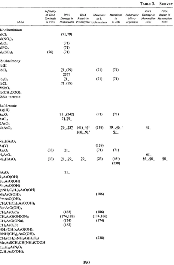
APPENDIX
2:
SURVEY
OF THE METALS
This is a complete listing of the short-term bioassay of metals, where the test systems are grouped
in the
16categories described in Appendix 1. Each test result (a chemical substance in a specific
mutagenicity assay) is represented by a number that refers to an entry in REFERENCES. Negative
test results are indicated by brackets around the entry number. Entry numbers without brackets are
used to indicate a positive result as reported by the author: Such results of a qualitative nature are
marked with a single underlining, whereas published quantitative data are indicated with two
underlinings. In those cases where it is felt that the data establish a dose-response
(or
dose-effect)
relationship, the citation is marked with three underlinings.
(To avoid confusion, single-digit cita-
tions appear as 5, -x-,
-x-,
two digits citations appear as
g ,
xx-,
x&-,
and three digit citations as
q,
gx,
xxx).
A question mark indicates that the published result is thought to be equivocal. Addi-
tional comments on test results have been placed in the right column of the tables. No attempt has
been made to evaluate the test procedures as reported.
List of Abbreviations Used in Table
3
8azag
Balb/c 3Tc cells
BHK-21 cells
CAC3H/lOT 1/2 cells
CHO cells
E. coli
HEC
orSHE
HGPRT
L5178Y
cells
S .typhimurium
SCE
V-79 cells
8-azaguanine resistance
Mouse fetal cell line
Baby hamster kidney cell line
Chromosome aberrations
Mouse embryo cell line
Chinese hamster ovary cell line
Escherichia coli
Primary Syrian hamster embryo cells
Hypoxanthine-guanine phosphoribtosyl transferase
Mouse lymphoma cell line
Salmonella typhimurium
Sister chromatid exchange
Chinese hamster lung cell line
TABLE
3. SURVEY
Infideliiy D N A D N A
o f D N A D N A D N A Mutaiions Muralions Eukaryolic Damage in Repnir in
Synthesis Damage in Repnir in in S. in Micro- Mnmmnlinn Mammolian
M e l d in Viiro Prokaryoies Proknryoles typhimurium E. coli organisms Cells Cells
Sb/A ntimony SbIII SbCL SbiO, SbCI, WSb04 Sb(CHSO0)i SbNa tartrate A s / A rsenic As(II1) ASiO3 AsCL KAsOi NaAs02 3AsOs R,AsO(OH) BulAsO(OH) Ph,AsO(OH) M ~ A S O ( O H ) ~ Pr"AsO(OH), (pNH2CsH,)iASO(OH) CHICHCHzAsO(OH)~ Bu"AsO(OH)~ CH,AsO,Ca CH,AsO(OH)ONa CH,AsO(ONa), CH,AsO,Fe NHl(CH2)3AsO(OH)~ RNH(CHi),ASO(OH)i CH,(CHi),NH&(H,OJ MelAsSCH2CH(NH2)COOH C I ~ H ~ ~ A S N ~ O S C,H,AsO(OH)2
3 90
METAL-INDUCED MUTAGENICITY
OF THE
METALS
SCE in Mutolions M u / . ond
C A in Mummulion in Cell C A in
Mammoliun Cells Mummulion Truns- Mommuliun Higher
Cells in Vilro Cells formation Drosophila in Vivo Plunis Orher Comments
147' (1 76)
'
(183)184
169, 126
Dominant lethal46
Co-mutagen with91
UV-light41, 189,
6Q
Mutation in 169' hQ-(4l) mammalian cells 17242
Significantly higheractivity with trivalent compounds than with pentavalent compounds _-- 147 Review
__- 167 Spindle disturbances
(l89),& in mouse fibroblasts
_-- 174 Eukaryotic micro- R = methyl or ethyl organisms 167' Bu = butyl (1617) Ph = phenyl Pr' = propyl (174)' (174)' 167 147' 167'
391
TABLE
3
~ ~~Infideliry D N A D N A
o f D N A D N A D N A Mutations Mutarions Eukaryotrc Damage in Repair in
Synthesis Damage in Repair in in S . in Micro- Mammalian Mammaliar,
Metal in Virro Prokaryores Prokaryotes typhirnurium E. coll organisms Cells Cells
AdArsenic OOCsHjCHzAsO(OH)2 CsH5(CHz)iASO(OWz C I C ~ H ~ A S O ~ NOzCsHaASO, CH,CsH4As03 Acetylarsan Arsenic dimethyl thiocarbamate CH,CICc,H,ASO, Ba/Barium Ba" BaCI, Ba(N0A Ba(CH3C00)2 Be/Beryllium BeCI2 Bi/Bismuth BiC1, Bi(NO,),? 4BiN03(OH), Bi,O, Ca/Calcium Ca" CaCI, Ce/Cerium CeC1, Ce(N0A CdCesium cs2co3 CSCl CsNO, cs2so4 CSCI, CS(NO~), CS,(SO4)3 CHCsO, 182 (182) (182) (79) (79)
392
METAL-INDUCED MUTAGENICITY
(CONT.)
~~ ~
SCE in Muralions M u r . and
C A in Mammalian in Cell C A in
Mammalran Cells Mammalian Trans- Mammalian Higher
Cells in Vilro Cells formalion Drosophila in Vrvo Planrs Olhfv- Cornnienrs
147l 147' 147l 147'
___
17982-
302 (183)184 168 Review212
Enhancement of _ _ _ EMS-induced CA in183,184 barley root tips
2 ! l
(1 83) 184 (302,297) (57)183 183,184393
TABLE
3.
Infidelify DNA D N A
of D N A D N A D N A Mutations Muralions Eukaryotic Damage in Repair in
Synthesis Damage in Repair in in S. in Micro- Mammalian Mammalian
Metal in Vitro Prokaryotes Prokaryotes fyphimurium E. coli organisms Cells Cells
Cd/Cadmium Cd Cd'* CdC1, CdS CdSO, Cd(CH3COO)z Cd-serum 118,280
3
94
METAL-INDUCED MUTAGENICITY
(CONT.)SCE in Murotions Mur. ond
CA in Mommolion in Cell CA in
Mommolion Cells Mommolion Trons- Mommolion Higher
Cells in Vilro Cells formorion Drosophila in Vivo Plonls Ollier Comments
(277) (290) (81,751 72?(158) 80- 107,202 (47'34) 302 (1 171106
189
(iiG,121) (472,108) (158) (120,173) 118' (2 1 9,224) 104,105299,116
(283)103S3
183,144 185,231 194 122,264395
211i
266 63-,'78 6 7 A 7 1217,259
268.,2;98
276,295 ( 1 !!ti) ( 1:iii) 4.328
Inhibition of mito- chondrial biogenesis in yeast14, 104, 108
CA in mice lQj,116
CA in oocytes--
118
Enhancement of chr. breaks in virus infected cells-__
171 Mut. in algae217
Enhancement of EMS induced CA in barley roots120,
173,
219,
224
Domi-nant lethals in mice
277
Dominant lethals in rats259
CA in grasshopper266
Inhibition of RNA polymerase in vivo in rats268
Stimulation of DNA synthesis273
Decrease in repair capacity of pancreas cells---
149 Promoting effect on BP induced transfor- mation299
Inhibition of DNA synthesis in rats298
SCE, pos. in human lymphocytes; neg. inV-79 cells
67
Enhances chain initia- tion of RNA when overall RNA synthesis is inhibited63
Mispairing of poly- nucleotides in vitro292
Inhibition of human DNA polymerase284
Inhibition of RNA synthesis in Physarum polycephalurn (slime mold)276,
295 Stimulation ofRNA synthesis in CHO cells chick myoblasts in mice
45
Abnormal growth in gl',283 Abnormal sperm
47' Micronucleus15.5.
SCE in hamster cellsTABLE
3.
~ ~ ~~
Infidelily D N A D N A
o f D N A D N A D N A Mulalions Mutations Eukaryolic Damage in Repair in
Synfhesis Damage in Repair in i n S. in M i c r e Mammalian Mammalian
Metal in Vitro Prokaryotes Prokaryoles typhimurium E coli organisms Cells Cells
CrKhromium CrCL 26- Cr(II1) 21.. CrC1,
32-
(20,791 (1 53,242 ) C r ( N 0 J 3 C r 2 0 3 CrK(S0,)- 12H20 Cr(OH)SO, NazS04 Cr1(S0& Cr2(S04)3 K2S04 Cr(CH3C00)3 Cr(II1) glycine Cr-serum (NH,)CrO, (NH,),Cr20, CaCrO, C1,Cr02 CrO, PbCrO, PbCrO,.PbO chrome orange PbCrO,. PbMoO, PbCrOn-PbS04 chrome yellow K1CrO4 KICrlO, (71,153) (71) 20-153
188(206)' (15) (1 5 4 ) 2 1 ~ (215)2 (206)' (206)l (206)'
396
METAL-INDUCED MUTAGENICITY
(CONT.)SCE in Mutations M u t . and
C A in Mammalian in Cell C A in
Mammalian Cells Mammalian Trans- Mammalian Higher
Cells in Vilro Cells formation Drosophila in Vivo Plants
15L25-
(25,301 15-,2? (20.26) (31,37) (149) (30,37) (150,206) (206,227) (239,291) 291,278 155 (20,30) (30)171' (38) 183,184 -32' 185(217)206
206
(30127- (30,371 (206) (206) (206,158) (155) (206) (206) 20-,37- (30.37) (36,175) 2 4 233? (75,26) (206) (158) 27- 27-206
206 206 --_ 181 (36) 206 206 206 ___ 80 149' 15-926- (38)' 78,(170) 26 1 Other Comments-_
154,
188,
206'
Inhibition of DNA synthesis296
Stimulation of RNA synthesis in rats in vivo215'
Protein- DNAcrosslinking in nuclei
215'
Protein-DNAcrosslinking in human cells
12
Cr-nitrate from Merck was negative but from riedel De Haen positive44'
Contaminated with Cr(V1)45
Abnormal growth in muscle fibers29'
DNA breaks (296) 281 Modification of U . V . spectra of DNA in BHK cellsgfi-.
18
Inhibition of mito- chondrial biogenesis in yeast 281--_
168 Review--_
170 Mut. in 'r4 bacteri- ophage--
17' Pos. only in fluctua- tion test22'
29' DNA breaks44'
Pos. when dissolv 0,5N NaOH
24
188
206' Inhibition of DNA synthesis28
58
Micronucleus12
Spot test in mice 215' Prot. -DNA crosslinking in mouse nuclei 215' Prot.-DNA cross
linking in human cells
261
Binding to nucleic acids--_ 149 Promoting effect on BP induced transfor- mation
--_ 180 DNA cross links in rat liver and kidney _- 13 C.A. in rat bone
marrow cells
TABLE
3.
I n fidelity D N A D N A
of D N A D N A D N A Mututiom Mutations Eukuryotic Damage in Repair in
Synthesis Dumuge in Repair in in S . in Micro- Mammalion Mammalian
Melal in Vilro Prokaryotes Prokaryotes typhimurium E. coli orgunisms Cells Cells
Cr/Chromium ZnCrO, ZnCrO,. Zn(OH), 44-722- 206 Cro(C0)6 (44) Cr(H2O)sCl, (190) K,(Cr(CN)e.) (190) (Cr(NH,)4H20CI)C12 (190) (Cr(NH,)40x)N03* R O (190) (Cr(NH3),Cl1)CI (190) (Cr(en),)Cl, -3H20
1%
( 190) (Cr(pn),)CI3*3H,O1%
( 190) cis-(Cr(bipy),Ox)I *4H101%
19Q cis-(Cr(bipy),Cl,)CI*2H2OL9n
19q ~is-(Cr(phen)~Cl,)C1*2.5H,O19n
1%
(Cr(urea),)C1,*3H20 190 190 (Cr(NH,),CI)CL (i90) (Cr(NHJ5H2O)CI3 (190) tran~-(Cr(en),(sCN)~)SCN 19Q ( 190) (Cr(en),) (SCN),190
(190) (Cr(enMC1 (237) (CrOx(NH,), C1 (237) cis-Cr(pyr),F2)Br (237) zink yellow K,(Cr(Ox),)* 3H20 ( 190) ci~-(Cr(en)~CI,)Cl* HzO (190) coso4cos
amorph. CoS COMOO4 CO(OH), Co(CH,COO)2 (Co(NHJs)CI, Co-serum (Co(Pn),)Cl,+
(Co(En),)I, - (Co(En),)I, D-cis(Co(En),(NO,),)Br ~-cis(Co(En)~(NO,),)Br Other Co(II1) hexaco-ordinate complexes 232 (237) 232 232 232 232
504'
(304) Cu/Copper CUCl cu 2 s398
METAL-INDUCED MUTAGENICITY
(CONT.)--
SCE in Mulalions Mul. and
CA in Mammalian in Cell C A in
Mammalian Cells Mammalian Trans- Mammalian Higher
Cells in Vitro Cells formation Drosophila in Vivo Planrs Ot,ker Comments
Ox = oxalate en = ethylenediamine pyr = pyridine bipy = bipyridine phen = 1.10-phenan- throline pn = propylenediamine
42
Enhancement of RNA 45 Abnormal growth in Pf-(217) 67-(292) initiation--_
51 (296) chick myoblasts 74.37- (1 70)51
Inhibition of spon- 183,185 taneous mutation in 184,232 b. subtilis24-
-- 17Q Mut. inT4
bacteri- ophage220
Antimutagenic action on TA 98 and TA 1538292
Inhibition of human DNA polymerase 4.6296
Stimulation of RNAsynthesis in rats in vivo 204' Complexes of Co(II1) show behavior identical to similar complexes of Cr(II1) and Rh(lI1) Pn = Propylenediamine En = Ethylenediamine
62
Enhancement of initi- ation of RNA synthesis3 99
TABLE
3.
Infidelity D N A D N A
o f D N A D N A D N A Mufations Mufations Eukaryofir Damage in Repair in
Synthesis Damage in Repair in in S. in Micro- Mammalian Mammalian
Mefal in Vitro Prokaryotes Prokaryotes typhimurium E. coli organisms Cells Cells
c u s o , Cu(CH3COO)i Cu-bleomycin Cu-glycine Er/Erbium Er(NO,), Eu/Europium EuCI, Au/Gold HAuCL Ir/Iridium HJrCI, Fe/Iron FeCL Fe(NO3I1 FeSOI K4Fe(CNs FeCI, Fe(NO,), Fe2(SO& Fe103 113' 177
METAL-INDUCED MUTAGENICITY
(CONT.)_-
SCE in Mutations M u l . and
CA in Mamma/ian in Ce// C A i n
Mammalian Cells Mammalian Trans- Mammalian Higher
Cells in Vitro Cells formalion Drosophila in Vivo Planls Other Commenls
80- 72 80- 302 67-,(296)
111
Cu" is co-mutagen 183,18459
Mut. in micrococcus 185,233 aureus with creatine (57) -_ 161 - Inhibition of B. sub- (59)294 tilis transformation 292,161193
Inhibition of DNA synthesis287
Enhancement of C.A.287
induced by isoniazid in CHO cells292
Inhibition of human DNA polymerase294
Enhancement of C.A. induced by ascorbate in CHO cells 296 Stimulation of RNAsynthesis in rats in vivo
183,184
2!%!
282 Enhancement of C.A.induced by isoniazid in CHO cells
294 Enhancement of C.A.
(I:j!j) induced by ascorbate in
CHO cells
y-rays in mice seed
155
SCE in hamster cells294(155)
202:
Only co-mut. withTABLE
3.
Infidelity D N A D N A
of D N A D N A D N A Mutations Mutations Eukaryotic Damage in Repair in
Synthesis Damage in Repair in in S. in Micro- Mammalian Mammalian
Metal in Vitro Prokaryofe Prokaryotes typhimurium E. coli organisms Cells Cells
Fe/lron K3Fe(CN6 Fe dextran Fe-edta Fe-dimethyldithiocarbamate La/Lanthanum LaCI, La(NO4, Pb/Lead Pb powder Pb” PbC12 PbO PbSO, PbO2 Pb,O4 Pb(CH3COO)z Li/Lithium Li? LiCI LiNO, (241) (71,79) (71,791 (17) (17)
402
METAL-INDUCED MUTAGENICITY
(CONT.)---
SCE In Mulalions M U ~ . and
CA in Mammalian in Cell CA in
Mammalian Cells Mammalian Trans- Mammalian Higher
Cells in Vitro Cells formalion Drosophila in Vivo Plants Olhm Commcnls
183,184
60-
(97) (84,94) (66,101) (189) 98-(253) 96-(117) (298)' 208,303 (265)286 100 Mut. in algae 6i-Enhancement of RNA synthesis initiation 78 Inhibition of mito- chondrial biogenesis in yeast42'.
282
Abnormal sperm assay412.
91
Micronucleus99
Dominant lethal108 C.A. in bone marrow in mice
102 Early fetal death in mice 17Q Mut. in 7'4 bacteri- ophage
263
C.A. in lymphocytes from monkeys275
Induction of RNA synthesis in mouse kidney292
Inhibition of human DNA polymerase298
Pos. in human lymphocytes. Neg. in V-79 cells 164 Review302 Only Co-mut. with y-rays
-__
fiz
Enhancement of RNA(217)297 (67) synthesis initiation 183,184
$3
Mispairing of polynu- cleotides 219 Dominant lethal in (63,671 mice 17-(183) ?,@ Mut. in algae 125 Mut. T4 bacteri- ophage403
TABLE
3.
In fidelity DNA DNA
o f D N A DNA DNA Mutations Mutations Eukaryoric Damage in Repair in Synthesis Damage in Repair in in S . in Micro- Mammalian Mammalian Metal in Vifro Prokaryotes Prokaryores typhirnurium E. coli organisms Cells Cells
MnSO, 79- KMn04 (79)242? Mn(CH,COO), Mn-glycine Hg(CH,COO)z CH3HgCH, CH,HgCI CH,HgOH CH,Hg dicyandiamide C,H,HgCI C2H,-Hg-cysteine bis(C,Hs-Hg)HPO, C,H,HgBr C,H,HgCI C6HX3HgBr CH30CH,HgCl CH,0C,H5HgCI C6H5HgC1 ( 182) basic C6H5HgN0, C6H,HgOH CHjHgOCOCH, C6H,HgOCOCH,
71
-,IS-
(182) C6H,Hg dinaphtylmethane (182) disulfonate Mercaptomerin Mo/Molybdenum MoS, MoCl, H,MoO., MOO, CoMoO, K,MoO, Polymol ybdate (NH,)~MO,OM 19-404
19-METAL-INDUCED MUTAGENICITY
(CONT.)SCE in Mutations Mur. and
CA m Mummalion in Cell C A in
Mammalian Cells Mummalion Trans- Mammalian Higher
Cells in Vitro Cells formation Drosophila in Vivo Plants Oih,?r Comments
220 22Q 216
27A214)
59
Mut. in Micrococcus aureus 1?,(77) (219) (217,302) 67-,1194282
Enhancement of C.A. 80- 2QQn,;/96 induced by isoniazid in63
- CHO cells (57)183294
Enhancement of C.A. 185,233 induced by ascorbate in CHO cells125
296
Stimulation of RNA synthesis in rats in vivo (!9) (220)' (220)'229 157225
232 173?231
(47,283) 221,221 114,262 114'226
(220)' (59)2$!6? 228? ,268 292( 155) (155) 222,2222.1
3 2:28 112.8___
114' C.A. in mice 173 Dominant lethals59
Mut. in Micrococcus aureus228
Abnormal mitosis in Hela cells22Q
Effects on mouse ovary229
Early embryo death in mice222,
223
Colchicine mitosis in human lymphocytes212
Aneuploid cells198
Inhibition of DNA synthesis42
Sperm abnormality and micronucleus in mice268
Stimulation of DNA synthesis283
Sperm abnormality in mice292
Inhibition of human DNA polymerase 296 Stimulation of RNAsynthesis in rats, in vivo
155
SCE in hamster cellspip = piperdine
___
191 DNA synthesis inhibitionTABLE
3.
In fidelity DNA DNA
o f D N A DNA DNA Mutations Mufalions Eukaryofic Damage in Repair in Synthesis Damage in Repair in in S . in Micro- Mammalian Mammalian Metal in Vitro Prokaryotes Prokaryofes typhimurium E. coli organisms Cells Cells
Nd/Neodymium NdN03 Nb/Niobium NbC1, OdOsrnium
oso,
Pd/Palladium cis-(PdCI,(N,H,)) cis-(Pd(Q3 (pip13 PdCI2 Ni/Nickel Ni powder NiCo3 NiC1, NiO Ni,O, Ni,Se, (NiS) unspec. Ni,Sl NiS Amorphous NiS NiS0, (191)' 191' K1Ni(CN4 Ni(CH,COO), Ni(CO), Ni-serum Ni-propylenebis Ni-dimethyldithiocarbamate dithiocarbamate254'
METAL-INDUCED MUTAGENICITY
(CONT.)
--
SCE in Mulalions M U ~ . and
CA m Mammalran in Cell CA m
Mammalian Higher
Cells in Vitro Cells formation Drosophila in Vivo Plants Other Comments
Mammalian Cdls Mammalian Trans-
-- 183 184 --_ 151
214
236
12-,163
(75)236,298'
15_5(298)'3'0.1
(3194)9
DNA breaks and DNA- protein crasslinks __- 196 Hostmediated assay28
Inhibition of mito- chondrial biogenesis in yeast6
Abnormal mitosis in muscle cells in rat embryo1'
2
240
Inhibition of DNA synthesis1'
DNA breaks __- 170 Mutation in T4 bac- teriophage42
Abnormal growth in chick myoblasts242'
25Q'
After reduction with LiAIH,254
Strand breaks288
Inhibition of RNA polymerase in hepatic nuclei; in vivo rats292
Inhibition of human DNA polymerase 298 Pos. in human lymphocytes neg. in V-79 cells149
Promoting effect on BP induced transfor- mation thesis in mammalian cells__
151
Mn dust reduced the transferring activity -_304 DNA repair syn-
TABLE
3.
Infidelity D N A D N A
of D N A D N A D N A Mufalions Mutafions Eukuryolic Damage in Repair in
Synthesis Damage in Repair in i n S. in Micro- Mammalian Mammalian
Metal in V i m Prokaryofes Prokaryofes typhimurium E. coli organisms Cells Cells
NH~(P~(NHJ)CL) PtCI(NH3)j'
+
PtCLNH3' (Pt(NH3)jCl)CI K(PtNH3)CL) Pt(NH&CIz C~S-(P~(NH,)~CI. trans-(Pt(NH3),CI4 cis-(Pt(NH3),(H,o),)(N0,), trans(Pt(NH,),(H,O)d (NO,), MePtCls" cis(Pt(en)ClJ (Pt(en)3CI4 (Pt(en)(H,O)~)(NO3)2 (Pt(en) (NHA)CI, (Pt(dien)CI)Cl Pt(NH,),-oxalate Pt( NH3),-malonate (Pt(CHMpn)CL (Pt(CH,),(pn)) malonate cis-Pt(CsHI.(NH2),CI~ trans ( - ) ddcp trans (+) ddcp cis-sulfato - 1,2 diamine cyclohexane PtII SHP trans (-) SHP trans ( + ) SHP cis-Pt (py),CL ~is(PtCl,(pip)~)H20 cis(PtCI),(N,H,),)Cl, cis(Pt(C1,) (me-pipz),) Pt(CsHm(NHAWO3)* ddcp 71 (71) 237 243131,134
128 246245
126,122 234,237 232(246) 245? 122(234) 237 235 235 126 235 (126) 138,138' 138 138 138 (138)' K/potassium KCI KCN KOCN KOH (76,241) (71)408
138155
138 138 (138) 138 234234
234 234 234138
135
191'
191'
(191)' (177)METAL-INDUCED MUTAGENICITY
(CONT.)SCE in Mutations Mut. and
C A in Mammalian in Cell C A in
Mummalian Cells Mammahan Trans- Mommalian Higher
Cells In Vitro Cells formarion Drosophila in Vivo Plants Other Comments
(136,132)
133,129
138,132
138,125.
_--
136,72IS-
(3) 183138’
DNA synthesis inhibition linking260 DNA- DNA cross-
en -ethylenediamine pn - propylenedi- amine py -pyridine pip - piperdine me-pipz- methylpipera-
191
DNA synthesis dien H,N-CH,-CH,-NH- zine inhibition CHZ-CHZ-NH, (297) (67) 62 Enhancement of RNA 184 initiation409
TABLE
3.
Injdelity D N A D N A
of D N A D N A D N A Mutations Mufalions Eukoryotic Damage in Repair in
Synthesis Damage in Repair in in S . in Micro- Mammalian Mammalian
Metal in Vitro Prokoryotes Prokaryoles typhirnurium E. coli organisms CeNs Cells
~ -~ K/potassium KH,PO, (76) KzHPOa (76) K(CH,COO) (76) K-sorbate KNO, (71) KHSO, Rh/Rhodium Rh(NOJ RhClz RhCI, (Rh(NH&Cl)CL (R~(NHJ),CL)CI RhCL(HzO), Rh(CH,CN),CIJ (Rh(en),)Cl, cis(Rh(en),C1,)Cl trans( Rh(en),Cl,)Cl ( R h ( ~ n ) ~ ) c L cis(Rh(trien)CI,)Cl Rh(pyr),CL mer(Rh(~yr),(SCN)d f a c ( R h ( ~ ~ M s c N ) J trans(Rh(pyr).,Br,)Br trans(Rh(pyr),Cl,)CI cis( Rh(bipy),Cl,)CI (Rh(bi~y)~)Cl, (Rh(phen)XI, cis(Rh(phen),Cl,)Cl trans(Rh(3-pic).,CI2)Cl Ligands alone: K,(Rh(ox)3) trien 3-pic en PYr bipy CHXN phen Pn Rb/Rubidium RbCl Rb(NO4 RbC1z Ru/Ruthenium RuCL RuCL(DMSO), (Ru(NWsC1)CL (Ru(NHJ,Ado)Br, (Ru(NH&)CI *3HzO -71 (79) -71 (237,43) 43-23? 43-23? 43-232 43-(237) 4 3 2 3 2 (43,237) (43) 43-23? (237) (43,237) (43) (43,237) 4 3 2 3 2 43-,237 43-232 (43) (43,237) 43-23? 43-23? 43- (43)? (43) (43) 43-? (43) (43) (43) (71) (76) (79) 71 71 (237,43) 43-23? 43-23? 4 3 2 3 2 (43,237) 4 3 2 3 2 (43,237) (43) 43-23? (237) (43,237) (43) (43,237) 43.232 4 3 2 3 2 43-232 (43) (43,237) 4 3 2 3 7 43-232
410
METAL-INDUCED MUTAGENICITY
(CONT.)SCE in Mutotions Mut. and
C A in Mammalian in Cell C A in
Mummulion Cells Mammolian Trans- Mammolian Higher
Cells m Vitro Cells jormation Drosophila in Viva Plunfs Other Commenls
en - ethylenediamine pn - propylenediamine trien - diethylenetriamine pyr
-
pyridine bipy - bipyridine phen-
phenanthroline 3-pic-
3-Picoline ox -oxalate 184 39 There is Q progressive increase in revertants from 1 to 941
1
TABLE
3.
In fidelity DNA DNA
of DNA DNA DNA Mutations Mutations Eukaryolic Damage in Repair in
Synthesis Damage in Repair in in S. in Micro- Mammalian Mammalian
Metal in V i m Prokaryotes Prokatyoter typhimurium E. coli organisms Cells Cells
Ru/Ruthenium (Ru(NH,),Gua)CI, (Ru(NH3)4l-rneCyt) (BF& (Ru(NH,)~I~o)CI, (Ru(NH,),(l ,3-Me1Xan))C1, (Ru(NHJ~~GuO)CL (M NH ,) ~H Y P) C L Se/Selenium Na,Se Se Se(IV) SeO, H,SeO, K,SeO, Na,SeO, Se(V1) H,SeO, K,SeO, NaSeO, Selenocystine Selenocystamine Selenomethionine Si/Silicon Si,N, SiO, N a S i F N a S i 0