Lake City). Knowledge of the presentation, histopathology, and microbiology for each type of infection is important for proper care of the patient. The presentation, etiology, and current management of these diseases are presented. Cellulitis
Cellulitis is a painful, erythematous infec-tion of the dermis and subcutaneous tissues that is characterized by warmth, edema, and advancing borders (Table 1). Cellulitis com-monly occurs near breaks in the skin, such as surgical wounds, trauma, tinea infections
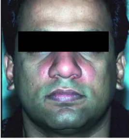
(Figure 1), or ulcerations, but occasionally
B
acterial skin infections are the28th most common diagnosis in hospitalized patients.1 Cellulitis, impetigo, and folliculitis are the most common bacterial skin infections seen by the family physician. The percentage of office visits for cellulitis was 2.2 percent, and for impetigo, it was 0.3 percent, in a cohort of almost 320,000 health plan mem-bers (data taken from primary physician diag-nosis codes from January 1, 1999 to December 1, 1999 for Intermountain Health Care, Salt
Family physicians frequently treat bacterial skin infections in the office and in the hospi-tal. Common skin infections include cellulitis, erysipelas, impetigo, folliculitis, and furun-cles and carbunfurun-cles. Cellulitis is an infection of the dermis and subcutaneous tissue that has poorly demarcated borders and is usually caused by Streptococcus or Staphylococcus species. Erysipelas is a superficial form of cellulitis with sharply demarcated borders and is caused almost exclusively by Streptococcus. Impetigo is also caused by Streptococcus or Staphylococcus and can lead to lifting of the stratum corneum resulting in the commonly seen bullous effect. Folliculitis is an inflammation of the hair follicles. When the infection is bacterial rather than mechanical in nature, it is most commonly caused by Staphylo-coccus. If the infection of the follicle is deeper and involves more follicles, it moves into the furuncle and carbuncle stages and usually requires incision and drainage. All of these infections are typically diagnosed by clinical presentation and treated empirically. If antibiotics are required, one that is active against gram-positive organisms such as peni-cillinase-resistant penicillins, cephalosporins, macrolides, or fluoroquinolones should be chosen. Children, patients who have diabetes, or patients who have immunodeficiencies are more susceptible to gram-negative infections and may require treatment with a sec-ond- or third-generation cephalosporin. (Am Fam Physician 2002;66:119-24. Copyright© 2002 American Academy of Family Physicians.)
Common Bacterial Skin Infections
DANIEL L. STULBERG, M.D., MARC A. PENROD, M.D., and RICHARD A. BLATNY, M.D. Utah Valley Family Practice Residency, Provo, Utah
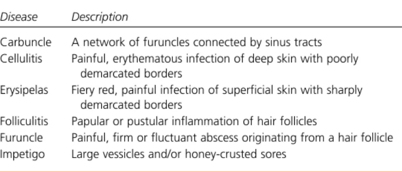
TABLE 1
Descriptions of Bacterial Skin Infections
Disease Description
Carbuncle A network of furuncles connected by sinus tracts Cellulitis Painful, erythematous infection of deep skin with poorly
demarcated borders
Erysipelas Fiery red, painful infection of superficial skin with sharply demarcated borders
Folliculitis Papular or pustular inflammation of hair follicles
Furuncle Painful, firm or fluctuant abscess originating from a hair follicle Impetigo Large vessicles and/or honey-crusted sores
FIGURE 1. Cellulitis secondary to tinea infection. CARING FOR COMMON SKIN CONDITIONS
This article is one in a series coordinated by Daniel L. Sulberg, M.D., director of dermatology curricu-lum at the Utah Valley Family Practice Resi-dency, Provo, Utah.
presents in skin that appears normal. Patients may have a fever and an elevated white blood cell count. Cellulitis can occur on any part of the body. Among the patients in the cohort above, the most common sites of cellulitis were the legs and digits, followed by the face, feet, hands, torso, neck, and buttocks (data taken from primary physician diagnosis codes from January 1, 1999 to December 1, 1999 for health plan members of Intermountain Health Care, Salt Lake City).
In otherwise healthy adults, isolation of an etiologic agent is difficult and unrewarding. If the patient has diabetes, an immunocompro-mising disease, or persistent inflammation, blood cultures or aspiration (some physicians inject sterile nonpreserved saline before aspira-tion) of the area of maximal inflammation may be useful.2-4 For infection in patients without diabetes, empiric treatment with a penicillinase-resistant penicillin, first-genera-tion cephalosporin, amoxicillin-clavulanate (Augmentin), macrolide, or fluoroquinolone (adults only) is appropriate.5Limited disease can be treated orally, but more extensive dis-ease requires parenteral therapy. Marking the margins of erythema with ink is helpful in fol-lowing the progression or regression of celluli-tis (Figure 2).Outpatient therapy with injected ceftriaxone (Rocephin) provides 24 hours of parenteral coverage and may be an option for some patients. The patient should be seen the following day to reassess disease progression.
Most cases of superficial cellulitis improve within one day, but patients who exhibit thickening of the dermis usually take several days of parenteral antibiotics before signifi-cant improvement occurs. Antibiotics should be maintained for at least three days after the resolution of acute inflammation.5Adjunctive therapy includes the following: cool com-presses; appropriate analgesics for pain; tetanus immunization; and immobilization and elevation of the affected extremity.6
A parenteral second- or third-generation cephalosporin (with or without an aminogly-coside) should be considered in patients who
have diabetes, immunocompromised patients, those with unresponsive infections, or in young children.5The patient may also require a plain radiograph of the area or surgical debridement to evaluate for gas gangrene, osteomyelitis, or necrotizing fasciitis.6
Recurrent episodes of cellulitis or undergo-ing surgery, such as mastectomy with lymph node dissection, can compromise venous or lymphatic circulation and cause dermal fibro-sis, lymphedema, epidermal thickening, and repeated episodes of cellulitis. These patients may benefit from prophylaxis with erythro-mycin, penicillin, or clindamycin (Cleocin).5,7 Periorbital cellulitis is caused by the same organisms that cause other forms of cellulitis and is treated with warm soaks, oral antibi-otics, and close follow-up.8 Children with periorbital or orbital cellulitis often have underlying sinusitis.9If the child is febrile and appears toxic, blood cultures should be per-formed and lumbar puncture considered.
Haemophilus influenzae type b (Hib) in young children was a significant concern until the widespread use of the Hib vaccine and cover-age with a parenteral third-generation cephalosporin was used routinely. Recently, some researchers have recommended no longer routinely covering for H. influenzae.8-10 Orbital cellulitis occurs when the infection passes the orbital septum and is manifested by proptosis, orbital pain, restricted eye move-ment, visual disturbances, and concomitant sinusitis. Complications include abscess for-mation, persistent blindness, limited eye movement, diplopia, and, rarely, meningitis.11 This ocular emergency requires intravenous antibiotics, otorhinolaryngology, and ophth-almologic consultation.12
Perianal cellulitis is caused by group A beta-hemolytic streptococcal infection and occurs most often in children. A study13of children FIGURE 2. Inked margins of cellulitis.
with perianal cellulitis found a mean age of onset of 4.25 years. Ninety percent of patients presented with dermatitis, 78 percent with itching, 52 percent with rectal pain, and 35 percent with blood-streaked stools. Despite 10 days of oral antibiotics (primarily penicillin or erythromycin), the recurrence rate was high at 39 percent. If there is recurrence, the presence of an abscess should be considered, with
nee-dle aspiration of the site for bacteriology being more accurate than a skin swab.14
Erysipelas
Erysipelas (Figure 3), also known as St. Anthony’s fire, usually presents as an intensely erythematous infection with clearly demar-cated raised margins, and often with associated lymphatic streaking (Table 1).Common sites are the legs and face.15Most cases do not have an inciting wound or skin lesion and are pre-ceded by influenza-like symptoms. The inci-dence of erysipelas is rising, especially in young children, the elderly, persons with diabetes, alcoholic persons, and patients with compro-mised immune systems or lymphedema.6
Erysipelas is caused almost exclusively by beta-hemolytic streptococcus and thus can be treated with standard dosages of oral or intra-venous penicillin. However, most physicians treat this infection the same as cellulitis, which is outlined earlier. Adjunctive treatment and complications are the same as for cellulitis. Impetigo
Impetigo is most commonly seen in chil-dren aged two to five years and is classified as bullous or nonbullous (Table 1).The nonbul-lous type predominates and presents with an erosion (sore), cluster of erosions, or small vesicles or pustules that have an adherent or oozing honey-yellow crust. The predilection for the very young can be remembered by the common lay misnomer, “infant tigo.” Impetigo usually appears in areas where there is a break in the skin, such as a wound, herpes simplex infection, or maceration associated with angular cheilitis (Figure 4),but Staphylo-coccus aureus can directly invade the skin and cause a de novo infection.6
The bullous form of impetigo presents as a large thin-walled bulla (2 to 5 cm) containing serous yellow fluid. It often ruptures leaving a complete or partially denuded area with a ring or arc of remaining bulla (Figure 5). More than one area may be involved and a mix of bullous and nonbullous findings can exist.
Skin Infections
FIGURE 3. Erysipelas.
FIGURE 4. Nonbullous impetigo.
Nonbullous impetigo was previously thought to be a group A streptococcal process and bul-lous impetigo was primarily thought to be caused by S. aureus.Studies16,17now indicate that both forms of impetigo are primarily caused by S. aureus with Streptococcus usually being involved in the nonbullous form. If the infection is a toxin-producing, phage group II, type 71 Staphylococcus (the same toxin seen in Staphylococcus scalded skin syndrome, a medical emergency where large sheets of the upper epidermis slough off), large bullae will form as the toxin produces intradermal cleav-age.18 Otherwise, smaller bullae develop and the honey-crusted lesions predominate.
A study19published in 1990 concluded that topical mupirocin (Bactroban) ointment is as effective as oral erythromycin in treating impetigo. However, because the lesions of bul-lous impetigo can be large and both forms of impetigo can have satellite lesions, an oral antibiotic with activity against S. aureus and group A beta-hemolytic streptococcal infec-tion is warranted in nonlocalized cases. Because of developing resistance, erythro-mycin is no longer the drug of choice.17 Azithromycin (Zithromax) for five days and cephalexin (Keflex) for 10 days have been shown to be effective and well-tolerated.20 Dicloxacillin (Pathocil), oxacillin (Pros-taphlin), first-generation cephalosporins, or amoxicillin-clavulanate are also acceptable
alternatives.5 Broad-spectrum fluoroquino-lones have also been shown to be effective, and several have been approved by the U.S. Food and Drug Administration for treating skin and soft tissue infections.21These medications have excellent skin penetration and good bioavailability, but no generic forms are cur-rently available, and they are only approved for use in adults.
As with other diseases involving Strepto-cocci, there is a small chance of developing glomerulonephritis, especially in children aged two to six years. Presenting signs and symptoms of glomerulonephritis include edema and hypertension; about one third of patients have smoky or tea-colored urine. Streptococcal glomerulonephritis usually resolves spontaneously although acute symp-toms and problems may occur.22
Impetigo can be spread by direct person-to-person contact, so appropriate hygiene is war-ranted. Nasal carriage ofS. aureus has been implicated as a source of recurrent disease and can be reduced by the topical application of mupirocin twice daily for five days.23
Folliculitis
Hair follicles can become inflamed by phys-ical injury, chemphys-ical irritation, or infection that leads to folliculitis (Table 1). Classifica-tion is by the depth of involvement of the hair follicle. The most common form is superficial folliculitis that manifests as a tender or pain-less pustule that heals without scarring.24The hair shaft will frequently be seen in the center of the pustule. Multiple or single lesions can appear on any skin bearing hair including the head, neck, trunk, buttocks, and extremities. Associated systemic symptoms or fever rarely exist.S. aureus is the most likely pathogen; however, commensal organisms such as yeast and fungi occasionally appear, especially in immunocompromised patients. These lesions typically resolve spontaneously. Topical ther-apy with erythromycin, clindamycin, mupiro-cin, or benzoyl peroxide can be administered to accelerate the healing process.25
The Authors
DANIEL L. STULBERG, M.D., is director of dermatology curriculum at the Utah Valley Family Practice Residency in Provo, Utah. Dr. Stulberg received his medical degree from the University of Michigan Medical School, Ann Arbor, where he also completed a family practice residency. He also publishes a bimonthly e-mail dermatology quiz. MARC A. PENROD, M.D., is a third-year and chief resident at the Utah Valley Family Practice Residency. He received his medical degree from the University of Virginia School of Medicine, Charlottesville.
RICHARD A. BLATNY, M.D., is in private family practice in Fairbury, Neb. He received his medical degree from the University of Nebraska College of Medicine, Omaha. Dr. Blatny completed a family practice residency at the Utah Valley Family Practice Residency. Address correspondence to Daniel L. Stulberg, M.D., Utah Valley Family Practice Resi-dency, 1134 N. 500 West, Provo, UT 84604 (e-mail: [email protected]). Reprints are not available from the authors.
Staphylococci will occasionally invade the deeper portion of the follicle, causing swelling and erythema with or without a pustule at the skin surface. These lesions are painful and may scar. This inflammation of the entire fol-licle or the deeper portion of the hair folfol-licle (isthmus and below) is called deep folliculitis. Oral antibiotics are usually used in the treat-ment and include first-generation cephalo-sporins, penicillinase-resistant penicillins, macrolides, and fluoroquinolones.
Gram-negative folliculitis usually involves the face and affects patients with a history of long-term antibiotic therapy for acne. Pathogens include Klebsiella, Enterobacter, and Proteus species. It can be treated as severe acne with isotretinoin (Accutane), but use of isotretinoin is associated with major side effects, including birth defects.26
“Hot tub” folliculitis is caused by Pseudo-monas aeruginosa contamination of under-treated water in a hot tub or whirlpool. Multi-ple pustular or papular perifollicular lesions appear on the trunk and sometimes extremities within six to 72 hours after exposure (Figure 6),
and mild fever and malaise may occur. Lesions in the immunocompetent patient typically resolve spontaneously within a period of seven to 10 days.25Treatment is directed at prevention by appropriately cleaning the whirlpool or hot tub and maintaining appropriate chlorine lev-els (bromine and copper solutions are less common alternatives) in the water.
Furuncles and Carbuncles
Furuncles and carbuncles occur as a follicu-lar infection progresses deeper and extends
out from the follicle (Table 1). Commonly known as an abscess or boil, a furuncle is a tender, erythematous, firm or fluctuant mass of walled-off purulent material, arising from the hair follicle. These lesions may occur any-where on the body, but have a predilection for areas exposed to friction. Furuncles rarely appear before puberty. The pathogen is usu-ally S. aureus. Typically, the furuncle will develop into a fluctuant mass and eventually open to the skin surface, allowing the purulent contents to drain, either spontaneously or fol-lowing incision of the furuncle.
Carbuncles are an aggregate of infected hair follicles that form broad, swollen, erythema-tous, deep, and painful masses that usually open and drain through multiple tracts. Con-stitutional symptoms, including fever and malaise, are commonly associated with these lesions but are rarely found with furuncles. With both of these lesions, gentle incision and drainage is indicated when lesions “point” (fluctuant or boggy with a thin shiny appear-ance of the overlying skin); caution should be taken to not incise deeper than the pseudo capsule that has been built at the site of infec-tion. Loculations should be broken with a hemostat. The wound may be packed (usually with iodoform gauze) to encourage further drainage. In severe cases, parenteral antibi-otics such as cloxacillin (Tegopen), or a first-generation cephalosporin such as cefazolin (Ancef), are required.27The physician should be aware of the potential for gas-containing abscesses or necrotizing fasciitis, which require immediate surgical debridement. Final Comment
The majority of bacterial skin infections are caused by the gram-positive bacteria
Staphy-Skin Infections
Nasal carriage of Staphylococcus aureus has been implicated as a source of recurrent impetigo and can be reduced by the topical application of mupirocin.
FIGURE 6. Folliculitis caused by contamination of undertreated water in a hot tub or whirlpool.
lococcus and Streptococcus species. Antibi-otics are used empirically with consideration for resistance patterns. Current antibiotic rec-ommendations include penicillinase-resistant penicillins, first-generation cephalosporins, azithromycin, clarithromycin, amoxicillin-clavulanic acid, or a second-generation fluoro-quinolone in the skeletally mature patient. Gram-negative coverage with a second-, third-, or fourth-generation cephalosporin is usually indicated in children under three years and in patients with diabetes or who are immunocompromised.
The photographs in Figures 1, 2, 4, and 5 were pro-vided by Utah Valley Family Practice Residency, Provo, Utah. The photographs in Figures 3 and 6 were provided by Richard Usatine, M.D., of the Uni-versity of California-Los Angeles, Los Angeles, Calif. The authors indicate that they do not have any con-flicts of interest. Sources of funding: none reported.
REFERENCES
1. Elixhauser A, Steiner CA. Most common diagnoses and procedures in U.S. community hospitals, 1996, summary. HCUP Research Note. Agency for Health Care Policy and Research, Rockville, Md. Retrieved August 2001, from: www.ahrq.gov/data/hcup/ commdx/commdx.htm.
2. Kielhofner MA, Brown B, Dall L. Influence of under-lying disease process on the utility of cellulitis nee-dle aspirates. Arch Intern Med 1988;148:2451-2. 3. Leppard BJ, Seal DV, Colman G, Hallas G. The value
of bacteriology and serology in the diagnosis of cellulitis and erysipelas. Br J Dermatol 1985; 112:559-67.
4. Epperly TD. The value of needle aspiration in the management of cellulitis. J Fam Pract 1986;23: 337-40.
5. Gilbert DN, Moellering RC, Sande MA. The Sanford guide to antimicrobial therapy 2000. 30th ed. Hyde Park, Vt.: Antimicrobial Therapy, 2000:39. 6. Trubo R, Bisno AL, Hacker SM, Roaten SP Jr. Today’s
strategies for bacterial skin infections. Patient Care 1997;31:78-94.
7. Olszewski WL. Episodic dermatolymphangioadeni-tis (DLA) in patients with lymphedema of the lower extremities before and after administration of ben-zathine penicillin: a preliminary study. Lymphology 1996;29:126-31.
8. Schwartz GR, Wright SW. Changing bacteriology of periorbital cellulitis. Ann Emerg Med 1996; 28:617-20.
9. Barone SR, Aiuto LT. Periorbital and orbital cellulitis in the Haemophilus influenzae vaccine era. J Pedi-atr Ophthalmol Strabismus 1997;34:293-6. 10. Ambati BK, Ambati J, Azar N, Stratton L, Schmidt
EV. Periorbital and orbital cellulitis before and after the advent of Haemophilus influenzae type B vac-cination. Ophthalmology 2000;107:1450-3. 11. Weiss A, Friendly D, Eglin K, Chang M, Gold B.
Bacterial periorbital and orbital cellulitis in child-hood. Ophthalmology 1983;90:195-203. 12. Martin-Hirsch DP, Habashi S, Hinton AH, Kotecha B.
Orbital cellulitis. Arch Emerg Med 1992;9:143-8. 13. Kokx NP, Comstock JA, Facklam RR. Streptococcal
perianal disease in children. Pediatrics 1987;80: 659-63.
14. Brook I. Microbiology of perianal cellulitis in chil-dren: comparison of skin swabs and needle aspira-tion. Int J Dermatol 1998;37:922-4.
15. Chartier C, Grosshans E. Erysipelas: an update. Int J Dermatol 1996;35:779-81.
16. Barton LL, Friedman AD. Impetigo: a reassessment of etiology and therapy. Pediatr Dermatol 1987; 4:185-8.
17. Misko ML, Terracina JR, Diven DG. The frequency of erythromycin-resistant Staphylococcus aureus in impetiginized dermatoses. Pediatr Dermatol 1995; 12:12-5.
18. Edlich RF, Horowitz JH, Nichter LS, Silloway KA, Morgan RF. Clinical syndromes caused by staphylo-coccal epidermolytic toxin. Compr Ther 1985;11: 45-8.
19. McLinn S. A bacteriologically controlled, random-ized study comparing the efficacy of 2% mupirocin ointment (Bactroban) with oral erythromycin in the treatment of patients with impetigo. J Am Acad Dermatol 1990;22(5 pt 1):883-5.
20. Kiani R. Double-blind, double-dummy comparison of azithromycin and cephalexin in the treatment of skin and skin structure infections. Eur J Clin Micro-biol Infect Dis 1991;10:880-4.
21. Lentino JR, Augustinsky JB, Weber TM, Pachucki CT. Therapy of serious skin and soft tissue infec-tions with ofloxacin administered by intravenous and oral route. Chemotherapy 1991;37:70-6. 22. Sanjad S, Tolaymat A, Whitworth J, Levin S. Acute
glomerulonephritis in children: a review of 153 cases. South Med J 1977;70:1202-6.
23. Raz R, Miron D, Colodner R, Staler Z, Samara Z, Keness Y. A 1-year trial of nasal mupirocin in the prevention of recurrent staphylococcal nasal colo-nization and skin infection. Arch Intern Med 1996;156:1109-12.
24. Jaworsky C, Gilliam AC. Immunopathology of the human hair follicle. Dermatol Clin 1999;17:561-8. 25. Sadick NS. Current aspects of bacterial infections
of the skin. Dermatol Clin 1997;15:341-9. 26. Ortonne JP. Oral isotretinoin treatment policy. Do we
all agree? Dermatology 1997;195(suppl 1):34-40. 27. Stone SP. Unusual, innovative, and long-forgotten