MANIPULATION AND EXPLOITATION OF THE HOST CELL BY KAPOSI’S SARCOMA-ASSOCIATED HERPESVIRUS
Louise Caroline Giffin
A dissertation submitted to the faculty of the University of North Carolina at Chapel Hill in partial fulfillment for the degree of Doctor of Philosophy in the Department of
Microbiology and Immunology in the School of Medicine.
Chapel Hill 2015
Approved by: Blossom Damania Nancy Raab-Traub Kristina De Paris Dirk Dittmer
© 2015
ABSTRACT
Louise Caroline Giffin: Manipulation and Exploitation of the Host Cell by Kaposi’s Sarcoma-Associated Herpesvirus
(Under the direction of Blossom Damania)
Kaposi’s sarcoma-associated herpesvirus (KSHV) is a human
gamma-herpesvirus that is the causative agent of three human malignancies: Kaposi’s sarcoma, primary effusion lymphoma, and multicentric Castleman’s disease. KSHV causes a lifelong infection for which there is no known cure, and the cancers associated with KSHV typically have poor prognosis and no established treatment protocol. KSHV is a large DNA virus that encodes over eighty open reading frames that have diverse functions, including viral proteins that thwart the host immune system and that alter cellular growth pathways to promote viral persistence, with the incidental effect of inducing cellular transformation.
KSHV expresses several homologs of human proteins, including a homolog of interleukin 6 (hIL-6) called viral interleukin 6 (vIL-6). vIL-6 is a predominantly
endothelial cells. We also found that vIL-6 significantly upregulates the expression of genes associated with cellular movement including the adhesion factor called
carcinoembryonic antigen-related cell adhesion molecule 1 (CEACAM1). We
determined that vIL-6 increases CEACAM1 expression via STAT3 signaling, and that CEACAM1 promotes vIL-6-mediated migration of endothelial cells. Both de novo and latent KSHV infection were also found to upregulate CEACAM1.
To further elucidate how KSHV modulates the cellular environment, we performed kinome profiling to identify cellular kinases that are differentially activated during latent and lytic KSHV infection in primary effusion lymphoma. Kinases regulate almost all cellular processes, and dysregulated kinase activation can drive
I dedicate this dissertation to my grandmother, Helen Louise Giffin, for giving me my love of science and teaching me to always be curious.
And to my family, Neil, Mom, Dad, and Nik for their love and support.
ACKNOWLEDGEMENTS
I am thankful to my advisor, Blossom Damania. She has shaped me as a scientist, and has taught me how to approach the most challenging questions. In the lab, she gave me both freedom to explore and guidance and support. Her passion for science is inspirational, and I will be indebted to her for my training as a scientist and a professional.
I am so thankful to have spent my graduate years amongst amazing lab mates. I came in as a very shy first year student, but everyone helped me come out of my shell. It is a special privilege to wake up every morning and look forward to seeing the people you work with. Thank you to every one of you for all the fun chats, unrestrained over-sharing of life issues, delicious baked goods, and of course, scientific banter. I’d especially like to thank Dr. Patrick Dillon for mentoring me as a rotation student way back in the day, Dr. Aadra Bhatt for all her guidance and help as the next-most-senior graduate student in the lab and for her endless experimental knowledge, Dr. Sarah Jacobs for her always-practical attitude and her plethora of cute dog and kid pictures, and Dr. John West for always being selfless and extremely helpful in the lab, for putting up with my constant heckling, and for being one of the best listeners and advice-givers I’ve met in graduate school.
I appreciate my dissertation committee, Drs. Nancy Raab-Traub, Ron
insight and advice for finishing up my graduate work and figuring out my next step. I am also thankful to Dixie Flannery and Verita Hinton for keeping the gears always turning in both the Microbiology and Immunology Department and Blossom’s lab.
Lastly, I would like to thank my family and friends for helping me survive graduate school. My father said grad school will be no fun at all, but thanks to the support from all of you, he’s been proven wrong. Thanks to my best running buddies, Fatima and Shelly, for keeping me active and healthy and up-to-date on department gossip. Only you guys could make me look forward to an early morning 10-mile training run! Thanks to Dan for being a great friend, and for always being there when I’m in the mood for a classy cocktail. John and Shelly, it’s been so much fun getting to know you two over the past couple of years, and I’m looking forward to all the exciting times to come with future refinishing projects! And finally, thank you to my family for always providing endless love and support. Neil, you’re the best brother I could ask for and I’m so proud of you for finding direction in your career – your enthusiasm and commitment towards your work motivates me every day. Mom and Dad, you did a perfect job raising me, and I know that I’ve accomplished my graduate degree because of all your hard work in making me who I am. I’m so thankful for all the opportunities you’ve given me in my life. You’ve prepared me for anything and everything. I love you both. And Nik, you are my rock. My life wouldn’t be the same without you, so thank you for always supporting me, for
TABLE OF CONTENTS
LIST OF TABLES ... x
LIST OF FIGURES...xi
LIST OF ABBREVIATIONS ... xii
CHAPTER 1: KSHV: PATHWAYS TO TUMORIGENESIS AND PERSISTENT INFECTION ... 1
OVERVIEW ... 1
INTRODUCTION ... 2
MALIGNANCIES AND SYNDROMES LINKED WITH KSHV INFECTION ... 3
VIRAL LATENCY AND ASSOCIATED PROTEINS ... 10
THE KSHV LYTIC CYCLE ... 16
LYTIC KSHV PROTEINS INVOLVED IN CELL GROWTH AND SURVIVAL ... 17
KSHV’S ACTIVATION AND EVASION OF THE HOST IMMUNE RESPONSE ... 23
CONCLUSIONS ... 34
CHAPTER 2: MODULATION OF KSHV VIL-6 FUNCTION BY HYPOXIA UPREGULATED PROTEIN 1 ... 36
OVERVIEW ... 36
INTRODUCTION ... 37
METHODS ... 40
RESULTS ... 47
CHAPTER 3: KSHV VIRAL INTERLEUKIN 6 MODULATES ENDOTHELIAL CELL MOVEMENT BY UPREGULATING CELLULAR GENESINVOLVED
IN MIGRATION ... 71
OVERVIEW ... 71
INTRODUCTION ... 72
METHODS ... 75
RESULTS ... 79
DISCUSSION ... 93
CHAPTER 4: ALTERATION OF THE HOST CELL KINOME BY LYTIC AND LATENT KAPOSI’S SARCOMA ASSOCIATED HERPESVIRUS INFECTION ... 99
OVERVIEW ... 99
INTRODUCTION ... 100
METHODS ... 103
RESULTS ... 106
DISCUSSION ... 109
CHAPTER 5: SUMMARY, CONCLUSIONS, AND FUTURE DIRECTIONS ... 114
GENERAL SUMMARY ... 114
KSHV-ASSOCIATED MALIGNANCIES AND MECHANISMS OF PATHOGENESIS ... 115
MODULATION OF KSHV VIRAL INTERLEUKIN 6 FUNCTION BY HYPOXIA UPREGULATED PROTEIN 1 ... 117
KSHV VIRAL INTERLEUKIN 6 MODULATES ENDOTHELIAL CELL MOVEMENT BY UPREGULATING CELLULAR GENES INVOLVED IN MIGRATION ... 120
ALTERATION OF THE HOST CELL KINOME BY LYTIC AND LATENT KAPOSI’S SARCOMA ASSOCIATED HERPESVIRUS INFECTION ... 125
LIST OF TABLES
Table 1.1: Characteristics of KSHV-associated malignancies ... 5
LIST OF FIGURES
Figure 1.1: KSHV encodes a number of proteins that contribute to cell
growth and transformation. ... 18
Figure 1.2: KSHV evasion of the host interferon response. ... 28
Figure 2.1: vIL-6 binds the ER chaperone protein hypoxia upregulated protein 1 (HYOU1)... 49
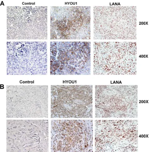
Figure 2.2: HYOU1 is expressed in tissue from KSHV-associated tumors. ... 51
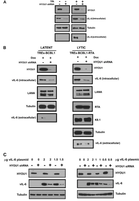
Figure 2.3: HYOU1 increases endogenous vIL-6 levels. ... 53
Figure 2.4: HYOU1 enhances vIL-6-induced STAT3 signaling. ... 56
Figure 2.5: HYOU1 facilitates vIL-6-induced migration of endothelial cells. ... 59
Figure 2.6: HYOU1 is required for vIL-6-induced endothelial cell survival in serum starved conditions. ... 62
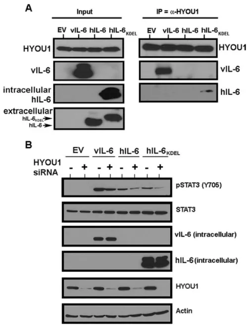
Figure 2.7: HYOU1 interacts with and influences the signaling of hIL-6. ... 65
Figure 3.1: vIL-6 modulates expression of host genes associated with cell movement. ... 81
Figure 3.2: vIL-6 upregulates long isoforms of CEACAM1 in endothelial cells. ... 84
Figure 3.3: vIL-6, but not hIL-6, upregulates CEACAM1 in a STAT3- dependent manner. ... 86
Figure 3.4: CEACAM1 facilitates vIL-6-mediated endothelial cell migration. ... 89
Figure 3.5: CEACAM1 is upregulated during de novo and latent KSHV infection of endothelial cells and during reactivation in PEL. ... 91
Figure 4.1: Viral kinases are isolated by MIB/MS. ... 107
Figure 4.2: Analysis of specific kinase activation in latent and lytic TREx-BCBL1 PEL. ... 108
LIST OF ABBREVIATIONS
AIDS Acquired immune deficiency syndrome
APC Antigen presenting cell
ATP Adenosine triphosphate
BCBL Body cavity-based lymphoma
BCR B cell receptor
BEC Blood endothelial cells
CAMK Calcium/calmodulin-stimulated protein kinase
CCL2 Chemokine C-C motif ligand 2
CDK Cyclin-dependent kinase
CEACAM1 Carcinoembryonic antigen-related cell adhesion molecule 1
cGAS Cyclic GMP/AMP synthase
CMV Cytomegalovirus
CTL Cytotoxic T lymphocyte
DMSO Dimethyl sulfoxide
DNA Deoxyribonucleic acid
EBV Epstein-Barr virus
ELISA Enzyme linked immunosorbent assay
ER Endoplasmic reticulum
FAK Focal adhesion kinase
FBS Fetal bovine serum
GTP Guanosine triphosphate
HEK Human embryonic kidney
hIL-6 Human interleukin 6
HAART Highly active anti-retroviral therapy
HIF Hypoxia-inducible factor
HIV Human immunodeficiency virus
HSV Herpes simplex virus
hTERT Human telomerase reverse transcriptase
HUVEC Human umbilical vein endothelial cell
HYOU1 Hypoxia upregulated protein 1
IFN Interferon
IHC Immunohistochemistry
IKK IκB kinase
IL Interleukin
IL-6R Interleukin 6 receptor
IP Immunoprecipitation
IPA Ingenuity Pathway Analysis
IRF Interferon regulatory factor
IRIS Immune reconstitution inflammatory syndrome
ITAM Immunoreceptor tyrosine activation motif
ITIM Immunoreceptor tyrosine inhibitory motif
JAK Janus kinase
KICS KSHV inflammatory cytokine syndrome
KS Kaposi’s sarcoma
KSHV Kaposi’s sarcoma-associated herpesvirus
LANA Latency-associated nuclear antigen
LEC Lymphatic endothelial cells
MAPK Mitogen-activated protein kinase
MCD Multicentric Castleman’s disease
MIB/MS Multiplex inhibitor bead/mass spectrometry
miRNA MicroRNA
mRNA Messenger RNA
mTOR Mammalian target of rapamycin
NFAT Nuclear factor of activated T cells
NFκB Nuclear factor kappa B
NHL Non-Hodgkin lymphoma
NK Natural killer
NLR NOD (Nucleotide-binding and oligomerization, leucine right repeat)-like receptor
NTC Non-targeting control
ORF Open reading frame
PAMP Pathogen-associated molecular patterns
pDC Plasmacytoid dendritic cell
PEL Primary effusion lymphoma
PI3K Phosphoinositide 3-kinase
PRR Pattern recognition receptor
qPCR Quantitative Real time polymerase chain reaction
RCA Regulator of complement activation
RLR RIG-I-like receptor
RNA ribonucleic acid
RRV Rhesus rhadinovirus
RTA Replication and transcription activator
RTK Receptor tyrosine kinase
SDS-PAGE Sodium dodecyl sulfate polyacrylamide gel electrophoresis
SH Src homology
siRNA Small interfering RNA
SNP Single nucleotide polymorphism
STAT3 Signal transducer and activator of transcription 3
STING Stimulation of interferon-dependent genes
TCR T cell receptor
TLK2 Tousled-like kinase2
TLR Toll-like receptor
TNF Tumor necrosis factor
TPA Tetradecanoyl phorbol acetate
TRAF TNF receptor associated factor
Tyr Tyrosine
UTR Untranslated region
vFLIP Viral FADD-like caspase-8 inhibitory protein
vGPCR Viral G protein-coupled receptor
vIL-6 Viral interleukin 6
vIRF Viral interferon regulatory factor
VKORC1v2 Vitamin K epoxide reductase complex subunit 1 variant 2
vPK Viral protein kinase, ORF36
CHAPTER 1: KSHV: PATHWAYS TO TUMORIGENESIS AND
PERSISTENT INFECTION1,2
OVERVIEW
Kaposi’s sarcoma associated herpesvirus (KSHV; also known as human herpesvirus 8) is the etiological agent of Kaposi’s sarcoma, primary effusion
lymphoma, and multicentric Castleman’s disease. These cancers often occur in the context of immunosuppression, which has made KSHV-associated malignancies an increasing global health concern with the persistence of the AIDS epidemic. KSHV has also been linked to several acute inflammatory diseases. KSHV exists between a lytic and latent lifecycle which allow the virus to transition between active
replication and quiescent infection. KSHV encodes a number of proteins and small RNAs that are thought to inadvertently transform host cells while performing their functions of helping the virus persist in the infected host. KSHV also has an arsenal of components that aid the virus in evading the host immune response, which help the virus establish a successful lifelong infection. In this comprehensive review, we will discuss the diseases associated with KSHV infection, the biology of latent and
1
Louise Giffin and Blossom Damania. Manuscript was written by LG and edited by BD.
2
lytic infection, and individual proteins and microRNAs that are known to contribute to host cell transformation and immune evasion.
INTRODUCTION
Kaposi’s sarcoma-associated herpesvirus (KSHV) was identified in 1994, and is the eighth and most recently discovered human herpesvirus. Shortly after, KSHV was found to be the etiological agent of three human malignancies including its namesake, Kaposi’s sarcoma, as well as two B cell lymphoproliferative diseases called primary effusion lymphoma and multicentric Castleman’s disease. Recently, KSHV has also been associated with several acute inflammatory diseases. KSHV infection typically occurs in the context of immunosuppression; thus
KSHV-associated cancers have become a global public health concern alongside the acute inflammatory disease syndrome (AIDS) epidemic.
Like all herpesviruses, KSHV infection transitions between a quiescent latent and replicative lytic life cycles. KSHV encodes an arsenal of viral proteins and non-coding RNAs that are thought to manipulate the host cell environment to directly or indirectly drive pathogenesis and viral persistence. Unlike other human pathogens such as bacteria, fungi, and parasites, viruses must enter the host cell to replicate and rely on the function of many host factors to successfully propagate. Because of this intimate relationship between host and pathogen, viruses have evolved in close contact with cellular components and thus have developed unique ways of
to encode homologs of cellular proteins. Some of these homologs allow the virus to activate or inactivate key host pathways to the virus’ advantage. Dissecting the function and role of individual viral components has helped create a picture of how KSHV induces disease. Understanding the mechanisms by which KSHV maintains a persistent infection and/or drives tumor development will help uncover potential therapeutic targets for the treatment of KSHV-associated malignancies.
MALIGNANCIES AND SYNDROMES LINKED WITH KSHV INFECTION
Kaposi’s sarcoma: The classical form of Kaposi’s sarcoma was first
described in 1872 as a pigmented sarcoma of the skin by the Hungarian
dermatologist, Moritz Kaposi (1). KS incidence rates started to increase dramatically with the onset of the AIDS epidemic in the 1980’s (2). The correlation between HIV-infected individuals and KS suggested an infectious agent was involved. About a decade later, representational difference analysis used by Chang and Moore
identified novel gammaherpesvirus DNA sequences in KS lesion biopsies (3). In the years following the discovery of KSHV, PEL and MCD were also found to be
causally linked to this human herpesvirus (4-6) (Table 1).
almost exclusively in young African children and cause significant mortality (12, 13). AIDS-associated, or epidemic KS has become the most common type of KS in the past three decades, and is the most aggressive form of the disease (2, 14, 15). AIDS-associated KS is considered an AIDS-defining illness (16). KS is currently the most common malignancy associated with HIV infection, and therefore is the most frequent cancer in many Sub-Saharan countries (17-21). The fourth type of KS is iatrogenic/post-transplant KS, which is associated with the use of immune
suppressive therapy for the prevention of organ transplant rejection (22-24). Interestingly, it was found that this type of KS occurs more often in KSHV-infected recipients rather than negative recipients that receive an organ from a KSHV-positive donor (25, 26).
KS lesions typically occur cutaneously on upper and lower extremities or on mucosal surfaces; however, lesions may also involve lymph nodes or may occur on visceral organs such as lung and spleen (22, 27). The immune status of the host and lymph node involvement are important factors in patient prognosis. KS disease progresses through six stages called the patch, plaque, nodular, lymphadenopathic, infiltrative, and florid stage (13, 28). KS lesions are highly angiogenic and as a result are usually red, purple, or brown in color. Additionally, the lesion vasculature is leaky, which allows for extravasation of erythrocytes and infiltration of inflammatory cells (29). KS tumor cells are of endothelial cell origin, and the primary
(BEC) can induce expression of lymphatic endothelial cell (LEC) markers and vice versa. This transcriptional reprogramming results in poorly differentiated endothelial cells that express mixed lineage markers such as CXCR4, CD34, VEGFR3, LYVE1, and PROX1 (34-36). As opposed to a metastatic dissemination, KS lesions typically arise independently of one another; however this oligoclonality is not universal, and situations of monoclonal KS have also been reported (37, 38).
Disease Presentation Lineage and
primary tumor cell Clonality KSHV genomes Kaposi’s sarcoma (KS) Highly angiogenic. Lesions can be
found on skin, visceral organs,
or mucosal surfaces
Endothelial cell origin; tumor cells
are spindle cells with mixed blood
and lymphatic endothelial cell markers Oligoclonal lesions >99% of tumor cells contain KSHV genomes Primary effusion lymphoma (PEL) Non-Hodgkin lymphoma; B-cell expansion in
body cavity
B cell; CD20-; markers resemble
partially differentiated
plasma cells
Monoclonal Each tumor cell has 50-100 copies of
the KSHV genome Multicentric Castleman’s disease (MCD) Plasmablastic variant of MCD
B cell; IgM λ-restricted plasmablasts
Typically polyclonal
Unknown
Table 1.1: Characteristics of KSHV-associated malignancies
Primary Effusion Lymphoma: Shortly after its association with KS, KSHV
pericardial, pleural, and peritoneal body cavities (39). Unlike KS, PEL is a monoclonal population of B cells as evidenced by clonal immunoglobin gene rearrangements, and each tumor cell has a high KSHV copy number ranging from 50-100 genomes per cell (40). Morphologically, PEL share features of both
immunoblastic and anaplastic large-cell lymphomas (41). Most PEL express CD45 and activation markers including CD30, CD38, and CD7, and epithelial membrane antigen (EMA) (42). Interestingly, PEL expresses plasma cell markers such as CD138, VS38c, and MUM-1/IRF4, but has relatively low expression of B-cell
associated antigens, suggesting that PEL resembles partially differentiated plasma cells rather than mature B cells (42-44). Unlike other NHLs, PEL typically does not exhibit c-myc rearrangements or mutations in the ras, bcl2, or p53 genes (39, 42). PEL is frequently coinfected with the Epstein Barr virus (EBV) (42).
Although PEL is characterized by a malignant serous effusion lacking a solid tumor mass, cases of solid PEL have also been reported (45). These tumors
typically present as an extracavitary lymphoma in extranodal or lymph node tissue and are composed of immunoblastic-like cells. These solid PEL are also KSHV positive, and have similar morphology, immunophenotype, immunoglobulin gene rearrangements to classical PEL (41).
also be an accurate predictor of clinical outcome of PEL patients (47). The level of immune suppression and the amount of circulating CD4+ lymphocytes may also contribute to the aggressiveness of PEL.
Multicentric Castleman’s disease: Around the same time as PEL, the
plasmablastic variant of MCD was also found to be associated with KSHV infection (4, 5). MCD also exists in a hyaline variant that is not associated with KSHV (48). MCD is an uncommon disseminated lymphadenopathy characterized by an
abnormal proliferation of IgM λ-restricted plasmablasts within the mantle zone of B
cell follicles (4, 49). MCD is considered non-neoplastic since the plasmablasts are typically polyclonal, however monoclonal B cell expansions have been observed (50). The plasmablasts are large with a vesicular nucleus containing one or more nucleoli (51). Systemic symptoms and inflammation as well as involvement of multiple organs often accompany MCD diagnosis (48, 52).
KSHV coinfection is observed in almost all HIV-positive MCD, although only a small proportion of cells in affected lymph nodes typically harbor the virus.
Interestingly, KSHV infection in MCD is quite lytic as compared to KS and PEL (53, 54). KSHV is detected in less than 40% of HIV-negative MCD cases (4, 48, 52, 55); however, in patients coinfected with HIV and KSHV, MCD tends to be very
endothelial growth factor (VEGF) (52, 58-60). In KSHV+ MCD, expression of the virally-encoded IL-6 (vIL-6) cytokine likely exacerbates inflammation and disease progression. vIL-6 can enhance cytokine signaling and further increase human IL-6 and VEGF expression (61-64). A cohort of plasmablastic MCD patients with
detectable vIL-6 expression were found to have a rapidly fatal clinical course as compared to vIL-6-negative MCD patients, suggesting the importance of cytokine signaling in MCD progression (55).
KSHV-associated Inflammatory Cytokine Syndrome: In the past few
years, several studies have reported patients that present with MCD-like
inflammatory symptoms but lack lymphadenopathy or other pathological evidence of true MCD (65). These patients typically have elevated cellular and viral cytokine levels, including human IL-6, IL10, C-reactive protein, and the viral cytokine vIL-6 (65). As compared to KS patients, high KSHV viral loads are also observed, indicative of a lytic or reactivated KSHV infection (27, 65). Concurrent KS is
frequently observed in these patients as well. Because of the systemic inflammatory symptoms, the proposed name for this disease is KSHV inflammatory cytokine syndrome or KICS. It differs from the chronic immune activation disease sometimes seen in HIV patients because two requirements for a KICS diagnosis include
detection of high KSHV viral load and vIL-6 cytokine levels (54).
There has been some controversy as to whether KICS is truly a distinct
point (54). Recently, a group investigated whether polymorphisms in the KSHV-encoded microRNAs (miRNA) could be correlated with the development of KICS (66). They found that a higher percentage KSHV+ MCD and KICS patients had single nucleotide polymorphisms (SNPs) in the KSHV miRNA loci than KS patients or KSHV+/KS-negative control patients. They also utilized classification tree analysis to determine combinations of SNPs that may predict development of KSHV+ MCD and KICS. Another recent case study identified a KICS patient with high viral loads of both KSHV and the ubiquitous human herpesvirus 6A, suggesting a possible role for other pathogens in development of KICS (27).
KSHV Immune Reconstitution Inflammatory Syndrome: A small
percentage of patients that begin HAART to treat advanced HIV infection exhibit a rapid deterioration of their clinical status. This phenomenon is known as immune reconstitution inflammatory syndrome (IRIS). It is proposed that following immune reconstitution, the increase in functional CD4+ T cell populations causes an immune recognition and response to autoantigens or pathogens that were previously present but asymptomatic. Cases of IRIS have been reported against KSHV and other pathogens such as Mycobacterium tuberculosis, Mycobacterium avium,
Cryptococcus neoformans, and human cytomegalovirus (CMV) (67). In many
instances, treatment of the offending pathogen or use of anti-inflammatory drugs can improve prognosis. High morbidity is observed in patients experiencing KS flares following initiation of HAART (IRIS-KS), although administration of systemic
one study determined that IRIS-KS patients had a significantly higher CD4+ count at KS diagnosis following HAART initiation than patients who did not develop IRIS, and that the mean time to KS diagnosis following HAART was less than 2 months (70). They also found that patients receiving more potent HAART regimens were more prone to IRIS-KS development. Beginning HAART treatment prior to advanced HIV infection or diagnosis of AIDS-KS decreases the chance of IRIS-KS (69).
VIRAL LATENCY AND ASSOCIATED PROTEINS
Latency is the default lifecycle for KSHV following infection of a host cell. During latency, the latency associated nuclear antigen (LANA) circularizes and tethers the viral genome to the host chromosomes by simultaneously binding both the terminal repeats and host histones H2A and H2B (71-73). The viral genome is replicated by host machinery with each cell division, and therefore persists as it is passed to each daughter cell (74, 75). As mentioned, only a small portion of the viral genome is actively transcribed during latency, and this region is known as the
have been investigated in depth to understand the mechanism by which KSHV causes disease. In this section, we will discuss a few elements of the latency locus.
LANA: LANA is encoded by ORF73 and is KSHV’s major latency protein. It is
responsible for tethering the viral episome to the host genome via the terminal repeats and histone interactions (71, 73), which allows host machinery to replicate and distribute the latent genome to daughter cells (72, 74, 75). The phosphorylated DNA-damage response protein γH2AX and the cellular replication fork factors Timeless and Tipin are some of the many known cellular proteins that assist LANA in maintaining KSHV episomes (82-84). LANA has also been shown to positively and negatively affect the transcription of a number of host genes (85, 86). This is likely mediated through LANA’s interaction with many transcription factors (84). LANA can also autoregulate its expression by inducing transcription from the LANA promoter (87). Furthermore, LANA and the LANA homologue in rhesus rhadinovirus (RRV) can repress transcription of the viral lytic transactivator, RTA (replication and transcription activator, ORF50) to help maintain latency (88-90).
LANA has several mechanisms by which it can promote host cell survival and proliferation. LANA can bind and inhibit p53 to reduce activation of p53-dependent reporter genes and cause chromosomal instability (91, 92). LANA can also bind and inactivate the tumor suppressor Rb leading to increased E2F-dependent reporter gene activation (93). Furthermore, LANA induces cytoplasmic β-catenin
accumulation by binding and sequestering GSK-3β in the nucleus (94), thus allowing
to increase telomerase expression, which increases the lifespan of infected cells (97). Finally, B cell-specific expression of LANA in a transgenic mouse model led to follicular hyperplasia, increased germinal center formation, and lymphomas,
implicating LANA as a key player in KSHV-associated lymphomagenesis (81). vCyclin: vCyclin is another latently-expressed protein and is encoded by
ORF72. vCyclin shares sequence and functional homology with cellular cyclin D2 and can bind and activate the cyclin-dependent kinase CDK6 (98, 99). When in complex with cdk6, vCyclin can phosphorylate and inactivate the tumor suppressor Rb, the cdk inhibitor p27 (Kip), and the anti-apoptotic protein Bcl-2, collectively leading to cell cycle deregulation (100-102). vCyclin-cdk6 can also phosphorylate histone H1 and cdc25a (101). Interestingly, vCyclin transgenic mice develop
lymphomas deficient in p53 (103). This is likely because vCyclin can also bind cdk9 which induces p53 phosphorylation and cell cycle arrest, so only cells which have lost p53 can continue to divide and expand into a lymphoma (104).
vFLIP: KSHV K13 encodes vFLIP, which is a viral homolog of cellular FLIP
(FLICE [protein FADD-like interleukin-1 beta-converting enzyme, now called
caspase-8] inhibitory protein). vFLIP is expressed during latency, and contains two death effector domains that can associate with FADD and prevent the CD95 death receptor from activating the apoptosis-inducing protease caspase 8 (FLICE) (105). It was subsequently shown that vFLIP can bind procaspase 8 directly to prevent its cleavage into active caspase 8 (106). Furthermore, vFLIP persistently activates nuclear factor kappa B (NFκB) signaling through binding to IKKα, IKKβ, RIP, and the
vFLIP (109). In vivo studies demonstrate that vFLIP transgenic mice can develop lymphomas and B cell-derived tumors (110-112).
The Kaposins: ORF K12 encodes three transcripts that yield kaposin A, B,
and C (113). Kaposin is highly abundant in PEL, and is transforming in cell culture-based assays (114). Kaposin A was shown to interact with the ARF guanine nucleotide exchange factor cytohesin-1 to mediate cellular transformation and
activation of the ERK/MAPK pathway (115). Kaposin B plays a role in preventing the decay of cytokine mRNAs by binding and activating the p38/MAPK target kinase MK2 (116, 117). MK2 can inhibit the decay of mRNAs that contain AU-rich elements, which include cytokine mRNAs and the mRNA for PROX1. KSHV induces
reprogramming of blood vascular endothelial cells towards a lymphatic lineage through upregulation of PROX1, and the ability of kaposin B to stabilize PROX1 mRNA is critical for this process (118).
KSHV microRNAs: Similar to other members of the herpesvirus family,
KSHV encodes 12 viral pre-microRNAs (pre-miRNAs) that are processed by the host proteins Drosha and Dicer to generate mature miRNAs. The KSHV pre-miRNAs are transcribed from the latent Kaposin/K12 promoter. While 10 of the pre-miRNAs are located in a Kaposin intron, the remaining 2 are located in the Kaposin protein-coding region and the Kaposin 3’ UTR (119-121) . The 12 viral pre-miRNAs
miR-K12-10 and miR-K12-12 levels are further increased during lytic replication (121-123). In PEL cell lines, over 90% of the expressed mature miRNAs are KSHV miRNAs. Several studies comparing clinical samples of KS biopsies and PEL to cultured PEL cell lines report that the KSHV pre-miRNAs are expressed at even higher levels in vivo and that their sequences are highly conserved between patients (124, 125). Functional KSHV miRNAs are also found in the virion, along with mRNAs, cellular miRNAs, and other small RNA species (126).
A number of validated host and viral mRNA targets of the KSHV miRNAs have been identified. These targets are involved in a variety of viral and cellular processes including maintenance of viral latency, immune evasion, cell cycle
regulation, cell survival and proliferation, and apoptosis. miR-K9* has been shown to directly target the 3’ UTR of the ORF50/RTA mRNA to prevent lytic reactivation (127), and miR-K12-5 may also indirectly suppress the RTA transcript (128). Furthermore, miR-K12-4 can target the DNA methyltransferase repressor Rbl2 to epigenetically maintain latency (128), and miR-K12-1 targets the 3’ UTR of the NFκB repressor IκBαto enhance NFκB signaling and promote latency (129). Two
components of the TLR/IL-1R signaling cascade, IRAK1 and MyD88, were identified as targets of miR-K12-9 and miR-K12-5, respectively, which results in reduced IL-6 and IL8 inflammatory cytokine production (130). Furthermore, miR-K12-7 can
directly bind the 3’ UTR of the mRNA of MICB, the stress-induced natural killer (NK) cell ligand, to repress MICB translation and promote viral immune evasion by
infection may prime B cells for transformation by expression of miR-K12-11, which is an ortholog of cellular miR-155 (133-135). miR-K12-11 targets the host protein Jarid2 and both miR-155 and miR-K12-11 can induce expansion of splenic CD19+ B cells in vivo (133, 136). Additionally, miR-K12-11 and -1 can induce MAPK signaling, promigration factors, and cell invasiveness through indirectly suppressing the MAPK phosphatase DUSP1 (137).
The KSHV miRNAs also have several mechanisms of preventing apoptosis of host cells: miR-K12-10 variants inhibit TGF-β signaling by targeting the 3’ UTR of the TGF-β type II receptor (TβRII) (138) and miR-K12-10a suppresses the tumor
necrosis factor (TNF)-like weak inducer of apoptosis (TWEAK) receptor (TWEAKER) to prevent TWEAK-mediated caspase activation, apoptosis, and proinflammatory cytokine production (139). Furthermore, it was demonstrated that miR-K12-1, -3, and 4-3p can target the 3’ UTR of caspase 3 to downregulate this host protein and inhibit apoptosis (140). The KSHV miRNAs have a variety of cellular and viral targets, only a handful of which have been discussed here. Collectively, the miRNAs work to drive KSHV pathogenesis by promoting latency, providing favorable growth conditions, and preventing apoptosis of infected cells.
THE KSHV LYTIC CYCLE
The KSHV lytic cycle can ensue following primary infection or when a latently infected cell undergoes lytic reactivation. During the lytic cycle, a temporal
transcriptional cascade begins that results in expression of viral immediate early, delayed early, and late genes followed by the subsequent assembly and egress of progeny virions (144). As discussed earlier, a variety of cell stresses can induce reactivation (145-149). Ultimately, expression of the lytic transactivator, ORF50/RTA, is required to initiate this complex stage of the viral lifecycle. RTA expression alone is sufficient to drive lytic replication, and suppression of RTA prevents reactivation (150-153). RTA is an immediate-early gene, and it is part of a polycistronic transcript that also encodes K8 and K8.1. Other immediate early genes include ORF45 and K4.2 (154). RTA has an activation domain and a DNA-binding domain on opposite ends of the protein. The DNA-binding domain allows RTA to directly bind and activate numerous viral promoters and the two KSHV origins of lytic replication, OriLyt-L and OriLyt-R (155, 156). The activation domain allows RTA to interact with cellular transcription factors and chromatin modification complexes to promote viral gene transcription (157).
Delayed early genes are sensitive to cyclohexamide treatment because in order to be transcribed they require the function of the proteins encoded by
Following expression of the delayed early genes, viral DNA replication begins from OriLyt-L and OriLyt-R (158, 159). The viral replication machinery includes the viral DNA polymerase, helicase, polymerase processivity factor, primase, primase-associated factor, and single strand binding protein (160) and replication is thought to occur by a rolling circle mechanism similar to other herpesviruses. Viral DNA replication stimulates expression of the KSHV late genes, which mainly encode structural proteins such as the viral capsid and envelope proteins (161). Linear genomes are packaged into the newly forming capsids. KSHV ORF67 and ORF69 assist in nuclear egress (162, 163), and KSHV glycoprotein B is thought to play a role in viral maturation and egress from the host cell (164, 165).
LYTIC KSHV PROTEINS INVOLVED IN CELL GROWTH AND SURVIVAL
Lytic reactivation results in expression of all KSHV genes. As described earlier, several of the proteins encoded by the KSHV latency locus can drive cellular transformation. A number of proteins encoded by KSHV lytic genes also have pro-growth or transforming qualities, which are discussed in this section and
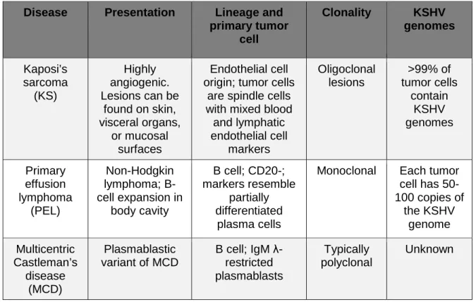
Figure 1.1: KSHV encodes a number of proteins that contribute to cell growth and
transformation. A) K1 is a transmembrane protein with a constitutively active
immunoreceptor tyrosine activation motif (ITAM) that activates signaling through
SH2-containing proteins. K1 expression results in production of VEGF and pro-inflammatory
cytokines. B) vGPCR is a constitutively active homolog of the IL8 receptor that results in
activation of numerous cell signaling pathways and transcription factors to increase
production of VEGF, VEGFR, and proinflammatory cytokines and chemokines. C) vIL-6 is a
functional homolog of human IL-6 that can signal through shared IL-6 pathways including
JAK/STAT, MAPK, and PI3K. This results in activation of multiple IL-6 response elements
and production of human IL-6 and VEGF. D) K15 is a transmembrane protein with several
tyrosine residues and SH2 and SH3 domains in its cytoplasmic tail that are critical for K15’s
signaling results in activation of numerous transcription factors and expression of
pro-inflammatory cytokines and chemokines and several human miRNAs that are involved in cell
motility.
K1: K1 is a single-pass transmembrane glycoprotein encoded by the first
open reading frame of KSHV (Figure 1.1 A). This protein is expressed on the cell and ER membranes and can be internalized to endosomes (166, 167). K1 is constitutively active and has a highly conserved intracellular immunoreceptor tyrosine-based activation motif (ITAM) on its C terminus (168-170). Upon K1 oligomerization, the ITAM becomes autophosphorylated and can activate various Src homology 2 (SH2)-containing signaling proteins including PI3K (p85)/Akt, PLCγ, Vav, Syk, Lyn, RasGAP, and Grb2 (171-173). Additionally, ITAM signaling results in activation of NFκB, nuclear factor of activated T cells (NFAT), Oct-2 and AP-1(173,
174).
Endothelial cells expressing K1 become immortalized in culture and primary marmoset T lymphocytes infected with a K1-expressing herpesvirus saimiri became immortalized to IL2-independent growth (175, 176). K1 can also induce focus formation in rat fibroblasts (176). In vivo, K1 transgenic mice display constitutively active NFκB and Src family tyrosine kinase signaling and a fraction of the mice
develop tumors (174) There are several aspects of K1 signaling that likely contribute to its transforming function. K1’s activation of Akt results in inactivation of the
and hsp40) were identified as K1 binding partners that are critical for both K1
expression and K1’s anti-apoptotic function (177). K1 also induces angiogenesis and VEGF production in primary human endothelial cells and cells derived from K1 transgenic animals (173, 175, 178). Furthermore, K1 signaling can induce secretion of inflammatory cytokines that are implicated in KS lesion development, including IL-6, GM-CSF, IL-1β, IL-8, and IL-10 (172, 174). A unique mechanism that K1 utilizes
to prolong the life of B cells is to downregulate surface expression of the B cell receptor (BCR) by binding the µ chain of the BCR to retain the complex in the ER (179). Overall, K1 is a multifunctional protein that can constitutively activate multiple pro-growth signaling pathways in KSHV infected cells.
Viral G protein Coupled Receptor (vGPCR): KSHV ORF 74 encodes the
vGPCR, which is a seven-pass transmembrane protein that shares homology with the human IL8 receptor (Figure 1.1 B) (180). This lytic protein has been detected at low levels in cultured reactivated PEL and in KS, PEL, and MCD clinical specimens (181). Conflicting reports have demonstrated that vGPCR has the ability to both sustain (182) and repress (183) RTA expression and lytic replication. Although vGPCR can bind the CXC and CC families of chemokines, it is constitutively active even in the absence of ligand (184-186). vGPCR activates a number of important signaling pathways, including PLC, PKC, MAPK, PI3K/Akt/mTOR, and NFκB (187).
Downstream signaling from these pathways activates the AP1, NFAT, NF-κB, HIF-1α, and CREB transcription factors which results in vGPCR-mediated production of
growth of endothelial cells in culture (188-190). It was also demonstrated that endothelial cell-specific expression of vGPCR can cause formation of KS-like angioproliferative lesions in mice (191). Similar to K1, vGPCR expression can also transform NIH3T3 fibroblasts, as well as rat kidney cells, which are then able to form tumors in nude mice (186). A line of transgenic mice expressing vGPCR in
hematopoietic cells developed angioproliferative lesions resembling KS at multiple organ sites (192). However, another study with a line of transgenic mice with vGPCR expressed ubiquitously from an SV40 promoter found that lesions mainly occurred on the tail and/or legs and that only a small fraction of tumor cells actually expressed vGPCR (193). Collectively, this research suggests a model by which vGPCR drives transformation of cells by inducing paracrine secretion of proinflammatory cytokines and angiogenic growth factors which can then work in concert with KSHV latent proteins to promote tumorigenesis.
Viral Interleukin-6 (vIL-6): KSHV ORF K2 encodes the vIL-6 cytokine
(Figure 1.1 C). vIL-6 is induced upon lytic replication, but it is also expressed at low levels during latency. Although vIL-6 has been detected in KSHV-associated
c-jun promoter IL-6 RE (JRE-IL-6) (62). Activation of these pathways leads to expression of hIL-6 (200) and VEGF (61). However, vIL-6 differs from the human cytokine in several regards. vIL-6 can signal intracellularly directly through the gp130 subunit of the IL6-R and does not require the extracellularly-located IL6-R gp80 subunit whereas hIL-6 requires both gp80 and gp130 (201-203); however, gp80 can still bind to vIL-6 and enhance signaling (63, 204, 205). Additionally, hIL-6 is
secreted much more efficiently than vIL-6, and a large portion of expressed vIL-6 is actually retained in the endoplasmic reticulum (ER) (206, 207). In the ER, vIL-6 interacts with the ER chaperone calnexin which impacts vIL-6 localization and intracellular retention (142). Furthermore, vIL-6 undergoes N-linked glycosylation which is required for its signaling activities (208).
vIL-6 expression transforms NIH3T3 fibroblasts and these cells form tumors in nude mice (61). vIL-6 expression can also induce growth in mouse hybridoma (198), PEL (209, 210), BAF (205), and Hep3B hepatoma (195) cell lines. In endothelial cells, vIL-6 expression induces proliferation, tubule formation, and neoangiogenesis (211, 212). Additionally, vIL-6 can help cells escape interferon (IFN)-induced growth arrest (209). Furthermore, transgenic mice expressing vIL-6 under the MHC class I promoter develop plasmablastic MCD-like disease, which is abrogated in the absence of endogenous IL-6 (213).
K15: KSHV K15 is encoded by the rightmost open reading frame of the virus
reactivation. The transcript is spliced to yield multiple K15 proteins with 4-12 transmembrane domains that localize to lipid rafts (215, 216). The short K15
cytoplasmic tail contains SH3 and SH2 signaling motifs and binding sites for TRAFs 1, 2, and 3 (215, 217). Several critical tyrosine residues within these motifs are constitutively phosphorylated by cellular Src family tyrosine kinases, which mediate activation of downstream signaling pathways. Pathways activated by K15 signaling include the Ras/MAPK, JNK/SAPK, and NFκB pathways as well as the NFAT/AP1 transcription factors (217-219). This signaling activates transcription of a number of cellular cytokines and chemokines including 6, 8, CCL20, CCL2, CXCL3, IL-1α/β, and Cox2 (217, 220). K15 can also downregulate signal transduction and intracellular calcium mobilization induced by the BCR, which may help the virus maintain latency (216). A potential mechanism by which K15 accomplishes this may be through K15’s interaction with the tyrosine kinase Lyn, which plays a role in the regulation of BCR signaling (219). Additionally, the K15 M allele induces cell motility, and this is dependent on K15-mediated upregulation of the human miRNAs miR-21 and -31 (221). K15 may contribute to KSHV-induced tumorigenesis through its ability to activate pro-growth signaling pathways, promote latency, and induce cell motility.
KSHV’S ACTIVATION AND EVASION OF THE HOST IMMUNE RESPONSE
adaptive immune response that are activated by KSHV infection as well as aspects that are suppressed by viral immune evasion techniques.
Immune Activation: Toll-like receptors (TLR) are innate pattern recognition
receptors that recognize pathogen-associated molecular patterns (PAMPs) and induce NFκB signaling and production of type I IFN and proinflammatory cytokines
(222). KSHV can activate TLR3 during infection of primary human monocytes, and this upregulates TLR3 expression and the production of IFNβ and CXCL10 (223,
224) which are then downregulated as latency is established (225). Although KSHV can reduce TLR4 activity in endothelial cells, TLR4 activation is still capable of inhibiting KSHV infection because cells lacking this receptor are more susceptible to infection (226). Thus, there is an initial TLR-mediated innate immune response to KSHV primary infection, but in many cases this response is subsequently
downregulated by the virus. KSHV is also sensed by IFN gamma-inducible factor IFI-16, which triggers inflammasome formation and subsequent production of IL-1β (227, 228). Additionally, KSHV infection can activate plasmacytoid dendritic cells (pDCs) which results in TLR9-mediated production of IFNα (229).
lytic and late lytic KSHV genes (235). In a cohort of seven KSHV+/HIV+ KS patients on HAART, KSHV-specific immune responses were detected in six of the seven patients (236). Interestingly, 100% of the non-progressor patients had KSHV-specific CD8+ cytotoxic T lymphocytes (CTLs) that simultaneously secreted IFNγ and TNFα in response to KSHV antigen whereas only 60% of the patients with progressive disease had a CD8+ CTL response (236). Although most studies of the adaptive immune response to KSHV have investigated the T cell response, KSHV infection also generates a humoral response to a variety of viral antigens (237).
Evasion of the Adaptive Immune Response: KSHV employs a variety of
mechanisms to evade KSHV-specific adaptive immune responses (reviewed in (238)). These techniques mainly involve repressing viral antigen presentation, T cell activation, B cell receptor (BCR)-mediated B cell activation, and B cell differentiation.
KSHV infection of B cells, dendritic cells (DCs), macrophages, and endothelial cells results in decreased expression of the major histocompatibility complex class I (MHC-I) (239, 240). MHC-I is critical for the presentation of viral antigens to the T cell receptor (TCR) of CD8+ T cells. KSHV K3 and K5 (also called modulator of immune recognition (MIR) 1 and 2, respectively) are capable of
resistant to recognition by KSHV-specific CD4+ T cells. LANA, which is expressed in all KSHV-infected cells, has an acidic central repeat domain that prevents its
antigenic processing to further hinder this process (247, 248). In addition to
repressing antigen presentation, KSHV infection also causes downregulation of the costimulatory molecules CD80, CD86, CD1a, and CD83 on antigen presenting cells (APCs) (249). K5 likely plays a role in this, because it has been shown to
downregulate CD86 and ICAM-1 (250). These costimulatory molecules are required for TCR-mediated activation of CTLs, so the downregulation of these proteins is a mechanism by which KSHV infection inhibits the adaptive T cell immune response.
As discussed previously, B cells are one of the main target cells of KSHV infection. B cells are a critical part of the adaptive immune response, and following binding of antigen to the B cell receptor (BCR), these cells proliferate and
differentiate into antibody-producing plasma cells or memory B cells (251).
Antibodies eliminate infection by binding to antigen that is either in the extracellular space or presented on the surface of infected cells. Antibody binding generally results in neutralization or phagocytosis of the pathogen or infected cell. If a B cell is unable to be activated through its BCR or unable to differentiate into a plasma cell, antibody production will not occur. One hypothesis is that KSHV targets these two aspects of B cell biology as a mechanism of adaptive immune evasion. The KSHV K5 protein can utilize its ubiquitin ligase activity to downregulate bone marrow stromal antigen 2 (BST-2, also called tetherin) which is an IFN-inducible protein that plays a role in B cell differentiation (252). As mentioned earlier, the KSHV K1
Furthermore, KSHV K15 is capable of disrupting signaling from the BCR and possibly accelerating BCR internalization to further reduce BCR-mediated B cell activation (216, 253). Collectively, this inhibition of both B cell differentiation and BCR signaling may help KSHV evade the B cell immune response.
Evasion of the Innate Immune Response: A large portion of the KSHV
genome is devoted to evading the innate immune response of the host. The innate immune functions targeted by viral proteins include interferon production, interferon regulatory factor (IRF) activation, natural killer (NK) cell activity, complement
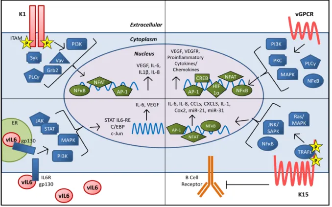
Figure 1.2: KSHV evasion of the host interferon response. KSHV encodes viral
interferon regulatory factors (vIRF 1-3) that antagonize the function of cellular IRFs, p300,
and NFκB to suppress production of type I IFN. ORF45, RTA, and KbZIP have also been
shown to interfere with IRF signaling. K3 and K5 are able to degrade the IFN3γR1 to
reduce antiviral IFNγ signaling through the JAK/STAT pathway. Viral infection and
expression of vGPCR reduces TLR4 expression, and RTA can induce degradation of the
TLR3 and TLR4 mediator TRIF. miR-K12-9 and 5 downregulate IRAK1 and MYD88 which
are also components of TLR signaling pathways. Reduction of TLR signaling results in
reduced expression of type I IFN. Finally, ORF64 is able to deubiquitinate RIG-I which
suppresses RIG-I mediated production of IFNβ.
persistence (Figure 1.2) (254). The virus encodes four homologs of the cellular IRFs called vIRF 1-4 (255). The cellular IRFs are a large family of transcription factors that drive expression of type I IFN (IFNαand β) and a variety of cytokines and
chemokines. Of the four KSHV-encoded vIRFs, only vIRFs 1, 2, and 3 have been shown to impact IFN signaling. vIRF1 can bind to and inhibit the transcriptional activities of IRF1, IRF3, and IRF7 (256, 257). Additionally, vIRF1 can bind and sequester the transcriptional coactivator p300 that is required for IRF1- and IRF3-mediated transcription of type I IFN (256, 257). vIRF2 is able to bind to cellular IRF1, 2, and 8 as well as NFκB RelA and p300 (258). vIRF2 is able to block type I IFN signaling and IFNα-, IFNλ-, and IRF1-dependent transactivation of the IFN
stimulated response element (ISRE) promoter (259). More recently, IRF3 was identified as a binding partner of vIRF2, and it was shown that this interaction both suppresses IRF3-mediated transcription of IFNβ and enhances
caspase-3-dependent degradation of IRF3 (260). vIRF3 can interact with cellular IRFs 3 and 7 which diminishes the DNA-binding abilities of IRF7 (261). vIRF3 can also interact with IRF5 to inhibit IRF5-mediated IFN promoter activation and production of type I IFN (262, 263). Recently, it was shown that vIRFs 1 and 2, but not vIRF3, are
capable of suppressing endogenous IFNβ message and protein expression following
environment for KSHV, and second, to promote cell survival to allow for persistence of the virus in the host.
KSHV ORF45 also influences the IFN response. It was demonstrated that KSHV ORF45 can interact with the inhibitory domain of cellular IRF7 (264). This interaction prevents IRF7’s phosphorylation and nuclear accumulation, which are both necessary for IRF7-mediated transcription of type I IFN (265). ORF45 also competes with IRF7 for phosphorylation by IKKε and TBK1 which reduces overall
levels of IRF7 phosphorylation (266). Infection of cells with an ORF45-null virus triggered a strong IRF7-dependent type I IFN response that rendered them resistant to subsequent vesicular stomatitis virus (VSV) infection (267). Interestingly, ORF45 is contained within the KSHV virion, which allows the virus to dampen the IFN response immediately upon infection (268). KSHV RTA can also act as an E3 ubiquitin ligase that induces the ubiquitination and degradation of IRF7 to reduce transcription of type I IFN genes (269). KSHV ORFK8 encodes a transcription factor KbZIP that can bind to the positive regulatory domain (PRD) I/III region of the IFNβ promoter to block IRF3-mediated IFNβ transcription (270).
In addition to inhibition of type I IFN, KSHV can also repress signaling by IFNγ. The K3 and K5 proteins are able to induce degradation of the IFN-γ receptor 1
(IFN-γR1) which normally triggers IFNγ-mediated activation of the JAK/STAT
pathway (271). Signaling through this pathway induces expression of a wide variety of antiviral genes, which is suppressed following reduction of IFN-γR1 expression by
utilizes a variety of mechanisms to evade IFN activation, suggesting the importance of avoiding this antiviral response in order for KSHV to persist in the host.
As mentioned earlier, the TLRs are pattern recognition receptors (PRR) that can be activated by invading pathogens. TLR activation triggers the production of antimicrobial cytokines and chemokines such as IFN, CCLs, and CXCLs through a variety of signaling proteins including NFκB, IRFs, and TRAFs. KSHV infection is
able to downregulate TLR4 expression partly through the actions of vGPCR and vIRF1, and this subsequently suppresses expression of TNF-α, IL1-β, IL-6, and IFNβ (226). Furthermore, it was recently discovered that the ubiquitin ligase activity of KSHV RTA may cause the degradation of TRIF (Toll-IL-1 receptor (TIR) domain-containing adaptor-inducing β-IFN), which is a critical mediator of TLR3- and TLR4-induced type I IFN production (272). As mentioned previously, the KSHV-encoded miRNAs miR-K12-9 and miR-K12-5 target IRAK1 and MYD88, which are both essential components of TLR and IL-1 receptor signaling pathways (130) .
to suppress RIG-I-mediated activation of the IFNβ promoter during reactivation
(276).
NLRs sense a variety of microbial ligands, and their activation results in the assembly of an inflammasome complex which activates caspase-1 to generate mature IL-1β and IL18 (277). Production of IL-1β and IL18 in response to infection can lead to hyperinflammatory caspase 1-mediated cell death, called pyroptosis. KSHV ORF63 has homology to parts of cellular NLRP1, but lacks the effector
caspase activation and recruitment (CARD) domain that is critical for inflammasome formation and function. ORF63 is able to interact with NLRP1 to prevent formation of both the NLRP1 and NLRP3 inflammasome and subsequent activation of caspase 1 (278). The function of ORF63 appears to be important for supporting viral gene expression and genome replication as well as suppressing IL-1β production.
A new cytosolic DNA sensor called cGAS was recently identified. cGAS activates the effector STING to induce IFN production and a subsequent antiviral response (279, 280). It was recently found that KSHV infection activates the
cGAS/STING pathway, and employs multiple mechanisms to dampen this activation (281). One of these mechanisms is expression of vIRF1, which blocks the
cGAS/STING-induced IFN response by disrupting the interaction between STING and the effector molecule TBK1.
-5, and -8 and CXCR-1, -2, and -4 (283). vCCL3 is an agonist for CCR4 (284). Collectively, binding of the viral chemokines to their respective cellular chemokine receptors is able to elicit a Th2-polarized response that is less cytotoxic to KSHV-infected cells than a Th1-polarized response (284, 285).
The complement pathway acts as a bridge between the innate and adaptive immune system, since activation of complement can occur in an antibody-dependent or independent mechanism. Furthermore, phagocytosis of complement-bound
pathogens or infected cells (opsonization) generates pathogen-derived antigens required to prime the adaptive immune system. Complement activation can occur through the classical, lectin, or alternative pathways which all result in the cleavage of complement component C3 into C3a and C3b by the C3 convertase (286). C3b can then be deposited onto the surface of pathogens or infected cells to facilitate lysis, neutralization, or phagocytosis. Since complement activation occurs through an amplifying cascade of proteolytic events, cellular regulators of complement activation (RCA) proteins keep this pathway in check to avoid hyperinflammatory responses (286). KSHV ORF4 encodes a structural and functional homolog to cellular RCA proteins called the KSHV complement control protein (KCP) (287, 288). KCP is able to prevent cleavage of C3 through accelerating the decay of the C3 convertase, by acting as an inhibitory cofactor to inactivate C3b and downstream complement molecules, and by preventing deposition of C3b onto target surfaces (289, 290). By evading the complement pathway, the virus is able to avoid
elimination of infected cells, and reduce the acquisition of viral antigens by phagocytes and APCs to inhibit the adaptive immune response.
As discussed in the adaptive immune evasion section, KSHV downregulates MHC-I expression on APCs. NK cells are designed to sense and kill cells displaying abnormal MHC-I levels through their leukocyte Ig-like receptor 1 (LIR1) and killer inhibitory receptor (KIR), which recognize endogenous MHC-I molecules on cells. To prevent the elimination of infected cells with reduced MHC-I, KSHV utilizes multiple mechanisms to inhibit NK cell function. In addition to downregulating MHC-I, KSHV K5 also downregulates surface expression of ICAM-1 and B7-2 (CD86) to avoid NK-mediated cell cytotoxicity (240, 242, 250). NK cell killing requires activation of the NKG2D and NKp80 receptors. As mentioned earlier, the KSHV miRNA miR-K12-7 targets the NKG2D ligand MHC class I-related chain B (MICB) 3’UTR. This results in decreased expression of this NKG2D ligand and effectively reduces NK cell killing ability (131). K5 also decreases the surface expression MICB and another NKG2D ligand, MICA, as well as the NKp80 ligand activation-induced C type lectin (AICL) (291). In these ways, KSHV has cleverly devised mechanisms to not only reduce activation of the adaptive immune system by downregulating MHC-I, but also to avoid the detrimental side effects of abnormal MHC-I levels on infected cells.
CONCLUSIONS
of several human malignancies. These cancers pose a large threat to global public health, particularly in areas that are still struggling with limited treatment options for HIV infection. Two decades of KSHV research has elucidated many of the
CHAPTER 2: MODULATION OF KSHV VIL-6 FUNCTION BY HYPOXIA
UPREGULATED PROTEIN 13
OVERVIEW
Kaposi’s sarcoma-associated herpesvirus (KSHV, also called human herpesvirus 8) is linked to the development of Kaposi’s sarcoma (KS), primary effusion lymphoma (PEL), and multicentric Castleman’s disease (MCD). KSHV expresses several proteins that modulate host cell signaling and deregulate cell growth. One of these proteins is viral interleukin-6 (vIL-6) which is a homolog of human interleukin-6. vIL-6 is able to prevent apoptosis and promote
pro-inflammatory signaling, angiogenesis, and cell proliferation. Although it can be secreted, vIL-6 is mainly an intracellular protein that is retained in the endoplasmic reticulum (ER). We performed affinity purification and mass spectrometry to identify novel vIL-6 binding partners and found that the cellular ER chaperone hypoxia upregulated protein 1 (HYOU1) interacts with vIL-6. Immunohistochemical staining revealed that both PEL and KS tumor tissues express significant amounts of
HYOU1. We also show that HYOU1 increases endogenous vIL-6 protein levels and that HYOU1 facilitates vIL-6-induced JAK/STAT signaling, migration, and survival in
3
Louise Giffin, Feng Yan, M. Ben Major, and Blossom Damania. Copyright © Journal of Virology, Aug 2014, 88(16):9429-41. BD and LG designed all
endothelial cells. Furthermore, our data suggest that HYOU1 also modulates vIL-6’s ability to induce CCL2, a chemokine involved in cell migration. Finally, we
investigated the impact of HYOU1 on cellular human IL-6 (hIL-6)signaling.
Collectively, our data indicate that HYOU1 is important for vIL-6 function and may play a role in the pathogenesis of KSHV-associated cancers.
INTRODUCTION
Kaposi’s sarcoma-associated herpesvirus (KSHV; Human Herpesvirus 8) is the causative agent of several human malignancies including Kaposi’s sarcoma (KS), primary effusion lymphoma (PEL), and multicentric Castleman’s disease (MCD) (3-6). These malignancies often occur in the context of immunosuppression, and as a result KSHV-associated malignancies have increased in incidence since the onset of the AIDS epidemic (2). KSHV is a member of the gamma-herpesvirus subfamily and has a double-stranded DNA genome that expresses over eighty open reading frames (ORFs) (40). KSHV infection usually exists in a latent state in which a small subset of the viral genome is expressed. When the virus undergoes lytic reactivation, all viral genes are expressed and progeny virions are produced.
It is thought that several latent and lytic genes contribute to modulation of host cell signaling to induce tumorigenesis. One of these genes is ORF K2 which encodes a viral homolog of human interleukin-6 (hIL-6) called viral interleukin-6 (vIL-6) (195-197). vIL-6 shares 25% identity and 63% similarity to hIL-6 at the amino acid level. vIL-6 is expressed at low levels in latently infected PEL and is highly
malignancies have detectable vIL-6 levels (55, 64, 194). vIL-6 expression transforms NIH3T3 cells, and vIL-6-expressing cells injected into mice form larger tumors as compared to control cells (61). Additionally, transgenic mice engineered to express vIL-6 under the MHC Class I promoter display a phenotype reminiscent of KSHV-associated plasmablastic MCD that is also dependent on mouse IL-6 expression (213). vIL-6 drives production of hIL-6 (200) and vascular endothelial growth factor (VEGF) (61) and can promote angiogenesis (211). Importantly, vIL-6 activates similar signaling pathways to the human cytokine, including the JAK/STAT, MAPK, and PI3K pathways (62, 198, 199).
vIL-6 differs from hIL-6 in several ways: hIL-6 must bind the IL-6 receptor (IL-6R, gp80) before activation of the gp130 signal transducer subunit, whereas vIL-6 can directly bind gp130 to induce signaling (201-203); however, involvement of gp80 can enhance vIL-6 signaling (205). Another difference is that hIL-6 is rapidly
secreted from cells whereas vIL-6 is primarily retained within the endoplasmic reticulum (ER) (206, 207). In this compartment, vIL-6 binds gp130 in a tetrameric complex to induce intracellular signaling (207). The cellular ER protein, calnexin, has been shown to interact with vIL-6 to stabilize vIL-6 folding and maintain its
intracellular distribution (142). The ER transmembrane protein vitamin K epoxide reductase complex subunit 1 variant 2 (VKORC1v2) was recently identified as an additional intracellular binding partner of vIL-6 (292, 293). vIL-6 binds to
VKORC1v2’s C terminus which is present in the ER lumen, but data suggest that this binding domain is not responsible for retention of vIL-6 in the ER.
abrogates vIL-6’s pro-growth phenotype in PEL cells independently of gp130 signaling (292). Furthermore, it was found that vIL-6 promotes PEL cell survival by suppressing the pro-apoptotic properties of the VKORC1v2 binding partner,
cathepsin D (294). This suggests that VKORC1v2 uses a mechanism independent of gp130 signaling to promote vIL-6 function and PEL cell survival.
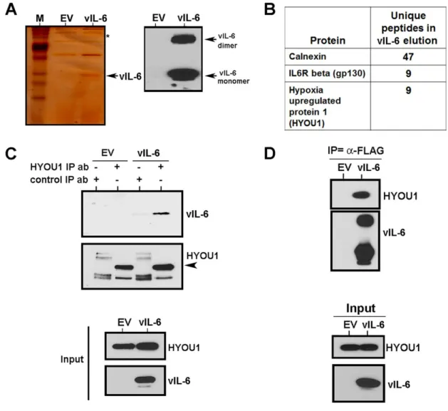
We performed affinity purification and mass spectrometry to identify novel binding partners of intracellular vIL-6. We found that a protein called hypoxia upregulated protein 1 (HYOU1; also called oxygen regulated protein 150 or
ORP150) is able to bind vIL-6. HYOU1 is an ER resident chaperone protein that is a member of the heat shock and ER stress protein families (295). HYOU1 is
expressed in many different cell types and can be upregulated by various cellular conditions including hypoxia and ER stress (295, 296). Furthermore, HYOU1 is upregulated in some human cancers including head and neck and breast cancer (297, 298). The HYOU1 transcript was originally cloned from astrocytes under hypoxic conditions (299), which makes it a relevant protein in KSHV biology since hypoxia plays a role in the KSHV lifecycle (300). Previous work has indicated that HYOU1 can suppress hypoxia-induced cell death (301) and induce angiogenesis by facilitating VEGF processing (302).
We found that HYOU1’s interaction with vIL-6 is important for vIL-6-induced intracellular STAT3 signaling and vIL-6 expression in PEL cells. Furthermore, we show that HYOU1 is required for several vIL-6 biological functions including
chemoattractant protein 1 or MCP1) in a HYOU1-dependent manner. CCL2 is implicated in the migration and metastasis of tumor cells and the extravasation of immune cells (303, 304). Finally, we investigated the impact of HYOU1 on cellular human IL-6 (hIL-6) signaling. Our results suggest that by modulating vIL-6 function HYOU1 may contribute to KSHV-associated tumorigenesis, making HYOU1 an attractive target for the treatment of KSHV-associated malignancies.
METHODS
Cell Culture and Generation of Stable Cell Lines
Human embryonic kidney (HEK) 293 and HEK293T cells were cultured in Dulbecco’s modified Eagle’s medium (Corning). BCBL1 PEL cells were cultured in RPMI 1640 medium (Corning) containing 0.05 mM β-mercaptoethanol. TREx BCBL1
and TREx BCBL1-RTA PEL cells (305) were cultured in RPMI 1640 medium (Corning) containing Tet System Approved FBS (Clontech) and 20 μg/mL
hygromycin B (Roche). hTERT-immortalized human umbilical vein endothelial cells (hTERT-HUVEC) were cultured in EBM-2 (Lonza) with the EBM-2 bullet supplement (Lonza) as described (178). All media were additionally supplemented with 10% heat inactivated fetal bovine serum (FBS), 1% penicillin-streptomycin (PS), and 1% L-glutamine. Charcoal filtered FBS was obtained from Life Technologies. Cells were transfected with XtremeGene HP transfection reagent (Roche) at a ratio of 2 µL
grown to 70% confluence and inoculated with lentivirus in the presence of 8 μg/mL
polybrene. Spinoculation was used for PEL cell transductions as previously
described (223). All transfections and transductions were incubated for 48-72 hours to allow for protein expression or knockdown. hTERT-HUVEC cells and HEK293 cells stably expressing empty vector or vIL-6 were generated by lentiviral
transduction. HEK293 cells stably expressing a non-targeting shRNA or a HYOU1 targeting shRNA plasmid (described below) were also generated by lentiviral transduction. For all stable cells, media were changed 24 hours post-transduction and the puromycin concentration was increased from 0.1 μg/mL to a final
concentration of 0.5 μg/mLfor hTERT-HUVEC and 1.0 μg/mL for HEK293 cells over
2 weeks.
Plasmids, Lentiviral vectors, shRNAs, and siRNAs
FLAG-tagged vIL-6 was cloned into the lentiviral vector pSuper-CMV puro (Invitrogen). All lentiviruses were produced using the ViraPower Lentiviral Expression System (Invitrogen) as per the manufacturer’s instructions.
Mass Spectrometry, Immunoprecipitations, and Western Blots
Twenty million 293T cells were transfected with pcDNA3 or vIL-6 expression vectors for 48 hours. Cells were harvested on ice in NP-40 lysis buffer (0.1% NP-40, 150 mM NaCl, 50 mM Tris HCl pH 8.0, 30 mM β-glycerophosphate, 50 mM NaF, 1
mM Na3VO4, 1 Roche protease inhibitor tablet per 50 mL) followed by one freeze-thaw cycle. Samples were clarified by centrifugation at 16 000 x g for 10 minutes and protein content was determined by Bradford assay (Bio-Rad). Equal amounts of protein were loaded on FLAG antibody-conjugated beads (EZview Red ANTI-FLAG M2 Affinity Gel; Sigma) and rocked at 4oC overnight. Beads were washed twice with lysis buffer followed by 2 washes with 50 mM NH4HCO3. Samples were eluted with 3x FLAG peptide (Sigma) diluted in 50 mM NH4HCO3 and 0.1% PPS Silent