Characterizing the Role of Crk in Central Nervous System Development in Drosophila
By Alison Bonner
Senior Honors Thesis
Department of Biology- Peifer Lab
University of North Carolina at Chapel Hill
April 20, 2018
Abstract
While determining the roles that different functional domains of Abelson tyrosine kinase
(Abl), a key developmental regulator and oncogene, during normal development, our lab was
surprised to find that a short, conserved motif (PXXP) in the linker region was more important
for Abl’s morphogenic roles than both kinase activity and F-actin binding. This finding led us to
hypothesize that Crk binding to Abl’s PXXP motif is critical for mediating Abl’s developmental
functions. Crk family proteins, including Crk and Crk-like (Crk-L), are a well-conserved family
of small adaptor proteins that play a role in cell adhesion, migration, and other biological
processes during normal development. Crk also plays a role in various cancers, including
invasive bladder cancer, and Crk-L is a key mediator of oncogenic forms of Abelson tyrosine
kinase (Abl) in Leukemia. Based on Abl’s well-defined roles in central nervous system (CNS)
patterning, we hypothesized that Crk might be required for proper CNS patterning. To test this
hypothesis, I used RNAi to deplete Crk both maternally and zygotically in embryos and asked
what effect, if any, this had on embryonic viability and CNS patterning. Using this approach, I
found that Crk is required for embryonic viability and that loss of Crk results in CNS patterning
defects. To better understand the mechanism(s) by which Crk may alter CNS patterning, I am
looking at the Robo/Slit repulsive axon guidance cues in the Crk knock down embryos to
determine if the CNS patterning defects observed are the result of loss of Crk affecting this
pathway. I found that localization of these proteins remains largely normal and their function
appears unaltered, indicating that this pathway is intact and does not require Crk. Preliminary
analysis suggests that zygotic loss of crk also results in partially penetrant CNS patterning
defects, mimicking what we see using an RNAi approach. We are continuing to characterize
morphogenic defects associated with loss of Crk to determine its Abl-dependent and –
independent roles. This work will help provide better insight into Crk’s roles during normal
I. Introduction
Development and tissue homeostasis require tight coordination of cell adhesion with
actin remodeling to allow cells to change shape and migrate. Both processes require the
assembly and activity of multi-protein signaling complexes, which include small adaptor
proteins1. Crk family proteins, including Crk and Crk-like (Crk-L), are a well-conserved family
of small adaptor proteins that play a role in cell adhesion, cell migration, and other biological
processes during normal development2. The mammalian Crk family is comprised of three
proteins-- Crk I, Crk II and Crk-L-- expressed from two gene loci3. crk is alternatively spliced
into crk I (minor form) and crk II (predominant form), while crk-L produces a single isoform3.
Crk proteins include a Src Homology 2 (SH2) domain and either one or two Src Homology 3
(SH3) domains connected by linker sequences1,3 (Fig. 1). Their SH2 domain allows for
interactions with upstream binding partners such as phosphorylated receptor tyrosine kinases and
focal adhesion complexes, while their SH3 domains mediate downstream interactions, typically
with effector molecules or other adaptor proteins4. Both upstream and downstream interactions
are controlled by phospho-regulation by tyrosine kinases, including Abelson tyrosine kinase
(Abl)3,5,6,7. Both Crk and Crk-L also play
critical roles in development and cancer.
Crk drives epithelial-to-mesenchymal
transitions in normal development, and is
overexpressed in many cancers, including
breast, ovarian, and bladder cancers, while
Crk-L is a key mediator of oncogenic forms
of Abl in Leukemia6-11.
SH2 SH2 SH2 SH2 Y221 Y207 Y251 Y215 Crk I Crk II Crk-L
Mammalian Crk Family
Drosophila Crk
Crk SH3 SH3 SH3 SH3 SH3 SH3 SH3
Crk I, Crk II, and Crk-L in mammals have many overlapping functions due to the
conserved structure and functional domains between the proteins. This redundancy makes it
difficult to study Crk function in mammals and cultured cells, because all three gene products
would have to be knocked down to demonstrate complete loss-of-function phenotypes12.
Knockout mouse models for Crk or Crk-L both die embryonically, but with different
developmental defects, indicating that the different gene products also have distinct,
non-overlapping roles13-17. As of yet, no double knockout has been conducted in mice. Drosophila
melanogaster has only one crk gene, resulting in one Crk protein, and thus is ideal for studying
the conserved function of Crk family proteins18.
While determining the roles that different functional domains of Abelson tyrosine kinase
(Abl), a key developmental regulator and oncogene, play during normal development, our lab
was surprised to find that a short, conserved motif (PXXP) in the linker region was more
important for Abl’s morphogenic roles than both kinase activity and F-actin binding in certain
contexts19. In mammals three proteins are known to bind Abl’s PXXP motif—Abi20,21, Crk22, and
Nck22—suggesting they may play a crucial role in Abl’s function during morphogenesis. Of
these three proteins, we wanted to know which, if any, work with Abl or are essential for Abl
function during morphogenesis. I used RNAi to knockdown abi, crk, or nck expression, and ask
if this resulted in phenotypes similar to maternal/zygotic Abl loss. Reducing abi or crk results in
increased embryonic lethality similar to Abl loss, while nck knockdown does not (Fig. 3). Given
that little is known about Crk function in Drosophila and Crk-L is the major downstream
mediator of oncogenic effects of BCR-Abl, we decided to focus in on Crk. We hypothesized that
Crk binding to Abl’s PXXP motif is critical for mediating Abl’s developmental effects. Based on
whether Crk is required for proper CNS patterning and, if so, on more extensively characterizing
Crk’s role in CNS patterning.
II. Methods
Fly stocks used
All stocks were maintained at on standard cornmeal agar media at room temperature or
25°C. All RNAi crosses were maintained at 25°C.
The following stocks, obtained from the Bloomington Drosophila Stock Center (NIH
P400D018537), were used in this study:
y1, sc*, v1; P{y+t7.7 v+t1.8=TRiP.HMC03964}attP40 (crk HMC RNAi);
y1, sc*, v1; P{y+t7.7 v+t1.8=TRiP.HMS01597}attP2 (abi RNAi);
y1, v1; P{y+t7.7 v+t1.8=TRiP.GL01519}attP2/TM3, Sb1 (nck RNAi)
We obtained y1, sc* v1; P{y+t7.7 v+t1.8=TRiP.HMJ2295}attP40/CyO (crk HMJ RNAi) from
the National Institute of Genetics (Mishima, Japan).
Additional stocks used include y,w (used as our wild-type laboratory strain); and w;
P(mat-tub-Gal4)mat67; P(mat-tub-Gal4)mat15 (subsequently referred to as matII; matIII); and
w; elav-Gal4/CyO.
RNAi experiments
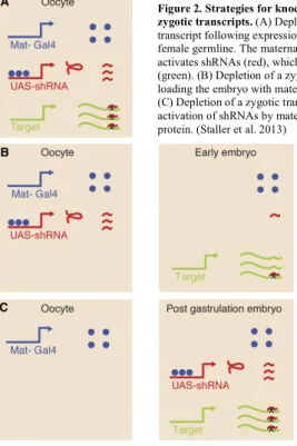
For all RNAi experiments, except CNS-specific knockdown, we wanted to knockdown
both maternal and zygotic mRNA and protein, as described by Staller et al24 (summarized in Fig.
2). To do so, we generated mothers carrying two copies of a strong, maternally contributed
GAL4 (matII; matIII) and a single copy of the UAS-shRNA targeting the transcript of interest. In
general, the genotype of the mothers used in the RNAi experiments was:
!"#!!!!"#!"#$%!&'$!"#$%&"'(!!"!"#$%$&#
!"#$$ ;
!"#$$$
We then crossed virgins of this
genotype to males carrying zero, one, or
two copies of the UAS-shRNA targeting
the transcript of interest, depending on
whether the insert was homozygous
viable. Using this approach, all embryos
had reduced maternal contribution of the
transcript of interest. Additionally, we
were able to alter the level of zygotic
knockdown by varying the number of
copies of UAS-shRNA present.
Measuring embryonic viability
To measure embryonic viability, crosses were set up in cups and allowed to lay eggs on
apple juice agar plates overnight (16-24 hours) at 25°C. Individual embryos were then
transferred to new plates, and the total number of embryos recorded (~200 embryos/individual
experiment). After forty-eight hours at 25°C, the number of embryos that have hatched and the
number of embryos that did not hatch were recorded. Embryos are expected to hatch after 24
hours, so after 48 hours, any eggs that have not hatched are considered dead. Percent viability
was then determined by dividing the number of embryos that hatched by the total number of
embryos placed on the plate. An un-popped cuticle prep was done on any unhatched eggs to
determine how many were unfertilized and the percent viability was adjusted accordingly. Figure 2. Strategies for knockdown of maternal and
Confocal microscopy of Central Nervous System
Embryos were dechorionated in 50% bleach for 5 minutes at room temperature. They
were then fixed in 1:1 4% formaldehyde:heptane for 20 minutes at room temperature and the
vitelline membrane removed by shaking embryos in 1:1 methanol:heptane. The embryos were
then rinsed three times in phosphate buffered saline (PBS) containing 0.1% Triton-X100 (PBT),
and blocked by incubating in PBT containing 1% normal goat serum (PNT) for 30 minutes at
room temperature. They were then incubated in primary antibody diluted in PNT either at room
temperature for 4 hours or overnight at 4°C. Following washing three times with PBT, embryos
were incubated in secondary antibody diluted in PNT either at room temperature for 2 hours or
overnight at 4°C.
Primary antibodies used were anti-BP102 (1:200), anti-FasII (1:100), anti-robo (1:100),
and anti-slit (1:10) and were obtained from the Developmental Studies Hybridoma Bank, created
by the NICHD of the NIF and maintained at the University of Iowa, Department of Biology,
Iowa City, IA, 52242. Secondary antibodies used were anti-mouse immunoglobulin G2a
(IgG2a), anti-mouse immunoglobulin G2b (IgG2b), anti-mouse immunoglobulin G1 (IgG1), and
anti-rat IgG conjugated to Alexa Fluor (AF) 488, AF568, or AF647 as indicated. The embryos
were mounted on glass slides in Aqua-Poly/Mount (Polysciences) and imaged on a Zeiss LSM-5
PASCAL confocal microscope. Images were processed using ImageJ25.
III. Results
Based on our lab’s finding that the PXXP motif within conserved region 1 (CR1) is more
essential for morphogenesis than both kinase activity and F-acting binding19, we wanted to look
at which of Abl’s PXXP-binding partners are required during embryonic morphogenesis. In
interact with Abl via this conserved PXXP motif. To
determine if any of these PXXP-binding partners is
essential for embryonic morphogenesis, I conducted a first
pass RNAi screen that should allow us to knockdown both
maternal and zygotic mRNA and protein levels. In
wild-type flies, it is not unusual for up to 10% to die
embryonically (in our wild-type control, 5.5% died, Fig. 3). The knockdown of nck resulted in
5.5% lethality, identical to the wild-type control (Fig. 3), though we did not determine if nck
RNAi effectively reduced Nck protein levels. Consequently, we decided not to continue further
with nck. In contrast, crk RNAi (HMC03964) showed a lethality of 87%, while abi RNAi
showed a lethality of 99% (Fig. 3). Here, I am focused on the effects of loss of crk on embryonic
morphogenesis.
Because of its location on the 4th chromosome, we lacked many of the sophisticated
genetic tools that would allow us to generate embryos lacking both maternal and zygotic crk.
There was only one extant crk
allele (crkKG00336; a P-element
insertion between the
transcription start site and start
codon), and it has not been fully
characterized. Because of this
limitation, we initially relied on
transgenic RNAi lines to reduce
maternal and zygotic crk levels. I Figure 3: A first pass RNAi-based screen revealed that reduction of abi or crk severely reduced larval viability, while nck had no effect on viability.
0% 10% 20% 30% 40% 50% 60% 70% 80% 90% 100% Wild-type
Control abi RNAi nck RNAi crk RNAi
Em b ryo ni c V ia b il it y
used two crk RNAi lines- HMC03964 (from here on
known as crk HMC RNAi) and HMJ2995 (from
here on known as crk HMJ RNAi), each of which
targets a different region of the crk transcript, to
ensure the phenotypes I observed were caused by
knockdown of crk and not the result of non-specific
off-target effects.
The controls used for viability experiments
carried the RNAi construct for the respective RNAi
lines, but did not have the Mat-GAL4, so the RNAi
was not produced. The HMC control and HMJ
control result in 97.9% viability and 97.5% viability, respectively (Fig. 4), which is unchanged
from wildtype where up to 10% die embryonically. Using the crk HMC RNAi line to deplete
maternal/zygotic crk strongly reduces embryonic viability. Crossing
!"#!!"#!"#$% (!"#!"#$)
!"#$$ ;
!"#$$$
! virgins to males that are heterozygous for the crk RNAi
(HMC03964) decreases viability to 6.1%, while crossing these virgins to crk RNAi homozygous
males further reduces viability to 1.1% (Fig. 4). Using the second crk HMJ RNAi line, I
observed 72.3% embryonic viability, which is decreased from wildtype, but less severe than the
HMC line (Fig. 4). This compares to 9.4% viability in maternal/zygotic loss of abl19.
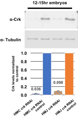
To ensure this increase in lethality compared to wild-type is the result of knockdown of
Crk protein levels, we conducted a Western blot to measure Crk protein levels in both RNAi
lines. The HMC line virtually eliminates Crk, while the HMJ line substantially reduces it (Fig.
HMC
crk RNAicontro
l 0.036 0.098 0.0 0.2 0.4 0.6 0.8 1.0 12-15hr embryos Crk le ve ls n o rm a liz ed to c o n tr o l HMC
crk RNAi
HMJ
crk RNAi
HMJ
crk RNAicontro
l
α- Tubulin
α-Crk
5). This demonstrates that both
RNAi lines result in decreased
levels of crk, but to different
extents, which may explain the
observed differences in
lethality.
Using these RNAi
reagents, I next looked at Crk’s
role in central nervous system
(CNS) patterning to compare
its role with Abl’s well-defined role in CNS patterning19,23. I used antibodies against BP102,
which labels all CNS axons26, and FasII, which labels a subset of axons in the longitudinal axon
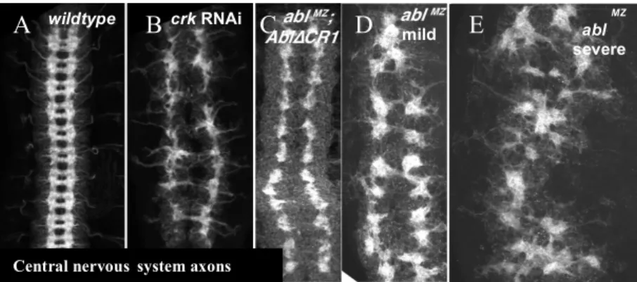
bundles27,28. The crk HMC RNAi line results in severely disrupted CNS patterning, similar to
70% of ablΔCR1 mutants and the milder phenotypes seen in 44% of abl null mutants (Fig. 6B
compared to 6C-D and Fig. 7). This similar phenotype, in which longitudinal axons form (albeit
wildtype crk RNAi
Central nervoussystem axons
abl
MZ
severe mild
Figure 6. crk RNAi results in CNS patterning defects similar to the minor class of mild phenotypes seen in of abl maternal/zygotic mutants or upon deletion of CR1. (A) wildtype. (B) crk RNAi (HMC03964). (C) abl with a deletion of the CR1 region
(AblΔCR1), (D-E) abl maternal/zygotic mutants. Wild-type central nervous systems exhibit a ladder-like appearance with longitudinal axon bundles making up the sides and commissural axon bundles making up the rungs (A). crk RNAi results in a range of phenotypes, including disorganized longitudinal axon bundles and failure to form commissural axon bundles (B). AblΔCR1 mutants fail to form commissural axon bundles, though the longitudinal axon bundles appear to form normally (C). ablMZ exhibit a less frequently observed, milder phenotype with disorganized longitudinal axons bundles and no commissural axon bundles (D), but the more frequent phenotype exhibits a severely disrupted CNS (E). (Panels A, C, D, and E reproduced from Rogers et al. 2016)
A B C D E
Figure 7. Loss of Crk results in Central Nervous Systerm (CNS) patterning defects. (A) Embryos of indicated genotype stained with antibodies against BP102 (green in left panels) and FasII (red in left panels, white in right panels). (B) Quantification of severity and penetrance of observed CNS patterning defects. Most HMJ RNAi embryos exhibit wild-type CNS patterning (B-blue) or a variety of minor CNS defects, including small gaps and incorrect midline crossing (center panels, B-orange). Most HMC RNAi embryos demonstrate severe CNS patterning defects (right panels, B-gray).
abnormally) and the commissural axons fail to form entirely, is more mild when compared to the
severe CNS patterning phenotypes seen in 56% of maternal and zygotic abl mutants (Fig. 6C
compared to 6E).
The crk HMC RNAi line most commonly results in severely disrupted CNS patterning
(85%), with many embryos lacking commissural axon formation and with abnormal longitudinal
axons (Fig. 7). A minority of crk HMC RNAi embryos (15%) develop minor CNS patterning
defects or have wild-type CNS patterning (Fig. 7). The crk HMJ RNAi line results in more mild
defects in CNS patterning (35% abnormal), mostly incorrect midline crossings, but also
including loss of commissures and small breaks in the longitudinal axon bundles of the ventral
nerve cord (Fig. 7). A small percentage (5%) of HMJ RNAi embryos exhibit severe CNS
patterning. However, the majority (55%) develop wild-type CNS patterning (Fig. 7). Despite the
difference in phenotype, the RNAi lines are consistent in that they both result in embryonic
lethality and affect CNS patterning, indicating that the defects are likely due to crk knockdown
as opposed to off-target effects in one of the RNAi lines.
Because the one extant crk mutant (crkKG00336)29 is a P-element insertion and has not been
fully characterized, we sought to create a null allele using CRISPR. In addition to deleting the
entire crk locus, this approach allowed us to incorporate an attP landing site, so we can target
rescue constructs back into the genomic locus, thereby conferring endogenous regulation of these
rescue constructs, including fluorescent protein tagged and FRT-flanked versions. We have
recently begun characterizing this mutant (crkΔattP), using the approaches we used with the RNAi
lines. We never see crkΔattP/crkΔattP adults, which suggests homozygous loss of crk is lethal. To
maintain this allele as a stock, we put it over a GFP-marked inverted fourth chromosome (In(4)
crkΔattPmutants, we first assessed embryonic viability of this stock. Because homozygosity for the
In(4) ciDciDpanciD chromosome is lethal, our base-line viability will be 75% as 25% of the
embryos will die from homozygosity of In(4) ciDciDpanciD (Fig. 8A, dotted red line). If the
crkΔattP mutation is embryonic lethal, we would expect 25% of the embryos to die from
homozygosity of crkΔattP. However in this assay, we observe only 35% embryonic lethality as
opposed to the 50% we would expect if crkΔattP mutants are embryonically inviable, indicating
that at least some, if not all, of the homozygous crk mutants survived embryogenesis (Fig. 8A).
Next, we measured pupal lethality; percent viability was determined by dividing the number of
adults that eclose by the total number of pupae that formed on the sides of the vial. Pupal
lethality experiments were done using crkΔattP heterozygotes (over a true wildtype chromosome)
crossed to themselves, so we would expect 25% to be wild-type, 50% to be heterozygotes, and
25% to be crkΔattP homozygotes. Here we observed that 25% of the pupae die, which corresponds
to the expected fraction that would be homozygous for crkΔattP (Fig. 8B). This suggests that
% E m br yo ni c vi ab il it y 0% 10% 20% 30% 40% 50% 60% 70% 80% 90% 100%
crkΔattP/In(4) ciD ciD panciD Embryonic Viability
N = 400
0% 10% 20% 30% 40% 50% 60% 70% 80% 90% 100% Pupal Viability % vi a b ili ty crk ΔattP
/+ x crk ΔattP /+ crk ΔattP /+ crk ΔattP wildtype &
Figure 8. crk
ΔattP
zygotic mutants do not die embryonically, but do exhibit pupal lethality. (A) Quantification of embryonic viability. The GFP-marked inverted fourth chromosome (In(4) ciDciDpanciD) is homozygous lethal, so the expected baseline embryonic viability is 75% (red dotted line). Embryonic viability of crk
ΔattP
zygotic mutants was 65%. (B)
Quantification of pupal viability. Pupal viability of crk ΔattP
zygotic mutants was 75%.
crkΔattP homozygotes primarily die as pupae, similar to abl zygotic mutants30.
Because of the CNS patterning phenotype observed in the crk RNAi embryos, I next
asked if crkΔattP mutants also exhibit CNS patterning defects. Here, I was able to distinguish
crkΔattP heterozygotes from crkΔattP homozygotes using a homozygous lethal, GFP marked fourth
chromosome (P{w[+mC]=ActGFP}unc-13[GJ]). Using this strategy, crkΔattP homozygotes are
readily identified by the absence of GFP. Preliminary examination of these embryos shows that,
while CNS patterning in crkΔattP heterozygotes and the majority (81%, n=17) of crkΔattP
homozygotes appear normal (Fig. 9A), homozygous mutants exhibit partially penetrant CNS
phenotypes (Fig. 9 B, C). Some embryos (6%, n=17) show defects similar to the more mild
defects seen when using crk RNAi line HMJ2995, including inappropriate midline crossing or
small breaks (Fig. 9). However, 12% (n=17) of embryos showed much more severe defects that
resembled the stronger crk RNAi line (HMC03964) or abl maternal zygotic mutants (Fig. 9).
The CNS phenotypes seen in both RNAi lines and our crk deletion mutant led us to ask
how Crk is participating in CNS patterning. During the development of the CNS, axons extend Figure 9. Heterozygotes for the crk deletion exhibit wild-type central nervous systems, while homozygotes for the deletion show partially penetrant CNS phenotypes. Embryos of the indicated genotype stained with antibodies against BP102 (green, A’-C’) and FasII (red, A”-C”). crkΔattP heterozygotes exhibit normal CNS patterning (A-A”). crkΔattP homozygotes show minor CNS defects, such as inappropriate midline crossing (B-B”), while some embryos exhibit severely disrupted CNS patterning (C-C”).
from the cell bodies along
either side of the
longitudinal tracks, and
either remain on the
ipsilateral side and extend
longitudinally or cross the
midline and extend along the
contralateral tract31.
Mediating this decision are a number of attractive and repulsive guidance cues along the midline,
which direct the axons to cross in certain places31. We started by asking whether Crk localizes to
the CNS, using a rescue construct tagged with monomeric Neon Green and a 3XFLAG tag
(mNG-3XFLAG-Crk), which is expressed from the endogenous locus. Using this line, we looked
at Crk localization in the CNS and found that Crk was enriched in axons and in cells at the
midline (Fig. 10- yellow arrows, presumably midline glia). The presence of Crk in axons
indicates that it is in the right location and cell type to participate in interpretation of axon
guidance cues.
I am currently looking at one type of axon guidance cue, a repulsive midline cue
mediated by the interaction between Slit (a repulsive ligand) and Robo (its receptor)32, to
determine if Crk plays a role in this signaling pathway. Previous work by other groups has
shown that Abl works downstream of Robo to negatively regulate Slit/Robo signaling33. Based
on these findings and our hypothesis that Crk may work with Abl to regulate CNS patterning, we
would expect Crk to also act downstream of Robo. However, given that Robo is also a substrate
for Abl kinase activity33, I sought to rule out the possibility that Crk could work upstream of Figure 10. Endogenously tagged Crk shows that Crk in enriched in axons and cells at the
midline. mNG-3XFLAG-Crk embryos with tagged Crk (green A, A”) and stained for BP102 (red A, A’). These embryos show localization of endogenous Crk in the embryonic CNS. Crk is enriched in axons and in cells at the midline (A”, yellow arrows).
A”
A’
Robo to regulate Robo or Slit localization by asking if Robo and Slit localization is altered by
loss of Crk.
In wildtype embryos, Robo is restricted to the longitudinal axons, which do not cross the
midline (Fig. 11A-A”). In 16/17 crk HMC RNAi embryos, where we get the strongest CNS
phenotypes, Robo restriction and enrichment in longitudinal axons is unchanged (Fig. 11B-C”),
suggesting that Crk is not required for proper localization of Robo within the CNS. I next wanted
to ask whether loss of Crk had any effect on localization of Robo’s ligand, Slit. In wildtype, Slit
is localized at the midline where it is secreted by midline glia (Fig. 11D-D”). In crk HMC RNAi
embryos, Slit localization to the midline is also largely normal, although I do observe minor
midline disruptions in 7/11 embryos (Fig. 11E-F”). This suggests that Crk is largely dispensable
for proper Slit localization. Interestingly, where I observe midline disruptions causing Slit to be
misplaced, I observed strong repulsion of axon bundles (arrows, Fig. 11E-F”), suggesting that
Slit-Robo- mediated repulsive signaling is intact and does not require Crk.
Next, I attempted central nervous system- specific knockdown of Crk using a GAL4
driver expressed in neurons, elav-GAL4, crossed to the crk HMC RNAi line or the crk HMJ
RNAi line. Embryos from both crosses exhibited central nervous systems with no noticeable
defects (data not shown). I also used an overexpression construct crossed to elav-GAL4 to
overexpress Crk in a CNS-specific manner. This also resulted in embryos with wild-type central
nervous systems (data not shown). Since Crk knockdown or overexpression in the axons did not
show an obvious phenotype, I began to look at whether the CNS phenotypes seen in Crk
knockdown embryos might be the result of Crk depletion in midline cells. I used slit-Gal4 to
deplete Crk in midline cells, which did not show an increase in embryonic lethality compared to
wildtype (data not shown). However, with tissue-specific knockdown, we have no way of
determining if Crk protein levels are actually reduced in that tissue, which could explain the lack
of noticeable phenotype. It is also possible that the knockdown achieved using this strategy is
occurring too late in morphogenesis to have an effect on CNS patterning.
IV. Discussion
Studies in cell culture and in mouse mutants suggest Crk is essential for cell adhesion,
cell migration, and other biological processes during normal development4. Loss of Crk can
result in disruption of these processes leading to developmental defects or cancer6-10. By
understanding how Crk works, both in normal development and in disease states, we can begin
them. We started studying Crk after our lab’s work on Abl revealed that Abl’s CR1, which
contains a conserved PXXP motif, is more essential for some aspects of development than both
the F-actin binding domain and kinase activity19. This finding led us to focus on better
understanding the role of Abl’s PXXP-binding partners during morphogenesis.
I screened three known CR1 binding partners in mammals, Abi20,21, Nck22, and Crk22, for
embryonic lethality and found that knockdown of abi and crk, but not nck, results in strong
embryonic lethality. nck knockdown resulted in 5.5% lethality (same as wild-type control),
whereas abi RNAi showed 99% lethality and crk RNAi showed 87% lethality. The efficacy of
the nck RNAi line in reducing nck transcript levels was not assessed, so we cannot rule out the
possibility that Nck has a role here. Given that little is known about Crk function in Drosophila
and Crk-L is the major downstream mediator of oncogenic effects of BCR-Abl, we decided to
focus on Crk’s roles in development, working with and independently of Abl. To study crk
function I have used two crk RNAi lines and a crk null allele we recently developed using
CRISPR.
The broadest assessment of phenotype is measuring lethality caused by knockdown of the
protein, so I started there. The first crk RNAi line (TRiP line HMC03964) exhibited an
embryonic lethality of 93.9% when crossed to males carrying one copy of the RNAi and 98.9%
when crossed to males with two copies of the RNAi. To check for off-target effects, I also
looked at a second crk RNAi line (TRiP line HMJ2995), which shows a lower, but still elevated,
embryonic lethality of 26.7%. Western data showed that the crk HMC line virtually eliminates
Crk, while the crk HMJ line substantially reduces it, but does not completely eliminate it, which
may explain the difference in lethality. Our zygotic crk CRISPR mutants (crkΔattP) did not show a
from the crk RNAi lines, indicating maternal contribution of crk is sufficient for embryonic
development, but not for survival to adulthood. We are currently also eliminating maternal
contribution by generating germline clones using a FLPout strategy34 with our crkΔattPallele that
will allow us further assess how complete elimination of Crk affects embryogenesis.
Based on Abl’s well-defined role in CNS patterning19,24, I next asked if Crk has a role in
CNS patterning by looking at the CNS of crk RNAi embryos and crkΔattPhomozygotes. The crk
HMC RNAi line shows CNS patterning defects where the longitudinal axons form abnormally,
and the commissural axons do not form at all. This is similar to the phenotype of AblΔCR1
mutants or the less frequent, milder phenotypic class observed in abl maternal/zygotic mutants.
The weaker crk HMJ RNAi line shows less severe CNS patterning defects where the CNS has
only small gaps or breaks or inappropriate midline crosses, resembling similar phenotypes seen
in abl zygotic mutants. The similarity of phenotypes (though not of severity) between the two
RNAi lines suggests that knockdown of crk is the cause of these phenotypes and they are likely
not the result of off-target effects. Preliminary examination of crkΔattP mutants shows phenotypes
more similar to the weaker crk RNAi line (small breaks and midline crosses); however, some
mutants exhibit severe CNS patterning defects similar to loss of Abl or deletion of CR1. The
similarity of these phenotypes to those of maternal/zygotic loss of abl suggests Crk and Abl may
be working together to modulate cell behavior during CNS patterning. However, more data is
needed to more definitively characterize the CNS patterning phenotypes seen in these animals.
Maternal contribution of crk in crkΔattP zygotic mutants may mask the defects caused by loss of
crk, so it would be helpful to look at CNS patterning after eliminating maternal contribution in
examination of embryos lacking maternal contribution of Crk shows severe developmental
defects that result in severe disruption of the embryos before the CNS begins to develop.
I next asked how Crk is mechanistically affecting CNS patterning during development.
During the development of the CNS, axons either remain on the ipsilateral side of the CNS and
extend longitudinally or cross the midline and extend along the contralateral tract, a decision that
is mediated by a number of attractive and repulsive guidance cues. Using an endogenous rescue
construct tagged at the N-terminus with monomeric Neon Green and a 3XFLAG tag
(mNG-3XFLAG-Crk), we were able to look at Crk localization in the CNS. We found that Crk localizes
both to the axons and to cells at the midline (presumably midline glia), which indicates that Crk
is in the right location to potentially play a role in axon guidance decision making. I am currently
asking if one of these midline guidance cues, a repulsive cue mediated by the interaction between
Slit and its receptor Robo, is altered by loss of Crk. Robo localization in the crk HMC line
appears to be normal, restricted to the longitudinal axons. Slit localization is also largely normal
with Slit localized to the midline, but does demonstrate some midline disruptions. These
disruptions correspond with strong axon bundle repulsion, indicating that Slit-Robo signaling is
likely intact, and that Crk is not required to mediate Slit/Robo-dependent repulsive guidance at
the midline. I am currently collecting more data to more definitively characterize these
phenotypes. Future work will need to be done to assess whether loss of Crk alters localization or
function of mediators of additional repulsive axon guidance cues as well as attractive axon
guidance cues at the midline.
To further assess this, we attempted to knock down crk in a CNS-specific manner, which
showed no effect on CNS patterning (data not shown). Overexpression of crk in a CNS-specific
knock down of Crk in midline cells did not result in an increase in embryonic lethality, but the
CNS patterning in these embryos has yet to be observed. However, we are unable to definitively
determine if Crk levels are reduced in these specific tissues, so the lack of phenotype observed
may be the result of insufficient knockdown or reduction of Crk too late in development to have
an effect.
We are also continuing to characterize the effects of loss of crk on overall morphogenesis
as well as what role crk plays in other developmental events in Drosophila. We have developed a
number of rescue constructs to be incorporated into our crk deletion mutant and are working to
characterize and quantify their ability to rescue loss of crk. Additionally, we are using the
mNG-3XFLAG-Crk rescue construct to look at Crk localization throughout the embryo during various
stages of morphogenesis. We also plan to assess how phenotypes seen upon loss of Crk are
modified by altering levels of Abl, and vice versa. By fully characterizing Crk’s role in
development, we can gain a better understanding of Crk’s role in cancer and other disease states
and create better treatments for these conditions.
V. Acknowledgements
I would like to thank Dr. Andrew Spracklen for being such a great mentor, for the years
of instruction, patience, and support in the lab, along with all the work he has put in to helping
me succeed. I am also grateful to Dr. Mark Peifer for giving me the opportunity to work in the
lab and for all his support in the time I have been there. I also appreciate the friendship and
guidance from the entire Peifer lab, and the help and advice of the other Honors Thesis students,
especially Annaleigh Powell, Sophia Shwartz, Judy Wang, and Carolyn Rapp. I would also like
VI. References
1. Birge et al. Crk and CrkL adaptor proteins: networks for physiological and pathological
signaling. Cell Comm and Signaling 7, 13-36 (2009).
2. Feller SM: Crk family adaptors-signalling complex formation and biological roles.
Oncogene 2001, 20:6348-6371.
3. Matsuda M, Tanaka S, Nagata S, Kojima A, Kurata T, Shibuya M: Two species of human
CRK cDNA encode proteins with distinct biological activities. Mol Cell Biol 1992,
12:3482-3489.
4. Kirsch KH, Georgescu MM, Shishido T, Langdon WY, Birge RB, Hanafusa H: The
adapter type protein CMS/CD2AP binds to the proto-oncogenic protein c-Cbl through a
tyrosine phos- phorylation-regulated Src homology 3 domain interaction. J Biol Chem
2001, 276:4957-4963.
5. Feller SM, Knudsen B, Hanafusa H: c-Abl kinase regulates the protein binding activity of
c-Crk. Embo J 1994, 13:2341-2351.
6. Rosen MK, Yamazaki T, Gish GD, Kay CM, Pawson T, Kay LE: Direct demonstration
of an intramolecular SH2-phosphotyrosine interaction in the Crk protein. Nature 1995,
374:477-479.
7. Kobashigawa Y, Sakai M, Naito M, Yokochi M, Kumeta H, Makino Y, Ogura K, Tanaka
S, Inagaki F: Structural basis for the transforming activity of human cancer-related
signaling adaptor protein CRK. Nat Struct Mol Biol 2007, 14:503-510.
KV, Griffin JD: The proto-oncogene product p120CBL and the adaptor proteins CRKL
and c-CRK link c- ABL, p190BCR/ABL and p210BCR/ABL to the phosphati-
dylinositol-3' kinase pathway. Oncogene 1996, 12:839-846.
9. Yang CC, Ogawa H, Dwinell MB, McCole DF, Eckmann L, Kagnoff MF: Chemokine
receptor CCR6 transduces signals that activate p130Cas and alter cAMP-stimulated ion
transport in human intestinal epithelial cells. Am J Physiol Cell Physiol 2005,
288:C321-328.
10.Cho SY, Klemke RL: Extracellular-regulated kinase activation and CAS/Crk coupling
regulate cell migration and suppress apoptosis during invasion of the extracellular matrix.
J Cell Biol 2000, 149:223-236.
11.Cabodi S, Moro L, Baj G, Smeriglio M, Di Stefano P, Gippone S, Surico N, Silengo L,
Turco E, Tarone G, Defilippi P: p130Cas interacts with estrogen receptor alpha and
modulates non-genomic estrogen signaling in breast cancer cells. J Cell Sci 2004,
117:1603-1611.
12.Isakov N: A new twist to adaptor proteins contributes to regulation of lymphocyte cell
signaling. Trends Immunol 2008, 29:388-396.
13.Guris DL, Fantes J, Tara D, Druker BJ, Imamoto A: Mice lacking the homologue of the
human 22q11.2 gene CRKL phenocopy neurocristopathies of DiGeorge syndrome. Nat
Genet 2001, 27:293-298.
14.Moon AM, Guris DL, Seo JH, Li L, Hammond J, Talbot A, Imamoto A: Crkl deficiency
disrupts Fgf8 signaling in a mouse model of 22q11 deletion syndromes. Dev Cell 2006,
15.Guris DL, Duester G, Papaioannou VE, Imamoto A: Dose-dependent interaction of Tbx1
and Crkl and locally aberrant RA signaling in a model of del22q11 syndrome. Dev Cell
2006, 10:81-92.
16.Park TJ, Boyd K, Curran T: Cardiovascular and craniofacial defects in Crk-null mice.
Mol Cell Biol 2006, 26:6272-6282.
17.Imaizumi T, Araki K, Miura K, Araki M, Suzuki M, Terasaki H, Yamamura K: Mutant
mice lacking Crk-II caused by the gene trap insertional mutagenesis: Crk-II is not
essential for embryonic development. Biochem Biophys Res Commun 1999, 266:569-574.
18.Galletta et al. Identification of a Drosophila homologue to vertebrate Crk by interaction
with MBC. Gene 228(1-2), 243-252 (1999).
19.Rogers et al. Abelson kinase acts as a robust, multifunctional scaffold in regulating
embryonic morphogenesis. Mol Biol Cell 27(16), 2613-31 (2016).
20.Dai, Z. & Pendergast, A. M. Abi-2, a novel SH3-containing protein interacts with the
c-Abl tyrosine kinase and modulates c-c-Abl transforming activity. Genes Dev 9, 2569-2582 (1995).
21.Shi, Y., Alin, K. & Goff, S. P. Abl-interactor-1, a novel SH3 protein binding to the
carboxy-terminal portion of the Abl protein, suppresses v-abl transforming activity.
Genes Dev 9, 2583-2597 (1995).
22.Ren, R., Ye, Z. S. & Baltimore, D. Abl protein-tyrosine kinase selects the Crk adapter as
a substrate using SH3-binding sites. Genes Dev 8, 783-795 (1994).
Cell Bio 155(7), 1185-1197 (2001).
24.Staller et al. Depleting gene activities in early Drosophila embryos with the
“maternal-Gal4-shRNA” system. Genetics 193(1), 51-61 (2013).
25.Abramoff et al. Image processing with ImageJ. Biophotonics International 11(7), 36-41
(2004).
26.Elkins et al. Genetic analysis of a Drosophila neural cell adhesion molecule: Interaction
of fasciclin I and Abelson tyrosine kinase mutations. Cell 60(4), 565-575 (1990).
27.Van Vactor et al. Genes that control neuromuscular specificity in Drosophila. Cell 73(6),
1137-1153 (1993).
28.Hummel et al. Drosophila Futsch/22C10 is a MAP1B-like Protein Required for Dendritic
and Axonal Development. Neuron 26(2), 357-370 (2000).
29.Ishimaru et al. PVR plays a critical role via JNK activation in thorax closure during
Drosophila metamorphosis. EMBO 23(20), 3984-3994 (2004).
30.Henkemeyer et al. The Drosophila Abelson proto-oncogene homolog: Identification of
mutant alleles that have pleiotropic effects late in development. Cell 51(5), 821-828
(1987).
31.Evans, T and Bashaw, G. Axon guidance at the midline: of mice and flies. Curr Op
Neurobio 20(1), 79-85 (2010).
32.Blockus, H and Chedotal, A. Slit-Robo Signaling. Development 143, 3037-3044 (2016).
33.Bashaw, G et al. Repulsive axon guidance: Abelson and enabled play opposing roles
downstream of the roundabout receptor. Cell 101(7), 703-715 (2000).
34.Blair, S. Genetic mosaic techniques for studying Drosophila development. Development