Post-Interventional Vascular Remodeling and Soluble
RP105 Modulates Neointima Formation
Jacco C. Karper1,2, Mark M. Ewing1,2,3, Margreet R. de Vries1,2,3, Saskia C. A. de Jager4, Erna A. B. Peters1,2, Hetty C. de Boer2,5, Anton-Jan van Zonneveld2,5, Johan Kuiper4, Eric G. Huizinga6, T. Harma C. Brondijk6, J. Wouter Jukema3, Paul H. A. Quax1,2*
1Department of Surgery, LUMC, Leiden, The Netherlands,2Einthoven Laboratory for Experimental Vascular Medicine, LUMC, Leiden, The Netherlands,3Department of Cardiology, Leiden University Medical Center, LUMC, Leiden, The Netherlands,4Department of Biopharmaceutics, Leiden University, The Netherlands,5Department of Nephrology, Leiden University Medical Center, LUMC, Leiden, The Netherlands,6Crystal and Structural Chemistry, Bijvoet Center for Biomolecular Research, Department of Chemistry, Utrecht University, Utrecht, The Netherlands
Abstract
Background:RP105 (CD180) is TLR4 homologue lacking the intracellular TLR4 signaling domain and acts a TLR accessory molecule and physiological inhibitor of TLR4-signaling. The role of RP105 in vascular remodeling, in particular post-interventional remodeling is unknown.
Methods and Results:TLR4 and RP105 are expressed on vascular smooth muscle cells (VSMC) as well as in the media of murine femoral artery segments as detected by qPCR and immunohistochemistry. Furthermore, the response to the TLR4 ligand LPS was stronger in VSMC from RP1052/2mice resulting in a higher proliferation rate. In RP1052/2mice femoral artery cuff placement resulted in an increase in neointima formation as compared to WT mice (49826974mm2 vs.19476278mm2,p = 0.0014). Local LPS application augmented neointima formation in both groups, but in RP1052/2mice this effect was more pronounced (1031661243mm2vs.42086555mm2,p = 0.0002), suggesting a functional role for RP105. For additional functional studies, the extracellular domain of murine RP105 was expressed with or without its adaptor protein MD1 and purified. SEC-MALSanalysis showed a functional 2:2 homodimer formation of the RP105-MD1 complex. This protein complex was able to block the TLR4 response in whole blood ex-vivo. In vivo gene transfer of plasmid vectors encoding the extracellular part of RP105 and its adaptor protein MD1 were performed to initiate a stable endogenous soluble protein production. Expression of soluble RP105-MD1 resulted in a significant reduction in neointima formation in hypercholesterolemic mice (25006573 vs.658161894mm2,p,0.05), whereas expression of the single factors RP105 or MD1 had no effect.
Conclusion:RP105 is a potent inhibitor of post-interventional neointima formation.
Citation:Karper JC, Ewing MM, de Vries MR, de Jager SCA, Peters EAB, et al. (2013) TLR Accessory Molecule RP105 (CD180) Is Involved in Post-Interventional Vascular Remodeling and Soluble RP105 Modulates Neointima Formation. PLoS ONE 8(7): e67923. doi:10.1371/journal.pone.0067923
Editor:You-Yang Zhao, University of Illinois College of Medicine, United States of America
ReceivedOctober 29, 2012;AcceptedMay 23, 2013;PublishedJuly 2, 2013
Copyright:ß2013 Karper et al. This is an open-access article distributed under the terms of the Creative Commons Attribution License, which permits unrestricted use, distribution, and reproduction in any medium, provided the original author and source are credited.
Funding:This study was (partly) performed within the framework of the Dutch Top Institute Pharma project D1-101. This project has been supported by the Foundation ‘‘De Drie Lichten’’ in the Netherlands. The funders had no role in study design, data collection and analysis, decision to publish, or preparation of the manuscript.
Competing Interests:The authors have declared that no competing interests exist. * E-mail: [email protected]
Introduction
In interventional cardiology restenosis remains a critical determinant of long-term efficacy of Percutaneous Coronary Interventions (PCI). Neointima formation is a common feature of restenosis and atherosclerosis and is characterized by proliferation and migration of vascular smooth muscle cells (VSMC) and extracellular matrix formation [1,2]. These processes are strongly mediated by inflammation and influx of inflammatory cells in the affected vessel wall [3]. Under hypercholesterolemic conditions this is accompanied by lipid accumulation in the vessel wall, thereby initiating a process of accelerated atherosclerosis. Previ-ously, we and others described an important causal role for Toll
Like Receptor 4 (TLR4) in neointima formation. Furthermore, TLR4 activation by local application of LPS had a stimulatory effect on neointima formation and accelerated atherosclerosis development[4–6]. LPS initiates TLR4 activation and the resulting inflammatory reaction causes a release of many pro-inflammatory cytokines that will affect the pathophysiological process of neointima formation strongly [7,8]. TLR4 is expressed on VSMC and is involved in their proliferation [9]. Previously, we were able to detect co-localization between endogenous TLR4 ligands (HSP60 and fibronectin-EDA) and TLR4 during vascular remodeling [4,6].
non-immune cells. It is the most robust and complex signaling TLR and can be activated by an array of ligands, including damage associated molecular patterns (DAMPs), which can for instance be degradation products of endogenous matrix molecules, necrotic cells, oxidized molecules and inflammation-specific molecules [10]. Activation of TLR4 by binding of LPS and or other ligands that become available after cell stress or tissue damage [11,12] is dependent on the presence of MD2, a TLR accessory molecule.
It is becoming more and more clear that accessory molecules play a key role in the complex TLR-signaling. Accessory molecules can be divided into molecules that reside in the endoplasmic reticulum, molecules that directly interact with TLR ligands or regulatory molecules present on the cell surface such as RP105 (CD180) [13]. RP105 is reported to be a physiological TLR4 inhibitor on myeloid cells.
In the presence of RP105 on myeloid cells activation of the TLR4 signaling pathway is directly reduced making RP105 one of the most important accessory molecules acting as a regulator of TLR4 signaling. Structurally, RP105 is highly homologous to TLR4, but lacks the intracellular Toll Interleukin Receptor (TIR) domain. TLR4 activation in cellular immunity, induced by e.g. its ligand LPS, is negatively regulated by RP105[14–16]. RP105 forms a complex with MD1, a MD2 homolog, in an unusual 2:2 homodimer [17,18]. This orientation is opposite to that of known ligand-induced TLR-homodimers. The functional consequence of this unusual dimer configuration is still under debate [17,18]. RP105/MD1 is thought to bind LPS and other TLR4 ligands, therefore RP105 is linked to several inflammatory processes including autoimmune diseases [16,19,20]. So far, the involvement and possible causal role of RP105 in vascular remodeling and other cardiovascular related events is unknown.
In the current study we demonstrate a functional role for RP105 in vascular remodeling during neointimal formation by the use knockout mice, cultured VSMC, purified proteins and by in vivo gene transfer mediated overexpression of soluble RP105 (solRP105) protein combined with the MD1 accessory protein.
Materials and Methods
Cell Culture and Cell Culture Immunostaining
Murine VSMCs explanted from aortas were cultured and incubated overnight with either PBS or LPS (1 ng/ml or 10 ng/ ml). Cells were either cultured for RNA isolation or were cultured on glass coverslips and fixed. The latter was followed by blocking with PBS, 3% BSA, 2% FCS for one hour at room temperature. After blocking, primary antibodies and isotype controls were incubated for one hour followed by washing and incubation with labeled secondary antibodies for one hour in the dark. TLR4 presence was demonstrated using TLR4 antibody (rabbit anti-human, Santa Cruz). RP105 was stained with RP105 antibody (rat anti-mouse, Abcam, United Kindom). A ZEISS 510 microscope was used for analysis and confocal microscopy.
Macrophage. Macrophages were derived from bone marrow from tibia and femur and seeded at a density of 500.000 cells/well in 6-wells plates and cultured for 7 days in RPMI GlutaMax (Gibco) supplemented with 100 U/ml penicillin/streptavidin, 25% Fetal Calf Serum (FCS) and 20mg/ml M-CSF (Myltec Biotech-nologies) as described previously [21].
Dendritic cells. Dendritic cells were derived from bone marrow from tibia and femur and seeded at a density of 1.500.000 cells/well in 6-wells plates and cultured for 10 days in RPMI Glutamin (Gibco) supplemented with 100 U/ml penicillin/ streptavidin, 10% Fetal Calf Serum (FCS) and 20mg/ml
GM-CSF (Myltec Biotechnologies). as described previously [22]. Cells were cultured in the presence of 1 ng/ml LPS.
RT-PCR
From VSMCs, total RNA was isolated using Tri-Reagent (Sigma-Aldrich) according to the manufacturer’s protocol. The expression levels of TLR4 and RP105 were analyzed by real time polymerase chain reaction (RT-PCR) (Taqman, Applied Biosci-ence) on non-stimulated VSMCs and VSMCs stimulated with LPS overnight. The relative mRNA expression levels were determined using GAPDH as housekeeping gene and the 2[2DDC(T)] method. Values were expressed as fold of unstimulated controls.
SMC Proliferation Assay
Murine SMCs, explanted from aortas, were subsequently cultured, characterized, and proliferation was measured using the 3H-Thymidine incorporation method as described previously [23]. Briefly, cells were seeded in a 24 well disk at a density of at least 100.000 cells per cm2. Next, cells were made quiescent for 48 hours in serum free medium followed by stimulation with LPS in full medium containing 10% FCS. Methyl-3H-thymidine incor-poration (Amersham, 0.25mCu per well) over a 16 hours period was measured by liquid-scintillation counting and compared to non stimulated control cells of WT and RP1052/2 VSMC in serum free medium. All experiments were done in triplicates.
Animals
Animal experiments were approved by the Institutional Committee for Animal Welfare of the Leiden University Medical Center (LUMC) and were performed according the regulatory guidelines. 10 week old male wild type (WT, C57/B6) and 10 week old male RP1052/2 animals (C57/B6 background, backcrossed for more than 10 generations) bred in our laboratory, were used. RP1052/2 mice were provided by K.Miyake, Tokyo University, Japan and described previously [24]. 10 week old male hypercholesteremic ApoE3Leiden mice were used to study the effect of soluble RP105 overexpression. ApoE3Leiden mice received a western-type diet that started 3 weeks before surgery and continued during the experiment. One week before surgery cholesterol levels in serum were determined (Roche Diagnostics, The Netherlands). All mice received water and food ad libitum.
Murine Model for Neointima Formation
Non-constricted polyethylene cuffs were placed around the femoral arteries as described previously [25] Mice were killed 3 weeks after cuff placement. The hypercholesterolemic mice were sacrificed after two weeks, since at this time point first signs of accelerated atherosclerosis and foam cells formation becomes detectable. For local TLR4 stimulation, TLR4 ligand LPS (1mg/
mL) (from Escherichia coli 055:B5, Sigma Aldrich, The Nether-lands) was dissolved in pluronic gel (F-127, Sigma Aldrich, The Netherlands) and administered inside the cuff [4].
Purification of solRP105 and solRP105-MD1
CMV-promoter. The cloning procedure introduces a triple-alanine sequence at the C-terminus of both proteins.
Protein expression and purification. Expression vectors encoding solRP105 and MD1 were transfected transiently, either individually or in combination, in HEK293-EBNA1-S cells. Culture supernatant was harvested 6 days post-transfec-tion. Supernatant was concentrated 10-fold using a Quixstand hollow fiber system (GE Healthcare) prior to diafiltration into 50 mM Tris-Cl, pH 8.0, 300 mM NaCl. RP105 and RP105-MD1 complex were purified using Ni-Sepharose/Streptactin-Sepharose tandem-affinity-purification and eluted in 50 mM Tris-Cl, pH 8.0, 300 mM NaCl supplemented with 5 mM desthiobiotin. MD1 was purified by Ni-Sepharose affinity purification in 50 mM Tris-Cl pH 8.0, 300 mM NaCl supple-mented with 250 mM imidazole followed by dialysis to buffer without imidazole. N-glycosylation of SDS-denatured MD1 was analysed by cleavage with PNGase (Roche) according to the manufacturer’s protocol. Protein quality was further assessed by size-exclusion chromatography on a Superdex 200 PC3.2/30 column (GE Healthcare) using 50 mM Tris-Cl, pH 8.0, 300 mM NaCl as the running buffer at 0.05 ml/min. The molecular weight of the solRP105-MD1 complex was deter-mined by size exclusion chromatography with multi-angle light scattering (SEC-MALS) using a WTC-030S5 column (Wyatt technologies) and 50 mM Tris-Cl, pH 8.0, 300 mM NaCl as the running buffer at 0.5 ml/min. Molecular weight was calculated by ASTRA software version 5.3.4.18 Wyatt technol-ogies).
In vivo Overexpression of Soluble RP105
In vivo overexpression of soluble RP105 (solRP105) was achieved by gene-transfer of the expression plasmids encoding the extracellular domain of RP105, MD1 or both plasmids together. A Luciferase encoding plasmid was used as control. Electroporation mediated gene transfer was performed 3 days before cuff placement by injecting 50mg of each plasmid per leg, either Luciferase, solRP105, MD1 or MD1 plus solRP105, into the calf muscles of both legs, followed by electroporation (8 pulses of 10 ms, field strength of 200 V/cm [Sq Wave Electroporator ECM 830, BTX] using Caliper Electrodes). Calf muscles were primed with an intramuscular injection containing 30mL of hyaluronidase (0.45 U/mL, Sigma) one hour before electropora-tion as described previously [26].
Whole Blood Assay
Blood from three C57/B6 mice was collected (180ml blood with 20ml heparin) and incubated with either solRP105 purified protein (7.5mg/ml, MW = 74706), solRP105-MD1 purified pro-tein complex (9mg/ml, MW = 90975) or PBS. Blood was stimulated overnight with 100 ng/ml LPS and subsequently the cytokine TNFawas measured by ELISA (BD biosciences).
Morphological Quantification
Mice were sacrificed 21d (or as stated otherwise) after surgery for histological analysis. Arterial segments were harvested after perfusion fixation (100 mm Hg) with 4% formaldehyde, fixed overnight and paraffin-embedded using an automated tissue processor (Leica, Germany). Cross-sections were made through-out the embedded cuffed arteries. Six representative sections per vessel segment were stained with Elastin von Giesson and Haematoxylin-Phloxine- Saffron (HPS) for histological and morphometric analysis (QWin, Leica, Germany). Neointima formation was defined as the area between lumen and the internal elastic lamina.
Immunohistochemisty on Vessels
Paraffin embedded sections (5mm) were de-paraffinized in xylene. Peroxidase activity was blocked by incubation in 0.3% (v/ v) H2O2in methanol for 20 min. Antigen retrieval was performed
and tissue sections were pre-incubated with 5% bovine serum albumin (BSA), followed by overnight incubation with detecting antibody for either RP105 antibody (rat anti mouse, Abcam, United Kingdom) or CD45 (rat-anti mouse). After washing in PBS, sections were incubated for 1 h with a secondary antibody, washed in PBS, incubated for 1 h with AB complex (Vector laboratories, The Netherlands) and visualized with Novared (Vector laboratories) or DAB (Dako, Denmark). Slides were counterstained with haematoxylin. Immunopositive areas of leukocytes were calculated as percentage of total either media or neointima area in cross-sections by morphometry (Qwin, Leica, Germany). To confirm the specificity of the IHC staining, parallel sections were incubated with 1% PBS/BSA alone without adding the primary antibody or with IgG isotype control or without first antibody. Sections were incubated with the secondary antibody, AB complex and were visualized with Novared. Controls were all negative.
Statistics
For the animal experiments, values are presented as mean6
standard error of the mean (SEM). Statistical significance was calculated in SPSS for Windows 17.0. Differences between groups were determined using a non-parametric Mann-Whitney test or One-Way ANOVA. In vitro data was statistically analyzed with a Student’s-T-test.
Results
RP105 and TLR4 are both Expressed by VSMC
Both TLR4 and RP105 mRNA are expressed in VSMC and as expected overnight incubation with LPS decreased mRNA expression for both TLR4 as described previously on leukocytes [27,28] as well as RP105 (Figure 1A–B). TLR4 and RP105 protein was detected by immunohistochemistry (IHC) and was also decreased after overnight LPS incubation (Figure1C–D). In non-stimulated cells co-localization between TLR4 and RP105 was sporadically detected (Figure 1E and Figure S1). To detect RP105 expression in the vessel wall IHC was performed on cross sections of murine femoral arteries. RP105 was expressed particularly in the media that consists of VSMC (Figure 1F). Furthermore, RP1052/2VSMC showed an increase in proliferation-index (fold of proliferation increase compared to unstimulated controls) compared to wild type controls upon LPS stimulation (Figure 1G).
RP105 is Present in the Arterial Wall and is Protective in Neointima Formation
LPS Induced Increased Neoinitma Formation and Caused Outward Remodeling in RP105 Deficient Mice
To initiate a stronger TLR4-driven vascular remodeling, the experiment presented above was repeated with local application of the TLR4 ligand LPS (1mg/mL in pluronic gel) around the cuffed artery. Local LPS application caused augmented neointima formation in both groups (RP1052/2 (n = 9) vs WT (n = 8)) and again significantly stronger neointima formation in RP1052/2 mice (1031661243mm2 vs. 42086555mm2, p = 0.0002) (figure 3A). Intima/media ratio after LPS application increased in both groups (1.0560.12 vs. 0.5260.07 p = 0.0037) (figure3B). Previously, we showed that TLR4 induced neointima formation via local LPS application caused outward remodeling [4,18]. Therefore, we compared the total vessel wall areas of both groups to study the outward remodeling response. Outward remodeling in the RP1052/2 mice (3331563945mm2 vs. 2140161140mm2, p = 0.027) (figure 3C) was significantly stronger compared to the controls.
Previously, RP105-mediated suppression of TLR4 signaling by plasmid overexpression in HEK-293 cells was shownin vitro[15]. So far we found negative effects of RP105 deficiency on vascular remodeling and therefore we hypothesized that RP105, by regulating TLR signaling, might have therapeutic potential in post-interventional vascular remodeling. Therefore we produced soluble RP105 protein, characterized it and tested its therapeutic potential.
Soluble RP105 Protein – Characterization
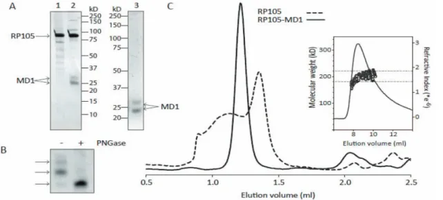
SDS-PAGE analysis of the purified proteins, shows that the solRP105-MD1 complex and MD1 preparations are .95% pure, whereas the solRP105 preparation shows some impurities (Figure 4A). solRP105 has the expected molecular weight of
,90 kD whereas MD1 shows as a double band at approxi-mately 23 and 26 kD (Figure 4A). Deglycosylation of denatured MD1 shows that the two bands in the MD1 preparation are due to differential glycosylation of the two N-glycosylation sites at positions Asn96 and Asn156 (Figure 4B). During size-exclusion chromatography, the solRP105-MD1 complex shows a single peak with an elution volume that is consistent with a hetero-tetrameric RP1052MD12 complex, as was observed in
the recently published crystal structures of the RP105-MD1 complex (16, 18). solRP105 shows a broad peak that is indicative of aggregation with a peak maximum at an elution volume that is consistent with monomeric RP105 (Figure 4C). The molecular weight of the complex was determined more accurately using SEC-MALS and yielding a value of 208611 kD, which is in good agreement with the expected molecular weight of 214 kD for fully glycosylated RP1052MD12
complex.
Soluble RP105 Protein – Functionality
We were able to show the increased TLR4 mediated inflammatory response in RP105 deficient myeloid cells (Figure S2). To see whether solRP105 or solRP105-MD1 could have effects on cytokine production by these circulating cells we performed anex vivowhole blood stimulation assay in the presence of these proteins. Blood was incubated with LPS in the presence of solRP105, solRP105-MD1 complex or PBS and TNFawas after measured after overnight LPS stimulation. solRP105-MD1 showed a marked decrease in TNFalpha production compared to controls (Figure 5).
Figure 1. RT-PCR of TLR4 (A) or RP105 (B) on wild type VSMC that were incubated with PBS, 1 ng/ml LPS or 10 ng/ml LPS. Immunostaining of TLR4 (green) or RP105 (red) or both (overlay) on wild type VSMC that were incubated with PBS (C) or 1 ng/ml LPS. (D) Colocalized TLR4 and RP105 staining on VSMC (E). RP105 staining in media area of a murine femoral artery (F). Proliferation of VSMC of WT and RP1052/2(G) Liquid-scintillation counting after 16 hours of WT and RP1052/2VSMC. VSMC of show a RP1052/2an increased proliferative response to LPS. VSMC were cultured in medium containing 10%FCS or 10%FCS with1 ng/ml LPS. Starved WT and RP1052/2VSMC were used as controls. White bars represent WT VSMC, black bars represent RP1052/2VSMC.
solRP105-MD1 Reduced Neointima Formation
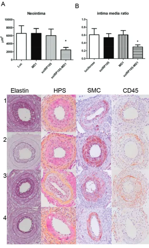
To see whether solRP105 could be a functional inhibitor of neointima formation in a more humanized model with mild foam cell formation in the neointima we applied our cuff model in ApoE3Leiden mice. Since RP105 also had strong effects on macrophages [15] important in foam cell formation [25] and the above described effects on VSMC proliferation we hypothesized that overexpression of the soluble form of the RP105 receptor, either alone or in complex with MD1, could have potential therapeutic benefits to reduce restenosis. Plasmids encoding solRP105, MD1 or luciferase (control) were injected in calf muscles followed by electroporation into the muscle cells 3 days before cuff placement in transgenic ApoE3Leiden mice that develop a diet induced hypercholesterolemia. This resulted in systemic overexpression of the recombinant proteins, as we have demonstrated previously [8,29,30]. Overexpression of both
solRP105 and MD1 proteins combined caused a significant reduction in neointima formation compared to luciferase control (25006573 vs. 658161894mm2, P,0.05) and a more beneficial intima media ratio (Figure 6A–D), whereas overexpression of either solRP105 or MD1 did not affect neointima formation.
Discussion
To our knowledge, the present study is the first to describe a role of RP105 in vascular disease. RP105 is expressed in cultured VSMC, as well as in the media. Furthermore, RP105 deficient mice develop less neointima after vascular injury and in these mice lacking the inhibitor TLR4 inhibitor R105, TLR4 activation via LPS clearly enhanced vascular remodeling more profoundly. Finally we demonstrated that overexpression of a soluble RP105/ MD1 protein complex strongly reduced neointima
forma-Figure 2. Neointima formation in WT and RP1052/2mice.Neointima formation after femoral artery cuff placement in RP1052/2and wild type
mice. Areas of femoral arterial sections were quantified by using 6 sequential sections per segment and are expressed in micrometers squared (mean6SEM). Increased neointima formation in RP1052/2mice compared to WT (wild type) controls (A). Increased intima/media ratio in RP1052/2 mice compared to WT controls (B). Representative pictures of Elastin von Giesson (C) HPS (D) anda-smooth muscle cell actin (F) of RP1052/2and WT controls. * = P,0.05 Arrows indicate the Internal Elastic Lamina (IEL) and the External Elastic Lamina (EEL).
tion.RP105 can be distinguished from other endogenous TLR regulators by being an extracellular TLR4 structural homolog with contrasting capabilities like the inhibitory effects on myeloid cells and activation of B cells [13]. Our study shows that RP105 protein is also expressed by vascular smooth muscle cells (VSMC). Others have shown RP105 presence on smooth muscle cells of the airway [31]. Additionally, we found via immunostaining as well as via RT-PCR that the expression of both receptors is down regulated upon overnight incubation with LPS as can be appreciated in figure 1. This is in line with previous observations in monocytes/ macrophages exposed to LPS, which show reduced responses to second stimulation with LPS. Furthermore, after an initial LPS stimulation, TLR4 mRNA expression was significantly decreased within a few hours and remained suppressed over 24 h. TLR4 presence at the cells surface rapidly decreased within 1 hour, with a gradual decrease after that [28]. In patients a similar situation can be observed where a reduction in TLR4 expression can be appreciated on monocytes at the end of cardiopulmonary bypass procedures [27]. While TLR4 might be down-regulated as a protective response, RP105 may just follow TLR4 expression as RP105 expression mimics that of TLR4 in human and mouse myeloid cells [15].
RP105 deficiency on VSMC caused an increase in proliferation upon incubation with TLR4 ligand LPS. A stimulating effect of TLR4 on VSMC proliferation was published previously [9]. This points towards a functional role of RP105 in vascular remodeling via its TLR4 inhibitory function, via its effects on VSMC in the pathophysiological process of neointima formation, as a common feature of restenosis. Femoral cuff placement in mice allows employing of a mild mechanically-induced inflammatory-based neointima formation. Lesions in mice consist primarily of VSMC and application of this model to RP1052/2and WT control mice leads to an increase in neointima formation in the RP1052/2 mice. This underscores the suggestion that RP105 is also functionally involved in the inhibition of post-interventional neointima formation, a process known to be regulated by TLR4 activation [4,5] and may play a role in the pathophysiological process of restenosis.
The influence of inflammation on restenosis is most prominent in the early phase after intervention. Interestingly, TLR4 was previously described to be involved in cardiovascular remodeling [5,6,32] and local application of LPS causes a strong increase in neointima formation after cuff placement [4]. RP105 deficient mice have a stronger pro-inflammatory response upon systemically delivered TLR4 ligand LPS [14]. In figure 3 we show that short term activation of TLR4 directly after cuff placement via local application of LPS causes a strong increase in neointima formation. As a result, the vessel seems to respond by compen-satory outward remodeling to secure the vessel patency [33]. In TLR4-deficient mice this response is inhibited upon local LPS application [4]. Due to a more prominent TLR4 activation in RP1052/2 mice we see that these mice have an increase in outward remodeling.
So far a protective role for RP105 on post-interventional remodeling is appreciated. This indicates potential for new strategies to use RP105 in a therapeutic approach. Therefore we focused on the development of recombinant protein expression. Divanovic et al showed that the extracellular region of RP105 is functional only when co-expressed with MD1 [15]. Similar to
Figure 3. LPS induced neointima formation.Neointima formation
after femoral artery cuff placement with LPS in RP1052/2and wild type mice. Areas of femoral arterial sections were quantified by using 6 sequential sections per segment and are expressed in micrometers squared (mean6SEM). Increased neointima formation in RP1052/2mice compared to WT controls after local LPS application (A). Increased intima/media ratio in RP1052/2mice compared to WT controls after local LPS application (B). Increased outward remodeling in RP1052/2 mice compared to WT controls after local LPS application (C)
Representative pictures of Elastin von Giesson (D), HPS (E) and a -smooth muscle cell actin (F) of RP1052/2and WT controls. * = P,0.05 Arrows indicate the Internal Elastic Lamina (IEL) and the External Elastic Lamina (EEL).
TLR4 surface expression that is dependent on MD2, RP105 surface expression was shown to be dependent on MD1 [34]. While we could express the soluble ectodomain of RP105 in the absence of MD1, we find that MD1 is necessary to prevent its aggregation. Monomeric RP105 protein is not capable of inhibiting the LPS response on whole blood while the RP105-MD1 complex did inhibit LPS induced TNFaproduction, a factor known to be important in neointima formation [7]. The exact mechanism of how RP105-MD1 regulates TLR4 signaling is still under discussion. However, it is suggested that the complex formation is crucial, since monomeric RP105 is highly unstable. It has been shown that RP105-MD1 interacts directly with
TLR4-MD2 [15]. The unusual 2:2 homodimer of the RP105-MD1 complex suggests two possible mechanisms for inhibition of TLR4-MD2 [18]; either (i) a lateral binding of TLR4-TLR4-MD2 to the RP105-MD1 complex which would involve direct MD1-MD2 interactions or, (ii) the formation of quasi-symmetrical TLR4-MD2/RP105-MD1 complexes reminiscent of the usual ligand-induced TLR homodimers. In both scenarios RP105-MD1 could block TLR4 homodimer formation and possibly prevent/destabi-lize LPS binding to MD2 [18]. However, the second mechanism was considered to be more plausible since it explains the regulation of the LPS response by RP105–MD1 regardless of LPS binding [18].
To set more focus on the therapeutic potential we initiated experiments to study the therapeutic potential of RP105 in a more humanized model having vessel damage combined with a diet induced hypercholesterolemia, i.e. cuff induced neointimal formation in hypercholesterolemic ApoE3*Leiden mice. No effect was seen of gene transfer of vectors expressing MD1 as a single factor. The absence of an effect of MD1 expression, as was also previously shown in vitro [15], demonstrates that the mechanism that leads to reduction in TLR4 signaling does not rely on MD1-MD2 interactions, thus the above-discussed scenario 1 is unlikely. Overexpression of solRP105 alone as a single factor did not alter neointima formation suggesting that the endogenously produced MD1 is not sufficient to stabilize RP105 by formation of complexes as is required in vitro (Figure 4). Combined overexpression of both proteins, however, resulted in the formation of a functional complex and led to a strong decrease in neointima formation. The soluble RP105/MD1 complex may act as a decoy receptor and as such have a therapeutic potential. Apparently, overexpression in vivo of solRP105 with MD1 at the site of production is essential to form a stable and effective RP105-MD1 complex for functional inhibition of neointimal formation. These results are also supportive for the quasi-symmetrical working mechanism of RP105-MD1, the above-discussed second scenario of RP105 mediated TLR4 inhibition.
Figure 4. Soluble RP105 protein – characterization.Purification and analysis of RP105, MD1 and RP105-MD1 complex. RP105 (lane 1) and
RP105-MD1 complex (lane 2) purified by nickel- and streptactin- tandem affinity purification and analysed by SDS-PAGE (10%) and subsequent silverstaining. Lane 3 shows MD1 purified by nickel-affinity purification and analysed by SDS-PAGE (12%) and subsequent Coomassie staining (A). SDS-PAGE analysis of PNGase deglycosylation of SDS denatured MD1 (B). Size exclusion chromatography (Superdex200 PC3.2/30) of the purified RP105 (dashed line) and RP105-MD1 complex (solid line). Molecular weight determination of the RP105-MD1 complex using SEC-MALS (inset). The dashed horizontal lines indicate a molecular weight of 180 kD and 220 kD, respectively (C).
doi:10.1371/journal.pone.0067923.g004
Figure 5. Soluble RP105 protein – functionality. Whole blood
Figure 6. SolRP105-MD1 reduced neointima formation. Neointima formation after femoral artery cuff placement with LPS in hypercholesterolemic APOE3Leiden mice. Areas of femoral arterial sections were quantified by using 6 sequential sections per segment and are expressed in micrometers squared (mean6SEM). Decreased neointima formation in mice that had overexpression of solRP105-MD1 (A). Intima Media ratio (B) Pictures of Elastin von Giesson (C) HPS (D)a-smooth muscle cell actin (E) and CD45 (F) of Luc(1) solRP105 (2) MD1 (3) solRP105-MD1 (4) * = P,0.05.
In summary, we demonstrate the presence of RP105 on cultured VSMC and in VSMC in vivo in media and neointima and the increased restenosis in RP1052/2mice indicate RP105 as a mediator of restenosis, probably as an inhibitor of TLR4. Furthermore, solRP105 significantly modulates the strong TLR4 driven vascular remodeling. Thus RP105 is an interesting new factor involved in the complex regulation of vascular remodeling and can be used to develop novel therapeutic strategies to inhibit neointima formation.
Supporting Information
Figure S1 Colocalized TLR4 and RP105 staining on VSMC shown by confocal microscopy.
(PDF)
Figure S2 TNFa levels produced upon LPS stimulation by dendritic cells (A) and macrophages (B) from WT and RP1052/2mice.* = P,0.05.
(PDF)
Acknowledgments
The authors thank K. Miyake for kindly providing the RP1052/2mice.
Author Contributions
Conceived and designed the experiments: JCK EGH TCB JWJ PHAQ. Performed the experiments: MME MRV SCJ HEO EAP HCB. Analyzed the data: AJZ JK EGH TCB. Contributed reagents/materials/analysis tools: AJZ JK. Wrote the paper: JCK TCB JWJ PHQ.
References
1. Jukema JW, Ahmed TA, Verschuren JJ, Quax PH (2012) Restenosis after PCI. Part 2: prevention and therapy. Nat Rev Cardiol 9: 79–90.
2. Jukema JW, Verschuren JJ, Ahmed TA, Quax PH (2012) Restenosis after PCI. Part 1: pathophysiology and risk factors. Nat Rev Cardiol 9: 53–62. 3. Monraats PS, Pires NM, Agema WR, Zwinderman AH, Schepers A, et al.
(2005) Genetic inflammatory factors predict restenosis after percutaneous coronary interventions. Circulation 112: 2417–2425.
4. Hollestelle SC, De Vries MR, Van Keulen JK, Schoneveld AH, Vink A, et al. (2004) Toll-like receptor 4 is involved in outward arterial remodeling. Circulation 109: 393–398.
5. Vink A, Schoneveld AH, van der Meer JJ, Van Middelaar BJ, Sluijter JP, et al. (2002) In vivo evidence for a role of toll-like receptor 4 in the development of intimal lesions. Circulation 106: 1985–1990.
6. Karper JC, De Vries MR, van den Brand BT, Hoefer IE, Fischer JW, et al. (2011) Toll-like receptor 4 is involved in human and mouse vein graft remodeling, and local gene silencing reduces vein graft disease in hypercholes-terolemic APOE*3Leiden mice. Arterioscler Thromb Vasc Biol 31: 1033–1040. 7. Monraats PS, Pires NM, Schepers A, Agema WR, Boesten LS, et al. (2005) Tumor necrosis factor-alpha plays an important role in restenosis development. FASEB J 19: 1998–2004.
8. Schepers A, Eefting D, Bonta PI, Grimbergen JM, De Vries MR, et al. (2006) Anti-MCP-1 gene therapy inhibits vascular smooth muscle cells proliferation and attenuates vein graft thickening both in vitro and in vivo. Arterioscler Thromb Vasc Biol 26: 2063–2069.
9. de Graaf R, Kloppenburg G, Kitslaar PJ, Bruggeman CA, Stassen F (2006) Human heat shock protein 60 stimulates vascular smooth muscle cell proliferation through Toll-like receptors 2 and 4. Microbes Infect 8: 1859–1865. 10. Kawai T, Akira S (2010) The role of pattern-recognition receptors in innate
immunity: update on Toll-like receptors. Nat Immunol 11: 373–384. 11. Kim HM, Park BS, Kim JI, Kim SE, Lee J, et al. (2007) Crystal structure of the
TLR4-MD-2 complex with bound endotoxin antagonist Eritoran. Cell 130: 906–917.
12. Shimamoto A, Chong AJ, Yada M, Shomura S, Takayama H, et al. (2006) Inhibition of Toll-like receptor 4 with eritoran attenuates myocardial ischemia-reperfusion injury. Circulation 114: I270–I274.
13. Akashi-Takamura S, Miyake K (2008) TLR accessory molecules. Curr Opin Immunol 20: 420–425.
14. Divanovic S, Trompette A, Atabani SF, Madan R, Golenbock DT, et al. (2005) Inhibition of TLR-4/MD-2 signaling by RP105/MD-1. J Endotoxin Res 11: 363–368.
15. Divanovic S, Trompette A, Atabani SF, Madan R, Golenbock DT, et al. (2005) Negative regulation of Toll-like receptor 4 signaling by the Toll-like receptor homolog RP105. Nat Immunol 6: 571–578.
16. Divanovic S, Trompette A, Petiniot LK, Allen JL, Flick LM, et al. (2007) Regulation of TLR4 signaling and the host interface with pathogens and danger: the role of RP105. J Leukoc Biol 82: 265–271.
17. Ohto U, Miyake K, Shimizu T (2011) Crystal Structures of Mouse and Human RP105/MD-1 Complexes Reveal Unique Dimer Organization of the Toll-Like Receptor Family. J Mol Biol. S0022–2836(11)01026-6.
18. Yoon SI, Hong M, Wilson IA (2011) An unusual dimeric structure and assembly for TLR4 regulator RP105-MD-1. Nat Struct Mol Biol 18: 1028–1035.
19. Freudenberg J, Lee AT, Siminovitch KA, Amos CI, Ballard D, et al. (2010) Locus category based analysis of a large genome-wide association study of rheumatoid arthritis. Hum Mol Genet 19: 3863–3872.
20. Tada Y, Koarada S, Morito F, Mitamura M, Inoue H, et al. (2008) Toll-like receptor homolog RP105 modulates the antigen-presenting cell function and regulates the development of collagen-induced arthritis. Arthritis Res Ther 10: R121.
21. Tanne A, Ma B, Boudou F, Tailleux L, Botella H, et al. (2009) A murine DC-SIGN homologue contributes to early host defense against Mycobacterium tuberculosis J Exp Med 206: 2205–2220.
22. Ait-Oufella H, Herbin O, Bouaziz JD, Binder CJ, Uyttenhove C, et al. (2010) B cell depletion reduces the development of atherosclerosis in mice. J Exp Med 207: 1579–1587.
23. Arkenbout EK, de Waard V, van Bragt M., van Achterberg TA, Grimbergen JM, et al. (2002) Protective function of transcription factor TR3 orphan receptor in atherogenesis: decreased lesion formation in carotid artery ligation model in TR3 transgenic mice. Circulation 106: 1530–1535.
24. Miyake K, Yamashita Y, Ogata M, Sudo T, Kimoto M (1995) RP105, a novel B cell surface molecule implicated in B cell activation, is a member of the leucine-rich repeat protein family. J Immunol 154: 3333–3340.
25. Lardenoye JH, Delsing DJ, De Vries MR, Deckers MM, Princen HM, et al. (2000) Accelerated atherosclerosis by placement of a perivascular cuff and a cholesterol-rich diet in ApoE*3Leiden transgenic mice. Circ Res 87: 248–253. 26. McMahon JM, Signori E, Wells KE, Fazio VM, Wells DJ (2001) Optimisation of
electrotransfer of plasmid into skeletal muscle by pretreatment with hyaluron-idase – increased expression with reduced muscle damage. Gene Ther 8: 1264– 1270.
27. Hadley JS, Wang JE, Michaels LC, Dempsey CM, Foster SJ, et al. (2007) Alterations in inflammatory capacity and TLR expression on monocytes and neutrophils after cardiopulmonary bypass. Shock 27: 466–473.
28. Nomura F, Akashi S, Sakao Y, Sato S, Kawai T, et al. (2000) Cutting edge: endotoxin tolerance in mouse peritoneal macrophages correlates with down-regulation of surface toll-like receptor 4 expression. J Immunol 164: 3476–3479. 29. Eefting D, Schepers A, De Vries MR, Pires NM, Grimbergen JM, et al. (2007) The effect of interleukin-10 knock-out and overexpression on neointima formation in hypercholesterolemic APOE*3-Leiden mice. Atherosclerosis 193: 335–342.
30. Eefting D, Seghers L, Grimbergen JM, De Vries MR, de Boer HC, et al. (2010) A novel urokinase receptor-targeted inhibitor for plasmin and matrix metalloproteinases suppresses vein graft disease. Cardiovasc Res 88: 367–375. 31. Zhang Y, Xu CB, Cardell LO (2009) Long-term exposure to IL-1beta enhances
Toll-IL-1 receptor-mediated inflammatory signaling in murine airway hyperre-sponsiveness. Eur Cytokine Netw 20: 148–156.
32. Timmers L, Sluijter JP, Van Keulen JK, Hoefer IE, Nederhoff MG, et al. (2008) Toll-like receptor 4 mediates maladaptive left ventricular remodeling and impairs cardiac function after myocardial infarction. Circ Res 102: 257–264. 33. Ward MR, Pasterkamp G, Yeung AC, Borst C (2000) Arterial remodeling.
Mechanisms and clinical implications. Circulation 102: 1186–1191. 34. Miyake K, Shimazu R, Kondo J, Niki T, Akashi S, et al. (1998) Mouse MD-1, a