viruses
Article
Human Norovirus Histo-Blood Group Antigen
(HBGA) Binding Sites Mediate the Virus Specific
Interactions with Lettuce Carbohydrates
Malak A. Esseili1,2,*, Xiang Gao1, Patricia Boley1, Yixuan Hou1, Linda J. Saif1, Paul Brewer-Jensen3, Lisa C. Lindesmith3 , Ralph S. Baric3, Robert L. Atmar4and Qiuhong Wang1,*
1 Food Animal Health Research Program, Ohio Agricultural Research and Development Center,
Department of Veterinary Preventive Medicine, The Ohio State University, Wooster, OH 44691, USA 2 Currently at Center for Food Safety, Department of Food Science and Technology, University of Georgia,
Griffin, GA 30223, USA
3 Department of Epidemiology, University of North Carolina, Chapel Hill, NC 27599-7435, USA 4 Department of Molecular Virology and Microbiology and Department of Medicine, Baylor College of
Medicine, Houston, TX 77030, USA
* Correspondence: [email protected] (M.A.E.); [email protected] (Q.W.); Tel.:+1-770-228-7313 (M.A.E.); +1-330-263-3960 (Q.W.)
Received: 8 August 2019; Accepted: 6 September 2019; Published: 8 September 2019
Abstract:Lettuce is often implicated in human norovirus (HuNoV) foodborne outbreaks. We identified H-like histo-blood group antigens (HBGAs) on lettuce leaves as specific binding moieties for virus-like particles (VLPs) of HuNoV GII.4/HS194/2009 strain. The objective of this study was to determine whether HuNoV-lettuce binding is mediated through the virus HBGA binding sites (HBS). Toward this objective, VLPs of historical HuNoV GII.4 strains (1987, 1997, 2002, 2004 and 2006) with known natural mutations in their HBS, two newly generated VLP mutants of GII.4/HS194/2009 (D374A and G443A) and a VLP mutant (W375A) of GI.1/Norwalk/1968 along with its wild type VLPs, which displays distinct HBS, were investigated for their binding to lettuce. ELISA revealed that historical GII.4 strains binding to lettuce was dependent on their HBGAs profiles. The VLP mutants D374A and G443A lost binding to HBGAs and displayed no to minimal binding to lettuce, respectively. The VLPs of GI.1/Norwalk/1968 strain bound to lettuce through an H-like HBGA and the binding was inhibited by fucosidase digestion. Mutant W375A which was previously shown not to bind to HBGAs, displayed significantly reduced binding to lettuce. We conclude that the binding of HuNoV GII.4 and GI.1 strains to lettuce is mediated through the virus HBS.
Keywords: Foodborne viruses; Human norovirus; Lettuce; Histo-blood group antigens; Virus-lettuce interactions; Virus-like particles; mutations; H-like antigens; virus HBGA-binding sites
1. Introduction
Human norovirus (HuNoV) continues to be the leading cause of foodborne disease outbreaks in the United States (US) [1]. It is estimated that 9.4 million foodborne illnesses occur annually in the US. The majority of these foodborne illnesses (58%) are caused by HuNoV with an estimated 15,000 hospitalizations and 150 deaths annually and over $2 billion in health-care costs [2,3]. Human norovirus is transmitted via the fecal-oral route, including direct person to person, consumption of contaminated food or water or contact with fomites. Infections with HuNoV cause gastrointestinal symptoms (diarrhea, vomiting, stomach pain and nausea) which are usually self-limiting in healthy adults but can be severe and prolonged in young children, elderly and immune-compromised
Viruses2019,11, 833 2 of 14
patients [4]. The virus is a small (27–40 nm in diameter), non-enveloped, single stranded RNA virus that belongs to theCaliciviridaefamily [5]. Human norovirus can be classified into at least five genogroups (GI–GV), which are further each subdivided into genotypes [5]. Infections with HuNoV are caused by both GI and GII strains. Among them, the GII.4 HuNoVs are the dominant strains causing the majority (~60–90%) of gastroenteritis outbreaks [6]. Currently, there are HuNoV vaccine candidates undergoing clinical trials [7–9]. However, in the absence of approved antivirals or vaccines against HuNoV, the virus continues to exert a significant global burden, estimated at ~$64 billion in direct (healthcare) and indirect (loss of productivity) costs [10].
Viruses2019,11, 833 3 of 14
2 (residues 390–395), which stabilizes the interactions along with several other residues proximal to those sites forming a pocket [22,24,25]. Studies with GI.1 HuNoVs revealed that HBGA binding occurs through residues 327, 375, 377, 378 to theβ-Gal of H-type HBGA or the GalNAc of A-type HBGA and through residues 329, 342, 344, 346, 375 and 380 toα-fucose of the H- or A-type HBGA [22,26]. For GI.1 HuNoVs, the amino acids at these positions are well conserved but for GII.4 HuNoVs only site 1 residues are conserved. Altered HBGA binding patterns, coupled with extensive antigenic variation in response to herd immunity, have resulted in the emergence of new variants of GII.4 HuNoV every 3–7 years [27]. For example, the first major HuNoV pandemic occurred in 1995 to 1997, which was replaced by subsequent pandemics in 2002, 2004, 2006, 2009 and 2012 [5].Variations in the surface exposed amino acids in the P2 regions correlate with altered HBGA binding capacity and antigenic variation that allows the virus to escape from existing protective immunity [21].
The objective of this study was to determine whether HuNoV binding to lettuce carbohydrates is mediated through the virus HBS. To accomplish this objective, we have assessed the binding of some of the historical GII.4 VLPs (Table1) to lettuce cell wall carbohydrates to determine whether the changes in the HBGA binding profiles affect their binding to lettuce. Then, using the HuNoV GII.4/HS194/2009 VLP strain that we characterized previously [16,18], we generated two VLP mutants with single amino acids substitutions at residues 374 and 443 located within the HBS. The VLP mutants were assessed for their binding to lettuce. In addition, VLPs for GI.1/Norwalk/1968 (referred to as GI.1), which has distinct HBS and its previously characterized mutant (W375A) that lost HBGA binding [22] were investigated for their potential binding to lettuce.
Table 1.A list of virus-like particles of GI.1 and GII.4 used in our study and their histo blood group antigen (HBGA) binding profiles as adapted from previous studies [21,22,24,28,29]. HBGA profile for GII.4.HS194 was generated in this study as described previously [30,31].
Genotype Year Strain GenBank
Accession No. VLP Variant VLP Name HBGA Profile
GI.1 1968 Norwalk M87661 Hu/US/1968/GI.1/Norwalk GI.1.1968 H1, H3 and A GII.4 1987 Camberwell AAK50355.1 Hu/GII.4/MD145-12/1987/US GII.4.1987 H3 and Ley
1997 US95/96 AFJ04707.1 Hu/GII.4/1997/USA GII.4.1997 H3, A, B, Le y
and Leb
2002 Farmington
Hill AFJ04708.1
Hu/GII.4/Farmington
Hills/2002/USA GII.4.2002 Le
y, H3 and B
2004 Hunter AAZ31376.2 Hu/GII.4/Hunter 284E/2004/AU GII.4.2004 None 2006 Minerva AFJ04709.1 Hu/GII.4/Minerva/2006/USA GII.4.2006 H3, B and A
2009 HS194 GU325839 Hu/GII.4/Minerva/2006/US GII.4.HS194 H3, B, H1, Le y,
A and Leb
2. Materials and Methods
2.1. Production and Purification of HuNoV VLPs
Viruses2019,11, 833 4 of 14
2.2. Generation of GII.4 Mutant VLP
The two recombinant baculoviruses for the expression of VLP mutants (D374A and G443A) of the HuNoV GII.4/HS194/2009/US strain were generated in this study. The Gateway entry vector (pENTRTM/SD/D-TOPO-HS194) containing the major capsid protein VP1 gene of WT HS194.2009 strain was used as a template. The QuikChange II XL site-directed mutagenesis kit (Agilent Technology, Santa Clara, CA, USA) was used to make the site mutation in the VP1 gene in the entry vector for the mutants. To mutate the D (aspartic acid) to A (Alanine) at amino acid 374 (D374A), forward and reverse primers were: 50-GGTACTGATACAGAAAATGCCTTTGAAACTCACC-30and 50-GGTGAGTTTCAAAGGCATTTTCTGTATCAGTACC-30, respectively. To mutate G (Glycine) to A (Alanine) at amino acid 443 (G443A), the forward and reverse primers were 50
-GCCCGGATGC AGCGCGTATCCCAACAACATGG-30 and 50-CCATGTTGGGATACGCGCTGCATCCGGGC-30, respectively. After confirmation by sequencing, LR recombination was performed with the entry vector and BaculoDirectTMLinear DNA to generate the recombinant baculovirus DNA carrying the mutant VP1 gene of HS194. The insect Sf9 cells were transfected by the recombination reaction products and the cells containing the recombinant baculovirus DNA were positively selected by ganciclovir. The expression of mutated HS194 capsid proteins in the Sf9 cells and culture supernatants were examined by immunoblot using the guinea pig antiserum against HS194 strain. The recombinant baculoviruses were propagated to prepare virus stocks with high titers for routine VLP expression. The production, purification and examination of GII.4.HS194 VLP mutants were performed as referenced above [16].
2.3. Extraction of Plant Cell Wall Material (CWM)
Extraction of lettuce leaves CWM was performed at 4◦C as described in our previous work [16]. Mature romaine lettuce heads were purchased from a local grocery store. Briefly, leaves were ground in liquid nitrogen to fine powder. The powder was then homogenized in 80% ethanol using polytron homogenizer (Cole-Parmer Instruments, Vernon Hills, IL, USA), followed by centrifugation for 20 min at 2500×g. The resulting pellet was subjected to a series of ethanol washes (80% twice and 100% once) and was then stirred for 2 h at 4◦C in 60 mL methanol: chloroform (1:1,v/v). The pellet was collected by filtration using Whatman® glass microfiber filters grade GF/A (Sigma, St. Louis, MO, USA), washed with ice-cold methanol: chloroform and finally with ice-cold acetone. The pellet was vacuum dried overnight at RT and the resulting CWMs were stored at 4◦C in a tightly sealed bottle under dark conditions.
Viruses2019,11, 833 5 of 14
(KPL, USA) for 10 min at RT. The reaction was stopped with 0.3 M sulfuric acid and the absorbance was read at 450nm. Controls included: (i) wells coated with CWM but no VLP added (named “No VLPs”); (ii) wells with CWM, VLP and secondary antibody but without primary antibody (named “No primary”); and (iii) PBS coated wells with VLP, primary and secondary antibodies added (named “No coating”).
2.5. Porcine Gastric Mucin and Human B-type HBGA Saliva ELISA
Purified porcine gastric mucin (PGM) type III (Sigma, USA) was used as a positive carbohydrate control since it was previously reported to contain primarily A, H and less of Leband Leyantigens [16,18,28,34]. In addition, the GII.4.HS194 VLPs were shown to bind to PGM through the A and H antigens [16]. For ELISA with historical GII.4 VLPs and GI.1 VLPs, we used PGM at 1mg/mL, a concentration experimentally determined to generate an absorbance double that of HuNoV VLP binding to CWM. The ELISA protocol described above was followed.
For assessing whether the two mutations at position 374 and 443 of GII.4.HS194 affected its HBGA binding profile, binding to PGM or B-type HBGA human saliva was tested. First, to determine whether the primary antibody (rabbit anti-GII.4 VLPs) can still detect the mutant VLPs, an ELISA plate was coated with 2µg/mL of each of the WT and mutant VLPs for 4 h at RT. Following an overnight standard blocking and washing steps, the primary antibody was serially diluted (1:10), added in duplicate to each VLP-coated wells and allowed to bind for 1 h at 37◦C while the secondary antibody (goat anti-rabbit IgG HRP-conjugated) was added at 1:2000 as described above. The primary antibody bound to WT and mutant VLPs without significant differences at all the tested dilutions; however, the binding signal for WT and mutant VLPs started to steadily decrease beyond the 1:8000 dilution (data not shown). Second, ELISA plates were coated with PGM at 5mg/mL or with a human blood type B saliva at 1:500 dilution. Briefly, 16µg/mL of each of the two mutants and the wildtype VLPs were serially diluted (1:2), added in duplicate to the wells and allowed to bind for 1 h at 37◦C. Following standard washings, blocking and incubations as described above, the primary antibody (rabbit anti-GII.4 VLPs) was added at 1:8000, while the secondary antibody (goat anti-rabbit IgG HRP-conjugated) was added at 1:2000. The ELISA plates were stopped and read at A450nm.
2.6. Detection of HuNoV VLP Binding to Lettuce Tissues Using Immunofluorescence Assay (IFA)
The VLP-lettuce tissue IFA protocol was followed as previously described [16,18]. Briefly, romaine lettuce heads were purchased from a local grocery store. The leaves were cut into small pieces (0.5×0.5 cm2), dehydrated in 30 mL of gradient ethanol series (70% (v/v), 90%, 95%, 100% and 100%) for 6 h and embedded in paraffin blocks. Five-micrometer sections were cut using a microtome and collected on glass slides (Fisher Scientific, Pittsburgh, PA, USA). Slides were dried at 37◦C for 2 h, deparaffinized in xylene for 15 min and rehydrated in 30 mL gradient of ethanol series (70%, 90%, 95%, 100%, 100%,v/v). Slides were incubated with cellulase R-10 (PhytoTechnology Laboratories, Lenexa, KS, USA) at a final concentration of 1µg/µL at 37◦C for 1 h. Following washing three times with 300 mL of PBS containing 0.05% Tween-20 (PBS-T), blocking was performed with 200µL of PBS supplemented with 1% bovine serum albumin (BSA). For fucosidase digestion, each slide was digested with 20 U
Viruses2019,11, 833 6 of 14
pig or anti-rabbit IgG (Invitrogen) at 1:700 dilution (green signal). Negative controls used included slides treated as above but without the VLPs. The slides were washed three times with 300 mL PBS-T after each incubation step. The plant chloroplast autofluorescence was shown in red while nuclei were stained with 4’,6-diamidino-2- phenylindole (DAPI) and shown in blue. All pictures were taken using the same exposure time.
2.7. Statistical Analyses
GraphPad Prism® version 5 (GraphPad Software, LaJolla, CA, USA) was used for statistical analyses. One-Way ANOVA followed by Tukey’s post hoc test was used to determine significant differences between the different treatments. Each treatment in each ELISA assay was run in triplicate and the ELISA was repeated at least twice to determine assay consistency. The VLP mutants (D37A and G443A) dose response ELISA with B-type saliva and PGM were only performed once with duplicate wells but with multiple VLP concentrations. The Alexa Fluor 488 green fluorescent intensity of all IFA images was measured by ImageJ software (https://imagej.nih.gov/ij/). The mean fluorescent intensity for WT VLPs (GII.4 or GI.1) was transformed to 100% and the intensity values of the different treatments were adjusted accordingly. Differences in means were considered significant when thep-value was
<0.05. Data were represented as mean±standard errors. Significantly different means were denoted by different alphabetical letters. The binding of VLPs to PGM or lettuce CWM was considered positive when the A450signal was significantly higher than the highest A450of control negative wells.
3. Results
3.1. Binding of Historical GII.4 VLPs to Lettuce CWM Correlated with Their PGM Binding
The binding of our previously characterized strain GII.4/HS194/2009 to PGM was not significantly different from that of the GII.4 historical strains of 1987, 1997 and 2006 (Figure1A). The GII.4.2002 VLPs showed significantly reduced signal in comparison to the 1987, 1997, 2006 and 2009 VLPs (Figure1A). However, the binding of GII.4.2004 VLPs to PGM was not statistically different from negative control wells (Figure1A). In a similar trend to PGM, the GII.4 of 1987, 1997, 2006 and 2009 exhibited a higher binding signal to lettuce CWM than that of GII.4 of 2002 and 2004 (Figure1B). The signal of GII.4.2002 and GII.4.2004 was not significantly different than that of the negative control wells (Figure1B).
Viruses 2019, 11, x FOR PEER REVIEW 6 of 14
using guinea pig anti-GII.4 NoV VLPs or rabbit anti-GI.1 VLP antisera (1:1000) and Alexa Fluor 488-conjugated goat anti-guinea pig or anti-rabbit IgG (Invitrogen) at 1:700 dilution (green signal). Negative controls used included slides treated as above but without the VLPs. The slides were washed three times with 300 mL PBS-T after each incubation step. The plant chloroplast autofluorescence was shown in red while nuclei were stained with 4’,6-diamidino-2- phenylindole (DAPI) and shown in blue. All pictures were taken using the same exposure time.
2.7. Statistical Analyses
GraphPad Prism® version 5 (GraphPad Software, LaJolla, CA, USA) was used for statistical
analyses. One-Way ANOVA followed by Tukey’s post hoc test was used to determine significant differences between the different treatments. Each treatment in each ELISA assay was run in triplicate and the ELISA was repeated at least twice to determine assay consistency. The VLP mutants (D37A and G443A) dose response ELISA with B-type saliva and PGM were only performed once with duplicate wells but with multiple VLP concentrations. The Alexa Fluor 488 green fluorescent intensity of all IFA images was measured by ImageJ software (https://imagej.nih.gov/ij/). The mean fluorescent intensity for WT VLPs (GII.4 or GI.1) was transformed to 100% and the intensity values of the different treatments were adjusted accordingly. Differences in means were considered significant when the p-value was <0.05. Data were represented as mean ± standard errors. Significantly different means were denoted by different alphabetical letters. The binding of VLPs to PGM or lettuce CWM was considered positive when the A450 signal was significantly higher than the highest A450 of control
negative wells.
3. Results
3.1. Binding of Historical GII.4 VLPs to Lettuce CWM Correlated with Their PGM Binding
The binding of our previously characterized strain GII.4/HS194/2009 to PGM was not significantly different from that of the GII.4 historical strains of 1987, 1997 and 2006 (Figure 1A). The GII.4.2002 VLPs showed significantly reduced signal in comparison to the 1987, 1997, 2006 and 2009 VLPs (Figure 1A). However, the binding of GII.4.2004 VLPs to PGM was not statistically different from negative control wells (Figure 1A). In a similar trend to PGM, the GII.4 of 1987, 1997, 2006 and 2009 exhibited a higher binding signal to lettuce CWM than that of GII.4 of 2002 and 2004 (Figure 1B). The signal of GII.4.2002 and GII.4.2004 was not significantly different than that of the negative control wells (Figure 1B).
Figure 1. Enzyme linked immunosorbent assay (ELISA) screening of historical HuNoV GII.4 virus-like particles (VLPs) for their binding to (A) Porcine gastric mucin (PGM) and (B) lettuce leaves cell wall material (CWM). Means with different letters differ significantly.
Viruses2019,11, 833 7 of 14
3.2. Loss of HBGA Binding Profile for GII.4 D374A and G443A VLP Mutants
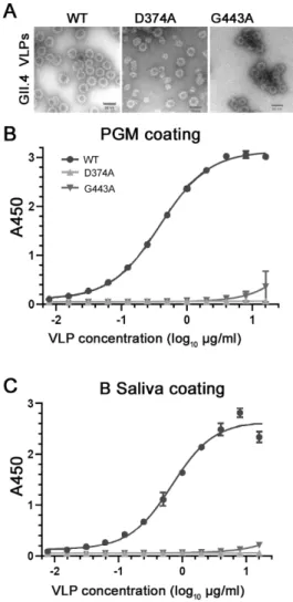
The VLPs of the two mutants D374A and G443A showed typical morphology and size (~40 nm in diameter) under TEM as those of the GII.4.HS194.2009 WT VLP (Figure2A). Besides the 40 nm particles, the VLPs of the mutant D374A also formed particles with a 27 nm in diameter (Figure2A). Neither VLP mutant showed binding to PGM (Figure2B) or the B-type HBGA human saliva (Figure2C) at all concentrations tested, with the exception of some binding of mutant G443A at high concentrations 10–16µg/mL withA450values of≤0.4. In contrast, the WT VLPs binding to PGM (Figure2B) or the B-type HBGA human saliva (Figure2C) increased with increasing VLP concentrations.
Viruses 2019, 11, x FOR PEER REVIEW 7 of 14
3.2. Loss of HBGA Binding Profile for GII.4 D374A and G443A VLP Mutants
The VLPs of the two mutants D374A and G443A showed typical morphology and size (~40nm in diameter) under TEM as those of the GII.4.HS194.2009 WT VLP (Figure 2A). Besides the 40 nm particles, the VLPs of the mutant D374A also formed particles with a 27 nm in diameter (Figure 2A). Neither VLP mutant showed binding to PGM (Figure 2B) or the B-type HBGA human saliva (Figure 2C) at all concentrations tested, with the exception of some binding of mutant G443A at high concentrations 10–16 µg/mL with A450 values of ≤ 0.4. In contrast, the WT VLPs binding to PGM
(Figure 2B) or the B-type HBGA human saliva (Figure 2C) increased with increasing VLP concentrations.
Figure 2. Characterization of the VLPs of HuNoV GII.4.HS194.2009 WT and its two mutants D374A and G443A by (A) transmission electron microscopy to show their sizes and morphology and by (B) PGM ELISA and (C) human B-type HBGA saliva ELISA to detect their binding to HBGA.
3.3. Amino Acid Residues 374 and 443 are Essential for GII.4 Binding to Lettuce
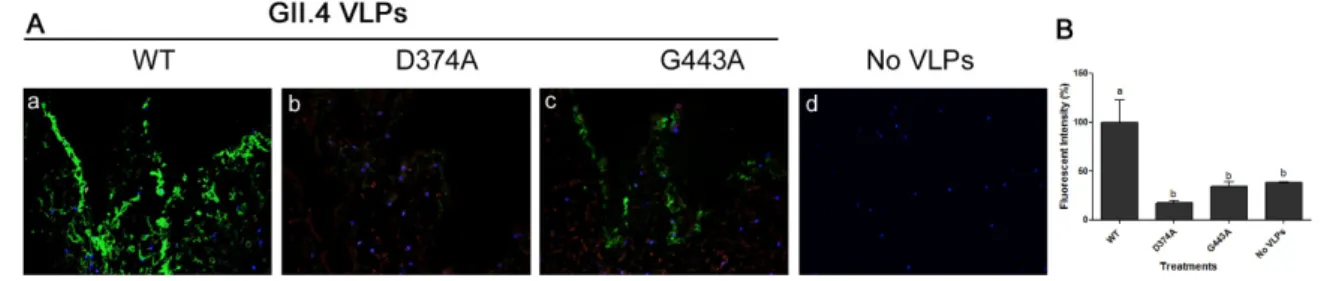
Mutation of WT GII.4.HS194.2009 at amino acid residue 374, abolished VLP binding to PGM, as its binding signal was similar to that of negative control wells (Figure 3A). However, mutation at the amino acid residue 443 significantly reduced the VLP binding to PGM by over 80%. The binding of mutant G443A VLPs to PGM was significantly higher than negative control wells (Figure 3A). Binding of the two mutant VLPs to lettuce CWM showed a similar trend as their binding to PGM (Figure 3B). Specifically, mutation of the WT at residue 374 abolished its binding to lettuce CWM, while mutation at residue 443 significantly reduced VLP binding by over 60%. Using IFA, the WT GII.4.HS194.2009 VLPs were shown to bind to lettuce tissues (Figure 4A a), while slides with no VLP added had no signal (Figure 4A d). The VLP mutant D374A lost its binding to lettuce tissues (Figure 4b), while mutant G443A showed weak IFA binding signal (Figure 4A c). Specifically, the fluorescent
Figure 2.Characterization of the VLPs of HuNoV GII.4.HS194.2009 WT and its two mutants D374A and G443A by (A) transmission electron microscopy to show their sizes and morphology and by (B) PGM ELISA and (C) human B-type HBGA saliva ELISA to detect their binding to HBGA.
3.3. Amino Acid Residues 374 and 443 are Essential for GII.4 Binding to Lettuce
Viruses2019,11, 833 8 of 14
(Figure4Ad). The VLP mutant D374A lost its binding to lettuce tissues (Figure4b), while mutant G443A showed weak IFA binding signal (Figure4Ac). Specifically, the fluorescent intensity of GII.4 WT was significantly different from that of D374A and G443A mutant VLPs and the negative control. Mutant D374A showed approximately 82% loss of fluorescent signal as compared to WT VLPs, while G443A showed a 65% loss in fluorescent signal (Figure4B).
Viruses 2019, 11, x FOR PEER REVIEW 8 of 14
intensity of GII.4 WT was significantly different from that of D374A and G443A mutant VLPs and the negative control. Mutant D374A showed approximately 82% loss of fluorescent signal as compared to WT VLPs, while G443A showed a 65% loss in fluorescent signal (Figure 4B).
Figure 3. ELISA showing the binding of HuNoV GII.4 WT VLPs and its two mutants D374A and G443A VLPs to (A) PGM and to (B) lettuce CWM. Means with different letters differ significantly.
Figure 4. (A) Immunofluorescence assay (IFA) showing the binding of (a) GII.4 WT VLPs and its two mutants (b) D374A and (c) G443A VLPs to lettuce tissues as well as (d) control negative with no VLP added, Magnification: 40×.. (B) Quantification of fluorescent intensity. Means with different letters differ significantly.
3.4. HuNoV GI.1 Binds to Lettuce Tissues Through a Fucose Moiety on H-like HBGA
The binding of GI.1 to lettuce was visualized using IFA (Figure 5Aa). Pre-treatment of the lettuce slides with monoclonal antibody against H-type HBGA eliminated the VLP IFA signal (Figure 5Ab), while treatment with fucosidase highly reduced the signal (Figure 5Ac). In contrast, a pre-treatment with lectins DBA (recognizing A-type HBGA) and LEA (recognizing GlcNAc) or LcH (recognizing α-D-Man/α-D-Glc) and BS-I (B-type HBGA) had no effect on the GI.1 VLP IFA signal (Figure 5A d and e). Specifically, the fluorescent intensity of GI.1 binding to lettuce tissues was only significantly different from that of anti-H HBGA and fucosidase pre-treatments but not from those of lectin pretreatments. Pre-treatment with anti-H HBGA and fucosidase reduced the fluorescent signal by approximately 92 and 67% as compared to that of GI.1 signal (Figure 5B).
Figure 3. ELISA showing the binding of HuNoV GII.4 WT VLPs and its two mutants D374A and G443A VLPs to (A) PGM and to (B) lettuce CWM. Means with different letters differ significantly.
Viruses 2019, 11, x FOR PEER REVIEW 8 of 14
intensity of GII.4 WT was significantly different from that of D374A and G443A mutant VLPs and the negative control. Mutant D374A showed approximately 82% loss of fluorescent signal as compared to WT VLPs, while G443A showed a 65% loss in fluorescent signal (Figure 4B).
Figure 3. ELISA showing the binding of HuNoV GII.4 WT VLPs and its two mutants D374A and G443A VLPs to (A) PGM and to (B) lettuce CWM. Means with different letters differ significantly.
Figure 4. (A) Immunofluorescence assay (IFA) showing the binding of (a) GII.4 WT VLPs and its two mutants (b) D374A and (c) G443A VLPs to lettuce tissues as well as (d) control negative with no VLP added, Magnification: 40×.. (B) Quantification of fluorescent intensity. Means with different letters differ significantly.
3.4. HuNoV GI.1 Binds to Lettuce Tissues Through a Fucose Moiety on H-like HBGA
The binding of GI.1 to lettuce was visualized using IFA (Figure 5Aa). Pre-treatment of the lettuce slides with monoclonal antibody against H-type HBGA eliminated the VLP IFA signal (Figure 5Ab), while treatment with fucosidase highly reduced the signal (Figure 5Ac). In contrast, a pre-treatment with lectins DBA (recognizing A-type HBGA) and LEA (recognizing GlcNAc) or LcH (recognizing α-D-Man/α-D-Glc) and BS-I (B-type HBGA) had no effect on the GI.1 VLP IFA signal (Figure 5A d and e). Specifically, the fluorescent intensity of GI.1 binding to lettuce tissues was only significantly different from that of anti-H HBGA and fucosidase pre-treatments but not from those of lectin pretreatments. Pre-treatment with anti-H HBGA and fucosidase reduced the fluorescent signal by approximately 92 and 67% as compared to that of GI.1 signal (Figure 5B).
Figure 4.(A) Immunofluorescence assay (IFA) showing the binding of (a) GII.4 WT VLPs and its two mutants (b) D374A and (c) G443A VLPs to lettuce tissues as well as (d) control negative with no VLP added, Magnification: 40×. (B) Quantification of fluorescent intensity. Means with different letters differ significantly.
3.4. HuNoV GI.1 Binds to Lettuce Tissues Through a Fucose Moiety on H-like HBGA
The binding of GI.1 to lettuce was visualized using IFA (Figure5Aa). Pre-treatment of the lettuce slides with monoclonal antibody against H-type HBGA eliminated the VLP IFA signal (Figure5Ab), while pre-treatment with fucosidase highly reduced the signal (Figure5Ac). In contrast, a pre-treatment with lectins DBA (recognizing A-type HBGA) and LEA (recognizing GlcNAc) or LcH (recognizing
Viruses2019,11, 833 9 of 14
Viruses 2019, 11, x FOR PEER REVIEW 8 of 14
intensity of GII.4 WT was significantly different from that of D374A and G443A mutant VLPs and the negative control. Mutant D374A showed approximately 82% loss of fluorescent signal as compared to WT VLPs, while G443A showed a 65% loss in fluorescent signal (Figure 4B).
Figure 3. ELISA showing the binding of HuNoV GII.4 WT VLPs and its two mutants D374A and G443A VLPs to (A) PGM and to (B) lettuce CWM. Means with different letters differ significantly.
Figure 4. (A) Immunofluorescence assay (IFA) showing the binding of (a) GII.4 WT VLPs and its two mutants (b) D374A and (c) G443A VLPs to lettuce tissues as well as (d) control negative with no VLP added, Magnification: 40×.. (B) Quantification of fluorescent intensity. Means with different letters differ significantly.
3.4. HuNoV GI.1 Binds to Lettuce Tissues Through a Fucose Moiety on H-like HBGA
The binding of GI.1 to lettuce was visualized using IFA (Figure 5Aa). Pre-treatment of the lettuce slides with monoclonal antibody against H-type HBGA eliminated the VLP IFA signal (Figure 5Ab), while treatment with fucosidase highly reduced the signal (Figure 5Ac). In contrast, a pre-treatment with lectins DBA (recognizing A-type HBGA) and LEA (recognizing GlcNAc) or LcH (recognizing α-D-Man/α-D-Glc) and BS-I (B-type HBGA) had no effect on the GI.1 VLP IFA signal (Figure 5A d and e). Specifically, the fluorescent intensity of GI.1 binding to lettuce tissues was only significantly different from that of anti-H HBGA and fucosidase pre-treatments but not from those of lectin pretreatments. Pre-treatment with anti-H HBGA and fucosidase reduced the fluorescent signal by approximately 92 and 67% as compared to that of GI.1 signal (Figure 5B).
Figure 5.(A) Immunofluorescence assay showing the binding of (a) GI.1 VLPs to the lettuce tissues without any pre-treatment and following pre-treatments with (b) anti-H HBGA antibody, (c) fucosidase enzyme or lectins (d) DBA/LEA and (e) LcH/BS-1, Magnification: 40×. (B) Quantification of fluorescent intensity. Means with different letters differ significantly.
3.5. Amino Acid at Position 375 is Important for GI.1 Binding to Lettuce
HuNoV GI.1 VLPs bound to PGM, while its mutant W375A exhibited significantly reduced binding (Figure6A). The binding of the mutant VLPs was reduced by ~85% and was not significantly different from the three negative controls. Binding of GI.1 and its mutant VLPs to lettuce CWM showed a similar trend to their binding to PGM (Figure6B). Specifically, GI.1 VLPs bound to lettuce while its mutant W375A VLPs, exhibited significantly reduced binding by ~55%. The binding of the mutant W375A was significantly higher than any of the three negative controls (Figure6B). Mutant W375A showed a weak IFA signal, which is nearly comparable to the negative control slide (Figure7Ab,c). Specifically, the fluorescent intensity of G1.1 WT VLP was significantly different than that of W375A mutant VLPs and the negative control. Mutant W375A showed approximately 75% loss of fluorescent signal as compared to WT VLPs (Figure7B).
Viruses 2019, 11, x FOR PEER REVIEW 9 of 14
Figure 5. (A) Immunofluorescence assay showing the binding of (a) GI.1 VLPs to the lettuce tissues without any pre-treatment and following pre-treatments with (b) anti-H HBGA antibody, (c)
fucosidase enzyme or lectins (d) DBA/LEA and (e) LcH/BS-1, Magnification: 40×. (B) Quantification
of fluorescent intensity. Means with different letters differ significantly.
3.5. Amino Acid at Position 375 is Important for GI.1 Binding to Lettuce
HuNoV GI.1 VLPs bound to PGM, while its mutant W375A exhibited significantly reduced binding (Figure 6A). The binding of the mutant VLPs was reduced by ~85% and was not significantly different from the three negative controls. Binding of GI.1 and its mutant VLPs to lettuce CWM showed a similar trend to their binding to PGM (Figure 6B). Specifically, GI.1 VLPs bound to lettuce while its mutant W375A VLPs, exhibited significantly reduced binding by ~55%. The binding of the mutant W375A was significantly higher than any of the three negative controls (Figure 6B). Mutant W375A showed a weak IFA signal, which is nearly comparable to the negative control slide (Figure 7A b and c). Specifically, the fluorescent intensity of G1.1 WT VLP was significantly different than that of W375A mutant VLPs and the negative control. Mutant W375A showed approximately 75% loss of fluorescent signal as compared to WT VLPs (Figure 7B).
Figure 6. ELISA showing the binding of HuNoV GI.1 WT VLPs and its mutant W375A VLPs to (A)
PGM and (B) lettuce CWM. Means with different letters differ significantly.
Figure 7. (A) Immunofluorescence assay showing the binding of (a) GI.1 WT VLPs and (b) its mutant W375A VLPs to lettuce tissue, as well as (c) control negative with no VLP added, Magnification: 40×.
(B) Quantification of fluorescent intensity. Means with different letters differ significantly.
4. Discussion
Our research group was the first to establish the involvement of carbohydrates in the specific binding of HuNoV GII.4 to lettuce [16]. We also were the first to discover that this binding is mediated through a fucose moiety in H-like HBGA, which is present in the cell wall hemicelluloses of lettuce
Figure 6.ELISA showing the binding of HuNoV GI.1 WT VLPs and its mutant W375A VLPs to (A) PGM and (B) lettuce CWM. Means with different letters differ significantly.
Viruses 2019, 11, x FOR PEER REVIEW 9 of 14
Figure 5. (A) Immunofluorescence assay showing the binding of (a) GI.1 VLPs to the lettuce tissues without any pre-treatment and following pre-treatments with (b) anti-H HBGA antibody, (c) fucosidase enzyme or lectins (d) DBA/LEA and (e) LcH/BS-1, Magnification: 40×. (B) Quantification of fluorescent intensity. Means with different letters differ significantly.
3.5. Amino Acid at Position 375 is Important for GI.1 Binding to Lettuce
HuNoV GI.1 VLPs bound to PGM, while its mutant W375A exhibited significantly reduced binding (Figure 6A). The binding of the mutant VLPs was reduced by ~85% and was not significantly different from the three negative controls. Binding of GI.1 and its mutant VLPs to lettuce CWM showed a similar trend to their binding to PGM (Figure 6B). Specifically, GI.1 VLPs bound to lettuce while its mutant W375A VLPs, exhibited significantly reduced binding by ~55%. The binding of the mutant W375A was significantly higher than any of the three negative controls (Figure 6B). Mutant W375A showed a weak IFA signal, which is nearly comparable to the negative control slide (Figure 7A b and c). Specifically, the fluorescent intensity of G1.1 WT VLP was significantly different than that of W375A mutant VLPs and the negative control. Mutant W375A showed approximately 75% loss of fluorescent signal as compared to WT VLPs (Figure 7B).
Figure 6. ELISA showing the binding of HuNoV GI.1 WT VLPs and its mutant W375A VLPs to (A) PGM and (B) lettuce CWM. Means with different letters differ significantly.
Figure 7. (A) Immunofluorescence assay showing the binding of (a) GI.1 WT VLPs and (b) its mutant W375A VLPs to lettuce tissue, as well as (c) control negative with no VLP added, Magnification: 40×. (B) Quantification of fluorescent intensity. Means with different letters differ significantly.
4. Discussion
Our research group was the first to establish the involvement of carbohydrates in the specific binding of HuNoV GII.4 to lettuce [16]. We also were the first to discover that this binding is mediated through a fucose moiety in H-like HBGA, which is present in the cell wall hemicelluloses of lettuce
Viruses2019,11, 833 10 of 14
4. Discussion
Our research group was the first to establish the involvement of carbohydrates in the specific binding of HuNoV GII.4 to lettuce [16]. We also were the first to discover that this binding is mediated through a fucose moiety in H-like HBGA, which is present in the cell wall hemicelluloses of lettuce leaves [18]. Because the virus is known to bind human HBGA through a number of amino acid residues present on the P2 subdomain of the major capsid protein (the HBGA binding sites), our hypothesis was that HuNoV binding to lettuce carbohydrates is mediated through the virus HBS. Our previous work was based on HuNoV strain GII.4.HS194.2009, which is a member of the Minerva/2006 subtype of the predominant genotype globally. For this reason, we first screened the binding potential of a number of GII.4 historical strains which accumulated natural mutations throughout the years that resulted in changes to their HBGA binding profiles (Table1) [21]. The premises was that if the binding to lettuce CWM is mediated through the virus HBS, then these natural mutants of GII.4 should bind to lettuce in a manner reflective of their HBGA binding profiles. Indeed, our results showed that historical GII.4 VLPs bound to lettuce CWM in a similar profile to their binding to PGM (used as a proxy for binding to HBGAs), indicating that the binding is dependent on the HBGA phenotype of each strain, that is, mediated through the virus HBS. Specifically, because the GII.4.2002 was shown to bind to Leyantigen and less efficiently to H3 (Table1), it was expected that it would bind less efficiently to PGM (which primarily has A and H antigens). Likewise, GII.4.2002 did not bind to lettuce leaves which primarily have H-like HBGA antigen [18]. The GII.2004 strain provided additional evidence that HuNoV binding to lettuce is mediated through the HBS. The VLPs of GII.4.2004 strain used in this study were shown not to bind to any known synthetic or saliva HBGA carbohydrates [21,24] and this is in agreement with our PGM-ELISA data which predicted the “no binding” result obtained with lettuce leaf CWM. There have been conflicting reports regarding the 2004 VLP variants, as two other groups using the P particles (expressed from the P domain) and VLP binding by surface plasmon resonance reported that strains from the GII.4.2004 bind to various HBGAs. This could be due to the different GII.2004 virus isolates (TCH05 and E1057) originally used in the preparation of the P particles or VLPs [25,35] versus the one used in our study which was commercially synthesized (Hunter284E.04O.AU) [29] and may not be 100% representative of the Hunter cluster. Regardless, the GII.4.2004 strain that we used in this study did not bind to any synthetic HBGA [21,24] and thus served along with GII.4.2002 VLPs as strains lacking binding to HBGA or efficient binding to the H antigen, respectively. The remaining historical GII.4 of 1987, 1997 and 2006 are known to bind to various HBGAs including the H antigen and therefore they were expected to show binding to PGM. Their binding to lettuce CWM in a manner reflective of their binding to PGM, provided further evidence in support of our hypothesis. Taken together, our results suggest that GII.4 HuNoVs binding to lettuce is mediated through the virus HBS and is dependent on their efficient binding to H-type HBGA.
Viruses2019,11, 833 11 of 14
both the side chain carboxyl and oxygen groups of aspartic acid residue at 374 form stable and strong hydrogen bonds with the hydroxyl groups of the fucose ring [36]. In contrast, there is a potential hydrogen bond between the backbone amide group of Glycine residue at 443 and the O atom of the fucose ring. Consistent with our PGM results, a mutation in residue 374 (D374A) of a GII.9 strain completely abolished its binding to H and Lewis antigens as confirmed by a saliva-based binding assay [37]. Similarly, a previous study in which a mutation at 443 (G443A) was performed in a GII.4 strain, showed that binding of VLP mutant G443A to HBGA glycoconjugates was absent or minimal as assayed by ELISA on synthetic carbohydrates linked to bovine or human serum albumin [35]. The minimal binding observed in our study for this VLP mutant (G443A) could be the result of using more biologically relevant HBGAs found in PGM which is known to have higher affinity to the virus than synthetic saccharides [36]. Taken together, our results showed that amino acid residues such as those at positions 374 and 443 located in the HBGA binding sites are important for HuNoV GII.4 binding to lettuce, thus providing additional evidence supporting our hypothesis.
Although HuNoV GII.4 is the predominant strain globally and is often implicated in leafy green outbreaks [38–41], the GI.1 is also implicated in HuNoV foodborne outbreaks [4]. The GI.1 HuNoV binds to similar HBGAs (A and H antigens) as the GII.4.HS194.2009 strain that we previously assessed (Table1); however, they differ considerably in their amino acid sequence at the HBS [22]. Interestingly, our results indicate that GI.1 binds to lettuce in a similar manner to GII.4.HS194.2009, that is, through a fucose moiety on an H-like HBGA and not through A or B antigen. The latter is supported by our previous results showing that lettuce tissues contain the H-like HBGA antigen but not the A- or B-type [18]. To determine whether GI.1 binding to lettuce CWM was mediated through its HBS, we utilized the previously characterized GI.1 VLP mutant (W375A) that was shown by surface plasmon resonance assay, to lose binding to both A and H-type HBGAs [22]. Our results showed that this mutant showed minimal binding to PGM that was not statistically different from that of the negative wells. However, it still bound to lettuce tissues, although the binding was significantly reduced by over half of that of the WT. This result may be explained by examining the complex interaction resolved through crystallography for GI.1 interacting with H-type HBGA pentasaccharide [22]. The latter study showed that although the tryptophan residue at 375 is conserved among the GI HuNoV, it does not provide hydrogen bonding to the fucose of the H-antigen. Rather, hydrophobic interactions are involved between Trp-375 and theα-Fuc of the H-type HBGA and this interaction is conserved and plays a critical role in conferring carbohydrate specificity [22]. In addition, synthetic carbohydrates such as the H pentasaccharride used in the crystallography or the H trisaccharide used in the surface plasmon resonance detection assays [22] may not completely represent the actual biologic H antigen present in PGM or that of H-like HBGA present in lettuce used in our study. This issue is a known limitation to many structural studies that utilize smaller saccharrides (such as tri or penta) and the P domain of the HuNoV. Natural HBGA have higher affinities to the virus than that of the P particles to the synthetic trisaccharides and this has been used to suggest that additional interactions must occur between the virus and natural HBGA beyond those identified in crystallography studies [36]. Therefore, mutant W375A may still interact with lettuce carbohydrates through other weaker interactions. Regardless, overall our results suggest that GI.1 interaction with lettuce is similar to that of GII.4 and is mediated in part through the virus HBGA binding sites, thus also supporting our initial hypothesis.
5. Conclusions
Viruses2019,11, 833 12 of 14
epidemiological investigation during norovirus foodborne outbreaks with genotyping data will allow for a better assessment of the relationship between circulating/emerging norovirus genotypes and lettuce and the impact on foodborne disease.
Author Contributions: Conceptualization, M.A.E., Q.W. and L.J.S.; Methodology, M.A.E., X.G. and L.C.L., Investigation, X.G., P.B., Q.W., Y.H. and P.B.-J., Formal Analysis, M.A.E., P.B.-J. and L.C.L.; Resources, Q.W., L.J.S., R.S.B. and R.L.A.; Writing—Original Draft Preparation, M.A.E.; Writing—Review & Editing, Q.W., L.J.S.; L.C.L., R.S.B. and R.L.A.; Visualization, M.A.E. and L.C.L., Supervision, M.A.E. and Q.W.; Project Administration, Q.W.; Funding Acquisition, Q.W.
Funding:This research was funded by USDA|National Institute of Food and Agriculture (NIFA) provided to Qiuhong Wang under grant number 2014-67017-21704 and NIH P01-AI057799 and USDA/NIFA 2011-68003-30395 (NoroCORE project) awards to Baylor College of Medicine.
Acknowledgments:We would like to acknowledge Baijun Kou and Frederick Neill who prepared and shipped the antiserum and GI.1 VLPs in Atmar’s lab, Xiaohong Wang in Wang’s lab who helped in routine VLP production and cell culture.
Conflicts of Interest: The authors declare no conflict of interest. The funders had no role in the design of the study; in the collection, analyses or interpretation of data; in the writing of the manuscript or in the decision to publish the results.
References
1. Dewey-Mattia, D.; Manikonda, K.; Hall, A.J.; Wise, M.E.; Crowe, S.J. Surveillance for Foodborne Disease Outbreaks—United States, 2009–2015.MMWR Surveill. Summ.2018,67, 1–11. [CrossRef] [PubMed] 2. Scallan, E.; Hoekstra, R.M.; Angulo, F.J.; Tauxe, R.V.; Widdowson, M.A.; Roy, S.L.; Jones, J.L.; Griffin, P.M.
Foodborne illness acquired in the United States—Major pathogens. Emerg. Infect. Dis. 2011, 17, 7–15. [CrossRef] [PubMed]
3. CDC. Burden of Norovirus Illness in the U.S. Available online: https://www.cdc.gov/norovirus/ trends-outbreaks/burden-US.html(accessed on 30 June 2019).
4. Hall, A.J.; Wikswo, M.E.; Pringle, K.; Gould, L.H.; Parashar, U.D.; Division of Viral Diseases, N.C.f.I.; Respiratory Diseases, C.D.C. Vital signs: Foodborne norovirus outbreaks—United States, 2009–2012.MMWR Morb. Mortal. Wkly. Rep.2014,63, 491–495. [PubMed]
5. Mallory, M.L.; Lindesmith, L.C.; Graham, R.L.; Baric, R.S. GII.4 Human Norovirus: Surveying the Antigenic Landscape.Viruses2019,11, 177. [CrossRef] [PubMed]
6. Hoa Tran, T.N.; Trainor, E.; Nakagomi, T.; Cunliffe, N.A.; Nakagomi, O. Molecular epidemiology of noroviruses associated with acute sporadic gastroenteritis in children: Global distribution of genogroups, genotypes and GII.4 variants.J. Clin. Virol.2013,56, 185–193. [CrossRef]
7. Atmar, R.L.; Baehner, F.; Cramer, J.P.; Lloyd, E.; Sherwood, J.; Borkowski, A.; Mendelman, P.M.; Group, N.O.R.S. Antibody persistence to two Virus-Like Particle norovirus vaccine candidate formulations in healthy adults: One-year follow-up with memory probe vaccination. J. Infect. Dis. 2019. [CrossRef] [PubMed]
8. Kim, L.; Liebowitz, D.; Lin, K.; Kasparek, K.; Pasetti, M.F.; Garg, S.J.; Gottlieb, K.; Trager, G.; Tucker, S.N. Safety and immunogenicity of an oral tablet norovirus vaccine, a phase I randomized, placebo-controlled trial.JCI Insight2018,3. [CrossRef]
9. Atmar, R.L.; Ramani, S.; Estes, M.K. Human noroviruses: Recent advances in a 50-year history.Curr. Opin. Infect. Dis.
2018,31, 422–432. [CrossRef]
10. Bartsch, S.M.; Lopman, B.A.; Ozawa, S.; Hall, A.J.; Lee, B.Y. Global Economic Burden of Norovirus Gastroenteritis.PLoS ONE2016,11, e0151219. [CrossRef]
11. European Food Safety Authority; European Centre for Disease Prevention and Control. The European Union summary report on trends and sources of zoonoses, zoonotic agents and food-borne outbreaks in 2010.Euro Surveill.2012,17, 10.
12. Gil, M.I.; Selma, M.V.; Suslow, T.; Jacxsens, L.; Uyttendaele, M.; Allende, A. Pre- and postharvest preventive measures and intervention strategies to control microbial food safety hazards of fresh leafy vegetables. Crit. Rev. Food Sci. Nutr.2015,55, 453–468. [CrossRef] [PubMed]
Viruses2019,11, 833 13 of 14
14. Esseili, M.A.; Gao, X.; Tegtmeier, S.; Saif, L.J.; Wang, Q. Abiotic stress and Phyllosphere Bacteria Influence the Survival of Human Norovirus and its surrogates on Preharvest Leafy Greens.Appl. Environ. Microbiol.2015, 10, 352–363. [CrossRef] [PubMed]
15. Esseili, M.A.; Chin, A.; Saif, L.; Miller, S.A.; Qu, F.; Lewis Ivey, M.L.; Wang, Q. Postharvest Survival of Porcine Sapovirus, a Human Norovirus Surrogate, on Phytopathogen-Infected Leafy Greens.J. Food Prot.2015,78, 1472–1480. [CrossRef] [PubMed]
16. Esseili, M.A.; Wang, Q.; Saif, L.J. Binding of human GII.4 norovirus virus-like particles to carbohydrates of romaine lettuce leaf cell wall materials.Appl. Environ. Microbiol.2012,78, 786–794. [CrossRef] [PubMed] 17. Esseili, M.A.; Meulia, T.; Saif, L.J.; Wang, Q. Tissue Distribution and Visualization of Internalized Human
Norovirus in Leafy Greens.Appl. Environ. Microbiol.2018,84, e00292-18. [CrossRef] [PubMed]
18. Gao, X.; Esseili, M.A.; Lu, Z.; Saif, L.J.; Wang, Q. Recognition of Histo-Blood Group Antigen-Like Carbohydrates in Lettuce by Human GII.4 Norovirus. Appl. Environ. Microbiol. 2016, 82, 2966–2974. [CrossRef]
19. Marionneau, S.; Cailleau-Thomas, A.; Rocher, J.; Le Moullac-Vaidye, B.; Ruvoen, N.; Clement, M.; Le Pendu, J. ABH and Lewis histo-blood group antigens, a model for the meaning of oligosaccharide diversity in the face of a changing world.Biochimie2001,83, 565–573. [CrossRef]
20. Ettayebi, K.; Crawford, S.E.; Murakami, K.; Broughman, J.R.; Karandikar, U.; Tenge, V.R.; Neill, F.H.; Blutt, S.E.; Zeng, X.L.; Qu, L.; et al. Replication of human noroviruses in stem cell-derived human enteroids. Science2016,353, 1387–1393. [CrossRef]
21. Donaldson, E.F.; Lindesmith, L.C.; Lobue, A.D.; Baric, R.S. Viral shape-shifting: Norovirus evasion of the human immune system.Nat. Rev. Microbiol.2010,8, 231–241. [CrossRef]
22. Choi, J.M.; Hutson, A.M.; Estes, M.K.; Prasad, B.V. Atomic resolution structural characterization of recognition of histo-blood group antigens by Norwalk virus.Proc. Natl. Acad. Sci. USA2008,105, 9175–9180. [CrossRef] [PubMed]
23. Debbink, K.; Donaldson, E.F.; Lindesmith, L.C.; Baric, R.S. Genetic mapping of a highly variable norovirus GII.4 blockade epitope: Potential role in escape from human herd immunity.J. Virol.2012,86, 1214–1226. [CrossRef] [PubMed]
24. Donaldson, E.F.; Lindesmith, L.C.; Lobue, A.D.; Baric, R.S. Norovirus pathogenesis: Mechanisms of persistence and immune evasion in human populations. Immunol. Rev. 2008,225, 190–211. [CrossRef] [PubMed]
25. Shanker, S.; Choi, J.M.; Sankaran, B.; Atmar, R.L.; Estes, M.K.; Prasad, B.V. Structural analysis of histo-blood group antigen binding specificity in a norovirus GII.4 epidemic variant: Implications for epochal evolution. J. Virol.2011,85, 8635–8645. [CrossRef] [PubMed]
26. Bu, W.; Mamedova, A.; Tan, M.; Xia, M.; Jiang, X.; Hegde, R.S. Structural basis for the receptor binding specificity of Norwalk virus.J. Virol.2008,82, 5340–5347. [CrossRef] [PubMed]
27. Debbink, K.; Lindesmith, L.C.; Donaldson, E.F.; Baric, R.S. Norovirus immunity and the great escape. PLoS Pathog.2012,8, e1002921. [CrossRef] [PubMed]
28. Lindesmith, L.C.; Debbink, K.; Swanstrom, J.; Vinje, J.; Costantini, V.; Baric, R.S.; Donaldson, E.F. Monoclonal antibody-based antigenic mapping of norovirus GII.4-2002.J. Virol.2012,86, 873–883. [CrossRef]
29. Lindesmith, L.C.; Donaldson, E.F.; Lobue, A.D.; Cannon, J.L.; Zheng, D.P.; Vinje, J.; Baric, R.S. Mechanisms of GII.4 norovirus persistence in human populations.PLoS Med.2008,5, e31. [CrossRef]
30. Lindesmith, L.C.; Donaldson, E.; Leon, J.; Moe, C.L.; Frelinger, J.A.; Johnston, R.E.; Weber, D.J.; Baric, R.S. Heterotypic humoral and cellular immune responses following Norwalk virus infection.J. Virol.2010,84, 1800–1815. [CrossRef]
31. Takanashi, S.; Wang, Q.; Chen, N.; Shen, Q.; Jung, K.; Zhang, Z.; Yokoyama, M.; Lindesmith, L.C.; Baric, R.S.; Saif, L.J. Characterization of emerging GII.g/GII.12 noroviruses from a gastroenteritis outbreak in the United States in 2010.J. Clin. Microbiol.2011,49, 3234–3244. [CrossRef]
32. Jung, K.; Wang, Q.; Kim, Y.; Scheuer, K.; Zhang, Z.; Shen, Q.; Chang, K.O.; Saif, L.J. The effects of simvastatin or interferon-alpha on infectivity of human norovirus using a gnotobiotic pig model for the study of antivirals. PLoS ONE2012,7, e41619. [CrossRef] [PubMed]
Viruses2019,11, 833 14 of 14
34. Tian, P.; Brandl, M.; Mandrell, R. Porcine gastric mucin binds to recombinant norovirus particles and competitively inhibits their binding to histo-blood group antigens and Caco-2 cells.Lett. Appl. Microbiol.
2005,41, 315–320. [CrossRef] [PubMed]
35. de Rougemont, A.; Ruvoen-Clouet, N.; Simon, B.; Estienney, M.; Elie-Caille, C.; Aho, S.; Pothier, P.; Le Pendu, J.; Boireau, W.; Belliot, G. Qualitative and quantitative analysis of the binding of GII.4 norovirus variants onto human blood group antigens.J. Virol.2011,85, 4057–4070. [CrossRef] [PubMed]
36. Cao, S.; Lou, Z.; Tan, M.; Chen, Y.; Liu, Y.; Zhang, Z.; Zhang, X.C.; Jiang, X.; Li, X.; Rao, Z. Structural basis for the recognition of blood group trisaccharides by norovirus.J. Virol.2007,81, 5949–5957. [CrossRef] 37. Chen, Y.; Tan, M.; Xia, M.; Hao, N.; Zhang, X.C.; Huang, P.; Jiang, X.; Li, X.; Rao, Z. Crystallography of a
Lewis-binding norovirus, elucidation of strain-specificity to the polymorphic human histo-blood group antigens.PLoS Pathog.2011,7, e1002152. [CrossRef] [PubMed]
38. Ethelberg, S.; Lisby, M.; Bottiger, B.; Schultz, A.C.; Villif, A.; Jensen, T.; Olsen, K.E.; Scheutz, F.; Kjelso, C.; Muller, L. Outbreaks of gastroenteritis linked to lettuce, Denmark, January 2010.Euro Surveill.2010,15, 6. 39. Makary, P.; Maunula, L.; Niskanen, T.; Kuusi, M.; Virtanen, M.; Pajunen, S.; Ollgren, J.; Minh, N.N.T. Multiple
norovirus outbreaks among workplace canteen users in Finland, July 2006. Epidemiol. Infect. 2009,137, 402–407. [CrossRef]
40. Oogane, T.; Hirata, A.; Funatogawa, K.; Kobayashi, K.; Sato, T.; Kimura, H. Food poisoning outbreak caused by norovirus GII/4 in school lunch, Tochigi prefecture, Japan.Jpn. J. Infect. Dis.2008,61, 423–424.
41. Schmid, D.; Stuger, H.P.; Lederer, I.; Pichler, A.M.; Kainz-Arnfelser, G.; Schreier, E.; Allerberger, F. A foodborne norovirus outbreak due to manually prepared salad, Austria 2006.Infection2007,35, 232–239. [CrossRef]
![Table 1. A list of virus-like particles of GI.1 and GII.4 used in our study and their histo blood group antigen (HBGA) binding profiles as adapted from previous studies [21,22,24,28,29]](https://thumb-us.123doks.com/thumbv2/123dok_us/8323874.2206832/3.892.118.779.622.813/table-particles-antigen-binding-profiles-adapted-previous-studies.webp)