1
Lucia Pasquato - Università di Trieste - vietata la riproduzione ai fini commercialiS
elf-
A
ssembled
M
onolayers (SAMs)
nanostructures are “all surface”
V
µ
l
3
S
µ
l
2
atoms or molecules at the surface of a material experience a different
environment from those in the bulk and thus have different free energies,
electronic states, reactivities, mobilities, and structures.
the physical properties of nanostructures depend to a much greater extent
on their surface and interfacial environment than do bulk material.
A. Ulman
Chem. Rev.
1996
,
96
, 1533-1554.
J. C. Love, L. A. Estroff, J. K. Kriebel, R. G. Nuzzo, G. M. Whitesides
Chem. Rev.
2005
,
105
, 1103-1169.
2
3
Self-assembled monolayers
are formed by simply immersing a substrate into a solution of the
surface-active material. The driving force for the spontaneous formation of 2D assembly includes
chemical bond formation of molecules with the surface and intermolecular interactions.
air-monolayer interface group
group-specific interactions
(H-bonding, dipolar)
intermolecular interactions
alkyl groups
chemisorption at the surface,
surface-active head group
Lucia Pasquato - Università di Trieste - vietata la riproduzione ai fini commerciali
2D-SAMs
metal substrate
piranha solution:
3:1 concentrated H
2
SO
4
: 30% H
2
O
2
4
2D-SAMs
5
2D-SAMs
6
2D-SAMs
7
Scanning tunneling microscopy (STM) is
a laboratory technique capable of obtaining
atomic-scale resolution images of surfaces.
Ernst Ruska
at the Friz-Haber Institut, Berlin
Gerd Binnig
and
Heinrich Rohrer
at the IBM
Rüschlikon laboratory have succeeded in
creating an instrument with stable vacuum
awarded in the 1986 Nobel Prize in physics
STM
si basa sul principio di
quantum tunneling
tip - conduttore
superficie metallica o di materiale semiconduttore
dalla superficie al tip ci può essere passaggio di elettroni
per effetto tunnel. A bassi voltaggi la variazione di corrente è
una funzione della local density of states (LDOS)
AFM
Atomic Force Microscopy, o Scanning Force Microscopy (SFM)
The AFM consists of a microscale cantilever
with a sharp tip (probe) at its end that is used to scan
the specimen surface. The cantilever is typically silicon
or silicon nitride with a tip radius of curvature on the
order of nanometers. When the tip is brought into
proximity of a sample surface, forces between the
tip and the sample lead to a deflection of the cantilever
according to Hooke's law. Depending on the situation,
forces that are measured in AFM include mechanical
contact force, Van der Waals forces, capillary forces,
chemical bonding, electrostatic forces, magnetic forces
(see Magnetic force microscope (MFM)),
9
(
3
´
3
)
R
30
Dubois, L.,
et al., J. Chem. Phys.
1993
,
98
, 678.
2D-SAMs
10
Schematic diagram depicting the arrangement of decanethiolates on Au(111) lattice when maximum
coverage of the thiolates is attained. (a) Structural model of the commensurate adlayer formed by
thiols on the gold lattice. The arrangement shown is a (√3x√3)R30
structure where the sulfur
atoms (dark gray circles) are positioned in the 3-fold hollows of the gold lattice (white circles, a ) 2.88
Å). The light gray circles with the dashed lines indicate the approximate projected surface area
occupied
by each alkane chain; the dark wedges indicate the projection of the CCC plane of the alkane chain
onto the surface. Note the alternating orientation of the alkane chains defines a c(4 x 2) superlattice
structure. The formal c(4 x2) unit cell is marked (long dashes); an equivalent 2√3 x3 unit cell is
marked by lines with short dashes. The alkane chains tilt in the direction of their next-nearest
11
Calcoli Ab-Initio
DFT-based MD dimostrano che:
1.
Sono stabili strutture in cui un singolo atomo di oro coordina due atomi di
zolfo (motivo RS-Au-RS)
Stabilizzate dalla presenza di ADATOM
2.
Sono stabili strutture a ponte (Au-S-Au)
Stabilizzate dalla presenza di VACANZE
sperimentalmente: photoelectron diffraction (PED) 250-630 eV
GIXRD
Grazing Incidence X-Ray Diffraction
16
2D-SAMs
17
2D-SAMs
Lucia Pasquato - Università di Trieste - vietata la riproduzione ai fini commerciali
surface analysis and spectroscopic/physical characterization
RAIRS, reflectance absorption infrared spectroscopy
Raman spectroscopy
XPS: X-ray photoelectron spectroscopy
HREELS: high-resolution electron energy loss spectroscopy
NEXAFS: near edge X-ray absorption fine structure spectroscopy diffraction
helium atom scattering
X-ray diffraction
contact angle goniometry
optical ellipsometry
SPR: surface plasmon resonance spectroscopy
mass spectrometry
18
2D-SAMs
5.78 Å
5.24 Å
The p(2x2) adsorption scheme of fatty acids on AgO.
The filled circles are the carboxylate carbon atoms,
while the small, hollow circles are carboxylate oxygen atoms.
C
n
H
2n+1
COOH on AgO
The alky chains are in all-trans extended configuration,
and are tilted 26.7
from the surface normal.
driving force
: formation of a surface salt between the
carboxylate anion and a surface metal cation, it is an acid-base
reaction.
O O O O O O O O O O O O
O O
O O
O O
O O
19
2D-SAMs
Lucia Pasquato - Università di Trieste - vietata la riproduzione ai fini commerciali
alkylsilanes on: SiO
2
, Al
2
O
3
, quartz, glass, mica,
ZnSe, GeO, Au
driving force
: in situ formation of polysiloxane
Si
O
O
O Si
Si
O
O
Si
O
Si
O
O Si
Si
O
O
O
O
Si
O
O
O
O
4.4 Å
A schematic description of a polysiloxane at the monolayer-substrate surface.
a tilt angol of about 10
from the
normal has been observed
The reproducibility of alkyltrichlorosilane monolayers is still a problem since
the quality of the monolayer formed is very sensitive to reaction conditions.
20
2D-SAMs
modification of surface properties
construction of self-assembled multilayers
SiCl3
CO2CH3
trace H2O
Si CO2CH3
Si CO2CH3
Si CO2CH3
O O O
O O O
LiAlH4
THF
Si CH2OH
Si CH2OH
Si CH2OH
O O O
O O O
OH surface 1 1 Si CH2 Si CH2 Si CH2
O O O
O O O
O O O
Si O Si O Si O CO2CH3CO2CH3CO2CH3
21
2D-SAMs
Lucia Pasquato - Università di Trieste - vietata la riproduzione ai fini commerciali
organosulfur adsorbates on gold
SH
S S
S
O
S
S
N
S
S
alkylthiol dialkyl disulfide dialkyl sulfide alkyl xanthate dialkylthiocarbamate
also on: Ag, Cu, Pt, Hg, Fe,
nanoparticles as:
g
-Fe
2O
3, GaAs, InP
S-H 87,26 (363)
S-S 54,32 (226)
bond bond energy, Kcal/mol,
(KJ/mol)
22
Folkers, John P.; et al.
Langmuir
1995
,
11
, 813.
(RSH on Pd: |a| ≈ 10
°
)
23
2D-SAMs
Lucia Pasquato - Università di Trieste - vietata la riproduzione ai fini commerciali
BAGNABILITA'
Perchè un tessuto asciuga bene l'acqua mentre un altro sembra rifiutarla? Perchè l'acqua si
raccoglie in grosse gocce su di una superficie unta e forma invece un film aderente su di una
superficie pulita? Una goccia di un liquido che venga deposta su di una superficie solida vi
aderisce in modo maggiore o minore a seconda della natura del liquido e di quella del solido.
Per comprendere questo fenomeno bisogna considerare che le molecole di un liquido sono
soggette ad una
forza di coesione
che le mantiene unite le une alle altre, ma esiste anche una
forza di adesione
che rappresenta la forza con cui le molecole del liquido aderiscono alla
superficie di un materiale con cui vengono in contatto. Quando le forze di adesione sono grandi
rispetto alle forze di coesione, il liquido tende a bagnare la superficie, quando invece le forze di
adesione sono piccole rispetto a quelle di coesione, il liquido tende a "rifiutare" la superficie. A
questo proposito si parla di bagnabilità fra liquidi e solidi. Per esempio, l'acqua bagna il vetro
pulito, ma non bagna la cera.
1 -
Misura dell'angolo di contatto.
Deponete una goccia di un liquido su di una superficie liscia
di un solido. A seconda della bagnabilità del liquido nei confronti di quel solido, la goccia formerà
un determinato angolo di contatto con il solido. In riferimento alla figura 10, se l'angolo di
contatto è inferiore a 90 , il solido viene definito bagnabile, se l'angolo di contatto è maggiore di
90 , il solido viene definito non bagnabile. Un angolo di contatto pari a zero indica completa
bagnabilità. Per misurare l'angolo di contatto usate un goniometro ed un righello. Fare una
fotografia del profilo della goccia rende più comoda e precisa la misura.
25
misura dell angolo di contatto
Surface scientists use a contact angle goniometer to measure
contact angle, surface energy and surface tension.
ELLIPSOMETRY
Ellipsometry
is an optical technique for investigating the dielectric properties
(complex refractive index or dielectric function) of thin films.
Ellipsometry can be used to characterize composition, roughness, thickness (depth),
crystalline nature, doping concentration, electrical conductivity and other material
properties. It is very sensitive to the change in the optical response of incident radiation
that interacts with the material being investigated.
Ellipsometry measures the change of polarization upon reflection or transmission and
compares it to a model. The exact nature of the polarization change is determined by the
27
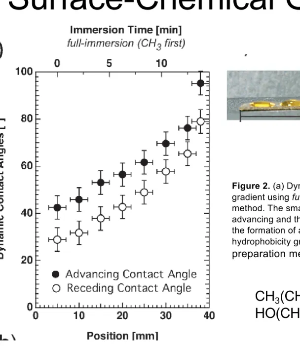
Changing Functional Groups
Low
High
Surface Polarity
Carboxylic acid group
Methyl group
Surface-Chemical Gradients
Figure 2. (a) Dynamic contact angles along a hydrophobicity
gradient using
full-immersion (CH3-first)
as the preparation
method. The small hysteresis of less than 15
between the
advancing and the receding contact angles is an indication for
the formation of a full monolayer. (b) Water droplets along a
hydrophobicity gradient using
full-immersion (CH3-first)
as the
preparation method.
CH
3
(CH
2
)
11
SH
HO(CH
2
)
11
SH
29
2D-SAMs
Lucia Pasquato - Università di Trieste - vietata la riproduzione ai fini commerciali
surfaces with different funtional groups have been reported, for example:
CF
3
, CH=CH
2
, C≡ CH, Cl, Br, CN, OH, OCH
3
, NH
2
, N(CH
3
)
2
, SO
3
H,
31
decanethiol self-assembled monolayers on Au(111)
2D-SAMs
Schematic one-dimensional energy diagram showing the effect
of chain length on the barrier for activation of the S-H bond of
n-
alkanethiols on gold. For short chain lengths (
n
0-2), the transition
from a physisorbed state to a chemisorbed state is an activated
process.
in UHV
33
2D-SAMs
Lucia Pasquato - Università di Trieste - vietata la riproduzione ai fini commerciali
defects on SAMs
34
2D-SAMs
surface modifications
35
2D-SAMs
36
2D-SAMs
surface modifications
N N
N R N
N N R R N N N NN N R R R R R N O O HS R N O O S R O
H2N
R N R S S R' HS R S S R O
HN NH2 R
O H
O HN N
R
Selected methods for SAMs functionalization via the formation of activated species.
2D-SAMs
surface modifications
Surface Bound Reactive Group Activated Species Reagent Product OH
O
O O
O R NH
2 N H O R OH O O N O O O
R NH2
N H O R OH O O O F F F F
R NH2
N H O R OH O O O F F F F
F R NH2
N H O R OH OH H
O R NH
2 H N R OH Si Cl O O O R OH Si O O O O R
39
2D-SAMs
Lucia Pasquato - Università di Trieste - vietata la riproduzione ai fini commerciali
surface modifications
OH
ClSO
3H
OSO
3H
O
O
O
HO
RO
O
O
O
OH
Cl
O
O
OH
O
3
O
O
NH
O
3
R
1. C
2O
2Cl
22. RNH
2O
Si
O
C
18H
37O
O
Si
H
3C
C
18H
37CH
3POCl
3R-N=C=S
C
18H
37(CH
3)
2SiCl
3C
18H
37SiCl
3O
O
O
CF
3O
(CF
3CO)
2O
P
HO
O
OH
NH
S
R
43
protein fibrinogen (Figure 2D).16As a final control, we prepared a monolayer presenting biotin but that did not incorporate the
quinone propionic ester tether. For this monolayer, application of a potential of-700 mV for 3 min had no effect on the amount of streptavidin that associated, and the monolayer also remained resistant to non-specific protein adsorption (data not shown). These data establish that ligands tethered to a monolayer with the quinone propionic ester can be selectively released without compromising the inertness of the monolayer or the activity of ligands that are not tethered to the monolayer through the quinone propionic ester.
The dynamic substrate described here embodies a general strategy for preparing biosurfaces that offer unprecedented, real-time control over the properties and functions of biosurfaces. Central to this strategy is the structural order inherent to SAMs, which allow the structures and environments of ligands to be tailored with molecular-scale control. Also important is the use of inert SAMs that prevents non-specific interactions at the interface. The incorporation of appropriate redox-active moieties that can be electrochemically switched by applying potentials to the gold provides a non-invasive and selective means to modify the structure and properties of the monolayer. This strategy to manipulate interfacial properties has the advantage that it is less invasive than alternate strategies that use heat, light, or reagents to alter properties. Electrically active films also offer advantages for efficient integration with microelectrical systems, including the use of microelectrode arrays and patterned monolayers to build surfaces that combine multiple functions.
We believe that this method will be most important for engineering tailored substrates for mechanistic studies in cell biology. The interactions of adherent cells, in vivo, with the insoluble extracellular matrix are mediated by the binding of cell-surface receptors to peptide and carbohydrate ligands of the matrix. Many cellular functionssincluding metastasis and migrationsare regulated by changes in the composition of ligands present in the matrix. The surface chemistry approach described here makes possible the preparation of substrates that present multiple ligands, but which can be triggered to selectively release a single class of ligand, and will provide new opportunities in experimental cell biology.
Acknowledgment. We are grateful for support provided by the National Science Foundation (BES-9980850) and the Materials Research Science and Engineering Center (DMR-9808595).
JA000419P
(15) Ellipsometric characterization of a monolayer that presented the biotin quinone propionic ester at a density of 25% (to increase signal contrast) showed that the thickness decreased by 5 Å after electrochemical treatment, consistent with release of biotin from the surface. Grazing-angle FTIR spectra were inconclusive, presumably because of the disordered structure of the oligo-(ethylene glycol) groups and the biotin quinone propionic ester groups.
(16) The use of more extreme potentials (-1100 mV for 5 min) gave mono-layers that were no longer inert to non-specific protein adsorption (Figure 2E), and lower potentials (-600 mV for 3 min) gave incomplete cleavage of biotin.
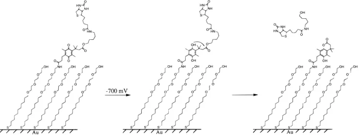
Figure 1. Design of a self-assembled monolayer (SAM) that selectively releases the ligand biotin upon application of a reductive potential. The ligand is tethered to the alkanethiolate through a quinone propionic ester and is present at a density of 1%. On application of a potential of-700 mV to the underlying gold film, the quinone is reduced to the hydroquinone, which then undergoes rapid lactonization with release of biotin. The tri(ethylene glycol) groups in the monolayer prevent the non-specific adsorption of protein.
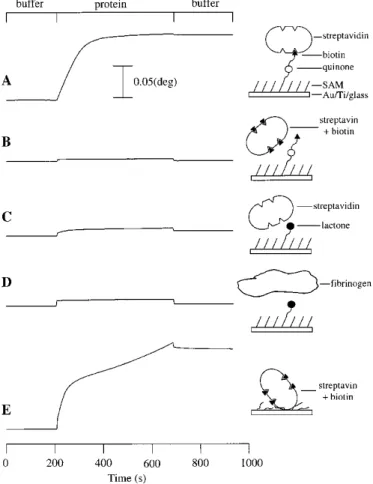
Figure 2. SPR data showing the association of proteins with the SAMs (see text for an explanation of the data). The change in resonance angle (∆θ) is plotted on the vertical axis: the scale bar applies to all data, which are offset for clarity. (A) Streptavidin (60 nM) associated with a surface presenting biotin. (B) Streptavidin pre-incubated with biotin (140
µM) showed no association with the surface, demonstrating that the inter-action is biospecific. (C) Application of a potential of-700 mV for 3 min to the monolayer, prior to the binding experiment, resulted in the release of ligand and a 95% reduction in streptavidin binding. (D) Fibrinogen (0.5 mg/mL) did not adsorb to the SAM following electrochemical treat-ment, demonstrating that the SAM remained inert to protein adsorption. (E) Application of a potential of-1100 mV for 5 min caused association of streptavidin presaturated with biotin, showing the surface was damaged and was no longer resistant to non-specific protein adsorption.
4236 J. Am. Chem. Soc., Vol. 122, No. 17, 2000 Communications to the Editor
Biomolecular Surfaces that Release Ligands under
Electrochemical Control
Christian D. Hodneland and Milan Mrksich*
Department of Chemistry, Uni
V
ersity of Chicago
Chicago, Illinois 60637
Recei
V
ed February 3, 2000
The design of any material that will contact a biological
environment requires that the surface of the material be tailored
to have desired interactions with molecules, proteins, or cells of
the contacting biological fluid. Applications that require precise
control over these interactions have benefited from the use of
self-assembled monolayers (SAMs) of alkanethiolates on gold
because these structurally ordered films offer unprecedented
flexibility in modifying surfaces with ligands and other moieties.
These characteristics were important in developing monolayers
that are inert in biological fluids
s
in that they prevent protein
adsorption and cell adhesion
s
which provided methods for
patterning the positions and shapes of attached cells.
1The
attachment of ligands to these inert SAMs gives surfaces to which
proteins and other receptors selectively bind. Monolayers
present-ing peptide ligands, for example, have been used to control the
adhesion of cells,
2and monolayers presenting oligonucleotides
have been used for probing gene expression in cells.
3A new challenge in biointerfacial science is to design
dynamic
substrates that can alter, in real-time, the display of ligands and,
hence, the interactions of proteins and cells with the substrate.
4We previously demonstrated a dynamic SAM that could be
switched from a state that is initially inert to a state that permits
the Diels
-
Alder mediated immobilization of ligands, which in
turn provides a strategy to activate the selective binding of proteins
to a substrate.
5Here, we describe a new class of dynamic
electroactive monolayer that can selectively
release
immobilized
ligands.
The monolayer shown in Figure 1 was designed to release the
ligand biotin when a reductive potential is applied to the
underlying gold. This dynamic property derives from the quinone
propionic ester that tethers the biotin to the monolayer. Previously,
quinone propionic esters and amides have been used as protecting
groups for alcohols and amines, respectively, because mild
chemical reduction of the quinone affords the hydroquinone,
which rapidly lactonizes with liberation of an alcohol or amine.
6The two methyl groups at the benzylic position together with the
proximal methyl group on the ring
s
collectively referred to as
the “tri-methyl lock”
s
serve to increase the rate of the
lactoniza-tion reaclactoniza-tion, and therefore, the release of ligand.
7For our
pur-poses it is essential that the monolayers remain inert to the
non-specific adsorption of protein
s
both before and after release of
the ligand. Accordingly, the monolayers used here present the
electroactive tether at low density (approximately 1% of total
alkanethiolate) surrounded by tri(ethylene glycol) groups because
the latter are highly effective at preventing non-specific adsorption
of protein.
8We first established that the quinone propionic ester moiety
could undergo
electrochemical
reduction and subsequent
lacton-ization to give the desired products in high yield. We prepared a
model quinone propionic ester (
1
) and electrolyzed a solution of
this molecule (1 mM, 5 mg total in 1:1 THF:H
2O containing 200
mM KCl) at
-
900 mV (all potentials reported here are relative
to a Ag/AgCl/KCl reference) until the current decayed to 1% of
its initial value.
9Isolation and characterization of the reaction
products showed that
1
was converted to the corresponding lactone
and alcohol in quantitative yield.
10We next describe release of biotin from the monolayer shown
in Figure 1. We used surface plasmon resonance (SPR)
spectros-copy to measure the biospecific association of streptavidin with
the monolayer and to demonstrate the loss of binding after biotin
was electrochemically released.
11,12The following sequence was
used to measure the association of proteins to the monolayers:
buffer (PBS, pH 7.4) was flowed over the monolayer for 3 min
to establish a baseline; a solution of protein in the same buffer
was flowed over the surface for 8 min to observe binding; buffer
was again flowed for 4 min to quantitate the amount of protein
that remained bound. Figure 2A shows that streptavidin (60 nM)
bound to a monolayer presenting the quinone propionic ester
substituted with biotin.
13The change in resonance angle (
∆θ
) after
streptavidin was flowed over the substrate corresponds to a final
protein density of 1.1 ng/mm
2. The protein remained irreversibly
bound because of the high affinity of the streptavidin
-
biotin
complex. When the streptavidin was mixed with biotin (140
µ
M)
before introduction to the SAM, there was no binding of the
protein to the surface, demonstrating that the interaction is
biospecific (Figure 2B). The reduction and subsequent
lacton-ization and release of biotin was triggered by application of a
potential of
-
700 mV for 3 min.
14,15SPR showed that the amount
of streptavidin that bound to the resulting SAM decreased by 95%
(Figure 2C). Importantly, the electrochemical treatment did not
damage the monolayer or compromise its resistance to
non-specific adsorption of several proteins, including the “sticky”
(1) (a) Mrksich, M.; Dike, L. E.; Tien, J. Y.; Ingber, D. E.; Whitesides, G. M.Exp. Cell Res.1997,235,305-313. (b) Chen, C. S.; Mrksich, M.; Huang, S.; Whitesides, G. M.; Ingber, D. E.Science1997,276,1345-1347.
(2) (a) Roberts, C.; Chen, C. S.; Mrksich, M.; Martichonok, V.; Ingber, D. E.; Whitesides, G. M. J. Am. Chem. Soc. 1998, 120, 6548-6555. (b) Houseman, B. T.; Mrksich, M.J. Org. Chem.1998,120, 6548-6555.
(3) Bamdad, C.Biophys. J.1998,75,1997-2003.
(4) Early examples of materials with dynamic function include: (a) Lau, A. N. K.; Miller, L. L.J. Am. Chem. Soc.1983,105, 5271-5277. (b) Ding, Z.; Long, C. J.; Hayashi, Y.; Bulmus, E. V.; Joffman, A. S.; Stayton, P. S. Bioconjugate Chem.1999,10,395-400.
(5) Yousaf, M. N.; Mrksich, M.J. Am. Chem. Soc.1999,121, 4286-4287. (6) (a) Carpino, L. A.; Triolo, S. A.; Berglund, R. A.J. Org. Chem.1989, 54,3303-3310. (b) Wang, B.; Liu, S.; Borchardt, R. T.J. Org. Chem.1995, 60, 539-543. (c) Zheng, A.; Shan, D.; Wang, B.J. Org. Chem.1999,64, 156-161.
(7) Milstien, S.; Cohen, L. A.J. Am. Chem. Soc.1972,94, 9158-9165.
(8) Mrksich, M.; Whitesides, G. M. ACS Symp. Ser. Chem. Biol. Appl. Polyethylene Glycol1997,680,361-373.
(9) Bulk electrolysis was performed under an argon atmosphere using a BAS CV-50W potentiostat in a standard cell with a vitreous carbon working electrode of large surface area, a coiled platinum wire counter electrode, and a Ag/AgCl/KCl reference electrode.
(10) The reaction mixture was purified using column chromatograpy to afford the lactone, which was characterized by1H and13C NMR and TLC.
(11) For examples of the use of SPR to measure biospecific association of proteins with SAMs, see: (a) Houseman, B. T.; Mrksich, M.Angew. Chem., Int. Ed.1999,38, 782-785. (b) Mrksich, M.; Grunwell, J. R.; Whitesides, G. M.J. Am. Chem. Soc.1995, 117, 12009-12010. (c) Spinke, J.; Liley, M.; Guder, H. J.; Angermaier, L.; Knoll, W.Langmuir1993,9, 1821-1825.
(12) Monolayers were prepared by immersing gold-coated glass slides in ethanolic solutions containing a mixture of a symmetric disulfide of an alkane-thiol substituted with tri(ethylene glycol) and an unsymmetric disulfide of an alkanethiol substituted with the tri(ethylene glycol) and the biotin quinone propionic ester. The unsymmetric disulfide was synthesized in 29 steps from commercially available reagents. All intermediates gave satisfactory1H NMR
and MS spectra.
(13) SPR measures the angle of light (θ) reflected from the backside of the gold substrate that is a minimum in intensity. Changes in this angle (∆θ) are linearly related to the index of refraction of the solution above the surface and therefore to the density of adsorbed protein (∆θof 0.10°)1 ng/mm2).
Experiments show a change inθimmediately following protein injection due to differences in refractive index between the two solutions.
(14) Electrochemistry was performed in buffered water (PBS, pH 7.4) using the gold substrate as the working electrode, a platinum wire as the counter electrode, and a Ag/AgCl/KCl reference electrodesprior to mounting the substrate in a cartridge for analysis by SPR.
4235
J. Am. Chem. Soc.
2000,
122,
4235
-
4236
10.1021/ja 000419p CCC: $19.00 © 2000 Am er ica n Ch em ica l Societ y P u blish ed on Web 04/14/2000
Biomolecular Surfaces that Release Ligands under
Electrochemical Control
Christian D. Hodneland and Milan Mrksich*
Department of Chemistry, UniVersity of Chicago Chicago, Illinois 60637 ReceiVed February 3, 2000
The design of any material that will contact a biological environment requires that the surface of the material be tailored to have desired interactions with molecules, proteins, or cells of the contacting biological fluid. Applications that require precise control over these interactions have benefited from the use of self-assembled monolayers (SAMs) of alkanethiolates on gold because these structurally ordered films offer unprecedented flexibility in modifying surfaces with ligands and other moieties. These characteristics were important in developing monolayers that are inert in biological fluidssin that they prevent protein adsorption and cell adhesionswhich provided methods for patterning the positions and shapes of attached cells.1 The attachment of ligands to these inert SAMs gives surfaces to which proteins and other receptors selectively bind. Monolayers present-ing peptide ligands, for example, have been used to control the adhesion of cells,2and monolayers presenting oligonucleotides have been used for probing gene expression in cells.3
A new challenge in biointerfacial science is to designdynamic
substrates that can alter, in real-time, the display of ligands and, hence, the interactions of proteins and cells with the substrate.4 We previously demonstrated a dynamic SAM that could be switched from a state that is initially inert to a state that permits the Diels-Alder mediated immobilization of ligands, which in turn provides a strategy to activate the selective binding of proteins to a substrate.5 Here, we describe a new class of dynamic electroactive monolayer that can selectivelyreleaseimmobilized ligands.
The monolayer shown in Figure 1 was designed to release the ligand biotin when a reductive potential is applied to the underlying gold. This dynamic property derives from the quinone propionic ester that tethers the biotin to the monolayer. Previously, quinone propionic esters and amides have been used as protecting groups for alcohols and amines, respectively, because mild chemical reduction of the quinone affords the hydroquinone, which rapidly lactonizes with liberation of an alcohol or amine.6 The two methyl groups at the benzylic position together with the proximal methyl group on the ringscollectively referred to as the “tri-methyl lock”sserve to increase the rate of the lactoniza-tion reaclactoniza-tion, and therefore, the release of ligand.7For our pur-poses it is essential that the monolayers remain inert to the non-specific adsorption of proteinsboth before and after release of the ligand. Accordingly, the monolayers used here present the electroactive tether at low density (approximately 1% of total alkanethiolate) surrounded by tri(ethylene glycol) groups because
the latter are highly effective at preventing non-specific adsorption of protein.8
We first established that the quinone propionic ester moiety could undergoelectrochemicalreduction and subsequent lacton-ization to give the desired products in high yield. We prepared a model quinone propionic ester (1) and electrolyzed a solution of this molecule (1 mM, 5 mg total in 1:1 THF:H2O containing 200 mM KCl) at-900 mV (all potentials reported here are relative to a Ag/AgCl/KCl reference) until the current decayed to 1% of its initial value.9 Isolation and characterization of the reaction products showed that1was converted to the corresponding lactone and alcohol in quantitative yield.10
We next describe release of biotin from the monolayer shown in Figure 1. We used surface plasmon resonance (SPR) spectros-copy to measure the biospecific association of streptavidin with the monolayer and to demonstrate the loss of binding after biotin was electrochemically released.11,12The following sequence was used to measure the association of proteins to the monolayers: buffer (PBS, pH 7.4) was flowed over the monolayer for 3 min to establish a baseline; a solution of protein in the same buffer was flowed over the surface for 8 min to observe binding; buffer was again flowed for 4 min to quantitate the amount of protein that remained bound. Figure 2A shows that streptavidin (60 nM) bound to a monolayer presenting the quinone propionic ester substituted with biotin.13The change in resonance angle (∆θ) after streptavidin was flowed over the substrate corresponds to a final protein density of 1.1 ng/mm2. The protein remained irreversibly bound because of the high affinity of the streptavidin-biotin complex. When the streptavidin was mixed with biotin (140µM) before introduction to the SAM, there was no binding of the protein to the surface, demonstrating that the interaction is biospecific (Figure 2B). The reduction and subsequent lacton-ization and release of biotin was triggered by application of a potential of-700 mV for 3 min.14,15SPR showed that the amount of streptavidin that bound to the resulting SAM decreased by 95% (Figure 2C). Importantly, the electrochemical treatment did not damage the monolayer or compromise its resistance to non-specific adsorption of several proteins, including the “sticky”
(1) (a) Mrksich, M.; Dike, L. E.; Tien, J. Y.; Ingber, D. E.; Whitesides, G. M.Exp. Cell Res.1997,235,305-313. (b) Chen, C. S.; Mrksich, M.; Huang, S.; Whitesides, G. M.; Ingber, D. E.Science1997,276,1345-1347.
(2) (a) Roberts, C.; Chen, C. S.; Mrksich, M.; Martichonok, V.; Ingber, D. E.; Whitesides, G. M. J. Am. Chem. Soc. 1998, 120, 6548-6555. (b) Houseman, B. T.; Mrksich, M.J. Org. Chem.1998,120, 6548-6555.
(3) Bamdad, C.Biophys. J.1998,75,1997-2003.
(4) Early examples of materials with dynamic function include: (a) Lau, A. N. K.; Miller, L. L.J. Am. Chem. Soc.1983,105, 5271-5277. (b) Ding, Z.; Long, C. J.; Hayashi, Y.; Bulmus, E. V.; Joffman, A. S.; Stayton, P. S.
Bioconjugate Chem.1999,10,395-400.
(5) Yousaf, M. N.; Mrksich, M.J. Am. Chem. Soc.1999,121, 4286-4287. (6) (a) Carpino, L. A.; Triolo, S. A.; Berglund, R. A.J. Org. Chem.1989,
54,3303-3310. (b) Wang, B.; Liu, S.; Borchardt, R. T.J. Org. Chem.1995,
60,539-543. (c) Zheng, A.; Shan, D.; Wang, B.J. Org. Chem.1999,64,
156-161.
(7) Milstien, S.; Cohen, L. A.J. Am. Chem. Soc.1972,94, 9158-9165.
(8) Mrksich, M.; Whitesides, G. M.ACS Symp. Ser. Chem. Biol. Appl. Polyethylene Glycol1997,680,361-373.
(9) Bulk electrolysis was performed under an argon atmosphere using a BAS CV-50W potentiostat in a standard cell with a vitreous carbon working electrode of large surface area, a coiled platinum wire counter electrode, and a Ag/AgCl/KCl reference electrode.
(10) The reaction mixture was purified using column chromatograpy to afford the lactone, which was characterized by1H and13C NMR and TLC.
(11) For examples of the use of SPR to measure biospecific association of proteins with SAMs, see: (a) Houseman, B. T.; Mrksich, M.Angew. Chem., Int. Ed.1999,38, 782-785. (b) Mrksich, M.; Grunwell, J. R.; Whitesides, G. M.J. Am. Chem. Soc.1995,117, 12009-12010. (c) Spinke, J.; Liley, M.; Guder, H. J.; Angermaier, L.; Knoll, W.Langmuir1993,9, 1821-1825.
(12) Monolayers were prepared by immersing gold-coated glass slides in ethanolic solutions containing a mixture of a symmetric disulfide of an alkane-thiol substituted with tri(ethylene glycol) and an unsymmetric disulfide of an alkanethiol substituted with the tri(ethylene glycol) and the biotin quinone propionic ester. The unsymmetric disulfide was synthesized in 29 steps from commercially available reagents. All intermediates gave satisfactory1H NMR
and MS spectra.
(13) SPR measures the angle of light (θ) reflected from the backside of the gold substrate that is a minimum in intensity. Changes in this angle (∆θ) are linearly related to the index of refraction of the solution above the surface and therefore to the density of adsorbed protein (∆θof 0.10°)1 ng/mm2).
Experiments show a change inθimmediately following protein injection due to differences in refractive index between the two solutions.
(14) Electrochemistry was performed in buffered water (PBS, pH 7.4) using the gold substrate as the working electrode, a platinum wire as the counter electrode, and a Ag/AgCl/KCl reference electrodesprior to mounting the substrate in a cartridge for analysis by SPR.
4235
J. Am. Chem. Soc.2000,122,4235-4236
10.1021/ja 000419p CCC: $19.00 © 2000 Am er ica n Ch em ica l Societ y P u blish ed on Web 04/14/2000
44
protein fibrinogen (Figure 2D).
16As a final control, we prepared
a monolayer presenting biotin but that did not incorporate the
quinone propionic ester tether. For this monolayer, application
of a potential of
-700 mV for 3 min had no effect on the amount
of streptavidin that associated, and the monolayer also remained
resistant to non-specific protein adsorption (data not shown).
These data establish that ligands tethered to a monolayer with
the quinone propionic ester can be selectively released without
compromising the inertness of the monolayer or the activity of
ligands that are not tethered to the monolayer through the quinone
propionic ester.
The dynamic substrate described here embodies a general
strategy for preparing biosurfaces that offer unprecedented,
real-time control over the properties and functions of biosurfaces.
Central to this strategy is the structural order inherent to SAMs,
which allow the structures and environments of ligands to be
tailored with molecular-scale control. Also important is the use
of inert SAMs that prevents non-specific interactions at the
interface. The incorporation of appropriate redox-active moieties
that can be electrochemically switched by applying potentials to
the gold provides a non-invasive and selective means to modify
the structure and properties of the monolayer. This strategy to
manipulate interfacial properties has the advantage that it is less
invasive than alternate strategies that use heat, light, or reagents
to alter properties. Electrically active films also offer advantages
for efficient integration with microelectrical systems, including
the use of microelectrode arrays and patterned monolayers to build
surfaces that combine multiple functions.
We believe that this method will be most important for
engineering tailored substrates for mechanistic studies in cell
biology. The interactions of adherent cells, in vivo, with the
insoluble extracellular matrix are mediated by the binding of
cell-surface receptors to peptide and carbohydrate ligands of the
matrix. Many cellular functions
s
including metastasis and
migration
s
are regulated by changes in the composition of ligands
present in the matrix. The surface chemistry approach described
here makes possible the preparation of substrates that present
multiple ligands, but which can be triggered to selectively release
a single class of ligand, and will provide new opportunities in
experimental cell biology.
Acknowledgment. We are grateful for support provided by the National Science Foundation (BES-9980850) and the Materials Research Science and Engineering Center (DMR-9808595).
JA000419P
(15) Ellipsometric characterization of a monolayer that presented the biotinquinone propionic ester at a density of 25% (to increase signal contrast) showed that the thickness decreased by 5 Å after electrochemical treatment, consistent
-Figure 1. Design of a self-assembled monolayer (SAM) that selectively releases the ligand biotin upon application of a reductive potential. The ligand is tethered to the alkanethiolate through a quinone propionic ester and is present at a density of 1%. On application of a potential of-700 mV to the underlying gold film, the quinone is reduced to the hydroquinone, which then undergoes rapid lactonization with release of biotin. The tri(ethylene glycol) groups in the monolayer prevent the non-specific adsorption of protein.
Figure 2. SPR data showing the association of proteins with the SAMs (see text for an explanation of the data). The change in resonance angle (∆θ) is plotted on the vertical axis: the scale bar applies to all data, which are offset for clarity. (A) Streptavidin (60 nM) associated with a surface presenting biotin. (B) Streptavidin pre-incubated with biotin (140
µM) showed no association with the surface, demonstrating that the inter-action is biospecific. (C) Application of a potential of-700 mV for 3 min to the monolayer, prior to the binding experiment, resulted in the release of ligand and a 95% reduction in streptavidin binding. (D) Fibrinogen (0.5 mg/mL) did not adsorb to the SAM following electrochemical treat-ment, demonstrating that the SAM remained inert to protein adsorption. (E) Application of a potential of-1100 mV for 5 min caused association of streptavidin presaturated with biotin, showing the surface was damaged and was no longer resistant to non-specific protein adsorption.
4236
J. Am. Chem. Soc., Vol. 122, No. 17, 2000
Communications to the Editor
protein fibrinogen (Figure 2D).
16As a final control, we prepared
a monolayer presenting biotin but that did not incorporate the
quinone propionic ester tether. For this monolayer, application
of a potential of
-
700 mV for 3 min had no effect on the amount
of streptavidin that associated, and the monolayer also remained
resistant to non-specific protein adsorption (data not shown).
These data establish that ligands tethered to a monolayer with
the quinone propionic ester can be selectively released without
compromising the inertness of the monolayer or the activity of
ligands that are not tethered to the monolayer through the quinone
propionic ester.
The dynamic substrate described here embodies a general
strategy for preparing biosurfaces that offer unprecedented,
real-time control over the properties and functions of biosurfaces.
Central to this strategy is the structural order inherent to SAMs,
which allow the structures and environments of ligands to be
tailored with molecular-scale control. Also important is the use
of inert SAMs that prevents non-specific interactions at the
interface. The incorporation of appropriate redox-active moieties
that can be electrochemically switched by applying potentials to
the gold provides a non-invasive and selective means to modify
the structure and properties of the monolayer. This strategy to
manipulate interfacial properties has the advantage that it is less
invasive than alternate strategies that use heat, light, or reagents
to alter properties. Electrically active films also offer advantages
for efficient integration with microelectrical systems, including
the use of microelectrode arrays and patterned monolayers to build
surfaces that combine multiple functions.
We believe that this method will be most important for
engineering tailored substrates for mechanistic studies in cell
biology. The interactions of adherent cells, in vivo, with the
insoluble extracellular matrix are mediated by the binding of
cell-surface receptors to peptide and carbohydrate ligands of the
matrix. Many cellular functions
s
including metastasis and
migration
s
are regulated by changes in the composition of ligands
present in the matrix. The surface chemistry approach described
here makes possible the preparation of substrates that present
multiple ligands, but which can be triggered to selectively release
a single class of ligand, and will provide new opportunities in
experimental cell biology.
Acknowledgment. We are grateful for support provided by the National Science Foundation (BES-9980850) and the Materials Research Science and Engineering Center (DMR-9808595).
JA000419P
(15) Ellipsometric characterization of a monolayer that presented the biotin quinone propionic ester at a density of 25% (to increase signal contrast) showed that the thickness decreased by 5 Å after electrochemical treatment, consistent with release of biotin from the surface. Grazing-angle FTIR spectra were inconclusive, presumably because of the disordered structure of the oligo-(ethylene glycol) groups and the biotin quinone propionic ester groups.
(16) The use of more extreme potentials (-1100 mV for 5 min) gave mono-layers that were no longer inert to non-specific protein adsorption (Figure 2E), and lower potentials (-600 mV for 3 min) gave incomplete cleavage of biotin. Figure 1. Design of a self-assembled monolayer (SAM) that selectively releases the ligand biotin upon application of a reductive potential. The ligand is tethered to the alkanethiolate through a quinone propionic ester and is present at a density of 1%. On application of a potential of-700 mV to the underlying gold film, the quinone is reduced to the hydroquinone, which then undergoes rapid lactonization with release of biotin. The tri(ethylene glycol) groups in the monolayer prevent the non-specific adsorption of protein.
Figure 2. SPR data showing the association of proteins with the SAMs (see text for an explanation of the data). The change in resonance angle (∆θ) is plotted on the vertical axis: the scale bar applies to all data, which are offset for clarity. (A) Streptavidin (60 nM) associated with a surface presenting biotin. (B) Streptavidin pre-incubated with biotin (140
µM) showed no association with the surface, demonstrating that the inter-action is biospecific. (C) Application of a potential of-700 mV for 3 min to the monolayer, prior to the binding experiment, resulted in the release of ligand and a 95% reduction in streptavidin binding. (D) Fibrinogen (0.5 mg/mL) did not adsorb to the SAM following electrochemical treat-ment, demonstrating that the SAM remained inert to protein adsorption. (E) Application of a potential of-1100 mV for 5 min caused association of streptavidin presaturated with biotin, showing the surface was damaged and was no longer resistant to non-specific protein adsorption.
4236
J. Am. Chem. Soc., Vol. 122, No. 17, 2000
Communications to the Editor
45
Deval, J.,
et al., J. Micromech. Microeng.
2004,
14
, 91.
Reversible Hydrophobic/Hydrophilic
Surfaces
Reversible Hydrophobic/Hydrophilic
Surfaces
Change Potential
Lahann, J.,
et al., Science.
2003
,
299
, 371.
47
Sun, T., et.al., Angew. Chem.
2004
, 116, 361.
Controlling Wettability
surface of poly(N-isopropylacrylamide)
nature materials VOL 4 SEPTEMBER 2005, 704
ammide fumarica
49
Figure 4 A photo-responsive surface based on switchable fluorinated molecular shuttles. a
,
Light-switchable rotaxanes with the fluoroalkane region (orange) exposed (
E
-
1
) were physisorbed onto a
SAM of 11-MUA on Au(111) deposited on either glass or mica to create a polarophobic surface,
E
-
1
.11-MUA.Au(111).
b
, Illumination with 240–400 nm light isomerizes some of the
E
olefins to
Z
(giving a 50:50 ratio at the photostationary state if the reaction on the surface mirrors that in solution),
causing a nanometre displacement of the rotaxane threads in the
Z
-shuttles which encapsulates the .
uoroalkane units leaving a more polarophilic surface,
E/Z
-
1
.11-MUA.Au(111). Photoemission spectroscopy
data are consistent with the molecular shuttles lying parallel to the Au surface (but they are not directionally
aligned or necessarily linear as depicted for clarity in this cartoon).
50
Figure 2. Light-driven directional transport of a 1.25 μl diiodomethane drop across the surface of monolayer
of molecular machines, both flat (a)-(d) and up a twelve degree incline (e)-(h). This extrapolation across 6
orders of magnitude in length scales from mechanical motion at the molecular level to macroscopic transport
is truly remarkable - the equivalent of millimetre motion of components in a machine working to raise an
object to over twice the height of the CN Tower, the world s tallest building. The work done by the monolayer
51
2D-SAMs
Lucia Pasquato - Università di Trieste - vietata la riproduzione ai fini commerciali