ALUMINUM SPECIATION AND BIOAVAILABILITY:
INVESTIGATIONS OF EQUILIBRIA BETWEEN ALUMINUM AND
CITRATE SPECIES IN PLASMA USING COMPUTER SIMULATION AND"AL-NMR
Abstract:
Knowledge of the chemical species that aluminum forms in vivo allows assessment of
its potential availability to tissues and, thus, prediction of toxicity. The major binders
of aluminum in serum are transferrin, an iron-transport protein (accounts for "90% of
aluminum in serum), and a number of ions, particularly phosphate and citrate. In this
work, a number of published equilibrium models describing the speciation of
aluminum and citrate were prepared and used to predict equilibrium species over a
range of pH. Qualitative NMR investigations confirmed the existence of an
asymmetrical trinuclear aluminum species (Al3Cit3(OH)4'^) that appeared rapidly (in
less than one minute after mixing the solution) and persisted over a wide range of pH.
A model by Ohman (1988) consisting of equilibrium constants for the
aluminum-citrate-hydroxide system was found to fit the observed data better than a
number of other published models describing the same system, based on predictions
made using Titrator, an equilibrium speciation program. This choice was strengthened
by the observation that the starting pH of actual solutions agreed better with the
Ohman's model than with the others. Ohman's equilibrium model was then
extrapolated (again, using Titrator) down to physiologic concentrations of aluminum
and citrate, which predict only charged (nonlipophilic) species at plasma pH and a
small amount of neutral aluminum-citrate complex at low pH. Passive uptake into
tissues requires the existence of an uncharged aluminum species — these results
suggest that, based on the aluminum-citrate system, aluminum will not be taken up
passively from serum as a citrate complex, although it could if bound to some other
ligand not considered here (such as phosphate). Furthermore, although concentrations
of aluminum and citrate relevant to gut contents were not considered here, the
presence of a neutral aluminum-citrate complex at gastrointestinal pH may be
Contents
Abstract... ii
List of figures... v
List of tables... vi
L Introduction... 1
II. Background... 3
III. Techniques for analysis of aluminum and its speciation in biological
media ... 7IV. Gross absorption of aluminum in the digestive tract... 10
V. Mechanisms of aluminum absorption from gastrointestinal tract ... 13
VI. Aluminum in blood... 16
VII. Kinetics of uptake by cells... 22
Vm. Pharmacokinetics ... 23
IX. Tissue distribution... 29
X. Equilibrium modeling ... 33
XI. Methods... 40
XII. Results... 47
XIII. Discussion ... 71
XIV. Conclusions... 82
XV. Acknowledgments... 85
XVI. References... 86
"'>'-'?^J^'?~-^.'-Figures
Change in pH of aluminum-citrate system over time according to Ohman
(1988)... 35
NMR spectrum of AKH^O)/^ ... 44
NMR spectrum of Al(OH)4- ... . . ... 45
NMR spectra of equimolar aluminum-citrate solution (0.1 M) titrated over a
range of pH... 49
Change of pH over time after addition of base to aluminum-citrate solutions . . 58
Kinetic NMR experiment... 59
Shift of ma.jor NMR signal over time, equimolar aluminum-citrate, pH ~4 . . . . 60
Gregor & Powell model: aluminum-citrate solutions titrated from pH 2-10 ... 62
Martin model: aluminum-citrate solution titrated from pH 2-10 ... 66
Ventunni & Berthon model: aluminum-citrate solution titrated from pH 2-10 . . 67
Trinuclear aluminum species (Al3Ci3(OH)4') postulated by Ohman (1988)... 75
Symmetrical trinuclear aluminum species (Al3Ci3(OH),6'') postulated by
Ohman (1988) ... 79
SS«?-..f
Tables
Concentrations of components in serum available to bind with aluminum
(from Harris, 1992)... 17
Major resonances observed Irom aluminum-citrate solutions ... 47
Resonances observed with an equimolar (0.1 M) aluminum-citrate solution
over a broad range of pH ... 47
Predicted starting pH, from simulations based on literature constants, for
Al:Ci 1:1, 0.1 M solution, compared to our measurements... 72
I. Introduction
Aluminum is the most common metal on earth and some exposure to it, whether via
ingestion or inhalation, is inevitable. Its toxicity (particularly neurotoxicity) is
currently hotly debated. One of the central issues to be resolved with regard to
toxicity is the chemical forms of aluminum present in the blood. This is important
because the various species of aluminum differ widely in their charge state and
solubility, which can determine how much of the metal is available to susceptible
tissues. The physiologic behavior of aluminum is particularly difficult to assess primarily because of a lack of a suitable radionuclide but also due to various other
analytical problems, not the least of which is contamination of samples by ubiquitous
aluminum-containing dusts.
Citrate, a common component of the human diet, binds aluminum and enhances the absorption of aluminum from the gastrointestinal tract. Once in the plasma, aluminum
is carried largely on transferrin, an iron-binding protein, with some 10% remaining in
a small-molecule form; citrate may still play a role here. The relative availabiUty to
tissues of the protein-bound compared to the soluble forms of aluminum is unclear.
Analytical chemists have developed equilibrium models in an attempt to characterize
the plasma chemistry of aluminum, particularly with regard to citrate. These models
were derived largely on the basis of theoretical curve-fitting of titiration data with very
on the physiologic disposition of aluminum, describes experiments using "Al-nuclear
magnetic resonance as a direct probe of aluminum species, and compares the
II. Background
Exposure to some aluminum is unavoidable and occurs for the most part orally,
through food and water, although oral intakes are small because aluminum as a
component of minerals (the usual form of the element in nature) is of limited
solubility. Inhalation constitutes a small fraction of aluminum exposure to the general
population, though it can be significant in some occupational settings. Total daily
intake of aluminum is 30 to 50 mg on average to the population at large (Ganrot,
1986), of which less than 1 mg is from water (Jones and Bennett, 1986).
The species of aluminum (that is, the chemical forms that it takes) in the body have
important implications for the balance between the metal's uptake into tissues and its
excretion. Aluminum is very insoluble except at exfremes or pH or in the presence of
chelating ligands. To be taken up passively by tissues, it must be in the form of a
neutral molecule or complex; to be taken up actively, in contrast, it appears that
aluminum must be bound to a protein for which a specific receptor exists.
Insoluble aluminum hydroxide, which constitutes some antacids, for example, is
Al(OH)3 (s). This mineral can form from the precipitation of aluminum from aqueous
solutions. When dissolved, and at low pH, aluminum is in the form of AP"^ or
Al(OH)2+, Al(OH)3''(aq), and Al(OH)4') via successive loss of protons from the
associated water molecules. Aluminum can also form complexes with small ions.
Citrate (Ci), a triprotic carboxylic acid, binds aluminum with high affinity to form a
variety of mono- and polynuclear aluminum species and mixed hydroxo species, such
as those listed here:
AlCi" AlCiH^
AlCi^
AlCiOH-AlCi(OHV
AljCijCOH),*- '
AljCijCOHV ^
Al^Ci^COHV
Aluminum also appears to complex strongly with phosphate to form AIPO4, A1HP04^,
A1H2P04'% Al2P04'^ AhVO.iOUV, and Al2P04(OH)3''. Citrate and phosphate appear
to be the most physiologically important small-ion ligands of aluminum. In serum,
one or two aluminum ions can associate with each molecule of transferrin, which canbe taken up specifically by cells.
Some over-the-coimter pharmaceuticals such as antacids and buffered aspirin contain
sufficient aluminum to increase the daily dose significantly. Many antacids consist of
a mixture of Al(OH)3 and other hydroxides, such as magnesium. Some
increase over the average exposure from food and drinking water alone. Patients with renal insufficiency are often administered large quantities of aluminum-containing antacids in order to bind excess phosphate; the resulting AIPO4 is an insoluble form
that makes the phosphate more easily excretable via the feces.
Aluminum has been measured in U.S. groundwaters at a range of 0.014 to 0.29 mg/1 and in untreated surface waters at 0.016 to 1.17 mg/1. Aluminum sulfate is often
added as a flocculant to surface waters, resulting in aluminum concentrations in
finished drinking water from 0.014 to 2 67 mg/1. Assuming average daily water consumption of 2 1 results in an average daily intake of aluminum from water of 0.2 mg (Orme & Ohanian, 1990).
Dialysis patients can be exposed to large amounts of aluminum from their dialysis fluid. This exposure has been responsible for notable episodes of neurotoxicity. Such
exposures have largely been eliminated, however, by deionization of fluids used to make up dialysis solutions.
Inhalation exposure of the general population to aluminum in dust amounts to 0.14
aluminum through dusts and aerosols. Blinder et al. (1991) found that welders
exposed to 3.0-8.9 mg aluminum/m' excreted 107-351 /ig/l aluminum in the urine
and 18-29 iig/g was found in the bone. Both workers had been welding for about 20
years.
Jones and Bennett (1986) estimate that ~3% of inhaled aluminum is absorbed into the
blood from the lung. Volunteers exposed to < 1 mg aluminum/m^ (RoUin et al..
1991b) experienced increased serum aluminum concentrations compared to controls,but urinary excretion was unchanged. Exposure of rabbits to 0.56 mg aluminum/m^
over 5 months led to 15.8-fold increased accumulation of aluminum in lung compared
to controls, 2.5-fold increase in brain, and 1.65-fold increase in kidney (RoUin et al..
1991a).
The study of aluminum speciation has presented a number of major difficulties to
researchers. No radionuclide of aluminum is readily available, obviating tracer
studies. Because of its ubiquity in the environment, contamination of samples (such as
that from dust particles) is difficult to avoid. Furthermore, the insolubility of some
III. Techniques for analysis of aluminum and its speciation in biological media
A. Atomic absorption
Total aluminum concentration of biological media is typically determined using atomic
absorption spectroscopy. This method alone, however, provides no information about
speciation. It is subject to contamination, since aluminum from dusts is difficult to
avoid. Spectral interference from other metals (such as vanadium) can be avoided by
judicious choice of absorption lines. The detection limit of flame atomic absorption
spectroscopy for aluminum is 30 ng/ml (1.1 fiM) (Skoog & Leary, 1992). The detection limit for elecfrothermal atomic absorption is considerably lower, 0.005
ng/ml, but this method has not been widely used in the applications described here.
B. High-energy-accelerator mass spectrometry
Day et al. (1991) described the use of ^*A1, a long-lived isotope (half-life ~10* years)
of aluminum, in a human tracer study performed with high energy accelerator mass
spectrometry. Meirav et al. (1991) performed a similar experiment in rats.
C. Filtration techniques
Gel filtration and ultrafilttation, combined with atomic-absorption spectroscopy, of
serum or simulated serum solutions (designed to contain all the important serum
ligands for aluminum) have been employed to distinguish between protein-bound and
non-protein-bound aluminum and, to a degree, among different protems bound to
represent protein-boimd aluminum, though it may not be possible to distinguish
between protein-bound and otherwise unfilterable aluminum.
A number of researchers have attempted to investigate how aluminum binds to serum
proteins using gel filtration techniques (Day eLal., 1991; Bertholf et al., 1984, 1985;
Rahman eLal., 1985; Favarato eLal., 1992a,b; Cochran eLal., 1985). This approach
has been problematic in a number of respects. Many columns avidly bind aluminum
themselves: Favarato et al. (1992a) recovered aluminum from different columns, by
washing with buffer containing desferrioxamine, in these amounts: TSK-GEL
HW-5SS, 75%; Sephadex G-200, 36%; Sephacryl S-500, 30%;and Bio Gel P-2, 24%.
In the absence of useful radionuclides of aluminum, the major analytical tool for
elucidating the distribution and pharmacokinetics of this element has traditionally been
atomic absorption spectroscopy (AAS). The limitations of this method have already
been discussed. Because of changes in speciation of plasma and aqueous solutions
with time, which tends to produce colloidal species that would not be expected in
vivo, filtration studies are often imreliable. Accelerator mass specfrometry requires
extremely specialized equipment and is furthermore prohibitively expensive. What is
needed is a method that can distinguish directly not only between protein-bound and
soluble species, but among species within both of these groups. Datta et al. (1990)
that the technique failed to distinguish among the small, soluble species, even those of different charge. Another method of studying aluminum kinetics separates aluminum
species into three categories, depending on the rate of reaction with ferron, a colorimetric reagent, but does not identify individual species (Hundt and O'Melia,
1988; Jardine and Zelazny, 1987).
Recently, nuclear magnetic resonance (NMR) directed at the ^'Al nucleus has been used to investigate aluminum species in solution. Unlike "C or 'H, "Al has a spin
quantum number greater than 1, resulting in broad signals when bound to
asymmetrically arranged ligands, which makes structural assignments difficult.
Theory and applications of ^'Al-NMR have been thoroughly reviewed by Akitt (1989).
Fatemi et al. (1991) reported preliminary data from "Al-NMR demonstrating
aluminum binding to transferrin, albumin, and citrate. For an aluminum-citrate
solution, these investigators found a double signal from pH 6.0 to pH 8.4 in the
region from about 0 to 10 ppm; at the highest pH, Al(OH)4" was detected as well.
Feng et al. (1990) detected a trinuclear aluminum-citrate species in solution over a range of intermediate pH whose identity was confirmed by x-ray crystallography.
Further information on earlier NMR studies can be found in the Discussion section,
IV. Gross absorption of aluminum in the digestive tract
A commonly cited estimate of absorption of total aluminum from the gut of 0.1 to
0.3% (Ganrot, 1986) was based in part on the assumption that urinary excretion
proportionally reflects absorption, which does not take into account tissue distribution,
as discussed below. An estimate closer to measured values is 1 % (based on data
reported by Jones and Bennett, 1986); results of Day et al. (1991) are also in
agreement with this estimate.
Some absorption of aluminum may occur in the stomach, as suggested by the results of Rodger et al. (1991); however, the majority of aluminum absorption is expected to occur in the intestine. The general, two-step absorption process in the gut is (1)
lumen— > mucosa and (2) mucosa— > bloodstream. Aluminum must be in the form of
neutral complexes in order to be absorbed by diffusion through the plasma membrane of cells. Ionic aluminum may be absorbed actively by specialized iron-absorption
pathways in the gut, as discussed below.
The most important aspect of the digestive tract with regard to its uptake of aluminum
is its change in pH, from 2-3 in the stomach to 3-8 in the small intestine. The low
pH of the stomach allows for complete dissolution of, for example, A1(0H)3 in
complexation and possible absorption. Studies of intestinal uptake that fail to specify or control for pH must therefore be considered unreliable.
A. Animal studies
Domingo et al. (1991) examined citrate and other organic acids (ascorbic, gluconic,
lactic, malic, oxalic, and tartaric) for their effect on aluminum absorption. These investigators administered aluminum to rats by gastric intubation and monitored
urinary and tissue aluminum. Each of these acids has fewer dissociable protons than citric acid, and, for the most part, promoted tissue accumulation of aluminum to a
lesser degree than citrate. Ascorbic acid's effect on tissue accumulation of aluminum was found in this study (though not in others) to be similar to that of citric acid.
Organic acid-induced urinary excretion of aluminum would provide further evidence
of increased gastiointestinal absorption, but in this experiment no significant changes in urinary aluminum were found as a result of administration of the organic acids
studied.
B. Human studies
The effect of gastric pH on absorption of aluminum was examined by Rodger et al.
(1991). These investigators administered either placebos or ranitidine, a drug that
suppresses production of stomach acids (and would, thus, elevate gastric pH), to healthy subjects and to patients with renal disease, prior to administration of Al(OH)3.
aluminum than those that received ranitidine, implying that low gastrointestinal pH
promotes absorption of aluminum.
It has been suggested that Alzheimer's disease (AD) patients might have a genetic
tendency to absorb excessive amounts of aluminum. To test this hypothesis, Taylor et
al. (1992) administered an aluminum citrate-containing drink to 20 AD patients and 20 age- and sex-matched controls, measuring blood aluminum before and after
consumption of the drink. The investigators found a greater increase in blood aluminum among younger AD subjects than controls, but not among the older AD subjects compared to their controls. This study could have been improved by a study population large enough to allow sufficient statistical power for stratified analysis or randomization. Furthermore, as has been reported elsewhere, there was considerable variability among measured serum aluminum concentrations, which could have been
either a contamination problem or the result of confounding by unstratitied exposure
variables.
The measurement of total aluminum concentration in urine before and after exposure is the simplest means of assessing absorption. Cobum et al. (1991) administered 950
mg calcium citrate four times daily and 2.4 g/day A1(0H)3 as an antacid to eight normal men. Baseline aluminum excretion was 0.02 + 0.004 /xmol/mmol creatinine;
factor of 5.3 to 11.1) when calcium citrate and A1(0H)3 were ingested together,
compared to AlCOH), alone. Similar results were obtained by Walker et al. (1990),
who foimd that sodium bicarbonate had less of an effect than sodium citrate on aluminum excretion; and Nolan et al. (1990), who found that calcium acetate
increased urinary aluminum excretion much less than did calcium citrate. Maximum urinary excretion of aluminum, after coadministration with calcium citrate, was 176 ±
103 fig/g creatinine/day compared to 6 + 3 when only Al(OH)3 was taken (Nolan et al., 1990).
House et al. (1992) examined risk factors for elevated serum aluminum in a group of 71 office workers and found higher serum aluminum to be associated with antacid
consumption and with the batch in which the sample was analyzed, which strongly
suggests blood-sample contamination problems. Consumption of cola drinks from aluminum cans appeared to be inversely related to serum aluminum, but this was
believed, again, to be an artifactual result of the high variability due to contamination.
V. Mechanisms of aluminum absorption from gastrointestinal tract
A. Role of active transport and iron absorption pathways
inhibit cellular respiration, sfrengthening the hypothesis of active fransport of
aluminum out of the intestine.
Since aluminum behaves similarly to iron in many biological systems (for example binding to transferrin, discussed below), AP^ may also be taken up via the specialized
iron absorption pathways found in the gut. As evidence for this hypothesis, iron
deficiency has been found to increase the absorption of aluminum both in rats
(Cannata et al.. 1991) and in renally impaired humans (Huang et al., 1992). Andersen et al. (1987), in confrast, found that administering exfra iron orally to
dialysis patients did not prevent absorption of aluminum from phosphate binders. Van der Voet et al. (1987), in an excised gut-section model, found that Fe^*^ enhanced disappearance of aluminum from the intestinal lumen, but did not increase systemic or portal blood aluminum; that is, it did not increase absorption overall. Fe^"^, in
confrast, did not cause disappearance of aluminum from the intestinal lumen.
An iron-binding protein has recently been identified in the duodenal mucosa of rats
and humans that may prove to bind to aluminum as well, and may thereby account for
part of the mechanism of aluminum's uptake from the gut (Conrad et al., 1992). The
protein, named mobilferrin after the Alabama city in which it was discovered, is
mobility, and amino acid composition. The dissociation constant for iron from
mobilferrin was found to be 8.92 x 10^. The physiologic role of this protein is not yet
known; to date it has only been isolated from duodenal homogenates. ^. Role of citrate
Citrate, the polyanion of triprotic citric acid, forms stable complexes with AP"^, including a neutral molecule (AlCi") that can be absorbed into the blood. Cifrate is
believed, for this reason, to be the most important small-molecule coraplexing species commonly found in the diet, and many experimental investigations have confirmed
this prediction. Furthermore, citrate is present in serum at 99 fiM, at least 100-fold
higher than the concentration of aluminum.
Van der Voet et al. (1989) found that citrate-enhanced absorption of aluminum was at
least in part an energy-dependent process, based on their observation that
administration of dinitrophenol, an uncoupler of oxidative phosphorylation, decreased
the citrate-mediated disappearance of aluminum from the intestinal lumen.
Froment et al. (1989), in an examination of the mechanism of enhancement of aluminum uptake by citrate, tested the hypothesis that citrate increases the
permeability of the tight junctions between intestinal epithelial cells. The investigators
isolated duodenal sections by transmission electron microscopy, infiltration of ruthenium red through the tight junctions was observed in the presence of aluminum citrate. In addition, aluminum citrate was found to decrease the transcellular electrical resistance of tight junctions. The investigators did not examine the effect of citrate
alone, however.
VI. Aluminum in blood
Because of the high concentration of potential ligands relative to the concentration of the metal, aluminum is expected to be soluble at blood concentrations up to at least 100 /ig/1. Ganrot (1986), based on more than 50 literature sources, reported that the most credible values for serum aluminum are in the range of 1 to 5 /xg/l, or 0.037-0.185 /iM, assuming that values much higher than this stem largely from
contamination. More recently, Wang et al. (1991) measured levels of aluminum in 63
Canadians by Zeeman atomic absorption spectroscopy and found an average of
0.06+0.05 ^M in serum and 0.20+0.10 /jlM in urine.
A. Nature and amounts of carrier molecules
Although many of the organic ligands found in the gut are also found in the blood
(Table I), the major plasma binder of aluminum is transferrin, an iron-transport protein that also specifically binds aluminum. Some nonspecific aluminum binding may also take place with albumin, and at least one other aluminum-binding protein has
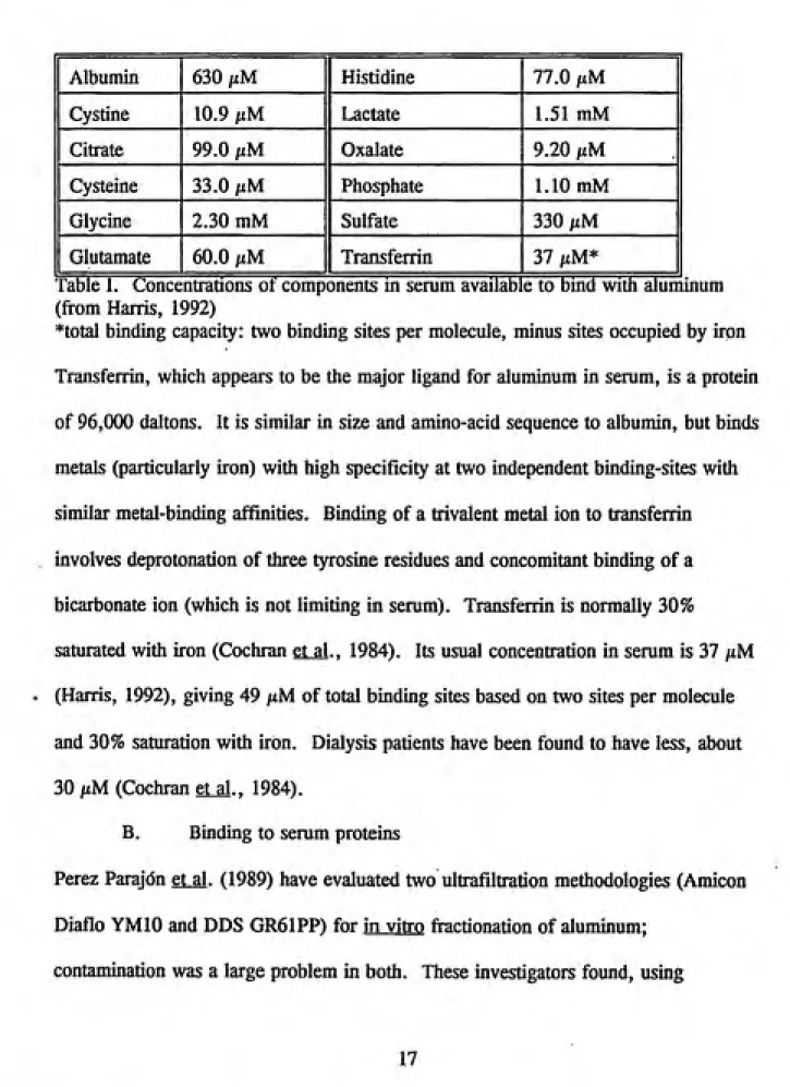
Albumin 630 ^M Histidine 77.0 fiM
Cystine 10.9 /xM Lactate 1.51 mM
Citrate 99.0 ^M Oxalate 9.20 fiM
Cysteine 33.0 fiM Phosphate 1.10 mM
Glycine 2.30 mM Sulfate 330 juM
Glutamate 60.0 nM Transferrin 31 fiM*
Table I. Concentrations of components in serum available to bind with aluminum
(from Harris, 1992)
*total binding capacity: two binding sites per molecule, minus sites occupied by iron
Transferrin, which appears to be the major ligand for aluminum in serum, is a protein
of 96,000 daltons. It is similar in size and amino-acid sequence to albumin, but binds
metals (particularly iron) with high specificity at two independent binding-sites with
similar metal-binding aifinities. Binding of a trivalent metal ion to transferrin
involves deprotonation of three tyrosine residues and concomitant binding of a
bicarbonate ion (which is not limiting in serum). Transferrin is normally 30%
saturated with iron (Cochran et al.. 1984). Its usual concentration in serum is 37 /xM
(Harris, 1992), giving 49 fiM of total binding sites based on two sites per molecule
and 30% saturation with iron. Dialysis patients have been found to have less, about
30 ^M (Cochran eLal., 1984).
B. Binding to serum proteins
Perez Parajdn et al. (1989) have evaluated two ultrafiltration methodologies (Amicon
Diatlo YMIO and DDS GR61PP) for in vitro fractionation of aluminum;
ultramicrofiltration with Amicon MPS-1 systems, that 8.3% of serum aluminum from normal subjects and 13.3% of that from uremic patients was ulfrafilfrable.
"Ultrafiltrable" as used here means able to pass through an ultrafilter, and refers to small-molecule species such as aluminum-cifrate species; nonultrafilterable species are primarily protein-bound but may also include insoluble or colloidal complexes of aluminum hydroxide, for example.
Yokel and McNamara (1988) found that ultratilterable aluminum was higher in the
serum of renally impaired rabbits than in serum from renally competent animals, after
incubation of aluminum with rabbit serum in vifro and after an intravenous dose of
100 fimoUkg. These investigators also compared the solubilities (in terms of their ultrafilterability) of different aluminum salts in bicarbonate buffer, and foimd that the
citrate salt of aluminum remained completely ulfrafiltrable up to a total aluminum
concentration of 1 mg/ml, while aluminum chloride, nifrate, and lactate salts
decreased in solubility when total aluminum exceeded 1 /xg/ml. The concentrations at
which aluminum was not totally ulfrafilterable were above the high end of the
expected range of serum concentrations. Two of the rabbits in this study were
reported to have died prematurely, possibly (according to the authors) from kidney
The similar sizes of transferrin and albumin make it difficult to separate these proteins
chromatographically, and failure to add bicarbonate to the elution buffer can decrease
or prevent binding of aluminum to transferrin. Many authors have assumed that
aluminum binds to both, although some evidence suggests that binding to albumin is
insignificant, as discussed below. Martin et al. (1987), based on weak binding in
vitro, knowledge of high competition in vivo for albumin binding sites, and generally low affinity of metal ions for albumin, argue against significant in vivo binding of aluminum to albumin. This would be particularly true under uremic conditions, where potential binding sites on albumin are likely to be occupied by other ligands that
accumulate in serum.
Day et al. (1991) administered a dose of 100 ng ^Al and about 1 /ig "Al (natural aluminum) in sodium citrate solution to a human volunteer. The highest plasma ^*A1
concentration measured was 0.3 ng/1 at 6 hours post-ingestion, which represents about
1 % of the administered dose (assuming a plasma volume of 3 1). The study
confirmed, by gel permeation chromatography and anion exchange chromatography at
pH 7.4, that 80% of aluminum in plasma was associated with transferrin, 15% existed
as other high-molecular-weight (>5 kDa) complexes (including albumin), and 5% as
redistribution of ionic aluminum among proteins, so the findings of protein-specific
binding are not reliable.
Favarato et al. (1992b) compared the protein binding of aluminum among renaUy
competent workers exposed and unexposed to aluminum and detected a novel protein,
dubbed albindin. This protein picked up more than 40% of serum aluminum after
treatment of the serum with desferrioxamine (a high-affinity chelator of aluminum)
and its amino-acid profile was distinct from that of transferrin or albumin (Favarato et
al., 1992a). The protein-binding profile of aluminum was most complex in the more
highly exposed group of workers (classified on the basis of total serum aluminum
content). Proteins were identified by polyacrylamide gel electrophoresis (SDS-PAGE),
a technique which did not resolve transferrin and albumin (Favarato et al., 1992b).
Cochran et al. (1985) eluted plasma (spiked with aluminum) from uremic patients on
Sephacryl S-300 and assessed the reproducibility of the aluminum/transferrin and
aluminum/albumin molar ratios in adjacent elution fractions. They found that the
aluminum-to-fransferrin ratio remained at about 0.12, whereas aluminum/albumin
varied from 0.024 to 0.002, providing evidence for an association between aluminum
and transferrin, but not albumin. Transferrin and albumin concentrations were
assessed by immunodiffusion, but the methods used were neither described nor
so the significance of the experiment's results is uncertain. Dialysis of purified
albumin against aluminum solution resulted in the association of 10% of the aluminum
with albumin, but when calcium and phosphate were added, the association between
albumin and aluminum could no longer be detected. There was no evidence of
association of aluminum with either a Sephacryl S-300 or Sephadex G-50 gel.
C. Role of transferrin
In vitro analytical chemical methods such as titration have been used to estimate the
affinity of aluminum (and other metals) for transferrin and other potential chelators
found in serum. Transferrin's affinity for iron is considerably higher than for
aluminum (e.g., log 62=40 for Fe^"^ and 31 for AP"^; Marques, 1991). Since
aluminum is present in small amounts in the blood relative to iron, and since iron
normally occupies only 30% of transferrin, aluminum binding to transferrin is not
expected to be limited by either levels of transferrin (concentration in blood 37 jxM;
Harris, 1992) or of iron.
Evidence for transferrin's role as an aluminum carrier is strengthened by a study that
showed aluminum levels in urine and brain to be increased in rats depleted of iron and
exposed orally to aluminum (Cannata et al.. 1991). This phenomenon occurs because
reduced iron stores lead to increased production of transferrin (Prasad, 1991;
'"«Ww«^Wfssps^S5''!E>«W*-'-^?-«^'«ar*'"?—?Sf^
that iron-depleted rat intestinal epithelial cells in vitro took up significantly more aluminum when exposed to transferrin-boimd aluminum than did normal cells.
Although useful as a predictor of uptake of aluminum into tissues in general, this
evidence is not specifically relevant to uptake by the intestine, since transferrin is not
found in the intestinal lumen.
VII. Kinetics of uptake by cells
Several investigators have examined the uptake kinetics of transferrin and iron by cells. Cole and Glass (1983) found that at iron concentrations up to 50 ^M, binding of iron to transferrin was not saturated. Iron uptake by mouse hepatocytes increased with increasing transferrin concentrations, but nonspecific uptake (pinocytosis and/or
diffusion) accounted for 10-20% of uptake. Dissociation constants of 0.081 fiM (for
suspended cells) and 0.29 fiM (for plated cells) were reported for dissociation of
transferrin from cells. Page et al. (1984) found similar results using cultured rat
hepatocytes. Cochran et al. (1991) examined the competition of aluminum and iron for transferrin binding and uptake in reticulocytes (immature red blood cells). Binding was significantiy stronger in these cells (K^^^S /xM) than Cole and Glass reported
for hepatocytes. Fiuthermore, radiolabeled aluminum-transferrin (5 fiM) was taken
up 1.8 times faster than radiolabeled iron-transferrin. Iron uptake reached a plateau
by 40 minutes; aluminum uptake, in contrast, continued to increase after 40 minutes.
by addition of unlabelled iron-transferrin) than by addition of aluminum-transferrin.
The authors postulate a post-uptake feedback mechanism to regulate uptake of iron,
which does not appear to have an effect on uptake of aluminum. McGregor et al. (1991) reported that aluminum was taken up to a greater degree from solutions of
AICI3 than of aluminum and citrate, possibly because, at physiologic pH, uncomplexed
aluminum would be expected to be hydrolyzed to colloidal Al(OH)3, in contrast with
aluminum citrate, which apparently does not form uncharged species at physiologic
pH (see section on speciation, above).
Interestingly, in contrast to the results of Cochran et al. (1991), in human
erythroleukemia K562 cells, transferrin-bound aluminum down-regulated the number
of cell-surface transferrin receptors to a greater degree than iron did (McGregor et al..
1990). It is possible that hepatocytes possess a regulatory mechanism for transferrin
receptors different from that in cultured leukemia cells, or there may be an
interspecies difference, since Cochran et al. used rat hepatocytes.
VIII. Pharmacokinetics
Upon gastrointestinal absorption (or intraperitoneal injection) aluminum travels via the
portal circulation into the liver, where it is thought to undergo a first-pass clearance;
of this process is evident from high levels of aluminum found in the liver relative to
peripheral organs (such as brain and bone) (Xu et al.. 1992).
Much of the work done thus far on aluminum speciation and distribution has looked at
exposures other than ingestion, such as intravenous or intraperitoneal injection, which
bypasses the firast-pass effect of the liver. Exposure to dialysis fluid can be
considered analogous to intravenous injection since the bloodstream is directiy
exposed, bypassing the first-pass effect of the liver.
A. Excretion routes
1. Urinary excretion
Aluminum is excreted primarily in the urine, as described above, and to a small
degree in the feces (largely as insoluble aluminum phosphate) by way of the portal
circulation, liver, and bile (< 1%; Xu et al.. 1991). The relative contributions of
each depend in part on such factors as solubility of aluminum, presence of dietary
constituents such as citrate and phosphate, renal competence, and dose of aluminum.
Braunlich et al. (1986) investigated the possibility that renal excretion of aluminum
was connected with nephrotoxic effects and found that aluminum administration in rats
p-ͣ
: ^5i^?e^^^^P^
aminohippuric acid, both of which were considered to represent toxicity.
2. Biliary excretion
Biliary excretion is apparentiy not a major route of clearance of aluminum. Xu et al.
(1991) found that it accounted for < 1% of doses administered to rats, whereas
urinary clearance accounted for between 9 and 17%.
B. Animal data
Fulton and Jeffery (1990) examined the effect of dietary citrate or ascorbate on the absorption of aluminum from drinking water and on its subsequent distribution and excretion. They found a dose-dependent increase in aluminum concentration in bone, stomach, intestine, and kidney; liver aluminum was 1.5-fold greater than controls, but there was no dose-response, and no uptake into brain was observed. Ascorbate and citrate increased the concentration of aluminum in tibia, plasma, urine, and feces. Plasma aluminum (about 0.7 fiM) was found to constitute less than 0.5% of total
blood aluminum, which is inconsistent with other studies and particularly surprising
given that transferrin is a plasma protein. Red-blood-cell aluminum as found not to
vary with dose of aluminum, whereas plasma aluminum did, which is consistent with
C. Human data
Alfrey et al. (1980) found that nondialyzed uremic patients experienced aluminum
burdens in liver at 6.3 times, spleen at 12 times, bone at 6.2 times, and brain at 1.7
times higher than renally competent patients; dialyzed uremics and, in particular,
dialyzed uremics suffering from encephalopathy, had tissue burdens many times higher
than nondialyzed uremics.
D. Uremia
Uremic (renally impaired) animals and humans seem to be more susceptible to aluminum accumulation and toxicity than renally competent subjects, even in the
absence of dialysis. Hosokawa and Yoshida (1989) performed 5/6 nephrectomy on
rats and collected serum three months later, without administering additional aluminum above the normal dietary exposure. They found that the average
concentration of aluminum in serum was more than 10 times that seen in renally intact rats, and the concentration in kidneys was about 10 times higher. Arieff et al. (1979)
obtained results similar to Alfrey's human data for deposition of aluminum into liver of nonuremic dogs and uremic compared with nonuremic rats but, in contrast, found
that brain aluminum was higher in nondialyzed than dialyzed chronic renal failure patients. The aluminum content of the dialysis fluid was not reported.
It would be instructive to perform a study in which long-term aluminum burden in
chronic low and moderate oral exposures, in combination with a short-term kinetic
study. Such a study would elucidate the effects of acute vs. chronic exposures and
kinetics on body burden.
There are a number of possible reasons for the observation that uremia leads to an
increased body burden of aluminum. The simplest explanation is that, in having
lower excretory capacity, uremics might simply not be able to rid themselves of
aluminum efficiently, allowing it more time to be distributed to peripheral tissues.
However, a number of recent studies have demonstrated that uremic animals actually
have higher renal (and systemic) clearance of aluminum than renally competent
animals (Yokel & McNamara, 1988; Meirav etal., 1991; Ittel eLal., 1991; Wilhelm
etal.. 1989).
The aluminum toxicity experienced by uremics may be iatrogenic, through exposure to
aluminum in dialysis fluid, which is equivalent to intravenous administration of
aluminum. This possibility has already been addressed in the clinical setting: dialysis fluid is now made from low-aluminum water, and patients appear to suffer less
aluminum toxicity as a result (Ganrot, 1986).
The increased excretion of aluminum in uremia suggests that the free, or ultrafiltrable,
uremia potential aluminum-binding sites on transferrin (and possibly albumin or some other protein) are more likely to be tied up with competing molecules that accumulate
in the blood. Renal clearance of aluminum would thus be expected to increase with
time after onset of uremia, as competing ligands accumulate, but this hypothesis has
not been investigated.
As discussed above, renal clearance of aluminum is higher in uremics than in normal,
renally competent individuals as investigated by pharmacokinetic studies. Given that
uremics also experience higher aluminum tissue burden and toxicity, and have a
higher non-protein-bound fraction of aluminum, it appears that this fraction might be
more available to tissues than the protein-bound fraction. The only other explanation is that tubular reabsorption of aluminum occurs to a greater degree in uremics, but
this possibility has not been examined.
'' ͣ ., "^
Chasteen (1977) reported that serum transferrin concentration in dialysis patients is
about 30 (iM. Given that serum aluminum concentrations are elevated in these
patients compared to normal individuals, this reduction in aluminum-binding capacity
IX. Tissue distribution
Bone, spleen, and liver are the primary organs of aluminum deposition (Ganrot, 1986). Patients with renal failure frequentiy suffer from osteomalacia and have been
shown to have deposits of aluminum in the bone. Recentiy, Goodman and O'Connor
(1991) found in chick embryos that 10 and 100 fiM aluminum decreased calcium
uptake into bone from 76 to 38% of controls, respectively, during 24-hour
incubations. Brown and Schwartz (1992) reported increased deposition of aluminum
in liver and spleen of rats in response to dietary iron depletion.
A. Brain
The blood-brain barrier is normally permeable only to small, uncharged molecules.
Since much of blood aluminum appears to be complexed with proteins, it is unlikely
to permeate the blood-brain barrier directiy, although transferrin-receptor-mediated
uptake is a possible mechanism (Roskams and Connor, 1990). In the presence of
other, smaller complexes, however, aluminum could cross the blood-brain barrier. A
case study was reported of several renal dialysis patients suffering from bone
aluminum-protein complexes, had been absorbed into the brain (Sherrard et al.. 1988). Yokel et al. (1991a) supported this hypothesis in a rat study using microdialysis
probes to monitor aluminum-desferrioxamine diffusion into various tissues, including the brain.
Allen and Yokel (1992) used in vivo microdialysis to compare the permeabilities of
aluminum and gallium through the blood-brain barrier (with a view to using gallium
as a model for aluminum) and found them to be dissimilar. Gallium, although
attractive because it possesses useable radioisotopes, therefore does not seem an appropriate model for aluminum penetration of the brain. Specifically, these
investigators found that aluminum permeates the brain primarily at the cerebral
capillaries, whereas gallium permeates equally at the cerebral capillaries and at the choroid plexuses. Aluminum's permeation was nonsaturable, suggesting a nonspecific absorption process such as diffusion rather than binding of aluminum-transferrin to cell-surface receptors. The absorption could, however, be a combination of specific and nonspecific processes and still appear nonsaturable overall, so involvement of transferrin cannot be ruled out.
Farrar gLal- (1990) found, using HPLC and gel electrophoresis, that binding of
normal humans. The authors postulate that reduced binding of gallium and,
presumably, aluminum to transferrin could lead to increased uptake by the metal
across the blood-brain barrier as a neutral citrate complex. As discussed above,
aluminum does appear to be taken up in a nonsaturable fashion (i.e., nonspecifically)
at least at the cerebral capillaries (Allen and Yokel, 1992).
Roskams and Connor (1990) examined the binding of aluminum-transferrin and
iron-transferrin to homogenized rat brain and found evidence of a receptor that binds both.
Dissociation constants (K^) were 5.7 nM for Fe-transferrin and 13.1 nM for
aluminum-transferrin. The affinity of this receptor for aluminum-transferrin, although
lower than that for Fe-transferrin, is still higher than the affinity of receptors in other
cells (such as hepatocytes and lymphocytes) for Fe-transferrin. Unfortunately,
homogenized rat brain is probably not a good model for the blood-brain barrier.
These authors suggested that aluminum's toxicity in the brain might involve, at least
in part, disruption of normal iron homeostasis and iron-dependent cellular processes in
the brain. Fleming and Joshi (1991) found that aluminum can interfere with the rate
of binding of iron to ferritin, an iron-storage protein. This finding supports Roskams'
'•^•^mm^^^^^^m^^^^^vm'
B. Fetus
Yokel (1985) found evidence to suggest placental transfer of aluminum. Gomez et al. (1991) reported that fetuses of pregnant rats exposed orally to A1(0H)3 concurrently with citrate experienced higher than normal skeletal malformations, demonstrating either that aluminum crosses the placental barrier directly or, possibly, that there is
some sort of indirect systemic effect on the mother.
Cranmer et al. (1986) found that intraperitoneal administration of AlCl, at doses from 100-200 mg/kg-day to gestating mice resulted in 6-10-fold increases over controls in placental and 2-3-fold increases in fetal aluminum. Oral gavage of 200 or 300 mg/kg-day led to less than a two-fold increase over controls in placental or fetal aluminum. No dose-response relationship was evident among any of the experimental
groups.
C. Milk
Yokel and McNamara (1985) found that, in rabbits, subcutaneous injection led to
excretion of 3.3% of the absorbed dose in milk, while intravenous administration led
to only 2.4%. The maximum serum concentration in the pups was 185 /iM after a
X. Equilibrium modeling
Equilibrium models, based on potentiometric data, allow a much more complete
(though theoretical) description of species formed in aqueous solutions of aluminum with its ligands, compared to the analytical techniques described in earher sections. In
this technique, fonnation (or equilibrium) constants are fit to data on change of pH as
a function of acid or base added to a solution. These constants can then be used '
(usually with a computer program) to predict equilibrium concentrations of the
postulated species under specified concentrations of aluminum and its ligands and at
different pH.
Berthon and Dayde (1992) described a model of aluminum speciation in the intestine
designed to compare the fraction of neutral aluminum species formed after
administration of either aluminum phosphate or aluminum hydroxide. This model
found A1(0H)3 to dissolve better than AIPO4 in the presence of organic acids, and to
complex with the acids better; therefore, aluminum administered orally as A^OH),
would be better absorbed in the gut than AIPO4 and thus lead to the observed
increased relative toxicity. Furthermore, this dissolution, complexation, and
Bertsch and Anderson (1989) studied the speciation of the aluminum-citrate system
using ion chromatography and found that only two peaks were eluted, one containing
hexaaquo (fully hydrated) aluminum and the other apparently containing neutral
aluminum citrate and AlHCit"^. Other singly charged species were not distinguishable from the second peak and, presumably, more highly charged species were
undetectable. Citrate complexes were highly sensitive to changes in ionic strength.
Ohman (1988) performed a titration study of aluminum-citrate speciation over time in
aqueous solution that followed pH as a function of added base. Speciation was
calculated by curve-fitting using published equilibrium constants. Two species (A1(H
'Cit)"' and A1(0H)H 'Cit)^) were found to exist at physiologic pH in fresh (that is,
unaged) solutions, and a third (Al3Ci3(OH)4'^) formed in somewhat older solutions.
On the basis of a similar pH-time study, Ohman (1988) first fitted stability constants
for AlCiH(H') and A10H(H"'Ci)^ at time 0 assuming that the concentration of Al3Ci3(OH)4'^ was zero. He then predicted corresponding concentrations of the
trinuclear species, based on the amount of hydroxide taken up, as evidenced by the
pH, then constructed distribution diagrams over time. In these models, the trinuclear
aluminum species appeared after 1 minute at around pH 6 and then came to
Fig. 1
fll
b)
cl
too lV.AI
80 60
A,"
<0 20
^--'li^"'^-^^
100 BO 60 to 20
-13.3.3
-13.3.3
-S.t.l 100 L.V.AI
eo -At'-60
-\ -3.1,1
to
- x^~^\^
20
-2,y\^^
^-*^'i'^~-—
Motekaitis and Martell (1984) examined potentiometrically the equilibrium between aluminum and a number of organic ligands, including citric acid, at metal:ligand ratios of 1:1 and 1:2. Equilibration times between additions of acid or base varied, but were not reported. Only three species were assumed, each of them a 1:1
aluminum:citrate species in a different protonation state.
Gregor and Powell (1986) assumed that citrate's hydroxy proton was available to complex with aluminum, and used a 5.3- or 6.3-fold excess of ligand over metal for
potentiometric determinations. K^-cit' ion pairing was taken into accoimt since KOH
was used as a titrating base. Eight aluminum-citrate species were assumed, but no
polynuclear species. "C-NMR was used to examine the identity of species, and
supported the possibility that the hydroxy proton was used in bridging.
Martin (1986) used adjusted equilibrium constants from the published literature to model the binding of aluminum and cifrate at ionic strengths from 0.1 to 0.16 M, and over a pH range of 1 to 9. He assumed no polynuclear species were involved. At
physiologic pH and a ratio of 100:1 citrate to aluminum, the species AlCiOH'" was
Venturini and Berthon (1989) examined the aluminum-citrate equilibrium at
physiologic ionic strength and devised a model containing seven citrate species, including a trinuclear and a binuclear aluminum species, and two hydroxy aluminum species. At physiologic pH and about 60:1 citrateialuminum, AlCijCOH)'^ and AlCi2(OH)2^ predominated, while the trinuclear aluminum species (stoichiometrically
equivalent to that postulated by Ohman) predominated at physiological pH at
citrate:aluminum ratios of about 5:3 and 6:1.
-ͣͣ'ͣͣͣ..ͣͣ { ^ .:-ͣͣ ͣ
Recentiy, a handful of studies have appeared arguing for aluminum-phosphate species as the predominant small-molecule form of aluminum in serum. Dayde et al. (1990)
an titrated acidic aluminum-chloride/orthophosphoric acid mixture at different mole
ratios over a wide range of pH to derive formation constants; the aluminum-phosphate species best fitting tiie model were taken to be AIPO4, A1HP04% A1H2P04*% MiPO^^\ Al2P04(OH)2^, and Al2P04(OH)3''. A model constructed from these formation
constants and those previously determined for aluminum citrate and hydroxide
predicted the predominant low-molecular-weight species, under physiologic conditions,
to be AKOH)," (51%), AIPO4'' (41.5%), and Al2P04(OH)2^ (7.2%).
Harris (1992) reported a largely different set of equilibrium constants for the
aluminum-phosphate system based on linear free-energy relationships. The two
developed a speciation model containing all ligands found in serum that are expected
to complex appreciably with aluminum, as well as metal ions (Zn^^, Ca^*, and Mg^^)
that compete with aluminum for binding sites on transferrin. The model was studied at aluminum and ligand concentrations typical of normal or uremic plasma and pH
7.4. The following assumptions were made: (1) the n-terminal binding site on
transferrin is 60% saturated with iron, which is not displaced by other metals; (2) aluminum does not bind to albumin; and (3) trinuclear aluminum citrate
(Al3Cit3(OH)4'^) and aqueous Al(OH)3 exist. Concentrations of organic ligands in normal serum were taken from the Geigy Scientific Tables, and formation constants (other than for phosphate) were adopted from a number of literature sources and
averaged when more than one was available. The principal species predicted from the
complete model were aluminum-transferrin (about 81%), AIPO4OH (about 16%), and a few hydrolyzed species. Harris' work is the first published so far to incorporate
cifrate and phosphate in the same equilibrium model, and the first to suggest that phosphate might be a more significant binder of aluminum than citrate. (See Appendix for a listing of the equilibria considered in this model.)
XI. Methods
The purpose of the experiments and modeling peif ormed here was to predict whether
any neutral, small-molecule complexes of aluminum and citrate exist in appreciable
quantities under physiologic conditions that could represent a source of aluminum in
plasma available for passive uptake of the metal into tissues. We attempted to identify
individual aluminum-citrate species using "Al-NMR as a direct probe. Further, we
characterized the aluminum-citrate system potentiometrically and stoichiometrically by
looking at starting pH and change of pH over time, and by titrating with strong base.
We then used an equilibrium-speciation computer program, called Titrator (Cabaniss,
1987), along with published sets of equilibrium constants, to predict distributions of
aluminum species in the aluminum-citrate system at mole ratios similar to those used
in the experiments. We compared the experiments with those of the models and selected a model that best fit the NMR spectra and the potentiometric data; this model
was then extrapolated to physiologic conditions to predict the distribution of aluminum
species expected in vivo.
The models described below were developed on the basis of equilibrium constants for
the aluminum-citrate system found in the literature (Gregor & Powell, 1986; Ohman, 1988; Venturini and Berthon, 1988; Martin, 1986). These models, as entered into the equilibrium-speciation program Titrator (Cabaniss, 1987), are found below. The
concentrations of components (aluminum and citrate). Simulated titrations from the
model over a broad range of pH were produced that were used to select a number of
distinct species to be sought by NMR investigations. More models were then
developed, and some rejected on the basis of the NMR results. A final model deemed
most suitable based on the evidence gathered was then selected in order to extrapolate
from the experimental conditions to physiologic conditions of pH and concentration.
Equilibrium constants were added to account for reaction of aluminum with ligands in
serum other than citrate.
A. Experimental
1. Aluminum and citrate solutions
Test solutions were prepared by adding either A1K(S04)2 or trisodium citrate salts (or
both; Aldrich Chemical Co., Milwaukee, WI) to distilled water and stirring overnight
to ensure that equilibrium had been reached. The pH of these solutions was
monitored with an Orion digital pH meter (Cambridge, MA).
2. Titrations
In order to establish the amount of acid or base required to reach a given target pH
and to reach an endpoint, titration curves were prepared by adding 1.0 or 0.5 M HCl
or (standardized) NaOH solution dropwise from a buret to a day-old aluminum-citrate
solution at the appropriate concentration. pH measurements were recorded upon
3. Serial pH-NMR titration
This experiment was designed to look, using NMR, at the succession of aluminum species over a range of pH. A 1:1 aluminum-citrate solution was prepared and then titrated gradually over a range of pH so that samples taken over that range could be examined by NMR. HCl was added to the 24-hour-old aluminum-citrate solution to
bring the solution to low pH, and then NaOH was added to adjust to high pH. Aliquots of the solution were removed at appropriate pH intervals and transferred to sample tubes. The pH in each sample tube was measured again at least 5 minutes
post-removal from the titration vessel in order to account for drift.
4. pH-time experiments
These experiments examined the behavior of the pH of aluminum-citrate solutions
over time after addition of a large volume of base. NaOH was added to equimoiar
aluminum-citrate solutions of 0.1 or 0.01, at mole ratios of 1 to 1.5 hydroxide to aluminum. pH was measured before adding base and at various points up to 24 hours afterward.
5. NMR investigations
The NMR instrument used was a Bruker MSL 360, which is designed to handle broad
signals such as those from "Al. The frequency used to detect the "Al nucleus was
Solutions for NMR experiments were decanted into specially modified
rubber-stoppered polystyrene test tubes instead of glass tubes, to avoid a possible background
aluminum signal.
a. Preparation of standards
The AP* standard was prepared by dissolving A1K(S04)2 in water and adding 1.0 M
HCl to reach pH ~2. The high-pH aluminum standard (Al(OH)4) was obtained by
dissolving the same salt in NaOH to pH "13. Both standards were used to calibrate
the NMR for each experiment. The AP^ standard had a shift of 0 ppm; Al(OH)4 was
found at 80 ppm relative to AP"^ (see Figs. 1 and 2).
h. Kinetic NMR experiments
In order to examine the formation of aluminum-citrate species over time, 1.25 ml of
NaOH were added to 10 ml of equimolar aluminum-citrate solution (0.1 or 0.01 M)
and mixed for 20 seconds, then 2 ml were rapidly removed to a sample tube and
inserted into the NMR, which began scanning within a minute after mixing of the base
was complete. pH was measured in the tube and in the mixing vessel after the NMR
experiment was completed.
B. Equilibrium modeling of the aluminum-citrate system
Titrator (Cabaniss, 1987) is a program for personal computers that calculates
equilibrium speciation of solutions from formation constants, total starting
Fig. 2a
flUH20J6i-3 IM H20
II
il
...« v<i "t. tUtf* lflLPXEP.BBl PPG:
SOLIDCTC.PC DATE 2-7-92 SF 93.839
oi 7aaa.aaa
SI 16384
TO 4896
SW 18518.519 HE/PT 2.261
RG NE NS TE 10 1 8 294 DW FW 02 DP 27.0 223B8 5528.BBB 16H DO 08 01 02 OS 04 i.aaas 4.3BBU i.aaau 10.000U 1.8000 LB GQ NC CX CY SR 1.002 0.0 I 20.00 12.00 2121.71
Cb p^f Z.io
^fW^
Fig. 2b
HL10MJ4 IN H20/NHQH
aii^<»i>^miiM ͣWWtWwtPWHOli mmtMmm** %«»«
JL^
liXiri^ymiW »l» i^i I i»i «t>n ^y^fO* »ͣ'ͣ ««» ͣ mi il»l'<'ͣlM>i m^ff^^f^^Wlillil^*flLEUP.002
PPG:
SOLIDCTC.PC
DATE 2-7-92
SF 93.839 01 7888.000 SI 16304 TO 4006 SV 18518.519 HZ/PT 2.261
RG NE NS TE 10 1 0 294 DV FW 02 DP 27.0 22300 5520.000 16H 00 00 01 02 03 04 i.aaas 4.30OU 1.000U 10.OOOU 1.O00U L8 GB NC CX cr SR 1.000 o.a i 20.00 12.00 2121.71
© pH X-i-fO
&if<~-fi—rA
of components. The program utilizes a Newton-Raphson algorithm to produce iteratively solutions to systems of equations. Titrator was used in these investigations
to simulate titration of equilibrium solutions over a range of pH; from these data
predominance diagrams were constructed relative to the total aluminum content of
each solution.
Sets of equilibrium constants used in modeling were from Gregor and Powell (1986); Martin (1986); Venturini and Berthon (1989); and Ohman (1988); equilibrium
constants and other details of the models can be found in the Appendix. Models were run on Titrator using the following conditions; 0.1 M total aluminum: 0.1 M total citrate (ionic strength = 0.2 M) or 0.01 M total aluminum: 0.1 M total citrate (ionic strength = 1.1 M). The physiologic conditions used in the final Ohman model were
ͣ
»J!I=-W?=55W!S^
XII. Results
A. Experimental results
Results from the first, exploratory NMR experiments are given in Table II. A total of
five resonances were found; a change in pH in the 10:1 citrate:aluminum solution
from ~4 to ~8 produces at least two different species. The peak seen in the 1:1
solution was broad enough to have obscured smaller peaks beneath it.
Table 11. Major resonances observed from aluminum-citrate solutions'
Solution pH Shift(s)
l.OMCi, 0.1 M Al 4.32 15.13; 16.46
1 l.OMCi, 0.1 MAI
7.87 25.70.1 MCi, 0.1 MAI 3.53 10.75; ca. 0 (2 peaks: AP+ and a
broad peak) |
Table III. Resonances observed with an equimolar (0.1 M) aluminum-citrate solution
over a broad range of pH.
1 pH
Shift(s) (relative to AP")1 2.58
0.056 (A13+)7.669 (broad, small to medium)
0.5
1 PH
Shift(s) (relative to AP^)1 ^'^
1
0.1099
ca. 0 (broadening toward base of A?^ peak)
ca. 7.5 (broadened shoulder on next peak (10.49);
remnant of 7.669 peak at
previous sample
10.48 (fairly large, broad) |
1 ^-^
0.1002 (APO 1
ca. 1 (broadening around base of AP"^ more obvious)
10.8713 (a lot bigger than prev. sample)
4.96 1.77
10.9195 (ca. twice as big as lower peak) ||
7.08
1.78 1
10.93 (ca. twice as big as lower peak) |
8.02 2.0049
10.8062 (ca. twice as big as lower peak)
1 8.98
2.039810.8057 (ca. twice as big as lower peak; looks a little
broader than earlier samples) ||
9.96 -.1780
10.748
79.86 (Al(OH),) 1
Table III shows chemical shifts observed in a 1:1 aluminum-citrate solution over a
range of pH values. There appear to be at most five species here: AP' at 0 ppm
(Fig. 1), two possible aluminum-citrate complexes at about 0.5 and 7.7 ppm, the
species with two resonances at 1 and 10 ppm (Fig. 3), and AKOH)/ at about 80 ppm (Fig. 2). Al(OH)4 appears at pH 9.96 (again, the actual pH was probably higher) in the second NMR experiment, but not in the 8.98 sample; it is probably formed
m;i.i r. 1. PH :!.5P
Fig. 3a
RLEXPC.001
PPG.
SOLIDCTC.PC DRTE 24-7-92
SF 93.839
01 6000.000
SI 16384
TD 4096
SW 23809.524
HZ/PT 2.906
RG 14
NE 1
NS 439
TE 294
DW 21.0
FW 28600
02 5520.000
DP 16H DO
00 1.0BBS
01 9.000U
02 1.00BU
03 10.000U
04 1.0B0U
LB 10.000
GB 0.0
NC 4
CX 20.00
CT 12.00
SR 797.48
.i..U I I PH < M
Fig. 3b
X a. a. ex flLtxpo.aai PPG: SOLIOCTC.PC OflTE 24-7-92 SF 93.839 01 6000.000 SI 16384 TO 4096 SW 23809.524 HZ/PT 2.906
RG IS
NE 1
NS 162
TE 294
OW 21 0
FW 20600 02 .5520.000
DP 16H DO
00 1.000S
01 9.000U
02 1 000U
03 I0.000U
L [ 1 1 I'H -rn+tf
Fig. 3c
flLEXPE.aai
PPG.
SOLIDCTC.PC DATE 24-7-92
fi
6000.00093.859 SI 1G384 TO 4096 SV 25809.524HZ/PT 2.906
RG 15
NE 1
NS 524 TE 294
DW 21 a
FW 28600
02 5520.aaa DP 16H 00
D0 1 aaas
Dl a.aaau D2 i.aaau D3 ta.a00u
Q4 i.a0au
LB la.aau
GB a.a
NC 2
CX 20.aa CT 12.aa SR 797.48
.i:..i, 1 1 I r-h i.'jL
Fig. 3d
flLtXPF.001 PPG.
SQLIOCTC.PC DATE; 24-7-92
SF 95.839 01 6000.000
SI 16384 TD 4096 SW 23809.524 HZ/Pr 2.906
CN
RG 15 >n
NE 1
NS 206 TE 294
OW 21 i FW 26600 02 5520.000
DP 16H 00 D0 1 0005
Dl 9.000U
D2 1 000U 03 10.0000
04 1 000U
LB 10.000
GB 0.0
NC 1
. 1 1.1 Pit > .J;t
Fig. 3e
flLtXPG.a01 PPG
50L10CTC.PC
DATE 24-7 -92
SF 93.859 01 6000.000 SI 16384 TO 4096
SW 25809.524 HZ/PT 2.906
RG 15 CO
Nt 1
NS P6
TE 294
OW 21 0
FW 28600 02 5520.000 DP 16H DO 00 1 h]00S 01 9.000U 02 1 000U 03 i0.a00u 04 1.000U
LB 10.000
GB 0.0
NC 1
CX 20.00 CY 12.00
Ill PH fi 112
Fig. 3f
OLtXPH.0ai PPG.
SOLIDCTC.PC DATE 24-7-92
SF 93.839 01 6000.000 SI 16584 TD 4098 SW 23809.524 HZ/PT 2.906 RG 16
NE 1
."$
NS 1.79 >n
TE 294
DW 21 0
FW 28600 02 5520.000 OP 16H DO 00 1.000S 01 9.000U 02 1.000U
05 10.000U
04 1 000U
LB 10.000
GB 0.0
NC 1
CX 20.00 cr 12.00
Fi8. 3g 1-1 I I PH -t Jrt
r. a. a.
flLExpj.aai
PPG;•
SOLIDCrC.PC DRTE 24-7-92
SF 93.839
01 6800.000
sr 163B4
TO 4096
SV 23809.524
HZ/PT 2.906
10
RG 16
<n
NE 1
NS 159
TE 294
DW 21.0 FW 28600
02 5520.000
OP 16H 00
00 1.000S
01 9.000(1
02 1 000U 03 10.000U 04 1.0B0U
LB 10.000
GB 0.0
NC 1
CX 20.00
cr 12.00
Fig. 3h
». ^«M!l»,«MM*^w*,PV''
flLEXPK.HBl PPG.
SOLIDCrC.PC DATE 24-7-92 SF 95.859
01 61100.000 SI 16584 FD 4096
SW 25809.524 HZ/PT 2.90B
RG 10
NE I
NS 264 TE 294
$
DW 21.0
FV 28600
02 S520.000
DP 16H 00
00 1.000S
01 9.000U
02 1.000U
03 10.000U
OH 1.00011
LB 10.000 GB
NC
0.0 2 CX 20.00 CT 12.00 SR 797.48
In the third non-kinetic NMR experiment we look at low pH values, but the resolution
was poor; there was one very broad peak downfield of the Al'"^ peak that moved from
about 8 to about 11 ppm, with another signal gradually growing on top of the AP"^
peak (data not shown).
B. Appearance of species over time
Base was added at ratios of 1.0, 1.25, and 1.5 to a 1:1 Al:Ci solution (0.1 M), which caused the pH to bounce up quickly, then somewhat more slowly drift downward, in the case of the 1.0 and 1.25 base:metal solutions, and upward in the 1.5 base:metal solution (Fig. 11). Most of the pH change took place after the first five minutes of mixing; this also occurred when the concentrations of base, metal and ligand were
reduced tenfold (Fig. 4).
When NMR was run on the aluminum:citrate:base solution, starting at about 55
seconds after addition of base, the major signal (ca. 10 ppm) that dominated
throughout the experiment had already appeared (Fig. 5; the sharp peak is Al(OH)4, as an internal standard). The shift did not change with time (r^ = 0.007; Fig. 6). Exact quantitation is not feasible at this point, despite the inclusion of an internal standard, because the areas of sharp and broad signals cannot be reliably compared
'^8- 4 Change in pH over time after addition of different volumes of base to equiraolar aluminumrcitrate
solutions.
p.
10
10
IA ___ 4
; t
D t
6
' If*
e
\
>--00
o
*
ͣ
0
-
10
l.O 0H:A1 (0.1 M
AltCll
20 30
time, min
40 50
1.25 0H:A1 (0.I M Al, CD
1.25 0H:A1 (O.Ol M
,7" * rr 1 t fT
!4
•Fig. 5
--^
KLKIII5.004
PPG:
SOLlDLTC.PC DHTE 19-8-92 SF 93.639 01 6000.000 SI 4096 TO 4096
SV 23B09.S24
HE/Ff 11 E26
(
\
RL 4 Nb' 1
NS 32 fE 294 Dtf 21.0 FW 28600
02 5520.000 DP I6H DO
D0 b00.B00H
01 10.BB0U 02 1.BB0U 05 20.B0BU
04 1 B00U
ON
'
1
LB 60.000 GB 0.0 NC -2
CX 20.00
cr 12.00
SR 2000.00
/.
![Fig. 1 fll b) cl too lV.AI8060A,"<020 ^--'li^"'^-^^100BO60to20 -13.3.3-13.3.3 -S.t.l100 L.V.AIeo-At'-60-\ -3.1,1to- x^~^\^20-2,y\^^ ^-*^'i'^~-— ^og[H']](https://thumb-us.123doks.com/thumbv2/123dok_us/8337839.2213711/39.1184.164.1123.226.809/fig-fll-cl-lv-ai-a-bo-aieo.webp)