Paclitaxel-Loaded In-Situ Forming Implants for Treatment
of Ovarian Cancer
Stephanie Kim
Senior Honors Thesis
Department of Chemistry
University of North Carolina at Chapel Hill
May 2017
Committee Members:
Advisor: Dr. S. Rahima Benhabbour
_________________________________
1 Abstract
Ovarian cancer causes more deaths than any other cancer of the female reproductive tract, often progressing undetected into advanced stages, in which treatment becomes difficult. The standard of care therapy for ovarian cancer is a cytoreductive surgery followed by a demanding schedule that typically involves 3-6 cycles of taxane (paclitaxel or docetaxel) and platinum compound (carboplatin or cisplatin) administered intravenously every three weeks.1
Subcutaneous injection of the in-situ forming implant (ISFI) system loaded with paclitaxel (PX) serves as an attractive alternative to the current standard of care treatment due to ease of
administration, long-acting and controlled release, and potential to enhance antitumor efficacy. The ISFI is a syringeable solution based on a water-miscible organic solvent, N- methyl-2-pyrrolidone, and biodegradable polymer, poly(lactic-co-glycolic acid), that precipitates upon injection to form an implant. When loaded with PX, the ISFI system releases the drug at controlled rates for weeks to months. The ISFI is a promising platform for PX delivery that potentially allows higher drug concentrations at the tumor site and mitigates systemic toxicity. Developmental studies for the optimization and characterization of PX-ISFI formulations included drug solubility, in vitro release studies, viscosity analysis, and stability studies. Solubility of PX in ISFI suspensions was increased by increasing the ratio of polymer. Release studies performed using three formulations of varying PLGA/NMP ratios demonstrated
controlled release patterns with burst release <10% within the first 24 hours. As solvent ratio increased, visually determined PX solubility and cumulative PX release with respect to total drug loaded increased. Decreased PX-loading resulted in faster release kinetics and increased stability. A 4-fold increase in viscosity of ISFI was observed after PX was loaded to 80% of maximum PX solubility in 1:4 and 1:8 PLGA/NMP formulations. All formulations investigated were
2 Introduction
Ovarian cancer causes more deaths than any other gynecologic cancer of the reproductive system, often progressing asymptomatically in the early stages and becoming more difficult to treat as it spreads throughout the pelvis and abdomen. The 5-year relative survival for all types of ovarian cancer is 46.5%.2 The standard of care treatment involves 3-6 cycles of intravenous (IV)
chemotherapy, consisting of a platinum compound (cisplatin or carboplatin) and a taxane (paclitaxel or docetaxel), administered every three weeks depending on the stage of cancer.1 Paclitaxel (PX) is a drug that interrupts microtubule function during mitosis and is FDA-approved to treat several types of cancers, including breast, pancreatic, and non-small cell lung cancers.3 Since there is currently no long-acting treatment for ovarian cancer, this study aims to introduce the PX-loaded in-situ forming implant (PX-ISFI) as an attractive alternative to the current standard of care therapy for ovarian cancer due to its ease of administration and sustained, controlled release of PX.
The ISFI is a drug delivery system that has recently gathered wide research interest for its biocompatibility, simple administration, and ability to provide long-acting, controlled release of drugs. The ISFI in this study is an example of the ATRIGEL® Delivery System, a syringeable solution consisting of a polymer, poly(lactic-co-glycolic acid) (PLGA), that can be co-dissolved with drug in a water-miscible solvent, N-methyl-2-pyrrolidone (NMP). Upon injection of the drug-loaded ISFI solution, the organic solvent diffuses out, leaving behind the hydrophobic polymer and drug to precipitate and form an implant.4 Solvent displacement, diffusion, and polymer degradation over weeks to months allow the drug that is entrapped within the implant to be released at a controlled rate. Drug release kinetics of ISFIs can be fine-tuned by modifying the selection of solvent, polymer composition, polymer molecular weight, polymer/solvent ratio, and drug-loading. Studies detailing the mechanisms and kinetics of the transition from ISFI solution to solid or semi-solid have been investigated.5,6
The ATRIGEL Delivery System has been approved by the FDA in ELIGARD®, a
marketed leuprolide acetate treatment for prostate cancer that is injected subcutaneously and lasts 1, 3, 4, or 6 months.7 ELIGARD contains the same PLGA (50:50 monomer ratio, MW 40 kDa8) used in the ISFI system of this study and thus serves as a model for developing the PX-ISFI system.
PX is a small, hydrophobic molecule (<1 µg/mL solubility in water at 25°C)3 that has high solubility in NMP (20 mg PX/mL NMP), a polar aprotic solvent. PX is metabolized by hydrolysis of its ester groups and degrades under acidic conditions by cleavage of its oxetane ring.9 Controlled release of PX by ISFIs provides sustained drug exposure to the tumor. Compared to IV therapy, subcutaneous administration of the drug-loaded ISFI near the tumor site delivers higher drug concentrations to the tumor, extends drug exposure, and reduces systemic toxicity.10
PLGA is a biodegradable copolymer that has been increasingly used in biomedical applications due to its thermal stability in the absence of water and controlled degradation into nontoxic products. PLGA is soluble in NMP and degrades in aqueous environments by
hydrolysis of ester bonds.11 Although many studies have been conducted on PLGA degradation and effects of solvent on PLGA,12,13,14 further studies regarding the interaction between the
ATRIGEL system and PX are needed.
3 PX in various polymer/solvent ratios of ISFI was examined to determine maximum
drug-loading. In vitro drug release studies were conducted to examine the effect of PX concentration and polymer/solvent ratio on drug release kinetics. PX-ISFIs were subjected to conditions with varying temperature and humidity to determine the stability and shelf-life of the formulations. Viscosities of ISFI formulations were measured prior to and after loading with PX to investigate whether the PX-ISFI solution was syringeable for injection.
Materials and Methods Materials:
50:50 Poly(DL-lactide-co-glycolide) (density 1.34 g/mL, IV 0.26-0.54 dL/g, Tg 45-50°C,
MW 40 kDa8) was purchased from LACTEL (Birmingham, AL, USA). PharmasolveTM
N-methyl-2-pyrrolidone was purchased from Ashland (Wilmington, DE, USA). Paclitaxel was purchased from LC Laboratories (Woburn, MA, USA).
Preparation of ISFIs and drug-loading:
PLGA was dissolved in NMP by sonicating the mixture for 20-30 minutes at 21°C to achieve desired ratio of PLGA to NMP in the ISFI (1:30, 1:8, and 1:4). PX was dissolved in the placebo ISFI solution by spinning on rotoevaporator with vacuum off in 40°C water bath to result in appropriate PX concentration (% w/w).
Determination of paclitaxel solubility in ISFI:
Placebo ISFI (1:4 and 1:8 PLGA/NMP) was oversaturated (visually translucent) with weighed quantity of PX. PX-ISFI mixture was centrifuged at 25°C and 14 rcf for 1 hour (1:8 PLGA/NMP) or 2 hours (1:4 PLGA/NMP). Supernatant was analyzed by HPLC to quantify PX concentration of saturated ISFI, using a WATERS Symmetry C18 5m column at 231 nm.
In vitro drug release:
Release medium (200 mL) consisted of 2% (w/w) solutol in 0.01 M phosphate buffer saline solution (pH 7.4). Four depot formulations (Table 1), each consisting of a specific PLGA /NMP ratio and loaded with PX (% w/w), were created. The depot of a known mass was quickly dispensed into the release media using a pipet. At specific time points, a 1-mL sample of release medium was collected and replaced with fresh medium. Release samples were analyzed by HPLC to quantify PX concentration using a WATERS Symmetry C18 5m column at 249 nm.
Rheology Analysis: ISFI Viscosity Measurement:
4 Accelerated Stability:
1:8 PLGA/NMP (40% and 32% w/w PX), 1:4 PLGA/NMP (36% and 28% w/w PX) formulations were stored in three conditions (room temperature and ambient humidity, 25°C and 60% humidity, or 40°C and 70% humidity) for one month. The percent (w/w) of PX loaded was 80% theoretical saturation of each formulation and then a further 80% saturation of these
concentrations based on preliminary solubility studies. Daily visual observations were made, and samples from each formulation and condition were analyzed weekly by HPLC with WATERS Symmetry C18 5mm column at 231 nm to determine PX concentration (% w/w). HPLC samples from unstable, heterogeneous mixtures were taken from the clear phase. Stability was measured over time by observing the change in PX concentration relative to the concentration at time 0.
Results
Determination of drug solubility in ISFI
Table 1. Solubility of paclitaxel in ISFI solutions (mg/mL and % w/w) ISFI Formulation
(PLGA/NMP)
Analytical* PX Concentration (mg/mL)
Theoretical† PX Concentration (% w/w)
1:4 0.397 ± 0.03 56.6 ± 1.0
1:8 0.362 ± 0.04 51.6 ± 5.7
*Analytical – by HPLC †
Theoretical – by weight
When the method described was used, both analytical and theoretical PX concentrations demonstrated that the 1:4 PLGA/NMP formulation dissolved more PX than did the 1:8
PLGA/NMP formulation. However, the PX-loaded ISFI formulations (Table 1) were suspensions rather than homogenous solutions, and therefore differed from the maximum PX-loading
determined visually (Table 2).
In vitro drug release
Four ISFI formulations with varying ratios of PLGA/NMP and PX-loading (Table 2) were dropped in 2% solutol and PBS solution, which imitated an in vivo injection site, to examine the effect of polymer/solvent ratio and drug loading on release kinetics.
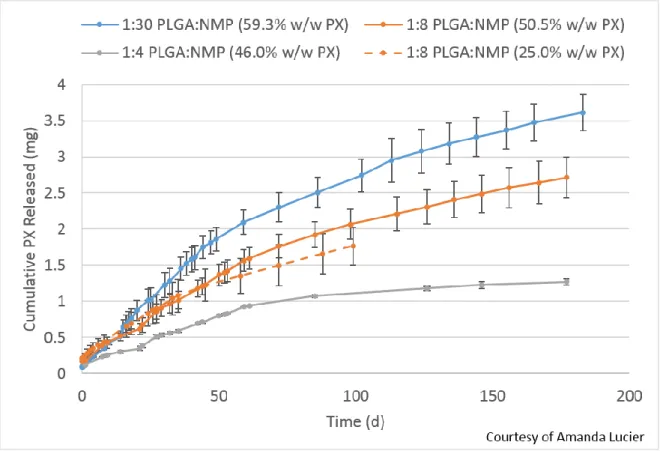
The effect of polymer/solvent ratio was examined by testing three ratios (1:4, 1:8, and 1:30) of PLGA/NMP at similar PX-loading (% w/w) near maximum drug solubility. At a common endpoint of approximately 99 days for PX-ISFI Formulations 1, 2, and 4 (Table 1), the larger the ratio of solvent, the larger the cumulative PX (% w/w) was released (Table 3).
5 studies,5,6 decreased drug-loading resulted in faster release kinetics. For this reason, although 25.0% w/w PX formulation was loaded with nearly half the amount of PX as the 50.5% w/w PX formulation, both depots released similar quantities of drug in milligrams in the first 99 days (Table 3). Figure 2 displays the similar controlled rates of PX release between these 1:8 PLGA/NMP formulations, especially within the first 45 days.
Table 2. Composition of depot formulations in release study
Formulation PLGA:NMP % (w/w) PX PLGA:NMP:PX
1 1:4 46.0 1:4:2.5
2 1:8 50.5 1:8:4.5
3 1:8 25.0 1:8:2.3
4 1:30 59.3 1:30:18.4
Within the first 24 hours of the study, all formulations showed burst releases of <10% of total drug loaded (Fig. 1) and no initial burst, characterized by a burst release of ≥20% within the first 24 hours. This indicated that a controlled amount of PX was released during implant formation, when solvent diffuses out of the depot, allowing the polymer and PX to precipitate.
6 Figure 2. Cumulative PX released (mg) with solid lines representing ISFI loaded at maximum PX saturation, determined visually.
Table 3. The average weight of depot and PX loaded, duration of release, and cumulative PX release (% and absolute) near common endpoint.
PLGA:NMP:PX (% w/w PX)
Depot Weight and PX Weight (mg)
Duration (days)
Total Cumulative PX Release (%)
Total Cumulative PX Release (mg)
1:4:2.5 (46.0%) 12.3 ± 1.8 (3.89 ± 0.6)
99 29 1.12
1:8:4.5 (50.5%) 16.9 ± 0.9 (5.66 ± 0.3)
98 36 2.07
1:8:2.3 (25.0%) 12.6 ± 0.6 (2.51 ± 0.1)
99 70 1.77
1:30:18.4 (59.3%) 14.4 ± 0.6 (5.36 ± 0.2)
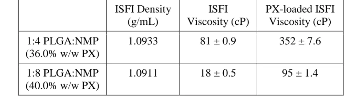
7 Rheology Analysis: ISFI Viscosity Measurement
Viscosity readings of the 1:8 PLGA/NMP ISFI and PX-ISFI (40.0% w/w PX) were measured with rotation speed of 2.0 RPM. However, the 0.5 RPM was used for 1:4 PLGA/NMP formulations because the 1:4 PLGA/NMP (36.0% w/w PX) solution was too viscous for the rheometer to provide a reading at 2.0 RPM. Both 1:4 and 1:8 PLGA/NMP formulations exhibited approximately a 4-fold increase in viscosity after PX was loaded to about 80% of PX saturation, determined visually (Table 4). All formulations investigated were syringeable with 18-gauge needles at 25°C.
Table 4. Viscosity measurements before and after drug-loading at 25°C and 2.0 RPM for 1:8 PLGA/NMP formulations and 0.5 RPM for 1:4 PLGA/NMP formulations.
ISFI Density (g/mL) ISFI Viscosity (cP) PX-loaded ISFI Viscosity (cP) 1:4 PLGA:NMP (36.0% w/w PX)
1.0933 81 ± 0.9 352 ± 7.6
1:8 PLGA:NMP (40.0% w/w PX)
1.0911 18 ± 0.5 95 ± 1.4
Accelerated Stability
Three storage conditions of varying temperature and humidity were tested on two ISFI formulations (1:4 and 1:8 PLGA/NMP) with varied drug-loading (% w/w). Increased
temperature and humidity provided accelerated exposure of conditions that may degrade
components in the PX-ISFI system to determine shelf life of the formulations. Room temperature and ambient humidity represented indoor storage conditions, the 25°C and 60% humidity
condition imitated indoor storage in humid environments, and the 40°C and 75% humidity condition tested stability of formulations near body temperature.
All PX-ISFI formulations investigated began as transparent, colorless, and homogenous solutions. Clear micelles formed as the solutions became unstable. The micelles grew larger and aggregated at the bottom of the clear solution, forming a translucent and viscous globule. This distinct phase separation indicated that the PX was precipitating out of solution. As the solvent evaporated over time, the formulation appeared as a white and waxy solid.
The relative percent PX in solution was expected to decrease with time due to PX
precipitating out of solution or degrading. Although this decreasing trend was generally observed over time, certain time points showed increases in relative percent PX (i.e. relative % PX
significantly greater than 100). The increase in relative percent PX was due to HPLC samples taken from heterogeneous, unstable solutions. Some HPLC samples of unstable formulations may have contained PX micelles that contributed to higher PX concentration measurements. However, HPLC analysis showed a single peak representing PX with 13.8 minute retention time. No extraneous peaks were present, indicating that PX was not degrading, but was rather
8 The estimated shelf life of formulations a, b, c, and d (Fig. 3) were 10 days, 21 days, 16 days, and 30 or more days. The 80% PX saturated formulas, a and c, were less stable and resulted in PX precipitating out of solution faster than the 60% or 64% PX saturated formulas, b and d. Considering the results of all three storage conditions, the 1:8 PLGA/NMP formulations maintained stability longer than the 1:4 PLGA/NMP formulations. An ongoing study of the 1:8 PLGA/NMP 40.0% w/w PX in 4°C demonstrated phase separation between days 8 and 13, indicating that shelf-life was not extended by decreasing the temperature of storage conditions.
9 Discussion
The maximum PX saturations of ISFIs (1:4 and 1:8 PLGA/NMP) determined by using the method in the experimental section were not consistent with those determined visually. After centrifugation, PX-ISFI supernatants were visually translucent suspensions rather than
homogeneous solutions. Although the 1:8 PLGA/NMP ISFI was expected to dissolve more PX than the 1:4 PLGA/NMP ISFI because of the larger ratio of solvent, the 1:4 PLGA/NMP ISFI demonstrated greater solubility. It was thus concluded that the higher ratio of polymer allowed a larger quantity of PX to be suspended in solution. Additional studies with modified method are needed to determine paclitaxel solubility in homogeneous ISFI solutions.
Although a concern with ISFI systems for drug delivery is large initial burst,
characterized by ≥ 20% drug release within 24 hours, the in vitro cumulative PX release study (Fig. 1) showed burst releases <10% in all formulations (Table 1). These results indicated that the PX-ISFI system demonstrates controlled drug release kinetics even during implant formation.
All four formulations (Table 1) demonstrated controlled release kinetics, and the larger the solvent ratio, the faster the PX was released and the larger the cumulative percent of PX was released. Both 1:8 and 1:4 PLGA/NMP ISFIs are ideal candidates for PX-ISFI systems because they minimize exposure to NMP compared to the 1:30 PLGA/NMP formulation while
demonstrating comparable release kinetics.
Similar quantities of PX released in the 1:8 PLGA/NMP formulations (50.5% and 25.0% w/w PX) indicated that similar drug doses can be delivered with reduced drug-loading due to faster release kinetics. This has profound clinical applications, as using less drug can reduce the cost of the final product without compromising the dose that the patient receives. PX-ISFI systems are tunable based on polymer/solvent ratio and drug-loading. Further release studies are needed to assess the effect of drug loading on release kinetics for the 1:4 PLGA/NMP ISFI.
Viscosity measurements of both 1:4 and 1:8 PLGA/NMP ISFI solutions increased significantly after PX was added. The 4-fold increase in viscosity of the PX-loaded ISFI (80% saturation) indicated that the hydrophobic drug was interacting with other components of the ISFI, likely the hydrophobic polymer. All formulations were syringeable with an 18-gauge needle at room temperature and are thus injectable. For in vivo studies and clinical use, ISFI solutions will be loaded at lower PX saturation, to maintain viscosity closer to that of the placebo ISFI for ease of injection.
10 Conclusion and future studies
Due to its long-acting and controlled release, the PX-ISFI system was investigated as an alternative to the standard of care treatment for ovarian cancer. ISFI systems have been shown to hold large quantities of PX while maintaining syringeability for injection. In vitro release studies of PX-ISFI systems demonstrated controlled release with minimal initial burst release and the ability to be fine-tuned by modification of polymer/solvent ratio and drug-loading. PX-loaded ISFI systems (1:4 and 1:8 PLGA/NMP) demonstrated stability at room temperature and ambient humidity for one week to at least one month. In order to reduce exposure to NMP, the 1:8 and 1:4 PLGA/NMP formulations will be tested further for optimization in vivo. Future studies include cytotoxicity assays, DSC analysis, and antitumor efficacy studies in an ovarian cancer xenograft mouse model.
Acknowledgements:
Dr. Rahima Benhabbour, Dr. Michel Gagné, and Amanda Lucier
Group members:
Roopali Shrivastava, Daijha Copeland, Clayton Swords, Gayane Paravyan, and Shamit Prabhu
References
1. American Cancer Society Available at: https://www.cancer.org/cancer/ovarian-cancer/treating/by-stage.html
2. National Cancer Institute Available at: https://seer.cancer.gov/statfacts/html/ovary.html 3. National Institutes of Health Available at:
https://pubchem.ncbi.nlm.nih.gov/compound/paclitaxel#section=Top
4. Wadee A, Pillay V, Choonara YE, et al. Recent advances in the design of drug-loaded polymeric implants for the treatment of solid tumors. Expert Opin. Drug Deliv. 2011;8(10):1323-1340.
5. Ahmed TA, Ibrahim HM, Samy AM, et al. Biodegradable Injectable In Situ Implants and Microparticles for Sustained Release of Montelukast: In Vitro Release,
Pharmacokinetics, and Stability. AAPS PharmSciTech. 2014;15(3):772-780. 6. Wang J, Xu J, Li J, et al. Improvement of the Antitumor Efficacy of Intratumoral
Administration of Cucurbitacin Poly(Lactic-co-Glycolic Acid) Microspheres Incorporated in In Situ-Forming Sucrose Acetate Isobutyrate Depots. J. Pharm. Sci. 2015;105(1):205-211.
7. U.S. Federal Drug Administration Available at:
https://www.accessdata.fda.gov/drugsatfda_docs/label/2007/021731s005,021488s010,02 1379s010,021343s015lbl.pdf
8. Arora S, Swaminathan SK, Kirtane A, et al. Synthesis, characterization, and evaluation of poly (D, L-lactide-co-glycolide)-based nanoformulation of miRNA-150: potential
11 9. Tian J, Stella V. Degradation of Paclitaxel and Related Compounds in Aqueous Solutions
III: Degradation under Acidic pH Conditions and Overall Kinetics. J. Pharm. Sci. 2010;99(3):1288-1298.
10. Krukiewicz K, Zak JK. Biomaterial-based regional chemotherapy: Local anticancer drug delivery to enhance chemotherapy and minimize its side-effects. Mater. Sci. Eng. C. 2016;62:927-942.
11. Félix Lanao RP, Jonker AM, Wolke JGC, Jansen JA, van Hest JCM, Leeuwenburgh SCG. Physicochemical Properties and Applications of Poly(lactic-co-glycolic acid) for Use in Bone Regeneration.Tissue Engineering Part B, Reviews. 2013;19(4):380-390. 12. Makadia HK, Siegel SJ. Poly Lactic-co-Glycolic Acid (PLGA) as Biodegradable
Controlled Drug Delivery Carrier. Polymers. 2011;3(4):1377-1397.
13. Avgoustakis, K. Polylactic-Co-Glycolic Acid (PLGA). Encyclopedia of Biomaterials and Biomedical Engineering. 2008;2(4):2259-2269.