The Effects of a One-Time Core Stability
Intervention Protocol on Anterior Pelvic Tilt During
a Dynamic Overhead Squat
By
Katherine Elizabeth Dyer
A thesis proposal submitted to the faculty of the University of North Carolina at Chapel
Hill in partial fulfillment of the requirements for graduation with honors in the
Department of Exercise & Sport Science in the College of Arts & Sciences.
Chapel Hill, NC
2014
Approved by:
Darin Padua, PhD, ATC
Joseph B. Myers, PhD, ATC
Abstract:
The Effects of a One-Time Core Stability Intervention Protocol on Anterior Pelvic Tilt During a Dynamic Overhead Squat Assessment
Dyer KE, Padua DA, Myers JB, Walker N, Bonney J: Sports Medicine Research Laboratory, De-partment of Exercise and Sport Science, University of North Carolina, Chapel Hill, NC
Table of Contents
Chapter 1
Introduction
4
Statement of the Problem
7
Hypothesis
8
Definition of Terms
9
Chapter 2
Review of Literature
11
Chapter 3
Methodology
Subjects
22
Apparatus/Equipment
24
Testing Procedures
25
Core Stability Protocol
30
Analysis and Data Reduction
35
Chapter 4
Results
37
Chapter 5
Discussion
39
Limitations and Future Research
44
Conclusion
47
Appendix
49
Chapter I
Introduction
Lower Back Pain is a prevalent on-going issue in the developed world with over
eighty-four percent of adults having reported symptoms in the United States alone (Deyo and Tsui-Wu,
1976). The condition of Lower Back Pain (LBP) can be very detrimental to productivity in the
work place, activities of daily living, and overall quality of life. Lower Back Pain is a pervasive
condition that is predictive of many other orthopedic injuries and muscular imbalances. Some
etiologies of Lower Back Pain may include vertebral disc pathology, spinal column arthropathy,
facet joint dysfunction, osteophytes encumbering nerves, spondylolisthesis, and spinal stenosis
(Corliss et al., 2013). Aside from these chronic, nagging issues, the condition of Lower Back
Pain can be very financially taxing in consideration of surgical costs, rehabilitation services, and
prophylactic measures which range from hundreds to tens of thousands of dollars in treatment
costs.
The condition of Lower Back Pain is an issue that has many derivations. They may range
from improper ergonomics in the work place, improper mechanisms when lifting heavy objects,
genetic abnormalities in boney alignment, changes in workout intensity and regimen, poor
pos-ture, excessive body weight, and physical inactivity. With so many factors accounting for Lower
Back Pain, there is an obvious need for a solution that is not only effective but economical and
long-term.
One of the main causes of Lower Back Pain with implications specific to both sedentary
and athletic populations is that of Anterior Pelvic Tilt (APT) (Sahrmann, 2002; Van Dillen et al.,
caused by muscular imbalances from physical inactivity and ergonomics associated with the
sed-entary work place. Improperly designed physical training regimens with overemphasis on
strengthening and neglect of stretching can also cause muscular imbalances that aggravate
Ante-rior Pelvic Tilt (Scholtes et al., 2009). The Pelvic Tilt Angle is more specifically described as the
angle made by an imaginary horizontal plane and a ray connecting the Anterior Superior Iliac
Spine and the Posterior Superior Iliac Spine, which are boney projections on the Iliac bone of the
pelvis. When this angle is greater than fifteen degrees, then the individual is qualified as having a
pelvis with Anterior Pelvic Tilt (Preece et al., 2008). This improperly tilted pelvis can cause
Lower Back Pain by putting irregular pressure on the spinal column with the anterior portion put
into excessive distraction and the posterior portion put into excessive compression (Hansraj et
al., 2001). To go along with the altered pressure on the spinal column, many movement patterns
in the lower extremity including increased trunk flexion, increased hip adduction, increased hip
internal rotation, and increased knee valgum have been noted to occur in tandem with Anterior
Pelvic Tilt, Lumbar Lordosis, and Lower Back Pain (Sahrmann, 2002). Not only is Anterior
Pel-vic Tilt associated with Lower Back Pain, but we see some concurrent kinematic deviations from
the norm in the lower extremity with APT during dynamic movement. These include increased
femoral internal rotation (Duval et al., 2010; Hruska, 1998; Ireland, 2002); hip adduction,
(Du-val, Lam, & Sanderson, 2010; Hruska, 1998; Ireland, 2002), and knee valgus (Padua et al., 2013,
Neumann, 2010). All of these kinematic deviations increase the individual’s overall risk for
inju-ry but can be altered by fixing the instigating cause of excessive Anterior Pelvic Tilt, which is
where the importance of a core protocol to increase neuromuscular control comes into play.
The emphasis of this study will not be on changes in boney alignment, which would
activation and neuromuscular control. As we have seen, Anterior Pelvic Tilt is influenced by
poor core stabilization. Such poor core stabilization can take the form of muscular imbalances
arising at the Lumbo-Pelvic-Hip Complex, of which a commonly seen example is that of Lower
Cross Syndrome. The muscular imbalance condition of Lower Cross Syndrome consists of
weakened or elongated hip extensors and abdominals, accompanied by overly strengthened or
tightened hip flexors and back extensors (Cibulka et al., 1986; Croisier, 2004; Comerford and
Mottram, 2001). More precisely, the underactive core musculature will be activated in static
po-sition and in dynamic activity during the Core Stability Intervention Protocol. In this study,
sig-nificant findings from changes in Anterior Pelvic Tilt will indicate that core stabilization may
influence Anterior Pelvic Tilt and have implications for the eradication of some Lower Back
Pain conditions. While there has been anecdotal evidence to back up the importance of a Core
Stability Program in the rehab setting, there has been little empirical evidence to say how core
stability affects Anterior Pelvic Tilt and subsequently Lower Back Pain. This study endeavors to
find empirical evidence in support of or in contrast to the efficacy of a one-time Core Stability
Intervention Protocol in decreasing Anterior Pelvic Tilt or an individual during a dynamic
Statement of the problem
Regardless of the pathological population, more and more individuals are suffering from
a multitude of conditions stemming from excessive anterior pelvic tilt. Anterior pelvic tilt can be
assessed clinically by palpation of the ASIS and PSIS of one side of an individual and comparing
that to a horizontal plane of 180 degrees. Said angle is compared to normative values which
in-clude the numerical limits of and fall into the category of anterior pelvic tilt if the angle is larger
than 15 degrees and enter the category of posterior pelvic tilt if that angle is fewer than 8
de-grees. Problems common to anterior pelvic tilt include chronic lower back pain, athletic
pubalgia, spondylosis, spondylolisthesis, foot pronation, patellar tendonitis, and general muscle
soreness and tightness among other conditions. In the clinical realm of athletic training, core
sta-bility routines including spinal stabilization and segmental stabilization have been utilized in an
attempt to fix the altered length-tension relationships, force couples, and arthokinematics that
arise at the hip, trunk, and leg with anterior pelvic tilt. Spinal stabilization is helpful in the
eradi-cation of chronic lower back pain (LBP) in athletic populations and sedentary individuals with a
prevalence of 70% of all patients suffering from LBP (Corliss et al., 2013). Low back pain is a
commonly occurring condition with Lumber Lordosis during static and dynamic activity (Deyo
and Tsui-Wu, 1976). Lumbar Lordosis can result from muscular imbalances including
tight/overactive back extensors and hip flexors, along with loose/underactive trunk flexors and
hip extensors that fit under the commonly used titles of “lower cross syndrome”and “cross
pel-vic syndrome.”Lack of neuromuscular control and core activation are co-existing conditions
with Lumbar Lordosis and LBP (Scholtes et al., 2009). An easy to perform, time-efficient, and
effective core stability program will have important implications for both sedentary and athletic
Hypotheses
-Research Question: Will individuals showing anterior pelvic tilt during a squat continue to
ex-hibit signs after implementation of a one-time core stability training protocol?
-Hypothesis 1: Anterior Pelvic tilt will be decreased during squat after the core stability training
protocol is implemented.
-Hypothesis 2:Trunk flexion will decrease relative to the individual’s pelvis after the core
stabil-ity training protocol is implemented.
-Hypothesis 3: Hip adductor displacement will decrease relative to the individual’s pelvis after
the core stability training protocol is implemented.
-Hypothesis 4: Hip internal rotation displacement will decrease relative to individual’s pelvis
af-ter the core stability training protocol is implemented.
-Hypothesis 5: Knee valgus and varus displacement will decrease relative to individual’s pelvic
Definition of terms
-Anterior Pelvic Tilt (APT): angle measure from ASIS to PSIS compared to horizontal plane w/
neutral averaging around 10-13 degrees, anterior pelvic tilt measuring >15 degrees, and posterior
pelvic tilt measuring <8 degrees
-Abdominal Drawing In Maneuver (ADIM): isometric contraction of the Transverse Abdominus
that includes verbal cueing with phrases such as “drawing your belly button in to meet back
bone”and “trying to fit into tight pair of jeans”while holding this position for 3-5 seconds,
re-laxing for another 3-5 seconds and repeating the series 10 times.
-Overhead Squat Assessment (OSA): test to measure biomechanical movement patterns in which
the subject holds their fully extended arms over their head (180 degrees shoulder abduction) with
feet facing straight forward and slightly farther than shoulder width apart. Subject squats into no
more than 90 degrees of knee flexion 5 times while to clinician evaluates their technique by
looking for pronation and ER at the foot, varus/valgus at the knee, excessive anterior pelvic tilt at
the hip, excessive trunk flexion, arms falling forward into flexion compared to start position.
-Peak Angle: largest difference measured in the angle between ASIS and PSIS compared to a
horizontal plane that the subject will go into during an Overhead Squat; different for each
indi-vidual
-Starting Angle: starting value for each individual as measured by the difference of the angle
-Descending Phase: the phase in which an individual starts their squat and whenever they go into
maximum flexion at the knee (no more than 90 degrees). Does not include the individual
Chapter 2
Literature Review
The “core”is the origin of movement and the center of gravity from which the
extremi-ties can operate efficiently and precisely and is essential to maintaining lifelong wellness and
improved Activities of Daily Living. An all-encompassing definition of the Lumbo-Pelvic-Hip
Complex, used interchangeably with the term “core,”takes into consideration the hip, pelvis, and
trunk as they work together to stabilize statically and dynamically. This core or LPHC will be
described as “the musculature that surrounds the lumbopelvic region and includes the
ab-dominals anteriorly, the paraspinals and gluteals posteriorly, the pelvic floor musculature
inferi-orly, the hip abductors and rotators laterally, and diaphragm superiorly” (Bliss & Teeple, 2005).
A properly balanced core (specifically consisting of the Transverse Abdominus, Multifidus,
Ex-ternal Obliques, and Pelvic Floor muscles) has appropriate length-tension relationships of
origi-nating and inserting musculature, force-couple relationships of these muscles, and efficiency in
resulting arthokinematics of the Lumbo-Pelvic-Hip Complex and kinetic chain (Hruska, 1998).
Spinal stabilization provided by a core stability program, helps the patient effectively use
neuro-muscular control, neuro-muscular strength, power, and endurance of their extremities while minimizing
risk of injury (Corliss et al., 2013).
Spinal stabilization is helpful in the eradication of chronic lower back pain (LBP) in
ath-letic populations and sedentary individuals, with a prevalence of 84% of all patients suffering
from LBP (Corliss et al., 2013). Low back pain is a commonly occurring condition with Lumber
Lordosis during static and dynamic activity (Deyo and Tsui-Wu, 1976). Lumbar Lordosis can
with loose/underactive trunk flexors and hip extensors that fit under the commonly used titles of “lower cross syndrome”and “cross pelvic syndrome.”Lack of neuromuscular control and core
activation are co-existing conditions with Lumbar Lordosis and LBP (Scholtes et al., 2009). One
of the main muscles of focus in this study’s core stability training protocol is the Transverse
Abdominus, which can be palpated about an inch medially and superiorly from the ASIS.
Re-search has shown that the Multifidi-- another muscle of interest in this study-- coincide in
activa-tion with the TA and funcactiva-tion to create a girdle of muscular support that works in a feed forward
mechanism to stabilize the spine in anticipation of extremity movement (Besier et al., 2001).
In-dividuals with LBP have been shown to have poor muscular endurance of the core stabilizers
resulting from a lack of neuromuscular control (Ebenbichler et al., 2001), but the implementation
of the drawing-in maneuver before core training has shown to significantly increase EMG
activi-ty and pelvic stabilization (Snijders et al., 1998). Stabiliactivi-ty in the lumbosacral region results from
cocontracting the TA and Multifidus (Richardson et al., 2002), which makes learning the
tech-nique essential to pain reduction and injury prevention.
Hip, lumbar, and pelvic movements happen concurrently in a combination of the three
planes including Sagittal (Flexion/Extension), Frontal (Abduction/Adduction), and Transverse
(Internal Rotation/External Rotation). When certain musculature is inhibited, a cycle of altered
joint kinematics, or arthrokinematics, is initiated with altered length tension relationships in the
muscle surrounding the joint. Altered force couples then result from these changed length tension
relationships and in turn cause different movement patterns at the joint, also known as altered
arthokinematics. These altered arthrokinematics put new tension and compression on joint
cap-sules and inserting musculature, putting these structures into precarious positions and loads
is substantially increased and can occur anywhere down the kinetic chain. These patterns need to
be altered in all planes of motion to reduce injury risk, but first need to start with the individual
learning neuromuscular control of their core musculature. Once neuromuscular control is
taught-- as is the aim of the core stability intervention programtaught--taught-- altered length tension relationships,
force couples, and arthrokinematics can be changed and the risk of injury can be substantially
reduced.
To start, the Lumbo-Pelvic-Hip-Complex has some commonly seen overactive or
tight-ened, and inhibited and loosened musculature that change normal movement patterns and can
cause injury (Chou, 2013). The commonly tightened musculature the directly affects the
Lumbo-Pelvic-Hip-Complex includes the iliopsoas, rectus femoris, tensor fascia latae, hip adductors
(brevis, longus, magnus, minimus; pectineus, gracilis, and obturator externus), erector spinae,
gastrocnemius, and soleus (Snijders et al., 1998; Richardson et al., 2002; Walker et al., 1987).
Typically elongated musculature includes the gluteus muscles (maximus and medius), biceps
femoris, semitendinosus, semimembranosus, transverse abdominus, multifidus, internal obliques,
and tibialis muscles (anterior and posterior) (Snijders et al., 1998; Richardson et al., 2002;
Walker et al., 1987). In combination, these tightened and loosened muscles contribute to the
condition commonly known as Lower Cross Syndrome which is associated with anterior pelvic
tilt, lumbar lordosis, lower back pain, and other injury to the lower extremity (Chou and Atlas,
2013).
Altered kinematics may originate from shortened musculature resulting in deficits in
range of motion and neuromuscular control. Excessive anterior pelvic tilt and subsequent lumbar
lordosis arise from tightened lattisimus dorsi and/or attached thoracolumbar fascia along with
Ichihashi, 2012; Neumann, 2010). Tightened triceps surae are correlated with knee valgus
(Pad-ua et al., 2012), and separate or combined with the aforementioned tightened structures can lead
to antagonistic inhibition that enhances anterior pelvic tilt as lumbar lordosis follows. These
in-hibited antagonists include the hamstrings, gluteus maximus, and most notably the rectus
abdominus (Workman, Docherty, Parfrey, & Behm, 2008; Neumann, 2010). These altered
an-tagonists play a role in the neuromuscular control of the Lumbo-Pelvic Hip Complex, affecting
hip adduction and femoral internal rotation which also influences anterior pelvic tilt and
increas-es the overall risk of injury to the lower extremity (Duval, Lam, & Sanderson, 2010; Hruska,
1998; Ireland, 2002).
Anterior pelvic tilt is of particular interest with its indications for several types of injuries
along the kinetic chain. Anecdotal evidence has shown that fixing an excessive anterior pelvic
tilt can substantially decrease chronic pain and injury, so developing a core stability intervention
program to accurately do this has enormous implications for decreasing the individual’s need for
long term rehabilitation services. This particular study will evaluate the efficacy of such a core
stability intervention program by using a dynamic overhead squat assessment to measure acute
changes in Lumbo-Pelvic-Hip-Complex neuromuscular control that will theoretically result in
changes in anterior pelvic tilt and lumbar lordosis.
Anterior pelvic tilt can cause a multitude of injuries. Knee injuries, particularly those to
the anterior cruciate ligament (ACL), occur en masse with around 80,000 to 250,000 injuries a
year for the physically active (Griffin et al., 2006). Other injuries that commonly occur as a
re-sult of anterior pelvic tilt include vertebral disc pathology, spinal column arthropathy, facet joint
dysfunction, osteophytes encumbering nerves, spinal stenosis (Corliss et al., 2013), chronic
pa-tellar tendonitis, and general muscle soreness and tightness to name a few (Chou, 2013; Chou
and Atlas, 2013). To go along with the lengthy list of orthopedic injuries arising from anterior
pelvic tilt, overall themes of loss of quality of life, large financial cost of surgery from resulting
injuries, and emotional cost when dealing with chronic pain or injury are serious issues for the
injured party (Krawciw and Atlas, 2013; Sowa and Delitto, 2013). With these real world
implica-tions, is it essential that more empirical research be done to solidify previously found anecdotal
evidence of the efficacy of a core stability intervention program.
There are many kinematic factors that predispose an individual to injury to the
Lumbo-Pelvic Hip Complex, trunk, and lower extremity when the core musculature is weak and unable
to perform its job of spinal stabilization. These kinematic risk factors include excessive hip
in-ternal rotation, knee valgus (Besier, Lloyd, Cochrane, & Ackland, 2001), hip adduction, trunk
flexion, and anterior pelvic tilt (Fields et al., 2013) which have been shown to increase the risk of
injury in the lower extremity and may also be concurrent with lack of neuromuscular control. It
has also been found that another risk factor for unspecified injury is that of poor core endurance,
especially trunk flexion endurance (Wilkerson et al., 2012). With the correlation of lower
ex-tremity injury and lateral torso flexion and torso rotation, it has been found that frontal plane
trunk position increases overall injury risk (Dempsey et al., 2012). The lower extremities are put
at biomechanically compromising and disadvantageous positions when the trunk departs from
the individual’s center of mass, in turn increasing injury rates particularly when the athlete has
other muscular weaknesses that don’t allow him to appropriately compensate for these kinematic
patterns.
It has been found that increased trunk motion arises from weak gluteus medius and
2012). When lateral trunk flexion occurs, hip adduction and foot abduction do as well which can
lead to altered lower extremity kinematics and subsequent injury because of improper hip
abduc-tion (Jamison et al., 2012). Leetun et al. found that in both genders, some
Lumbo-Pelvic-Hip-Complex factors contributing to injury included a substantially smaller amount of strength into
hip external rotation and hip abduction than uninjured counterparts (Leetun et al., 2004). It was
also discovered in a study by Zazulak et al. that injury could be predicted by deficits in trunk
neuromuscular control, particularly in females (Zazulak et al., 2007). Anterior pelvic tilt and
femoral internal rotation have an established relationship (Duval et al., 2010; Hruska, 1998;
Ire-land, 2002; Walker, Rothstein, Finucane, & Lamb, 1987), and a study by McKeon et al. (2009)
showed significantly more anterior pelvic tilt in women compared to men. Females already see
an increased rate of knee injury, and with such correlations between knee injury and lack of
Lumbo-Pelvic-Hip Complex control a core stability intervention program would do a lot to help
reduce risk of the lower extremity. Anterior pelvic tilt is associated with increased tibial external
rotation, foot pronation, femoral internal rotation, and lumbar lordosis, (Duval et al., 2010;
Hruska, 1998; Ireland, 2002). As we can see, anterior pelvic tilt is associated with a multitude of
lower extremity rotational changes that could be corrected with a core stability intervention
pro-gram.
Further evidence of the efficacy of the importance of core activation to lower extremity
kinematics is found in findings from Shirey et al. The study established that during a single leg
squat, subjects who consciously activated their core musculature were in turn able to decrease
hip frontal plane motion. Each of the fourteen subjects did the single leg squat task with and
dis-placement and knee flexion was found to have increased significantly, indicating that the core
plays a significant role in lower extremity kinematics (Shirey et al., 2012).
The above risk factors have been commonly evaluated using the Overhead Squat
As-sessment (Bell et al., 2008; Butler et al., 2010; Macrum et al., 2012; Padua et al., 2012). The
Overhead Squat Assessment task will be used to identify any changes in LPHC and lower
ex-tremity kinematics (Bell et al., 2008; Butler, Plisky, Southers, Scoma, & Kiesel, 2010; Macrum,
Bell, Boling, Lewek, & Padua, 2012; Padua et al., 2012) in a test, re-test method with the core
stability protocol being put into place between squatting trials. Any changes from excessive
ante-rior pelvic tilt to ranges of normal anteante-rior pelvic tilt after the core stability protocol may indicate
changes that took place due to conscious motor learning used to activate the core musculature
(Transverse Abdominus, Multifidus, External Obliques, and Pelvic Floor muscles) which is
nec-essary for normal pelvic tilt during dynamic movement. Changes in anterior pelvic tilt and
lum-bar lordosis may also result in other kinematic changes in the lower extremity, particularly
de-creased trunk flexion, femoral internal rotation, knee valgus, and hip adduction.
While the Overhead Squat Assessment is commonly used to assess movement pattern
dysfunction of the lower extremity (Bell et al., 2008; Macrum et al., 2012; Padua et al., 2012; B.
T. Zazulak et al., 2007), it only has theoretical implications for evaluating dysfunction of the
Lumbo-Pelvic-Hip-Complex. Excessive anterior pelvic tilt/lumbar lordosis are correlated with
weakened musculature of the deep Lumbo-Pelvic-Hip muscles, rectus abdominis, hamstrings,
and Gluteus Maximus and Medius. Tightened or overactive musculature putting the individual at
risk of increased anterior pelvic tilt/lumbar lordosis includes the hip flexors, erector spinae, and
latissimus dorsi. With weaknesses in the Lumbo-Pelvic-Hip Complex musculature as listed
exces-sive anterior pelvic tilt/lumbar lordosis. The Overhead Squat Assessment becomes as valid
as-sessment tool in that it accurately measures dysfunction of the lower extremity, ergo measuring
dysfunction in the LPHC. The only downfalls of the Overhead Squat Assessment are that it
doesn’t take into account an assessment of the internal and external obliques and quadratus
lumborum (QL), which make up the trunk’s lateral musculature. These muscles, particularly the
quadratus lumborum, are essential players in spinal stability occurring in the frontal plane (S.
McGill, Juker, & Kropf, 1996; S. M. McGill et al., 1999).
Today’s commonly used clinical interventions to increase core strength and endurance
usually include concepts of stretching with a purpose; addressing altered neuromuscular patterns
in order to regain proper movement patterns; and strengthening with musculature in the correct
position (Van Dillen and Sahrmann, 2009). The clinician can tailor these programs to the
indi-vidual depending on their initial ability to incorporate neuromuscular control into dynamic
movement. This may provide more of a challenge for some individuals and they may need more
coaching and time to practice appropriate activation of certain key musculature, particularly the
transverse abdominus and multifidus. The core stability intervention program can be done
sever-al times a week and as evidenced, can help with not only the aim of fixing excessive anterior
pelvic tilt, but many other orthopedic injuries regardless of injury and subsequent goals.
An elementary approach to core neuromuscular control can start with the individual in
hooklying position on the floor. From here, the clinician can help the individual learn when
ap-propriate contractions feel like and how to further incorporate them into smaller movements. The
core stability intervention program doesn’t require special equipment and can be done at home,
which allows the individual to get the most out of their rehabilitation services, which may be
in-expensive exercise ball or unstable objects found in the household such and pillows or furniture
can substitute for more expensive equipment and allow the individual to easily incorporate an
injury preventing, core strengthening and stability program into their everyday routine. The
indi-vidual can use this same equipment to progress through many levels of core stability once they
understand neuromuscular control or the core musculature.
Some common exercises put into place for the goal of core stabilization are as follows.
They are done in both supine and prone exercises in order to best teach and implement the
learn-ing of motor skills that use isometric co-contractions in more than one position (Richardson et
al., 2000). Each exercise is typically performed for three to four sets of eight to twelve
repeti-tions to help learn the motor skill and encourage strength and endurance gains (Clark et al.,
2012). While lying supine on the floor, typical exercises include toe taps, dying bug, hip-ups, and
hip-ups with knee extension. Toe taps are described as extending the knee slightly to bring one
foot 6 inches off the ground at a pace of 1 second up and 1 second back down and then
alternat-ing feet (Sandry, 2012). Dyalternat-ing bug is described as startalternat-ing with one arm extended to 135 degrees
while the contralateral leg is in 120 degrees of hip flexion with the foot off the ground (the
re-maining arm and leg are resting on the ground at this point). The subject then flexes the arm and
the hip to touch above the abdomen and after going back to resting point, starts the same series
with the opposite arm and leg. A one second up and one second down rhythm is followed
(Sou-za, Baker, and Powers, 2001). Hip-ups are described as starting by bringing the hip off the
ground so that a straight line is formed with the knees, hips, and shoulders. Follow a one second
up and one second down rhythm (Olson, 2013). Hip-ups with knee extension are described as
hip-ups with one knee being extended after the knees, hips, and shoulders are in line so that now
hooklying position and repeating with the opposite knee being extended. Follow a one second up
and one second down rhythm (Rosania, 2010). When in a prone position on the floor, some often
utilized exercises include a head lift, head rotations, hip extensions, and quadruped. The head lift
consists of the subject going into 45 degrees of cervical extension at a rhythm of one second up
and one second down with the arms resting at the subject’s sides (Park et al., 2013). Head
rotation consists of the subject going into 45 degrees of cervical extension and the going into 60
degrees of cervical rotation in either direction. The subject will then go back to the start position
and repeat the series but go into 60 degrees of cervical rotation on the opposite side. A rhythm of
one second into straight cervical extension, one second into cervical rotation, and one second
back to starting position will be followed (Park et al., 2013) Hip extension will consist of the
subject going into 20 degrees of hip extension by lifting one bent knee off the table at a rhythm
of one second up and one second down. The subject will continue this patterns and alternate legs
(Olson, 2013) The Quadruped consists of the subject getting on all fours while in a prone
posi-tion on the table. The subject will extend one arm off the table 90 degrees and extend the
oppo-site hip off the table 110 degrees off the table at the same time while using a one second up and
one second down rhythm. The subject will return to the all fours position before repeating the
sequence with the opposite arm and leg (Olson, 2013).
The main issue with a core stability intervention program is the individual’s ability to
in-corporate neuromuscular control of the core and Lumbo-Pelvic-Hip-Complex into dynamic
ac-tivity after success with the intervention program. The individual may also mistake certain
bio-feedback with tilting the pelvis posteriorly as the appropriate solution to their lower back pain or
as part of injury prevention, but this is inaccurate. Their pelvis may return to normal during
being able to incorporate this during activity. Diligence and positive reinforcement of
appropri-ate core muscular activation on the part of the clinician and copied by the individual can solve
this problem.
Biofeedback in the form of a placing a hand or Blood Pressure Cuff under the lumbar
vertebrae while in hook-lying position and attempting to activate the core musculature are good
tools in helping the individual learn appropriate force production for the musculature and pelvic
positioning during an acute treatment session. Even if an individual displays neuromuscular
con-trol during hook-lying and the core stability intervention program, they still may not be able to
convert this into changes in excessive anterior pelvic tilt during a dynamic Overhead Squat
As-sessment. A lack of change may be due to lack of practice, focus, or getting adjusted to the new
activity at hand (Overhead Squat Assessment AND core stability activation, as opposed to
simp-ly the Overhead Squat Assessment activity onsimp-ly). Strength deficits in the core, as well as lack of
mobility may account for lack of change in anterior pelvic tilt during a dynamic squat. These fall
out of the scope of this study unfortunately and can’t be accounted for as a result.
In summary, Anterior Pelvic Tilt has been found to be a marker of poor core stabilization
(Teyhen et al., 2005) and is influential in lower extremity biomechanics including increased
trunk flexion, femoral internal rotation, knee valgus, and hip adduction. A practical solution in
the physical therapy and athletic training settings is one that supports activation of the core
mus-culature in order to return pelvic tilt to normative values. Such returns to neutral pelvic alignment
would theoretically result in secondary improvements to the aforementioned kinematic patterns
of the lower extremity during dynamic movement, in turn decreasing overall risk for hip and
Chapter 3
Subjects
Subjects will include a convenience sample of 38 students at the University of North
Carolina who are either involved in any Varsity level athletic program or are any physically
ac-tive individuals of the general student body who fit the inclusion criteria. “Physically acac-tive”will
be defined according to the Center for Disease Control and Prevention’s 2008 Physical Activity
Guidelines for Americans, which recommends at least 150 minutes of moderate-intensity aerobic
activity (i.e. speed walking) and muscle strengthening activity at least 2 days a week, or 75
minutes of vigorous-intensity aerobic activity (i.e. jogging and running) and muscle
strengthen-ing activity at least 2 days a week (Centers for Disease Control and Prevention, 2011). Division 1
athletes are assumed to fulfill this requirement given their off-season and in-season scheduled
activity while anyone in the general student body will qualify if they have the above CDC
rec-ommended activity levels in a given week. Other inclusion and exclusion criteria are listed
below. Subjects have to meet four (3) of the five (4) inclusion criteria to participate in the study
-the subject will fit into ei-ther -the general student body or -the Division 1 student-athlete
popula-tion. Subjects will be excluded if they meet at least one (1) of the exclusion criteria.
Inclusion Criteria:
o No low back, hip, knee, ankle or shoulder injuries (ligament sprains, muscle strains, and any
chronic pain) within 6 months prior to the testing.
o Ages between 18 and 25.
o Athletes who play sports at the Division I level.
Exclusion Criteria-
o History of low back, hip, knee, abdomen, or shoulder surgery.
o Excessive femoral anteversion or retroversion.
Apparatus/Equipment
The apparatus will include one main pieces of technological equipment.
1. The electromagnetic motion capture system (MotionStar, Ascension Technology
Cor-poration, Burlington, VT) with 3 corresponding Ascension Flock of Birds electromagnetic
track-ers used to measure lower body kinematics. Sampling frequency will be set at 100 Hz
(Black-burn and Padua, 2009). The 3 Flock of Birds sensors will be placed on the subject’s dominant
side on the midpoint of the tibia; midpoint of the lateral thigh; and sacrum on S1 spinous process.
The Anterior Superior Iliac Spine and Posterior Superior Iliac Spine will be digitized. A segment
linkage model will be used, with the spinal sensor placed on the L4 spinous process, and spinal
column landmarks will be digitized at T12-L1 and C7-T1. A right hand coordinate system will
be used to establish a world and segment axis system in which the positive direction for the
x-axis is anterior, the y-x-axis is medial, and the z-x-axis is superior (Blackburn & Padua, 2008, 2009;
Padua et al., 2012). Motions of particular interest are anterior/posterior pelvic tilt, hip flexion,
Testing Procedures
Once the subject has been approved for testing in consideration of the inclusion and exclusion
criteria, the following seven testing procedures will be put into place.
1. Consent Form: The subject will read, sign, and date the consent form acknowledging
the purpose of the study; his role in the study; the testing protocol; the time commitment and
number of session associated with the study; any risks, benefits, and consequences
accompany-ing participation or droppaccompany-ing out of the study; privacy protection; and contact information for
questions, comments and grievances among other information for disclosure to the subject. See
the Appendices section for the Consent Form.
2. Set up Instrumentation: While the subject is reading over the consent form, the
clini-cian will ready the testing area by calibrating the x-, y-, z-axes of the the electromagnetic capture
system. Once the consent form has been read, signed, and dated the clinician will then prepare
the subject for data collection. The subject will stand on the platform. The clinician will then
fix-ate the 3 Ascension Flock of Bird electromagnetic sensors on the subject’s dominant side on the
midpoint of the tibia; midpoint of the lateral thigh; and sacrum on S1 spinous process. The
clini-cian will digitize the ASIS and PSIS as well. To secure each sensor, double sided tape, prewrap,
and athletic tape will be placed over each sensor with the exception of the sacral sensor which
will also be secured using a Velcro belt. The wires attached to each sensor will be gathered to the
lateral side of the subject according to their dominant leg which has the sensors attached to it in
order to avoid any tangling or catching of the wires or sensors during the dynamic Overhead
3. Describe the Squat Task: The clinician will then describe the squat task for the subject.
This script will be read before the first squatting session, which will occur before the core
stabil-ity intervention:
“Standing with your feet facing forward and shoulder width apart, raise your arms above
your head as if you are trying to touch the ceiling. From this position, you will go into a
squat-ting position by first bending at the waste to bring your chest lower your chest to the ground, by
then bending at the knee, and by then bending at the ankle to bring your knee closer to your toes.
Squat so that your knee does not go into more than 90 degrees of flexion under the instruction of
the clinician. Once you reach this position, you will go back the starting position with your hands
over your head. You will perform this squat at an even pace of 1-2 seconds on the descent phase
and 1-2 seconds on the ascent phase with 1 second of rest between each squat repetition. You
will get three practice squats if needed and five trial squats with the Flock of Birds sensors will
be recorded (Richardson et al., 2000). You will have 30 seconds of rest between the practice set
of squats and the trial set of squats (Clark et al., 2012). Do you understand the instructions as I
have read them? Do you have any questions?”
4. The subject will then perform one set 5 Overhead Squats to the beat of the metronome
(80 bpm) while on the Force plate of the MotionStar Ascension Technology and attached to the
Flock of Birds electromagnetic sensors. The subject will squat to 90 degrees of knee flexion each
time by utilizing the clinician’s cueing to ensure the accurate joint angle.
5. Teach TA Contractions: The subject will lay in the supine hook-lying position on the
padded clinical-use table and transverse abdominis activation will be taught by the investigator.
in-cluding “pull your belly button to your spine,”“make yourself as skinny as possible,”and
“swal-low your stomach,”and tactile cueing through palpation of the lower abdominals just medial to
the anterior superior iliac spines. Subjects will then be instructed on how to posteriorly tilt the
pelvis and flatten the lumbar spine against the table by “tucking your tailbone,”or “roll your hips
back.”
6. Core Stability Protocol: The Core Stability Protocol will then be put into place with
both supine and prone exercises in order to best teach and implement the learning of motor skills
that will need to use isometric co-contractions in more than one position (Richardson et al.,
2000). Each exercise may be performed for as many reps as it takes to relearn motor skill, but for
purposes of time and to prevent muscle fatigue we will limit it to 5 repetitions to teach each
indi-vidual and 3 extra if someone is having trouble co-contracting the TA and Mulfitidus and
per-forming the exercise at the same time (Clark et al., 2012). While lying supine on the padded
clin-ical-use table, the exercises will include toe taps, dying bug, hip-ups, and hip-ups with knee
ex-tension. Toe taps are described as extending the knee slightly to bring one foot 6 inches off the
table at a pace of 1 second up and 1 second back down and then alternating feet (Sandry, 2012).
Dying bug is described as starting with one arm extended to 135 degrees while the contralateral
leg is in 120 degrees of hip flexion with the foot off the ground (the remaining arm and leg are
resting on the ground at this point). The subject then flexes the arm and the hip to touch above
the abdomen and after going back to resting point, starts the same series with the opposite arm
and leg. A one second up and one second down rhythm is followed (Souza, Baker, and Powers,
2001). Hip-ups are described as starting by bringing the hip off the ground so that a straight line
is formed with the knees, hips, and shoulders. Follow a one second up and one second down
extended after the knees, hips, and shoulders are in line so that now one knee is also in line.
Bring the foot back to the ground before returning to the original hook-lying position and
repeat-ing with the opposite knee berepeat-ing extended. Follow a one second up and one second down rhythm
(Rosania, 2010). The subject will then move to a prone position on the padded clinical-use table
and perform head lifts, head rotations, hip extensions, and quadruped. The head lift will consist
of the subject going into 45 degrees of cervical extension at a rhythm of one second up and one
second down with the arms resting at the subject’s sides (Park et al., 2013). Head rotation will
consist of the subject going into 45 degrees of cervical extension and the going into 60 degrees
of cervical rotation in either direction. The subject will then go back to the start position and
re-peat the series but go into 60 degrees of cervical rotation on the opposite side. A rhythm of one
second into straight cervical extension, one second into cervical rotation, and one second back to
starting position will be followed (Park et al., 2013). Hip extension will consist of the subject
going into 20 degrees of hip extension by lifting one bent knee off the table at a rhythm of one
second up and one second down. The subject will continue this patterns and alternate legs
(Ol-son, 2013). The Quadruped consists of the subject getting on all fours while in a prone position
on the table. The subject will extend one arm off the table 90 degrees and extend the opposite hip
off the table 110 degrees off the table at the same time while using a one second up and one
se-cond down rhythm. The subject will return to the all fours position before repeating the sequence
with the opposite arm and leg (Olson, 2013). See Appendix 2 (Core Stability Protocol) for
de-tailed script instructions for each exercise.
7. Remeasure Squats: The clinician will instruct the subject to stand on the platform
where the squats were first measured. The clinician will not need to reattach each of the 3 Flock
attached during all the exercises. After the reading of the script below, the subject will perform
the Overhead Squat task. Upon completion and assurance that the data has been recorded
appro-priately, the sensors and all adhesive equipment will be removed and the subject will be free to
leave. This script will be read before the second squatting session, which will occur after the core
stability intervention:
“You will now perform 5 Overhead Squats in succession as you did prior to the Core
Stability Intervention Protocol. Standing with your feet facing forward and shoulder width apart,
raise your arms above your head as if you are trying to touch the ceiling. From this position, you
will go into a squatting position by first bending at the waste to bring your chest lower your chest
to the ground, by then bending at the knee, and by then bending at the ankle to bring your knee
closer to your toes. Squat so that your knee does not go into more than 90 degrees of flexion
un-der the instruction of the clinician. Once you reach this position, you will go back the starting
position with your hands over your head. You will perform this squat at an even pace of 1-2
se-conds on the descent phase and 1-2 sese-conds on the ascent phase with 1 second of rest between
each squat repetition. You will get three practice squats if needed and five trial squats with the
Flock of Birds sensors will be recorded. You will have 30 seconds of rest between the practice
set of squats and the trial set of squats. Do you understand the instructions as I have read them?
Core Stability Protocol
The core stability protocol will be put into place with both supine and prone exercises in order to
best teach and implement the learning of motor skills the subject will need to use during
isomet-ric co-contractions in more than one position (Richardson et al., 2000). Clinically, each exercise
is usually performed for as many reps as it takes to learn the motor skill, but for purposes of time
and to prevent muscle fatigue we will limit it to 5 to teach each individual with 3 extra
repeti-tions if the subject is having trouble co-contracting the transverse abdominis and mulfitidus and
performing the exercise at the same time (Richardson et al., 2000). Between each set of
exercis-es, the subject will rest 30-45 seconds (Clark et al., 2012). While lying supine on the floor, the
exercises will include toe taps, dying bug, hip-ups, and hip-ups with knee extension.
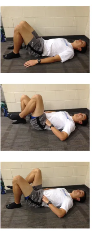
1. The following script will be read to describe the Toe Taps task for each individual:
“The first exercise is Toe Taps. Toe Taps are described as starting in the hook lying position
(back flat on ground with knees bent, feet flat on the ground, and hands at the side on the
ground), extending (straightening) the knee slightly to bring one foot 6 inches off the ground.
Slightly flex at the hip to bring one leg off the ground and lower the same leg by extending at the
hip at a pace of 1 second up off the ground and 1 second back down to the ground. Now do this
same motion while alternating which foot is lifted off the ground. (Sandry, 2012). Repeat this 5
times for each foot and 3 more if you feel you are unable to maintain the Transverse Abdominus
contractions we previously discussed.” See Figure 1 for visual example.
2. The following script will be read to describe the Dying Bug task for each individual:
“Dying Bug is described as starting in the hook lying position (back flat on ground with knees
contralateral (opposite sided) leg is in 120 degrees of hip flexion with the foot off the ground (the
remaining arm and leg are resting on the ground at this point). Flex the arm and the hip that are
not in contact with the ground to touch above the abdomen. Go back to resting point and then
start the same series with the opposite arm and leg. A one second up and one second down
rhythm is to be followed. (Souza, Baker, and Powers, 2001). Repeat this 5 times for each side
and 3 more if you feel you are unable to maintain the Transverse Abdominus contractions we
previously discussed.” See Figure 2 for visual example.
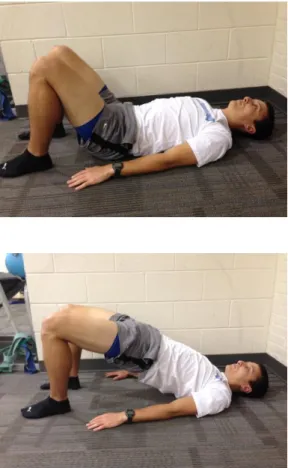
3. The following script will be read to describe the Hip-Ups task for each individual:
“Hip-Ups are described as starting in the hooklying position (back flat on ground with knees
bent, feet flat on the ground, and hands at the side on the ground), and then by bringing the hips
off the ground so that a straight line is formed with the knees, hips, and shoulders. Focusing on
pushing your hips through the ceiling by contracting your glutes will help with appropriate form.
Follow a one second up and one second down rhythm (Olson, 2013). Repeat this 5 times for each
foot and 3 more if you feel you are unable to maintain the Transverse Abdominus contractions
we previously discussed.” See Figure 3 for visual example.
4. The following script will be read to describe the Hip-Ups with Knee Extension task for
each individual:
“Hip-Ups with Knee Extension are described as Hip-Ups with one knee being extended after the
knees, hips, and shoulders are in line so that the lower leg is also now in line. Bring the foot back
to the ground before returning to the original hooklying position(back flat on ground with knees
bent, feet flat on the ground, and hands at the side on the ground). Repeating with the opposite
2010). Repeat this 5 times for each foot and 3 more if you feel you are unable to maintain the
Transverse Abdominus contractions we previously discussed.” See Figure 4 for visual example.
The subject will then move to a prone position on the floor and perform head lifts, head
rota-tions, hip extensions, and quadruped.
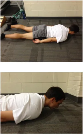
5. The following script will be read to describe the Head Lift task for each individual:
“The Head Lift will consist of the subject going into 45 degrees of cervical extension (lift you
head straight off the ground while your eyes are still looking down at the ground) at a rhythm of
one second up and one second down with the arms resting at the subject’s sides (Park et al.,
2013). Repeat this 5 times for each foot and 3 more if you feel you are unable to maintain the
Transverse Abdominus contractions we previously discussed.” See Figure 5 for visual example.
6. The following script will be read to describe the Head Lift with Rotation task for each
individual:
“Head Lift with Rotation will consist of the subject going into 45 degrees of cervical extension
and then going into 60 degrees of cervical rotation to either side (turn your head until your chin
reaches the middle of your shoulder on one side). The subject will then go back to the start
posi-tion with the head on the ground with eyes looking straight down into the ground and repeat the
series by going into 60 degrees of cervical rotation on the opposite side. A rhythm of one second
into straight cervical extension, one second into cervical rotation, and one second back to starting
feel you are unable to maintain the Transverse Abdominus contractions we previously
dis-cussed.” See Figure 6 for visual example.
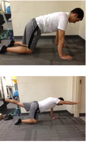
7. The following script will be read to describe the Hip Extension task for each individual:
“Hip Extension will start with the subject on all fours and eyes facing the ground. Go into 20
de-grees of hip extension (20 dede-grees past the leg being in straightened position) by lifting one bent
knee off the ground at a rhythm of one second up and one second down. The subject will
contin-ue this pattern and alternate legs (Olson, 2013). Repeat this 5 times for each foot and 3 more if
you feel you are unable to maintain the Transverse Abdominus contractions we previously
dis-cussed.” See Figure 7 for visual example.
8. The following script will be read to describe the Quadruped task for each individual:
“The Quadruped consists of the subject starting on all fours while in a prone position on the
ground. The subject will extend one arm off the table 90 degrees and extend the opposite knee
and hip off the table 20 degrees past the straightened hip position at the same time while using a
one second up and one second down rhythm. The subject will return to the all fours position
be-fore repeating the sequence with the opposite arm and leg (Olson, 2013). Repeat this 5 times for
each foot and 3 more if you feel you are unable to maintain the Transverse Abdominus
contrac-tions we previously discussed.” See Figure 8 for visual example.
The subject will then stand and the sacral sensor will be placed back on the patient and
recalibrated. The subject will then perform 5 consecutive overhead squats to the beat of a
metro-nome set at 80 bpm (Bolling et al., 2006). Kinematic data will be recorded with the
“Now stand with one foot on each force plate, with both feet facing forward as was done
before with the previous set of squats. You will perform 5 consecutive Overhead Squats to the
beat of the metronome as before with the only difference being that you will contract your
Transverse Abdominis as you did with the 8 practiced exercises from the Core Stability
Interven-tion Protocol. If you need to take a break it will be at the top of the squat and it needs to be very
quick (under half a second as you pause at the top of the squat) as you need to stay on beat with
the metronome for the ensuing squat. Ideally, you would perform the TA contraction throughout
the entire set of five squats but ultimately you just need to make sure you are contracting your
Transverse Abdominis during the descending phase and ascending phase of each squat with a
Analysis and Data Reduction
A paired samples t-test will be used in statistical analysis between each of the pelvic tilt
baseline values prior to the core stability training protocol and the pelvic tilt values for the same
population after the core stability training protocol (5 total paired samples t-tests). This will be
measured by looking at the descent phase of the squat only which will start with the first
move-ment of the subject towards a squat from static standing position to the body position when 90
degrees of knee flexion is attained during the squat. The ascending phase of the squat-- when the
subject extends the knee to return to the static standing position-- will not be calculated into any
statistical analysis. After the 5 trial squats are performed and data is collected for each of the 5
Independent Variables, an average for each Independent Variable will be attained. This process
will be repeated for the pre-Core Stability Protocol intervention set of 5 trial squats and for the
post-Core Stability Protocol intervention set of 5 trial squats. The averages for the pre and post
intervention scores for each Independent variable will then be compared for any significant
dif-ference.
DV1 -Trunk Flexion Displacement: Will consist of the difference in starting trunk flexion
angle as measured by the Flock of Birds sensors on the midpoint of the lateral thigh, the sacrum
on S1 spinous process (vertex), and the ASIS and the peak angle measured between the same
Flock of Birds sensors at the end of the squat when the knee is flexed at 90 degrees.
DV 2 -Anterior Pelvic Tilt Displacement: Will consist of the difference in starting pelvic
tilt angle as measured by the Flock of Birds sensors on the ASIS, the sacrum on S1 spinous
pro-cess (vertex) and the PSIS, and the peak angle measured between the same Flock of Birds
DV 3 -Hip Adduction Displacement: Will consist of the difference in starting hip
adduc-tion angle as measured by the Flock of Birds sensors on the ASIS, the midpoint of the lateral
thigh (vertex), and the midpoint of the tibia and the peak angle measured between the same
Flock of Birds sensors at the end of the squat when the knee is flexed at 90 degrees.
DV 4 -Hip Internal Rotation Displacement: Will consist of the difference in starting hip
rotational angle as measured by the Flock of Birds sensors on the ASIS, the midpoint of the
lat-eral thigh (vertex), and the midpoint of the tibia and the peak angle measured between the same
Flock of Birds sensors at the end of the squat when the knee is flexed at 90 degrees.
DV 5 -Knee Abduction Displacement: Will consist of the difference in starting knee
an-gle as measured by the Flock of Birds sensors on the ASIS, the midpoint of the lateral thigh
(ver-tex) and the midpoint of the tibia and the peak angle measured between the same Flock of Birds
Chapter 4
Results
Data was collected from 38 participants for the current study ranging in age from 18-25
(21.1 +/- 1.1 years) (Table 2). The participants average mass was 68.6 +/- 11.25 kg and their
av-erage height was 172.0 +/- 8.8 cm. Participants were physically active individuals from the
gen-eral population who fulfilled the Center for Disease Control and Prevention’s 2008 Physical
Ac-tivity Guidelines for Americans, which recommends at least 150 minutes of moderate-intensity
aerobic activity (i.e. speed walking) and muscle strengthening activity at least 2 days a week, or
75 minutes of vigorous-intensity aerobic activity (i.e. jogging and running) and muscle
strength-ening activity at least 2 days a week (Centers for Disease Control and Prevention, 2011). The
subjects also had no known history of injury within the last 6 months or any surgical proceedings
to the low back, hip, knee, ankle, abdomen, or shoulder. Craig’s Test for Femoral Anteversion
was performed to determine whether the individual qualified for the study (excessive femoral
anteversion or retroversion was a disqualifier). The participants all went through the exact same
procedure including two sets of 5 Overhead Squats before and after the Core Stability
Interven-tion Protocol consisting of teaching the participants to contract their Transverse Abdominus
dur-ing 8 core exercises.
Along with the 5 paired samples T-tests for each dependent variable, a 95% confidence
interval was utilized (p=.05) for the study. There were no significant differences between the pre-
and post-intervention measures of anterior pelvic tilt displacement (p=.455), hip internal rotation
displacement (p=.082), knee abduction (valgus) displacement (p=.317). However, there were
significant changes from pre- to post-intervention for both trunk flexion (p=.008) and hip
in trunk flexion displacement following the core stability intervention, which was then evaluated
further with the descriptive statistic Cohen’s d to measure effect size-- or the measure of strength
of the phenomenon. Cohen’s d was found to be 0.21 for trunk flexion, which is considered to be
weak and indicates that the average 2.39 degree decrease in trunk flexion was not actually
clini-cally significant. Hip adduction displacement was significantly decreased following the
interven-tion as individuals showed 1.12 degrees of hip adducinterven-tion displacement prior to the interveninterven-tion.
After the intervention, however, the participants displayed 7.92 degrees of hip abduction
dis-placement (for an absolute value of 9.04 total degrees of change). The magnitude of change in
hip abduction as measured by Cohen’s d was 2.31 and as such would be considered large and
Chapter 5
Discussion
The most important finding in this study was that Trunk Flexion and Hip Adduction
de-creased significantly. While there was no significant change in Anterior Pelvic Tilt, Hip Internal
Rotation, or Knee Abduction, there was significantly less Trunk Flexion and decreased Hip
Ad-duction when comparing pre to post treatment joint displacement means. The fact that both
Trunk Flexion and Hip Adduction saw significant statistical changes does not necessarily
trans-late to clinical significance in which we would see meaningful change in Trunk Flexion and Hip
Adduction in the real world. Such a meaningful change could indicate that there is enough of a
change in kinematic patterns that the individual is now less prone to certain biomechanical
movement patterns that predispose that individual to injury. Putting such a Core Stability
Inter-vention Protocol into place could change these kinematic patterns with lessened Hip Adduction
and theoretically lower injury risk and rates as a result. The significant difference in trunk flexion
(m = 2.40; SD = 5.26; p = 0.008; d = 0.21) does not look to be clinically significant when the
effect size was calculated, while the change in hip adduction (m = 9.03; SD = 5.08; p < 0.001; d
= 2.31) and its effect size shows that it could be clinically significant-- this has huge implications
for rehab programs if the goal is to try to lower hip adduction.
A larger hip adduction joint angle during dynamic activity is correlated with increased
knee valgum, which puts the knee in a predisposed position for injury (Duval, Lam, &
Sander-son, 2010; Hruska, 1998; Ireland, 2002). This results in a subsequently increased risk of injury at
the lower extremity, especially with the anterior cruciate ligament (ACL) of the knee (Sahrmann,
2002; Griffin et al., 2006). Knee injuries, particularly those to the ACL, occur en masse with
already see an increased rate of knee injury and with such correlations between knee injury and
lack of Lumbo-Pelvic-Hip Complex control (Fields et al., 2013), a core stability intervention
program could do a lot to help reduce risk of the lower extremity. While it is not known what the
direct effect of this Core Stability Intervention Protocol will be ACL injury rates, the
Lumbo-Pelvic-Hip-Complex, and the rate and type of lower extremity injuries including vertebral disc
pathology, spinal column arthropathy, facet joint dysfunction, osteophytes encumbering nerves,
spinal stenosis (Corliss et al., 2013), chronic lower back pain, athletic pubalgia, spondylosis,
spondylolisthesis, osteoarthritis, foot pronation, patellar tendonitis, and general muscle soreness
and tightness (Chou, 2013; Chou and Atlas, 2013), changes in hip adduction would significantly
change kinematic patterns, likely resulting in fewer of these complications. As was previously
discussed with the cycle of muscular imbalances leading to altered length tension relationships,
altered force couples, altered arthrokinematics, Lower Cross Syndrome, Anterior Pelvic Tilt,
Lumbar Lordosis, and Lower Back Pain at the Lumbo-Pelvic-Hip-Complex (LPHC) (Hruska,
1998), it is not a stretch to think that these patterns can be reversed by returning the altered
arthrokinematics to their appropriate movement patterns. Another benefit to an efficacious Core
Stability Intervention Protocol is that it could help lower the overall loss of quality of life, large
financial cost of surgery from the above orthopedic injuries, and emotional cost of having to deal
with chronic pain or injury (Krawciw and Atlas, 2013; Sowa and Delitto, 2013). For the above
reasons, it is an extremely important finding that hip adduction was significantly and clinically
lessened by following the Core Stability Intervention Protocol.
Finding an inexpensive, easy to use, and efficacious intervention backed by empirical
re-search is essential to help resolve these real world implications-- thankfully this intervention that
real kinematic change through this study. It is essential, however, that more empirical research
be done on the subject to quantify how much of a tangible effect this intervention has in the
clin-ical setting on reduction of injury rates. Further evidence speaking to the efficacy of core
activa-tion to lower extremity kinematics was found in a study from Shirey et al. The study established
that during a single leg squat, subjects who consciously activated their core musculature were in
turn able to decrease hip frontal plane motion (hip adduction/abduction). Each of the fourteen
subjects did the single leg squat task with and without the core musculature engaged. Subsequent
performance as measured by medial hip displacement and knee flexion was found to have
in-creased significantly, indicating that the core plays a significant role in lower extremity
kinemat-ics (Shirey et al., 2012). There was a previous foundation to say that activation of the core
mus-culature would affect lower extremity kinematics as seen in Shirey et al., but not necessarily a
foundation saying that this specific gathering of core exercises would be efficacious in doing the
same thing. Other studies done byBell et al., Butler et al., Macrum et al., and Padua et al. all
uti-lized a dynamic Overhead Squat Assessment to look at changes in lower extremity kinematics,
but did not necessarily find significant decreases in hip adduction from their studies that were
more focused on looking at the efficacy of an Overhead Squat Assessment as clinical movement
dysfunction assessment tool. Whereas we only previously had anecdotal evidence to speak to the
efficacy of a Core Stability Intervention Protocol like this, we now have empirical evidence to
say that this specific collection of core exercises works as an intervention in significantly altering
lower extremity kinematics, particularly that of Hip Adduction during an overhead squat task.
To understand the overall implications, we have to consider that excessive hip adduction
displacement during dynamic movement is known to be correlated with increased knee
adduc-tion displacement-- as we did with the Core Stability Intervenadduc-tion Protocol-- it would
theoretical-ly then be possible to lower the risk and rate of injury to the lower extremity, particulartheoretical-ly to the
knee and ACL. Overall, the findings of the study mean that this specific Core Stability
Interven-tion Protocol with these 8 collective exercises was efficacious in altering Lower Extremity
kine-matics, particularly that of hip adduction, during a dynamic overhead squat. We still need future
research to investigate what the actual influence of this Protocol is on injury to the lower
extrem-ity. We need more research to help answer if this Core Stability Protcol Intervention actually
changes the risk and rate of injury to the lower extremity in the real world.
Our original hypothesis and main motivation for carrying out the study was to look at
changes in Anterior Pelvic Tilt (APT). We saw no isolated changes in APT but there was a
sig-nificant change in trunk flexion, which would suggest APT should have changed. It may not
have changed because the intervention itself was only a one-time session and may not have been
potent enough as a result to elicit real change. As there was no requirement for the subject
popu-lation to present with lumbar lordosis to fit the inclusion criteria, we may have had subjects who
had no real room for change which would have then been measurable under our study.
Perform-ing future research that looks at a pathological population with APT and lumbar lordosis is
defi-nitely needed for future research to help us see what any potential changes the Core Stability
In-tervention Protocol could cause for such a pathological population. Had more emphasis also been
placed on maintaining proper pelvic alignment during each of the 8 Core Stability Intervention
Exercises and during the post-treatment squat session, we may have seen more change in
Anteri-or Pelvic Tilt as well. The main focus of the cueing relied on contracting the Transverse
Abdominis, with continual reminders to do so in each of the scripts for the 8 individual exercises