35
© 2017 by the Serbian Biological Society How to cite this article: How to cite this article: Kang MK, Bang A, Choi HO, Han SJ. Comparative analysis of two murine CDC25B isoforms. Arch Biol Sci. 2017;69(1):35-44.
Comparative analysis of two murine CDC25B isoforms
Min Kook Kang, Aera Bang, Hwa Ok Choi and Seung Jin Han*
Department of Biological Sciences, Inje University, 197 Inje-ro, Gimhae, Gyeongnam, 50834, Korea
*Corresponding author: [email protected]
Received: March 15, 2016; Revised: April 17, 2016; Accepted: April 19, 2016; Published online: July 28, 2016
Abstract: CDC25B phosphatase plays a pivotal role in the cell cycle process by dephosphorylating and activating the CDC2
kinase of maturation-promoting factor (MPF). In mice, two transcripts of Cdc25B are generated by the alternative
splic-ing of one gene. We compared the properties of these two forms of CDC25B. When the expression pattern of Cdc25B was
examined using RT-PCR, both forms were detected in almost all mouse tissues tested. The expression of two forms of the CDC25B protein in various mouse tissues was confirmed using Western blotting with generated isoform specific antibod-ies. CDC25B1 tends to accumulate more in the cytosol than CDC25B2 does, and they have different binding capacity for
14-3-3 proteins. CDC25B1 was more effective in dephosphorylating in vitro substrate para-nitrophenyl phosphate and
showed higher activity in the modified histone H1 kinase assay than CDC25B2. These results suggest that the two forms of CDC25B play different roles in cell cycle regulation.
Key words:Cdc25B; 14-3-3; localization; phosphatase activity; isoform
IntRoDuCtIon
Cell cycle progression is orchestrated by several cyclin-dependent kinases (CDKs). Activation of the CDKs requires both dephosphorylation of the inhibitory phosphates on Thr14 and Tyr15 by dual-specificity phosphatase CDC25 [1] and association with cyclins [2]. As a cell cycle regulator, CDC25 is involved in mi-tosis, meiosis [3], DNA damage and replication check-point release [4, 5]. In mammals, three members of the CDC25 family, CDC25A, CDC25B and CDC25C, are expressed differently in diverse tissues and during different developmental stages [6]. Although they have partially functional redundancy, they are involved in different cell cycle processes. CDC25A has a role in the G1/S phase transition, and CDC25C is involved in the G2/M transition [2, 7]. CDC25A-null mice are lethal during the peri-implantation period (embry-onic days 5-7) [8], but CDC25C-null mice display no appreciable phenotype, with normal fertility in both males and females [9]. In CDC25B-null female mice, the oocyte cannot undergo resumption of the meiotic progression [10]. The accumulation of CDC25B in mitosis triggers centrosomal microtubule nucleation and activates cyclin B/CDC2 in the centrosome for
mitotic entry [11]. Therefore, CDC25B is essential for the G2/M phase transition in the cells.
The human CDC25B gene consists of 15 exons, whereas the mouse Cdc25b gene is composed of 16 exons in chromosome 2.To date, more than four dif-ferent types of human CDC25B transcripts generated by the alternative splicing of their amino termini have been found; however, some of the human CDC25B transcripts are not translated into protein [12, 13]. The expression patterns of translated CDC25B isoforms are different in human HeLa cells [14]. The variants of CDC25B have different phosphatase activity when expressed in fission yeast, indicating the amino ter-minus of CDC25B is responsible for the regulation of its activity toward cyclin B/CDC2 [15].
36 Arch Biol Sci. 2017;69(1):35-44
its accumulation in cytoplasm [19]. Some CDC25B phosphorylation sites are involved in 14-3-3 binding to modulate the localization of CDC25B and its activity in the mitosis, G2 checkpoint and meiosis processes [20-25]. For instance, p38MAPK phosphorylates the S323 site upon ultraviolet (UV) irradiation and stabilizes 14-3-3 binding to block entry into mitosis [24, 26]. The subcellular localization of CDC25B is dependent on the combined effects of a nuclear localization sig-nal, a nuclear export sigsig-nal, as well as the interaction with 14-3-3 proteins. It is believed that phosphory-lated Ser-323 of human CDC25B is a major 14-3-3 dimer-binding site and pSer-151 and pSer-230 act as low-affinity 14-3-3 binding sites to cooperatively bind with the 14-3-3 dimer by creating an intramolecular bridge [21].In the mouse GV oocyte arrested in the prophase of meiosis I, PKA phosphorylates CDC25B on S321 (human S323 corresponding site), leading to enhanced binding with 14-3-3 [27] and the localization of CDC25B in the cytosol [25].
Given that the mouse is a major experimental ani-mal model, it is important to understand the similari-ties and differences in CDC25B protein between mice and humans. However, almost all experiments on the properties of CDC25B protein have mainly been per-formed with human CDC25B proteins; studies of the mouse ortholog are uncommon. In addition, there is little information about the differences between CDC25B isotypes. In this study, we provide some ev-idence for the expression of two splicing variants of mouse CDC25B and the properties of these proteins in the cell cycle.
MAteRIAls AnD MethoDs
Reverse transcription and polymerase chain reaction
Full-length mouse Cdc25b cDNAs were cloned with mouse lung mRNA using Superscript III reverse tran-scription (Thermo Fisher Scientific, Inc. Waltham, MA). Primers for mouse Ccd25b genes were 5’ gcc g-gatccgccacgatggaggtacccctg 3’ and 5’ aggctcgag atcact-ggtcttgcagcctgc 3’ with generated BamH I and Xho I restriction enzyme sites (italics in the sequence). To check the expression of Cdc25b genes in various
tis-sues, cDNAs were amplified with specific primers:
Cdc25b1 forward, 5’ cctcattccagctctgccca 3’, Cdc25b2
forward, 5’ ccagagaccaagatggaggagc 3’, and Cdc25b
reverse, 5’ ccgggctcagctcctctac 3’.
Cell culture
Hek 293, HeLa or Cos7 cells were grown in Dulbecco’s modified Eagle medium (DMEM) supplemented with 10% fetal calf serum (Thermo Fisher Scientific, Inc.), 30 mg/ml penicillin and 100 mg/mL streptomycin at 37°C under 5% CO2. For the expression of Cdc25B
constructs, cells were transfected using JetPEI trans-fection reagent (Polyplus Transtrans-fection Inc., Illkirch-Grafenstaden, France).
Western blot analysis
The various mouse tissues were washed with PBS and then homogenized in RIPA buffer (25 mM Tris-HCl pH 7.4, 150 mM NaCl, 1% NP40, 0.1% SDS) using a motor-driven homogenizer (IKA, Guangzhou, Chi-na). The lysates were incubated overnight with 0.1 µg of CDC25B antibody (sc326, Santa Cruz Biotech, Inc. Dallas, TX) at 4°C. After immunoprecipitation for 2 h using 30 µL of 50% protein G sepharose (GE Healthcare Life Sciences, Piscataway, NJ), the precipi-tates were washed with RIPA buffer and subjected to 8% SDS-PAGE. After transfer to PVDF membranes (Millipore Corp., Bedford, MA), immunoblotting was performed by incubating with a 1:1000 dilution of CDC25B1- or CDC25B2-specific antibody. The anti-bodies that can respectively recognize only CDC25B1 or the B2 specific region (Fig. 1A) were generated us-ing synthesized peptides (AbFrontier, Seoul, Korea).
subcellular localization of mouse Cdc25B in Cos-7 cells
14-3-3 co-immunoprecipitation
To investigate the ability of CDC25B to bind the 14-3-3 proteins, one of the HA-tagged Cdc25b genes and one of FLAG-tagged 14-3-3 genes were co-transfected into Hek293 cells at a 1:1 ratio. Cell lysate (500 µg) was immunoprecipitated with HA antibody (HA 1.1, Sigma-Aldrich, St. Louis, MO) at 4°C overnight. Af-ter the addition of protein G sepharose, the washed immune complexes were separated by SDS-PAGE. Co-precipitated 14-3-3 was detected with the FLAG antibody (F1804, Sigma-Aldrich).
Modified histone h1 kinase assay
To purify the inactive cyclin B/CDC2 complex, HeLa cells were treated with 10 mM of hydroxyurea for 18 h and immunoprecipitated with cyclin B antibody (H-85, Santa Cruz Biotech). After transfecting vari-ous mutants of Cdc25b into the HEK293 cells, the CDC25Bs were immunoprecipitated with HA anti-body and mixed with the cyclin B/CDC2 complex. The mixture was incubated for 30 min at 30°C in the phosphatase buffer (40 mM Tris-HCl, pH 8.0, 300 mM NaCl, 10 mM EDTA, 10 mM DTT). After being
38 Arch Biol Sci. 2017;69(1):35-44
spun down, the pellet was incubated at 30°C for 15 min with 50 µL of kinase buffer (50 mM Tris-HCl, pH 7.5, 10 mM MgCl2, 1 mM DTT, 50 µM ATP, 5 µCi/mL [γ-32P]ATP) with the substrate (10 µg histone
H1). The reaction was stopped by adding 4X Laem-mli buffer, and the samples were loaded on a 12.5% SDS-PAGE gel. The density of 32P incorporation was
compensated with the amount of added immunopre-cipitated protein after scanning and digitizing the im-age using the Imim-age J program.
expression and purification of Gst-fused Cdc25b and phosphatase assay
Mouse Cdc25b1 and Cdc25b2 were respectively cloned in the pGEX4T-2 vector and induced the expression of a fusion protein by adding 0.2 mM of isopropyl β-D-thiogalactoside (IPTG) in soluBL21 cells (Gen-lantis, Inc. San Diego, CA). For phosphatase assays, purified GST, GST-CDC5B1 and GST-CDC25B2 were incubated with 20 mM of para-nitrophenyl phosphate (pNPP, Sigma-Aldrich), 0.1% β-mercaptoethanol, 1 mM EDTA and 50 mM Tris-HCl (pH8.2) for 20 min at 30°C. The reaction was terminated by the addition of 160 µL 0.2 M NaOH, and absorbance was measured at 410 nm using an MQX200R Powerwave XS (BioTek, Winooski, VT).
Results
two forms of Cdc25b transcripts expressed in various mouse tissues
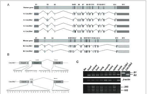
It appears mice have only two forms of protein coding
Cdc25b transcripts according to the Ensemble genome browser search result (ENSMUSG00000027330.9, Fig. 1A). We isolated two forms of mouse Cdc25b cDNA by RT-PCR and called the longer form Cdc25b1 and the shorter form Cdc25b2 (Fig. 1A and 1B, NCBI ac-cession numbers NP_001104545.1 and NP_075606.1 for proteins). These were generated by the alternative splicing of one Cdc25b gene. The 123 nucleotides of exon 7 in the Cdc25b1 transcript were replaced by 45 other nucleotides in the Cdc25b2 transcript by con-necting an alternative splice donor site in exon 6 with a splice acceptor site in exon 8 without exon 7 (Fig. 1B). The expression of two different transcripts in the
various mouse tissues was confirmed by RT-PCR with different sets of primers designed to distinguish two mRNAs (Fig. 1B). RT-PCR with primers for a specific region of Cdc25b1 yielded an amplicon with 136 base pairs (bp), while an 83 bp fragment was amplified with the Cdc25b2 specific primers. All the tested tissues expressed Cdc25b1 and Cdc25b2 transcripts (Fig. 1C).
two forms of CDC25B protein expressed in mice
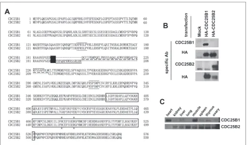
The deduced mouse CDC25Bs, which were trans-lated from two alternatively generated transcripts, had a common catalytic domain. The 41 amino acids in the CDC25B1 were replaced by 15other amino acids in CDC25B2 in the amino terminus (Fig. 2A). Therefore, we investigated whether two mouse Cdc25b
transcripts could be translated into the CDC25B pro-teins in various mouse tissues. Two antibodies that can respectively recognize only a CDC25B1 or a B2 specific region were produced with two synthesized peptides corresponding to the distinct sites between two proteins (Fig. 2A). The specificity of the gener-ated antibodies was monitored after the transfection of HA-Cdc25b1 or HA-Cdc25b2 into the Hek293T cells. The anti-CDC25B1 antibody could specifically detect overexpressed CDC25B1 but not CDC25B2 and vice versa (Fig. 2B). To confirm the transfection and expression of HA-tagged proteins, the HA anti-body was used after stripping (Fig. 2B, second and fourth panels). The expression of two proteins in the various mouse tissues was examined with these an-tibodies. Because the amount of CDC25B proteins is below the detectable range with these antibodies, the endogenous CDC25B proteins were immunopre-cipitated with the pan-specific CDC25B antibody, and immunoprecipitated CDC25B proteins were checked using the CDC25B1- or CDC25B2-specific antibody. In agreement with the RT-PCR results, CDC25B2 pro-tein (~62.9 kDa) was detected in all the mouse tissues checked. Cdc25B1 protein (~65.5 kDa) was also ex-pressed in several organs, especially in the heart and muscle (Fig. 2C).
two forms of CDC25B protein have different localization preferences in cells
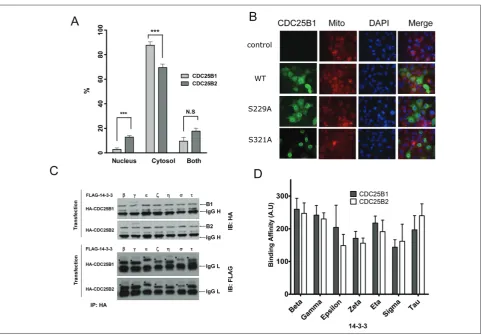
Cdc25b1 and Cdc25b2 were transfected into the Cos7 cells, the major position of the tagged proteins in the cells was checked and counted. Both fused proteins were mainly found in the cytosol, and some proteins were detected only in the nucleus or in both areas. When the distribution of each CDC25B was exam-ined, the tendency ratio of cytosol-localized CDC25B1 protein was significantly higher than that of CDC25B2 (Fig. 3A). This result suggests CDC25B1 and CD-C25B2 proteins have different localization preferences.
two forms of CDC25B protein bind 14-3-3 proteins
Mouse CDC25B1 contains a human CDC25B Ser-230 corresponding site (Ser-229), but this residue does
not exist in mouse CDC25B2 (Fig. 2A). To elucidate the importance of the Ser-229 site of CDC25B1 as a localization determinant, the localization of CDC25B1 was checked after the mutation of Ser-229 to alanine, which is not phosphorylated by any kinase(s). The S229A mutant tended to accumulate more in the nucleus than the wild-type CDC25B1 protein does (Fig. 3B). Most of the S321A protein was present in the nucleus, indicating Ser-321 residue is the major site to regulate the localization of CDC25B (Fig. 3B).
We assessed the difference between CDC25B1 and CDC25B2 to interact with each 14-3-3 protein of the seven isoforms of 14-3-3 [28]. One of the FLAG-tagged 14-3-3 proteins was co-transfected with one of the HA-tagged CDC25B genes, and
40 Arch Biol Sci. 2017;69(1):35-44
tation was performed with the HA antibody. When the co-precipitated 14-3-3 were monitored with the FLAG-specific antibody, all forms of 14-3-3 proteins were detected (Fig. 3C). The β, γ and τ forms of 14-3-3 interacted with CDC25Bs more strongly than the ζ and σ forms (Fig. 3C, 3D), indicating the affinity of 14-3-3 to CDC25Bs is different from the 14-3-3 pro-tein isoforms. It appears that there is some difference in binding affinity between CDC25B1 and CDCB2 for each 14-3-3 isoform, but it is not statistically sig-nificant (Fig. 3D).
two forms of CDC25B protein have different phosphatase activity
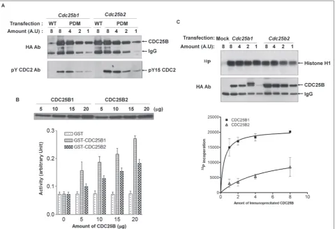
To better determine the difference between two forms of mouse CDC25B, we investigated the affinity of two CDC25B forms to the substrate cyclin B/CDC2. We conducted a substrate-trapping experiment [21] using a phosphatase-dead mutant for each form of CDC25B. The wild-type CDC25B dephosphorylated immedi-ately and dissociated from its substrate, tyrosine 15 residue phosphorylated Cdc2 (Fig. 4A, first and fifth
lane). Because the mutant has no dephosphorylation activity for the phosphate residue in the substrate, it binds stably and is easily co-immunoprecipitated with the phosphorylated form of CDC2 [29]. Therefore, the method well reflected the affinity of CDC25 phos-phatase enzymes for the substrate. The HA-tagged CDC25B was transfected, followed by immunopre-cipitation. The different amounts of the immune com-plexes were resolved by SDS-PAGE, and the co-im-munoprecipitated endogenous phosphorylated CDC2 was checked. Surprisingly, CDC25B2 had a stronger affinity to its substrate than CDCB1 (Fig. 4A).
Next, we examined the difference in activity be-tween two forms of mouse CDC25B using an in vitro
phosphatase activity assay. The activity was checked by incubating the purified GST-fused CDC25B1 or CDC25B2 with pNPP as a substrate [30]. The abil-ity of GST-CDC25B1 to dephosphorylate pNPP was greater than that of GST-CDC25B2 (Fig. 4B). To con-firm the result, we used a modified histone H1 kinase assay. Histone H1 is a well-known substrate for the cyclin B/CDC2 complex activated by CDC25B phos-phatase. Therefore, if histone H1 is incubated with the cyclin B/CDC2 complex after treatment with Cdc25B,
42 Arch Biol Sci. 2017;69(1):35-44
the phosphorylation degree of histone H1 could re-flect the ability of Cdc25B to activate the cyclin B/ CDC2 complex. When the phosphorylation state of histone H1 was checked, a lower amount of CDC25B1 induced more 32P incorporation into histone H1
com-pared to CDC25B2. Altogether, these results indicate CDC25B1 has a greater ability than CDC25B2 to ac-tivate the cyclin B/CDC2 complex.
DIsCussIon
CDC25B is implicated in cell cycle regulation in germ cells and somatic cells [27, 31], but its regula-tion mechanism has not been completely elucidated. As most of the experiments to elucidate the proper-ties of CDC25B have been performed with human CDC25B, we tried to better understand the common roles of CDC25B in cell cycle regulation by monitor-ing the characteristics of mouse CDC25B isoforms and comparing them with human ones. In this study, we isolated two spliced forms of mouse Cdc25b-
Cdc25b1 and Cdc25b2. The human CDC25B gene is transcribed into at least four transcripts [14]; however, only CDC25B2 and CDC25B3 proteins have been bio-chemically detected in human cells [13]. It is likely two transcripts are transcribed from one mouse Cdc25b
gene and both are translated into proteins in various mouse tissues (Fig. 1, 2). There are significant differ-ences in the expression pattern of Cdc25B1 as assessed by Western blot analysis and RT-PCR (Figs 1C and 2C, respectively). However, several reports demonstrated that the correlation between the expression levels of mRNA and protein can be poor, showing that the correlation is less than 40% [32, 33]. We believe that the expression of the CDC25B1 isoform might be a plausible example of that poor correlation. Recently, a truncated nuclear form of CDC25B missing the amino terminus was identified from human spermatozoa, and it was found to be specifically involved in G2/M checkpoint recovery [34]. We could not rule out the possible existence of a mouse counterpart for the shorter human CDC25B form, as several commercial antibodies have been able to recognize the small-sized proteins in Western blot using mouse tissue extracts (data not shown).
While yeast has only one CDC25 [35] and Dro-sophila melanogaster has two [36], most other
ani-mals have three (CDC25A, B and C). Three types of CDC25 proteins share common features in the phosphatase catalytic domain in their carboxyl half [37]. The cysteine in the HCXXXXXR motif plays a central role in human CDC25B [38], as is the case for the corresponding cysteine site (Cys483) of mouse CDC25Bs (Fig. 2A, 4A). The comparatively diverse amino-terminal domain of the CDC25s may be impli-cated in the regulation of catalytic activity or in sub-strate recognition [13]. Therefore, CDC25Bs that have different amino termini would have different affinities to substrate and phosphatase activity. As shown in Fig. 4, mouse CDC25B1 and CDC25B2, which have different amino termini, possess different affinities and different phosphatase activity compared to the in vivo substrate cyclin B/Cdc2 and the in vitro substrate pNPP. Several isoforms can also be generated by alter-native splicing,not only in CDC25B but also in other types of CDC25, such as CDC25C [39].
The two isoforms have different binding affinity with different 14-3-3s, which likely results in the dis-tinction in their localization and activity in the cells (Fig. 3). It is known that Ser-151 and Ser-230 in hu-man CDC25B3 has weak affinity to 14-3-3, whereas the Ser-323 site binds more tightly. It is believed one subunit of the 14-3-3 dimer binds Ser-323 and the other flip-flops either on Ser-151 or Ser-230 to form an intramolecular bridge. The binding prevents CD-C25B from accessing its substrate, the cyclin B/CDC2 complex [21]. In mouse CDC25B2, the Ser-230 cor-responding site is missing; therefore, it is likely the CDC25B2 isoform has a more rigid conformation to access its substrate, resulting in less activity.
Alternative splicing might also be an important mechanism in regulating stability. Human CDC25B is an unstable protein and degrades in a proteasome-dependent manner upon phosphorylation by the cy-clin A/Cdk1 complex [40]. The ubiquitination and subsequent degradation of a number of substrates has been shown to depend on the presence of a KEN box degron motif recognized by the APC/Ccdh1 E3 ligase
Why two different forms of Cdc25B are expressed in mouse tissue is still under investigation. One plau-sible explanation is that they are separately involved in the fine tuning of the cell cycle progression. It is necessary to knock down each transcript to elucidate their exact roles in the cell cycle.
Acknowledgments: This work was supported by the Research Year Grant funded by Inje University (20140022).
Authors’ contribution: MKK and AB contributed equally to this work. MKK, AB and HOC carried out the experiment and analyzed the data. SJH conceived the study, and participated in its design, data analyses and coordination. All authors read and approved the final manuscript.
Conflict of interest disclosure: No potential conflict of interest was reported by the authors.
ReFeRenCes
1. Gabrielli BG, Lee MS, Walker DH, Piwnica-Worms H, Maller JL. Cdc25 regulates the phosphorylation and activity of the Xenopus cdk2 protein kinase complex. J Biol Chem. 1992;267(25):18040-6.
2. Lammer C, Wagerer S, Saffrich R, Mertens D, Ansorge W, Hoffmann I. The cdc25B phosphatase is essential for the G2/M phase transition in human cells. J Cell Sci. 1998;111(Pt16):2445-53.
3. Gabrielli BG, De Souza CP, Tonks ID, Clark JM, Hayward NK, Ellem KA. Cytoplasmic accumulation of cdc25B phospha-tase in mitosis triggers centrosomal microtubule nucleation in HeLa cells. J Cell Sci. 1996;109 (Pt5):1081-93.
4. Sanchez Y, Wong C, Thoma RS, Richman R, Wu Z, Piwnica-Worms H, Elledge SJ. Conservation of the Chk1 checkpoint pathway in mammals: linkage of DNA damage to Cdk regula-tion through Cdc25. Science. 1997;277(5331):1497-501. 5. Zeng Y, Forbes KC, Wu Z, Moreno S, Piwnica-Worms H,
Enoch T. Replication checkpoint requires phosphoryla-tion of the phosphatase Cdc25 by Cds1 or Chk1. Nature. 1998;395(6701):507-10.
6. Wickramasinghe D, Becker S, Ernst MK, Resnick JL, Cen-tanni JM, Tessarollo L, Grabel LB, Donovan PJ. Two CDC25 homologues are differentially expressed during mouse devel-opment. Develdevel-opment. 1995;121(7):2047-56.
7. Boutros R, Dozier C, Ducommun B. The when and wheres of CDC25 phosphatases. Curr Opin Cell Biol. 2006;18(2):185-91.
8. Ray D, Kiyokawa H. CDC25A levels determine the bal-ance of proliferation and checkpoint response. Cell Cycle. 2007;6(24):3039-42.
9. Chen MS, Hurov J, White LS, Woodford-Thomas T, Piwnica-Worms H. Absence of apparent phenotype in mice lacking Cdc25C protein phosphatase. Mol Cell Biol. 2001;21(12):3853-61.
10. Lincoln AJ, Wickramasinghe D, Stein P, Schultz RM, Palko ME, De Miguel MP, Tessarollo L, Donovan PJ. Cdc25b phos-phatase is required for resumption of meiosis during oocyte maturation. Nat Genet. 2002;30(4):446-9.
11. De Souza CP, Ellem KA, Gabrielli BG. Centrosomal and cyto-plasmic Cdc2/cyclin B1 activation precedes nuclear mitotic events. Exp Cell Res. 2000;257(1):11-21.
12. Boutros R, Lobjois V, Ducommun B. CDC25B involvement in the centrosome duplication cycle and in microtubule nucle-ation. Cancer Res. 2007;67(24):11557-64.
13. Forrest AR, McCormack AK, DeSouza CP, Sinnamon JM, Tonks ID, Hayward NK, Ellem KA, Gabrielli BG. Multiple splicing variants of cdc25B regulate G2/M progression. Bio-chem Biophys Res Commun. 1999;260(2):510-5.
14. Baldin V, Cans C, Superti-Furga G, Ducommun B. Alter-native splicing of the human CDC25B tyrosine phospha-tase. Possible implications for growth control? Oncogene. 1997;14(20):2485-95.
15. Gabrielli BG, Clark JM, McCormack AK, Ellem KA. Hyper-phosphorylation of the N-terminal domain of Cdc25 regu-lates activity toward cyclin B1/Cdc2 but not cyclin A/Cdk2. J Biol Chem. 1997;272(45):28607-14.
16. Lindqvist A, Kallstrom H, Karlsson Rosenthal C. Characteri-sation of Cdc25B localiCharacteri-sation and nuclear export during the cell cycle and in response to stress. J Cell Sci. 2004;117(Pt 21):4979-90.
17. Woo ES, Rice RL, Lazo JS. Cell cycle dependent sub-cellular distribution of Cdc25B subtypes. Oncogene. 1999;18(17):2770-6.
18. Dutertre S, Cazales M, Quaranta M, Froment C, Trabut V, Dozier C, Mirey G, Bouche JP, Theis-Febvre N, Schmitt E, Monsarrat B, Prigent C, Ducommun B. Phosphorylation of CDC25B by Aurora-A at the centrosome contributes to the G2-M transition. J Cell Sci. 2004;117(Pt 12):2523-31. 19. Baldin V, Theis-Febvre N, Benne C, Froment C, Cazales M,
Burlet-Schiltz O, Ducommun B. PKB/Akt phosphorylates the CDC25B phosphatase and regulates its intracellular localisa-tion. Biol Cell. 2003;95(8):547-54.
20. Uchida S, Kuma A, Ohtsubo M, Shimura M, Hirata M, Nak-agama H, Matsunaga T, Ishizaka Y, Yamashita K. Binding of 14-3-3beta but not 14-3-3sigma controls the cytoplasmic localization of CDC25B: binding site preferences of 14-3-3 subtypes and the subcellular localization of CDC25B. J Cell Sci. 2004;117(Pt 14):3011-20.
21. Giles N, Forrest A, Gabrielli B. 14-3-3 acts as an intramolecu-lar bridge to regulate cdc25B localization and activity. J Biol Chem. 2003;278(31):28580-7.
22. Kumagai A, Yakowec PS, Dunphy WG. 14-3-3 proteins act as negative regulators of the mitotic inducer Cdc25 in Xenopus egg extracts. Mol Biol Cell. 1998;9(2):345-54.
23. Conklin DS, Galaktionov K, Beach D. 14-3-3 proteins asso-ciate with cdc25 phosphatases. Proc Natl Acad Sci U S A. 1995;92(17):7892-6.
24. Cui C, Ren X, Liu D, Deng X, Qin X, Zhao X, Wang E, Yu B. 14-3-3 epsilon prevents G2/M transition of fertilized mouse eggs by binding with CDC25B. BMC Dev Biol. 2014;14:33. 25. Meng J, Cui C, Liu Y, Jin M, Wu D, Liu C, Wang E, Yu B.
44 Arch Biol Sci. 2017;69(1):35-44
Cdc25B at its Ser321 in the release of the mouse oocyte from prophase I arrest. PLoS One. 2013;8(1):e53633.
26. Bulavin DV, Higashimoto Y, Popoff IJ, Gaarde WA, Basrur V, Potapova O, Appella E, Fornace AJ, Jr. Initiation of a G2/M checkpoint after ultraviolet radiation requires p38 kinase. Nature. 2001;411(6833):102-7.
27. Pirino G, Wescott MP, Donovan PJ. Protein kinase A regu-lates resumption of meiosis by phosphorylation of Cdc25B in mammalian oocytes. Cell Cycle. 2009;8(4):665-70.
28. Kjarland E, Keen TJ, Kleppe R. Does isoform diversity explain functional differences in the 14-3-3 protein family? Curr Pharm Biotechnol. 2006;7(3):217-23.
29. Lee KH, Tsutsui T, Honda K, Ohtake H, Omasa T. Over-expression of mutant cell division cycle 25 homolog B (CDC25B) enhances the efficiency of selection in Chinese hamster ovary cells. Cytotechnology. 2013;65(6):1017-26. 30. Parks JM, Hu H, Rudolph J, Yang W. Mechanism of Cdc25B
phosphatase with the small molecule substrate p-nitrophenyl phosphate from QM/MM-MFEP calculations. J Phys Chem B. 2009;113(15):5217-24.
31. Teng YN, Chung CL, Lin YM, Pan HA, Liao RW, Kuo PL. Expression of various CDC25B isoforms in human sperma-tozoa. Fertil Steril. 2007;88(2):379-82.
32. de Sousa Abreu R, Penalva LO, Marcotte EM, Vogel C. Global signatures of protein and mRNA expression levels. Mol Bio-syst. 2009;5(12):1512-26.
33. Vogel C, Marcotte EM. Insights into the regulation of protein abundance from proteomic and transcriptomic analyses. Nat Rev Genet. 2012;13(4):227-32.
34. Jullien D, Bugler B, Dozier C, Cazales M, Ducommun B. Iden-tification of N-terminally truncated stable nuclear isoforms of CDC25B that are specifically involved in G2/M checkpoint recovery. Cancer Res. 2011;71(5):1968-77.
35. Fantes PA. Isolation of cell size mutants of a fission yeast by a new selective method: characterization of mutants and implications for division control mechanisms. J Bacteriol. 1981;146(2):746-54.
36. Alphey L, Jimenez J, White-Cooper H, Dawson I, Nurse P, Glover DM. twine, a cdc25 homolog that functions in the male and female germline of Drosophila. Cell. 1992;69(6):977-88.
37. Gottlin EB, Xu X, Epstein DM, Burke SP, Eckstein JW, Bal-lou DP, Dixon JE. Kinetic analysis of the catalytic domain of human cdc25B. J Biol Chem. 1996;271(44):27445-9. 38. Reynolds RA, Yem AW, Wolfe CL, Deibel MR, Jr., Chidester
CG, Watenpaugh KD. Crystal structure of the catalytic sub-unit of Cdc25B required for G2/M phase transition of the cell cycle. J Mol Biol. 1999;293(3):559-68.
39. Bonnet J, Mayonove P, Morris MC. Differential phosphoryla-tion of Cdc25C phosphatase in mitosis. Biochem Biophys Res Commun. 2008;370(3):483-8.
40. Baldin V, Cans C, Knibiehler M, Ducommun B. Phosphory-lation of human CDC25B phosphatase by CDK1-cyclin A triggers its proteasome-dependent degradation. J Biol Chem. 1997;272(52):32731-4.