© 2019 by the Serbian Biological Society How to cite this article: Brkljačić J, Veličković N, Elaković I, Teofilović A, Vojnović- 417 Milutinović D, Đorđević A, Matić G. Fructose-enriched diet affects hepatic lipid
metabolism in young male and female rats in different ways. Arch Biol Sci. 2019;71(3):417-24.
Fructose-enriched diet affects hepatic lipid metabolism in young male and female rats in
different ways
Jelena Brkljačić*, Nataša Veličković, Ivana Elaković, Ana Teofilović, Danijela Vojnović Milutinović, Ana Đorđević and Gordana Matić
*Department of Biochemistry, Institute for Biological Research “Siniša Stanković” – National Institute of the Republic of Serbia,
University of Belgrade, 142 Despot Stefan Blvd., 11060 Belgrade, Serbia
*Corresponding author: [email protected]
Received: March 6, 2019; Revised: April 2, 2019; Accepted: April 2, 2019; Published online: April 9, 2019
Abstract: Anincrease in fructose consumption coincides with a rising incidence of metabolic disorders. Dietary fructose has been shown to affect hepatic lipid metabolism in a way that may lead to lipid deposition in the liver. In this study, we tested the hypothesis that the effects of fructose overconsumption on hepatic lipid metabolism differ between sexes. To that end we examined the effects of a high-fructose diet on the expression of key enzymes and transcription factors involved in the regulation of fatty acid oxidation and de novo lipogenesis in the liver of 12-week-old male and female Wistar rats. Immediately after weaning, the rats were subjected to a standard diet and 10% fructose solution or drinking water for 9 weeks. The fructose-enriched diet induced hypertriglyceridemia and increased hepatic de novo lipogenesis in both sexes, without lipid deposition in the liver. At the same time, visceral adiposity was observed only in female rats, while in males the treatment stimulated hepatic fatty acid oxidation. The fructose-enriched diet induced sex-specific effects on hepatic lipid metabolism in young rats. These results imply that male and female rats employ different strategies to cope with dietary fructose-related energy overload and to avoid lipid accumulation in the liver.
Keywords: dietary fructose; lipid metabolism; liver; rat; sex differences
Abbreviations: Acetyl-CoA carboxylase 1 (ACC1); carnitine palmitoyltransferase 1 (CPT1); fatty acid synthase (FAS); free fatty acids (FFA); peroxisome proliferator-activated receptor α (PPARα); PPARγ coactivator 1α (PGC-1α); sterol regulatory element binding protein 1c (SREBP-1c)
INTRODUCTION
Fructose is one of the most frequently used ingredients in the food industry. The huge rise in fructose con-sumption observed in the last decades mostly comes from increased consumption of refined sugars and not from naturally occurring fructose in fruits. This trend runs parallel with the increasing prevalence of obesity and related metabolic disorders, which has raised interest in the potential role of fructose as a factor contributing to the development of metabolic disorders. Some authors have suggested that fructose overconsumption is associated with increased risk of obesity, insulin resistance, metabolic syndrome, type 2 diabetes, cardiovascular disease and nonalcoholic fatty liver disease [1-3], with other authors not fully supporting these assertions [4-6]. Studies performed on
animals have shown that fructose supplementation can result in hypertension, insulin resistance, dyslipidemia and the accumulation of visceral and ectopic fat [7-9].
between de novo lipogenesis and fatty acid oxidation, and between hepatic lipid intake and secretion [11,12]. The expression of genes involved in fatty acid oxidation, including carnitine palmitoyltransferase 1 (CPT1), is stimulated by a transcription complex that is comprised of peroxisome proliferator-activated receptor α (PPARα), PPARγ coactivator 1α (PGC-1α) and lipin-1 [13,14].
Previous studies have reported sex-dependent dif-ferences in the susceptibility to and progression of meta-bolic disorders [15]. Females appear to be more prone to obesity, while males display higher rates of insulin resistance-related disorders and nonalcoholic fatty liver disease [16, 17]. Studies on adult rats have shown that male rats subjected to a high fat diet developed a greater degree of hepatic steatosis, while in females the disease phenotype was alleviated [17, 18]. Although nonalco-holic fatty liver disease is predominantly diagnosed in adults, pediatric liver diseases are a growing problem. As the immature young organism and adult differ largely in their metabolic and physiological profiles, it is of interest to evaluate the effects of a fructose-rich diet on hepatic lipid metabolism in the young population that consumes more fructose-enriched drinks as compared to adults. Furthermore, the majority of animal studies on diet-induced metabolic disorders were performed in males, while females are still underrepresented. As a consequence, it is not always clear whether the obtained results apply equally to both sexes.
In this study, we hypothesized that the effects of a fructose-rich diet on hepatic lipid metabolism differ between sexes. To test this hypothesis, we measured the expression of key enzymes and transcription fac-tors involved in the regulation of fatty acid oxidation and de novo lipogenesis in the livers of both male and female rats subjected to a fructose-enriched diet im-mediately after weaning.
MATERIALS AND METHODS Animals and treatment
Male and female 21-day-old Wistar rats were randomly divided into two experimental groups (9 animals per group) as follows: the control group (C) was fed with commercial standard chow and drinking water, and the fructose group (F) was fed with the same chow and 10% (w/v) fructose solution instead of drinking water. Both
experimental groups had ad libitum access to food and drinking fluid during 9 weeks. The concentration of fructose solution was chosen to be similar to the intake of sweet solutions typical for a Western diet [19]. The detailed composition of laboratory chow was previously published [20]. The animals were housed three per cage and kept in a temperature-controlled room (22±2°C) with a 12 h light/dark cycle (lights on at 07:00 h) and constant humidity. During 9 weeks of treatment, food and liquid intake was measured daily. Energy intake was calculated as the sum of calories ingested in the form of food and liquid. At the end of the treatment, all animals were killed by rapid decapitation after overnight fasting during which both experimental groups were provided only with drinking water. Female rats were killed in the diestrus phase of the estrous cycle, which was determined by analysis of vaginal smears. Body, liver and visceral adipose tissue mass were weighed immediately after sacrifice. All animal procedures were in compliance with the EEC Directive 2010/63/EU on the protection of animals used for scientific purposes, and were approved by the Ethical Committee for the Use of Laboratory Animals of the Institute for Biologi-cal Research “Siniša Stanković”, University of Belgrade.
Blood plasma preparation, tissue collection and determination of biochemical parameters
After overnight fasting, the animals were killed by rapid decapitation. The livers were perfused with cold 0.9% NaCl, quickly excised and stored in liquid nitrogen until use. Liver triglycerides were isolated from 100 mg of liver tissue by the modified Folch method [21] and analyzed according to Fletcher [22]. Blood glucose and triglyceride concentrations were determined in the blood immediately after sacrifice using MultiCare strips (Biochemical Systems International, Arezzo, Italia). Plasma was isolated by blood centrifugation at 1600xg for 10 min at room temperature and stored at -70°C for subsequent processing. The plasma level of FFA was determined by a modified version of Dun-combe’s [23] method.
Preparation of hepatic nucleosols and whole cell extracts
homog-enization buffer (20 mM Tris-HCl pH 7.2, 10% glycerol, 50 mM NaCl, 1 mM EDTA-Na2, 1 mM EGTA-Na2, 2 mM DTT, protease and phosphatase inhibitors). The homogenates were further processed to obtain nucleo-sols as previously described [24]. For preparation of whole cell extracts, the livers were homogenized with ice-cold RIPA buffer (50 mM Tris-HCl pH 7.2, 1 mM EDTA-Na2, 150 mM NaCl, 0.1% SDS, 1% Nonidet P-40, 0.5% sodium deoxycholate, 2 mM DTT, protease and phosphatase inhibitors). The homogenates were soni-cated (3x5 s, 1 A, 50/60 Hz), incubated for 60 min on ice with continuous agitation and frequent vortexing, centrifuged (16000xg, 20 min, 4°C) and stored at -70°C.
Sodium dodecyl sulfate polyacrylamide gel electrophoresis (SDS-PAGE) and immunoblotting
After boiling in Laemmli’s sample buffer, 40 µg of liver proteins were resolved on 7.5% or 10% SDS-polyacrylamide gels. Western transfer of proteins from gels to PVDF membranes was performed in 25 mM Tris buffer, pH 8.3, containing 192 mM glycine and 20% (v/v) methanol, at 135 mA overnight in a Mini Trans-Blot Electrophoretic Transfer Cell (Bio-Rad Laboratories, Hercules, CA, USA). The mem-branes were blocked by PBS (1.5 mM KH2PO4, 6.5 mM Na2HPO4, 2.7 mM KCl, 0.14 M NaCl, pH 7.2) containing 1% non-fat milk, incubated by rocking (1.5 h, room temperature) with the respective primary antibodies (Santa Cruz Biotechnology, Dallas, TX, USA): anti-PPARα (sc-9000), anti-PGC-1α (sc-13067), anti-lipin-1 (sc-98450) and anti-CPT1 (sc-139482). Beta-actin (AC-15, Sigma-Aldrich, St. Louis, MO, USA) was used as an equal load control. After washing with PBS containing 0.1% Tween 20, the membranes were incubated with appropriate alkaline phosphatase-conjugated secondary antibody (Amersham Pharmacia Biotech, Little Chalfont, UK). Immunopositive bands were visualized by the enhanced chemifluorescence (ECF) method and quantified by ImageQuant software (GE Healthcare, Little Chalfont, UK).
RNA isolation, reverse transcription and real-time PCR
Total RNA was extracted using TRI Reagent® (Am-Bion Inc., Austin, TX, USA). RNA was dissolved
in RNase-DNase-free water (Eppendorf, Hamburg, Germany) and its concentration and purity were tested spectrophotometrically (OD 260/280>1.8 was considered satisfactory). RNase inhibitor (Applied Biosystems, Foster City, CA, USA) was added and the samples were frozen at -70˚C until use. Prior to cDNA synthesis, DNA contamination was removed by DNase I treatment (Fermentas, Waltham, MA, USA). cDNA was synthesized from 2 µg of RNA. Reverse transcription was performed using the High-Capacity cDNA Reverse Transcription Kit (Applied Biosystems, Foster City, CA, USA), according to the manufacturer’s instructions, and cDNA was stored at -70°C until use. Quantification of target gene expression in the liver was performed by TaqMan® real-time PCR. Primers and probes for ACC1 (Rn00573474_m1), FAS (Rn 01463550_m1) and SREBP-1c (Rn01495769_m1) were obtained from Applied Biosystems Assay-on-Demand Gene Expression Products. HPRT1 (Rn01455646_m1) was used as a previously validated endogenous control.
Statistical analysis
A Student’s unpaired t-test (two-tailed) was used to compare the differences between the experimental groups. Statistical analyses were performed using GraphPad Prism v.5 (GraphPad Software, Inc., La Jolla, CA, USA). A probability level less than 0.05 was considered to be statistically significant. The data are presented as means±SEM.
RESULTS
The effects of a fructose-rich diet on physiological and biochemical parameters
P<0.05), the level of hepatic triglycerides remained unaltered after the treatment (Table 1).
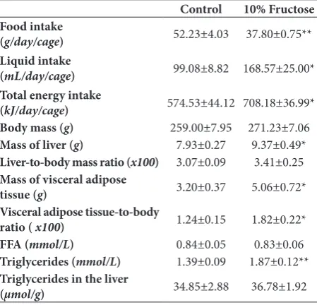
Similar to males, the fructose-fed females consumed less chow than females maintained on a standard diet (Table 2, P<0.01), and total energy intake was increased due to calories from the fructose solution (Table 2, P<0.05). As in males, fructose consumption also induced triglyceridemia (P<0.01) in females, but did not affect body mass and the liver to body mass ratio, nor the hepatic level of triglycerides. However, in contrast to males, in females the liver mass, visceral adipose tissue mass and visceral adipose tissue to body ratio were increased (Table 2, P<0.05), while plasma FFA levels remained unchanged after the long-term fructose diet (Table 2).
The effects of a fructose-rich diet on hepatic de novo lipogenesis and β-oxidation of fatty acids
To explore the effects of a long-term fructose-rich diet on hepatic de novo lipogenesis, we determined the mRNA level of transcription factor SREBP-1c and its target genes, ACC1 and FAS, coding for two rate-limiting lipogenic enzymes. In both sexes, statistically significant increases in mRNA levels of SREBP-1c (P<0.05 for males, P<0.01 for females), ACC1 (P<0.01 for males, P<0.05 for females) and FAS (P<0.05) were observed in fructose-fed rats when compared to the respective controls (Figs. 1 and 3). As for β-oxidation of fatty acids, statistically significant increases in the nuclear levels of transcription factors PPARα (P<0.05), PGC-1α (P<0.01) and lipin-1 (P<0.01), as well as in the level of their target gene, CPT1 (P<0.001), were observed in fructose-fed male rats when compared to males maintained on a standard diet (Fig. 2). In contrast to males, there was no significant difference in the protein levels of PPARα, PGC-1α, lipin-1 and CPT1 between female rats maintained on fructose-rich and standard diets (Fig. 4).
DISCUSSION
Fructose has been shown to affect lipid metabolism in a way that can result in obesity and hepatic lipid accumulation [2]. However, it has not been studied whether the fructose-related occurrence of fatty liver is sex-dependent. Under physiological conditions,
Table 1. Food, liquid and energy intake and physiological and bio-chemical parameters in male rats subjected to a fructose-rich diet.
Control 10% fructose
Food intake
(g/day/cage) 61.71±4.49 42.59±3.43*
Liquid intake
(mL/day/cage) 148.04±9.35 222.31±19.73*
Total energy intake
(kJ/day/cage) 678.76±49.35 850.88±42.41*
Body mass (g) 339±13 311±13
Mass of liver (g) 12.48±0.69 10.77±0.49
Liver-to-body mass ratio (x100) 3.65±0.09 3.48±0.15
Mass of visceral adipose
tissue (g) 2.59±0.40 3.16±0.64
Visceral adipose
tissue-to-body ratio ( x100) 0.75±0.10 1.01±0.17
FFA (mmol/L) 0.61±0.03 0.70±0.02*
Triglycerides (mmol/L) 1.63±0.10 2.07±0.08*
Triglycerides in the liver
(µmol/g) 20.40±1.20 16.30±1.50
The data are presented as the means±SEM (n=9 animals per group). Comparisons between fructose-fed and control rats were made by unpaired Student’s t-test. A value of P<0.05 was considered statistically significant. Asterisks indicate significant differences. *P<0.05.
Table 2. Food, liquid and energy intake, and physiological and biochemical parameters in female rats subjected to a fructose-rich diet.
Control 10% Fructose
Food intake
(g/day/cage) 52.23±4.03 37.80±0.75**
Liquid intake
(mL/day/cage) 99.08±8.82 168.57±25.00*
Total energy intake
(kJ/day/cage) 574.53±44.12 708.18±36.99*
Body mass (g) 259.00±7.95 271.23±7.06
Mass of liver (g) 7.93±0.27 9.37±0.49*
Liver-to-body mass ratio (x100) 3.07±0.09 3.41±0.25
Mass of visceral adipose
tissue (g) 3.20±0.37 5.06±0.72*
Visceral adipose tissue-to-body
ratio ( x100) 1.24±0.15 1.82±0.22*
FFA (mmol/L) 0.84±0.05 0.83±0.06
Triglycerides (mmol/L) 1.39±0.09 1.87±0.12**
Triglycerides in the liver
(µmol/g) 34.85±2.88 36.78±1.92
hepatic triglyceride levels are maintained by a precise balance between FFA uptake from plasma and de novo
lipogenesis vs. triglyceride disposal through fatty acid oxidation or lipoprotein secretion. The pathological lipid accumulation in the liver could occur as a con-sequence of an imbalance between these processes.
In the current study, the fructose-rich diet induced hypertriglyceridemia and increased hepatic de novo
lipogenesis in both male and female rats, without lipid deposition in the liver. However, this was concurrent with the development of adiposity only in females, while a parallel increase in hepatic fatty acid β-oxidation
Fig. 3. The effect of a 10% fructose diet on hepatic de novo lipo-genesis in young female rats.The levels of SREBP-1c, ACC1 and FAS mRNAs relative to HPRT mRNA in the liver of control (C) and fructose-fed (F) females was determined by TaqMan real-time PCR and presented as the fold-change with respect to the control. The values represent the means±SEM (n=9). All measurements were done in triplicate. Statistical significance (Student’s t-test) of differences between experimental groups: *P<0.05; **P<0.01.
Fig. 4. The effect of a 10% fructose diet on hepatic fatty acid oxida-tion in young female rats.Relative integrated optical density of the immunoreactive bands corresponding to PPARα, PGC-1α, lipin-1 and CPT1 in the livers of control (C) and fructose-fed (F) females was assessed by ImageQuant software, normalized to β-actin and expressed as the fold-change with respect to the control. The values represent the means±SEM (n=9).
Fig. 1. The effect of a 10% fructose diet on hepatic de novo lipo-genesis in young male rats. The level of SREBP-1c, ACC1 and FAS mRNAs relative to HPRT mRNA in the liver of control (C) and fructose-fed (F) males was determined by TaqMan real-time PCR and presented as the fold-change with respect to the control. The values represent the means±SEM (n=9). All measurements were done in triplicate. Statistical significance (Student’s t-test) of differences between experimental groups: *P<0.05; **P<0.01.
was exhibited in male rats. These results imply that male and female rats employed different strategies to cope with the energy overload originating from a fructose-rich diet.
In our study, elevated caloric intake related to fructose overconsumption was not followed by an increase in body mass. Similar findings were reported in some previously published studies [7,25-27], while other authors have reported an increase in body mass [28,29]. These discrepancies could be attributed to differences in experimental settings, such as treatment duration, fructose concentration and the form in which the fructose was consumed (liquid vs. solid food), as well as the age of the experimental animals. As young and adult organisms differ largely in their metabolic and physiological profiles, it could be assumed that responses to fructose overconsumption in youth and adulthood would be different. De Moura [25] reported that a high fructose intake is more effective at inducing signs of the metabolic syndrome, including glucose intolerance, insulin resistance, hypertriglyceridemia, dyslipidemia and high concentrations of total liver lipids in adult (90-day-old at the beginning of the treatment) than in young (28-day-old at the beginning of the treatment) male Wistar rats, implying protective mechanisms specific for young individuals.
One of the most consistently reported effects of fructose overconsumption is hypertriglyceridemia [7,25,28,29]. Our results showing elevated plasma tri-glycerides in both sexes indicate that the same applies to young rats independent of gender. The observed hypertriglyceridemia is likely a consequence of dietary fructose-related stimulation of hepatic de novo lipogen-esis. Namely, in accordance with other studies [26,29], fructose overconsumption upregulated the hepatic expression of lipogenic transcription factor SREBP-1c and lipogenic enzymes ACC1 and FAS, although lipid accumulation in the liver was not observed. As mentioned above, triglycerides could accumulate in the liver as a result of elevated hepatic lipid uptake, elevated
de novo lipogenesis and/or reduced fatty acid oxida-tion or lipid export. Fructose-induced hepatic lipid accumulation was mostly studied in adult male rats, and was attributed to increased lipogenesis concur-rent with reduced fatty acid β-oxidation [29, 30]. In our study, increased de novo lipogenesis in the liver of young fructose-fed male rats occurred simultaneously
with the induction of fatty acid oxidation, as evidenced by increased nuclear accumulation of PPARα, PGC-1α and lipin-1, and a parallel raise in CPT1 level.Together with triglycerides export, intensified fatty acid oxidation contributes to clearance of fructose-derived lipids from the liver in males. Upregulation of both lipid synthesis and catabolism in the liver was somewhat unexpected, but it has been reported in several studies in which the effects of antihyperlipidemic drugs were investigated in rodents [31,32]. For example, simultaneous induction of hepatic fatty acid oxidation and de novo lipogenesis was observed in mice treated with the PPARα agonist fenofibrate [31,32]. Stimulated β-oxidation might be a part of the metabolic response to increased influx of fatty acids to the liver, considering the elevated plasma FFA level in male fructose-fed rats. Namely, as demonstrated in our previously-published paper, a fructose-rich diet stimulated lipolysis in visceral adipose tissue, thus leading to elevated plasma FFA levels [33] rather than to visceral adipose tissue accu-mulation. Elevated triglycerides derived from fructose catabolism together with FFAs originating from adipose tissue lipolysis may expose young male rats to the risk of ectopic lipid accumulation and lipotoxicity [34]. Under lipid overload, the activation of β-oxidation could protect the liver from lipid accumulation. As reviewed in [34], several studies on different rodent models of metabolic syndrome demonstrated that the enhancement of fatty acid oxidation could attenuate or even completely prevent an ectopic accumula-tion of lipids and, consequently, development of the correlated disorder. Nonetheless, the possibility that prolonged fructose overconsumption might ultimately lead to hepatic lipid accumulation and contribute to the progression of metabolic disorders in later adult-hood cannot be ruled out.
the ability of adipocytes to accommodate the caloric surplus of overnutrition can minimize ectopic lipid accumulation [34]. Indeed, the TG content in the liver of fructose-fed female rats remained unaltered despite increased de novo lipogenesis. It seems that, since the level of FFAs in the plasma of fructose-fed females was not increased, their liver was not challenged by the lipid surplus to the extent it was in males, and thus hepatic fatty acid oxidation in female rats was not stimulated. It should be kept in mind that the visceral adipose tissue can protect from ectopic lipid deposition only to a certain extent, after which enlargement of this tissue poses a greater risk for the development of metabolic disturbances. This implies that prolonged fructose feeding could ultimately disrupt the ability of visceral adipose tissue to deal with large amounts of lipids, thus contributing to the development of further metabolic disturbances.
In conclusion, our results on young rats revealed sex-specific effects of a fructose-enriched diet on hepatic lipid metabolism. Although the fructose-enriched diet induced hepatic de novo lipogenesis and consequent hypertriglyceridemia in both sexes, males and females deployed different mechanisms to cope with the energy overload and avoid hepatic lipid deposition. In male rats, the diet regime stimulated hepatic fatty acid oxidation as part of the metabolic response to excess adipose-derived FFA, while in females, fructose-promoted adiposity prevented the release of FFA, thus creating circumstances that do not challenge hepatic fatty acid oxidation. These re-sults also imply that sex-related differences in plasma FFA levels emphasize the importance of a cross-talk between adipose tissue and the liver in adaptation to diet-induced energy overload.
Acknowledgments: This work was supported by the Ministry of
Education, Science and Technological Development of the Republic of Serbia, Grant III41009.
Author contributions: JB, AT, IE and DVM contributed to data acquisition, data analysis and data interpretation. JB, NV, AD and GM designed the study and contributed to manuscript writing. JB and IE wrote the paper. All authors approved the final version of the manuscript.
Conflict of interest disclosure: The authors report no conflict of interest.
REFERENCES
1. Jegatheesan P, De Bandt JP. Fructose and NAFLD: The Multifaceted Aspects of Fructose Metabolism. Nutrients. 2017;9(3):230.
2. Dekker MJ, Su Q, Baker C, Rutledge AC, Adeli K. Fructose: a highly lipogenic nutrient implicated in insulin resistance, hepatic steatosis, and the metabolic syndrome. Am J Physiol Endocrinol Metab. 2010;299(5):E685-94.
3. Johnson RJ, Segal MS, Sautin Y, Nakagawa T, Feig DI, Kang DH, Gersch MS, Benner S, Sánchez-Lozada LG. Potential role of sugar (fructose) in the epidemic of hypertension, obesity and the metabolic syndrome, diabetes, kidney disease, and cardiovascular disease. Am J Clin Nutr. 2007;86(4):899-906. 4. Rippe JM, Angelopoulos TJ. Relationship between Added
Sugars Consumption and Chronic Disease Risk Factors: Cur-rent Understanding. Nutrients. 2016;8(11):E697.
5. Lowndes J, Sinnett SS, Rippe JM. No Effect of Added Sugar Consumed at Median American Intake Level on Glucose Tol-erance or Insulin Resistance. Nutrients. 2015;7(10):8830-45. 6. Evans RA, Frese M, Romero J, Cunningham JH, Mills KE.
Chronic fructose substitution for glucose or sucrose in food or beverages has little effect on fasting blood glucose, insulin, or triglycerides: a systematic review and meta-analysis. Am J Clin Nutr. 2017;106(2):519-29.
7. Dupas J, Feray A, Goanvec C, Guernec A, Samson N, Bou-garan P, Guerrero F, Mansourati J. Metabolic Syndrome and Hypertension Resulting from Fructose Enriched Diet in Wis-tar Rats. Biomed Res Int. 2017;2017:2494067.
8. Kawasaki T, Igarashi K, Koeda T, Sugimoto K, Nakagawa K, Hayashi S, Yamaji R, Inui H, Fukusato T, Yamanouchi T. Rats fed fructose-enriched diets have characteristics of nonalco-holic hepatic steatosis. J Nutr. 2009;139(11):2067-71. 9. Crescenzo R, Bianco F, Coppola P, Mazzoli A, Valiante S,
Liv-erini G, Iossa S. Adipose tissue remodeling in rats exhibiting fructose-induced obesity. Eur J Nutr. 2014;53(2):413-9. 10. Miyazaki M, Dobrzyn A, Man WC, Chu K, Sampath H, Kim
HJ, Ntambi JM. Stearoyl-CoA desaturase 1 gene expression is necessary for fructose-mediated induction of lipogenic gene expression by sterol regulatory element-binding protein-1c-dependent and -independent mechanisms. J Biol Chem. 2004;279(24):25164-71.
11. Rebollo A, Roglans N, Baena M, Sanchez RM, Merlos M, Alegret M, Laguna JC. Liquid fructose downregulates Sirt1 expression and activity and impairs the oxidation of fatty acids in rat and human liver cells. Biochim Biophys Acta. 2014;1841(4):514-24.
12. Musso G, Gambino R, Cassader M. Recent insights into hepatic lipid metabolism in non-alcoholic fatty liver disease (NAFLD). Prog Lipid Res. 2009;48(1):1-26.
13. Vega RB, Huss JM, Kelly DP. The coactivator PGC-1 coop-erates with peroxisome proliferator-activated receptor alpha in transcriptional control of nuclear genes encoding mitochondrial fatty acid oxidation enzymes. Mol Cell Biol. 2000;20(5):1868-76.
15. Rochlani Y, Pothineni NV, Mehta JL. Metabolic Syndrome: Does it Differ Between Women and Men? Cardiovasc Drugs Ther. 2015;29(4):329-38.
16. Galipeau D, Verma S, McNeill JH. Female rats are protected against fructose-induced changes in metabolism and blood pressure. American journal of physiology Heart and circula-tory physiology. 2002;283(6):H2478-84.
17. Stoppeler S, Palmes D, Fehr M, Holzen JP, Zibert A, Siaj R, Schmidt HH, Spiegel HU, Bahde R. Gender and strain-spe-cific differences in the development of steatosis in rats. Lab Anim. 2013;47(1):43-52.
18. Ramdhave AS, Ojha S, Nandave M. Energy intake correlates with the levels of fatty acid synthase and insulin-like growth factor-1 in male and female C57BL/6 mice. Am J Transl Res. 2017;9(3):830-44.
19. Ventura EE, Davis JN, Goran MI. Sugar content of popular sweetened beverages based on objective laboratory analysis: focus on fructose content. Obesity. 2011;19(4):868-74. 20. Nestorov J, Glban AM, Mijuskovic A, Nikolic-Kokic A,
Ela-kovic I, VelicEla-kovic N, Matic G. Long-term fructose-enriched diet introduced immediately after weaning does not induce oxidative stress in the rat liver. Nutr Res. 2014;34(7):646-52. 21. Folch J, Lees M, Sloane Stanley GH. A simple method for the
isolation and purification of total lipides from animal tissues. J Biol Chem. 1957;226(1):497-509.
22. Fletcher MJ. A colorimetric method for estimating serum triglycerides. Clin Chim Acta. 1968;22(3):393-7.
23. Duncombe WG. The Colorimetric Micro-Determination of Non-Esterified Fatty Acids in Plasma. Clin Chim Acta. 1964;9:122-5.
24. Vasiljevic A, Velickovic N, Bursac B, Djordjevic A, Milu-tinovic DV, Nestorovic N, Matic G. Enhanced prereceptor glucocorticoid metabolism and lipogenesis impair insulin signaling in the liver of fructose-fed rats. J Nutr Biochem. 2013;24(11):1790-7.
25. de Moura RF, Ribeiro C, de Oliveira JA, Stevanato E, de Mello MA. Metabolic syndrome signs in Wistar rats submit-ted to different high-fructose ingestion protocols. Br J Nutr. 2009;101(8):1178-84.
26. Janevski M, Ratnayake S, Siljanovski S, McGlynn MA, Cam-eron-Smith D, Lewandowski P. Fructose containing sugars
modulate mRNA of lipogenic genes ACC and FAS and pro-tein levels of transcription factors ChREBP and SREBP1c with no effect on body weight or liver fat. Food Funct. 2012;3(2):141-9.
27. Vila L, Roglans N, Perna V, Sanchez RM, Vazquez-Carrera M, Alegret M, Laguna JC. Liver AMP/ATP ratio and fructo-kinase expression are related to gender differences in AMPK activity and glucose intolerance in rats ingesting liquid fruc-tose. J Nutr Biochem. 2011;22(8):741-51.
28. Mahmoud AA, Elshazly SM. Ursodeoxycholic acid amelio-rates fructose-induced metabolic syndrome in rats. PLoS One. 2014;9(9):e106993.
29. Ge CX, Yu R, Xu MX, Li PQ, Fan CY, Li JM, Kong LD. Betaine prevented fructose-induced NAFLD by regulating LXRalpha/ PPARalpha pathway and alleviating ER stress in rats. Eur J Pharmacol. 2016;770:154-64.
30. Roglans N, Vila L, Farre M, Alegret M, Sanchez RM, Vazquez-Carrera M, Laguna JC. Impairment of hepatic Stat-3 activa-tion and reducactiva-tion of PPARalpha activity in fructose-fed rats. Hepatology. 2007;45(3):778-88.
31. Oosterveer MH, Grefhorst A, van Dijk TH, Havinga R, Staels B, Kuipers F, Groen AK, Reijngoud DJ. Fenofibrate simulta-neously induces hepatic fatty acid oxidation, synthesis, and elongation in mice. J Biol Chem. 2009;284(49):34036-44. 32. Yan F, Wang Q, Xu C, Cao M, Zhou X, Wang T, Yu C, Jing
F, Chen W, Gao L, Zhao J. Peroxisome proliferator-activated receptor alpha activation induces hepatic steatosis, suggesting an adverse effect. PLoS One. 2014;9(6):e99245.
33. Bursac BN, Djordjevic AD, Vasiljevic AD, Vojnovic Miluti-novic D, Velickovic NA, Nestorovic NM, Matic GM. Fructose consumption enhances glucocorticoid action in rat visceral adipose tissue. J Nutr Biochem. 2013;24(6):1166-72. 34. Unger RH, Clark GO, Scherer PE, Orci L. Lipid homeostasis,
lipotoxicity and the metabolic syndrome. Biochimica et Bio-physica Acta (BBA) - Molecular and Cell Biology of Lipids. 2010;1801(3):209-14.