1521-0081/69/1/12–32$25.00 http://dx.doi.org/10.1124/pr.116.012948
PHARMACOLOGICALREVIEWS Pharmacol Rev 69:12–32, January 2017
Copyright © 2016 by The American Society for Pharmacology and Experimental Therapeutics
ASSOCIATE EDITOR: LYNETTE C. DAWS
Contrasting Regulation of Catecholamine
Neurotransmission in the Behaving Brain:
Pharmacological Insights from an Electrochemical
Perspective
Megan E. Fox and R. Mark Wightman
Department of Chemistry and Neuroscience Center, University of North Carolina, Chapel Hill, North Carolina
Abstract. . . 12
I. Introduction. . . 13
II. Building the Foundation for In Vivo Recordings . . . 13
A. Electrochemical Methods for Catecholamine Measurements. . . 13
B. Regulation of Extracellular Catecholamines in Brain Slices . . . 14
1. Release. . . 14
2. Uptake. . . 15
3. Autoreceptors.. . . 15
C. Catecholaminergic Plasticity . . . 15
D. Other Modulators of Catecholamine Release. . . 16
III. In Vivo Recordings in Anesthetized Animals . . . 17
A. Pharmacological Validation . . . 17
B. Differential Release of Catecholamines in Anesthetized Animals . . . 17
1. Dopamine. . . 17
2. Norepinephrine. . . 19
3. Mixed Catecholamines. . . 19
C. Adaptations in Catecholamine Function . . . 20
IV. Catecholamine Function in Awake Animals . . . 22
A. Spontaneous Fluctuations. . . 22
B. Intracranial Self-Stimulation . . . 22
C. Natural Rewards . . . 24
D. Aversion. . . 25
E. Social Interaction . . . 25
F. Drugs of Abuse . . . 25
1. Psychostimulants. . . 25
2. Cannabanoids. . . 26
3. Alcohol. . . 26
4. Opiates. . . 26
G. Norepinephrine–Dopamine Interactions . . . 27
V. Clinical Implications . . . 27
VI. Summary . . . 28
References . . . 29
Abstract——Catecholamine neurotransmission plays a key role in regulating a variety of behavioral and physio-logic processes, and its dysregulation is implicated in both
neurodegenerative and neuropsychiatric disorders. Over the last four decades, in vivo electrochemistry has enabled the discovery of contrasting catecholamine regulation in
Address correspondence to:Dr. R. Mark Wightman, Department of Chemistry, Venable Hall CB #3290, University of North Carolina, Chapel Hill, NC 27599-3290. E-mail: [email protected]
dx.doi.org/10.1124/pr.116.012948.
12
at Univ of North Carolina-Chapel Hill on August 16, 2019
Downloaded from
/content/suppl/2017/02/28/69.1.12.DC1.html
the brain. These rapid and spatially resolved measure-ments have been conducted in brain slices, and in anes-thetized and freely behaving animals. In this review, we describe the methods enabling in vivo measurements of dopamine and norepinephrine, and subsequent findings
regarding their release and regulation in intact animals. We thereafter discuss key studies in awake animals, demonstrating that these catecholamines are not only differentially regulated, but are released in opposition of each other during appetitive and aversive stimuli.
I. Introduction
The catecholamine neurotransmitters dopamine and nor-epinephrine modulate a variety of behavioral and physiologic processes through their actions on postsynaptic receptors, and their dysregulation underlies the pathophysiology of many disease states. Deficits in dopamine and norepineph-rine are a hallmark of neurodegenerative disorders such as Alzheimer’s (Weinshenker, 2008) and Parkinson’s disease (Schapira, 2009), and adaptations in catecholamine signaling are thought to contribute to the development of psychiatric disorders such as depression and drug addiction (Koob and Volkow, 2010; Chaudhury et al., 2015). Despite making up a relatively small proportion of synapses (Brady et al., 2005), catecholamine neurons can signal through volume trans-mission (Garris et al., 1994; Cragg et al., 2001) to affect a wide variety of downstream targets and influence communication throughout the entire brain. Over 40 years ago, Ralph Adams envisioned using electrochemistry in vivo to measure oxidizable neurotransmitters (Kissinger et al., 1973). This technique has enabled researchers to measure neurotrans-mitter release and uptake in brain slices, anesthetized, and freely behaving animals to more precisely determine their actions on neural communication and behavior. Although several electrochemical techniques have been applied to the challenge, fast-scan cyclic voltammetry (FSCV) has been more widely applied due to its fast time resolution and chemical selectivity over other measurements.
It is impossible to cover the last four decades of in vivo electrochemistry in a single review. Therefore, this re-view has three goals. First, we will provide a general overview of FSCV and the pioneering work in brain slices that led to measurements in intact animals. Second, we will focus on the use of anesthetized animals combined with pharmacology to reveal differences in regulation of dopamine and norepinephrine. Third, we will highlight studies in freely moving animals that reveal contrasting roles for these catecholamines in modulating behavior.
II. Building the Foundation for In Vivo Recordings
A. Electrochemical Methods for Catecholamine Measurements
Electrochemical methods have enabled researchers to measure dynamic fluctuations of catecholamines in
brain tissue (Robinson et al., 2008). These methods rely on the oxidation or reduction of the molecule of interest at a solid electrode, and the resultant current provides a quantitative measure of catecholamine concentrations. Electrochemical measurements have a time resolution sufficient to study both release and uptake, and can be combined with pharmacology to probe local mecha-nisms governing extracellular neurotransmitter con-centrations. The two most commonly employed methods are FSCV and constant-potential amperometry.
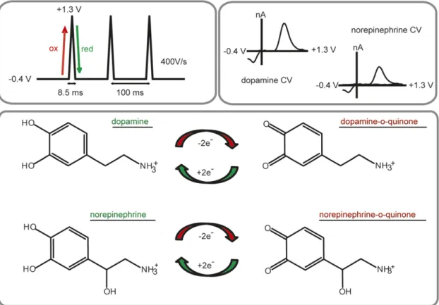
In FSCV, a potential sweep is applied to an electrode at a rapid scan rate (100–1000 V/s) to oxidize and reduce electroactive species. The current from this potential sweep results in a characteristic cyclic voltammogram (Fig. 1) and can be converted into concentration esti-mates for the species of interest with in vitro calibration factors and multivariate analysis techniques (Keithley et al., 2009; Rodeberg et al., 2015). FSCV measurements are typically conducted at glass-encased, carbon-fiber microelectrodes in which the sensor is 5–10 mm in diameter, and with an active length of 50–150 mm. The small size of the sensor allows for minimal tissue damage, as well as excellent spatial selectivity (Peters et al., 2004). Voltammetric measurements are typically conducted at fast sampling frequencies by repeating the potential sweep every 20–200 ms, allowing for the detection of single release and uptake events.
Constant-potential amperometry (CPA) measure-ments are also conducted at carbon-fiber microelec-trodes; however, this method differs from FSCV in that it uses a single potential to oxidize or reduce the molecule of interest. The simplicity of this approach results in better time resolution than FSCV because it is only limited by the sampling frequency. Similar to CPA, carbon-fiber microelectrodes are used in chro-noamperometry, which also relies on a single electrol-ysis potential. However, chronoamperometry differs from CPA in that the potential is stepped periodically instead of constantly applied. It is important to note that in both CPA and chronoamperometry, any mole-cule that is electroactive at that given potential will be detected at the electrode. Thus, amperometric measure-ments have poor chemical selectivity as compared with FSCV. To combat this, researchers have turned to chem-ically treated electrodes to enhance analyte selectivity
(Gerhardt et al., 1984). For a detailed comparison of FSCV, chronoamperometry, and CPA, we refer the reader to a recent review (Bucher and Wightman, 2015).
In addition to electrochemical detection of endoge-nous catecholamine release, catecholamines can also be exogenously applied to examine the kinetics of neuro-transmitter clearance. This approach has been used in brain slices (Falkenburger et al., 2001), as well as in anesthetized (Cass et al., 1992; Zahniser et al., 1999) and freely moving animals (Gerhardt et al., 1999; Sabeti et al., 2002). Exogenous application is often used in amperometric methods because it confirms the identity of the measured species. However, the disappearance of applied catecholamines reflects information about the diffusion of the analyte away from the electrode surface, as well as clearance due to uptake. Therefore, care must be taken when interpreting data obtained by this methodology.
Due to the chemical selectivity of FSCV over amper-ometry, FSCV has been the more widely applied electro-chemical technique for measuring both release and uptake of catecholamines. A commonly used voltam-metric waveform for detection of catecholamines sweeps from 20.4 to +1.3 V at 400 V/s, applied every 100 ms (Fig. 1). However, it is important to note that dopamine and norepinephrine differ structurally by a single hydroxyl group, and current voltammetric waveforms
cannot distinguish between cyclic voltammograms of the two catecholamines in vivo. Thus, we and others have turned to a histologic and pharmacological signal validation method that is discussed further in Pharma-cological Validation. Although this method ensures the catecholamine signal is purely dopamine or norepineph-rine, it typically precludes measurements in regions containing mixed catecholamines, such as the prefron-tal cortex. However, recent advances in optogenetics (Deisseroth, 2015) and chemogenetics (Roth, 2016) allow for the excitation or inhibition of specific cell populations. Although dopamine and norepinephrine cannot be differentiated electrochemically, it is possible that these selective stimulation methods will further extend the reach of FSCV for catecholamine measure-ments throughout the brain. For the time being, the electrochemical signal needs to be validated pharmaco-logically to claim that the measured current is solely dopaminergic or noradrenergic in nature.
B. Regulation of Extracellular Catecholamines in Brain Slices
1987; Palij et al., 1990). Early studies focused on dopamine, and FSCV in brain slices allowed researchers to determine that extracellular concentrations were a balance of release and uptake, with metabolism operating on a slower time scale (Near et al., 1988; Sulzer et al., 2016). Dopamine release per stimulation pulse ranges from;15 nM in the basolateral amygdala to;90 nM in the dorsal striatum (Garris and Wightman, 1994a, 1995). These values are in stark contrast to norepinephrine release, which reaches only;2 nM in the dorsal lateral geniculate, and ;7 nM in the anteroventral thalamus (Mitchell et al., 1994; Garris and Wightman, 1995). Additionally, maximal norepinephrine efflux only weakly depends on stimulation current above 250 mA, unlike striatal dopamine, which does not approach saturation even with stimulation currents of 450mA (Kennedy et al., 1992; Miles et al., 2002). Longer pulse trains are required to elicit norepinephrine release in slices containing the bed nucleus of the stria terminalis (BNST), and the kinetics of norepinephrine release are slower as com-pared with dopamine (Kennedy et al., 1992; Miles et al., 2002). These differential release profiles may be due to the differences in vesicular release rate between norepi-nephrine and dopamine (Chiti and Teschemacher, 2007), or differential dependence on N-type calcium channel activity (Mitchell and Adams, 1993). Despite evidence that both catecholamines are released from similar sized vesicles (Bergquist and Ludwig, 2008; Papke et al., 2012), the mechanism underlying differential release kinetics in slices is unknown.
2. Uptake. After catecholamines are released, they are cleared from the extracellular space by neuronal transporters. The rate of uptake is often approximated using t1/2, the time it takes to clear half of the
neurotransmitter concentration from the extracellular space. As measured by t1/2, uptake rates differ vastly
between dopamine and norepinephrine. For dopamine, t1/2 values are,0.1 second in both dorsal and ventral
striatum; for norepinephrine t1/2 values exceed 1.0
second (Garris and Wightman, 1994a, 1995; Mitchell et al., 1994).
The primary clearance mechanism for dopamine is mediated by the dopamine transporter (DAT), which obeys Michaelis–Menten kinetics (Wightman et al., 1988). Uptake rates are heterogeneous in subregions of the striatum (Trout and Kruk, 1992; Jones et al., 1996; Siciliano et al., 2014; Salinas et al., 2016), which might be attributed to differences in striosome and matrix compartments (Salinas et al., 2016). Researchers have manipulated DAT expression to determine its con-tribution to extracellular dopamine concentrations in brain slice preparations, and genetic deletion of DAT prolongs the life of extracellular striatal dopamine by 300-fold (Jones et al., 1998). Conversely, overexpression of DAT results in a 50% faster dopamine uptake rate, and an overall reduction in evoked dopamine concentra-tions (Salahpour et al., 2008).
The primary clearance mechanism for norepineph-rine is the norepinephnorepineph-rine transporter (NET), but non-NET mechanisms may play a larger role in norepi-nephrine clearance. The lifetime of ventral BNST (vBNST) norepinephrine clearance is only prolonged sixfold in NET knockout mice (Xu et al., 2000; Miles et al., 2002), and both organic cation transporters (OCTs) and DAT are expressed in the vBNST (Miles et al., 2002; Gasser et al., 2009). Although DAT has a higher affinity for dopamine over norepinephrine, DAT knockout mice exhibit a slight reduction in vBNST norepinephrine clearance rate (Miles et al., 2002). However, DAT is not likely a major contributor to norepinephrine clearance in animals with normal NET function because pharma-cological blockade of DAT in rats does not affect norepi-nephrine clearance rate in the vBNST (Palij and Stamford, 1992). However, in cases of prolonged signal-ing, DAT or OCTs may serve as an additional mechanism for norepinephrine clearance.
Although the main substrates for DAT and NET are dopamine and norepinephrine, respectively, the role of catecholamine transporters is further complicated in regions receiving both dopaminergic and noradrenergic innervation. Catecholamine transporters are notori-ously promiscuous (Daws, 2009), and NET serves as the primary clearance mechanism for both dopamine and norepinephrine in the cortex (Moron et al., 2002). Thus, care must be taken when choosing pharmacolog-ical agents for signal validation, as discussed below in
Pharmacological Validation.
3. Autoreceptors. Extracellular catecholamine con-centrations are also governed by autoreceptor control of release. Circuitry is mostly severed in a slice prepara-tion, which serves as an advantage when examining regulation of release at catecholamine terminals. Any effects on release exerted by receptor agonists/antagonists can thus be attributed to direct actions on terminals. The principal autoreceptor for dopamine is the D2 (Beaulieu and Gainetdinov, 2011). D2 receptors maxi-mally inhibit dopamine release within 500–1000 ms, and the time course varies between dorsal and ventral striatum (Phillips et al., 2002). D2 receptors become markedly desensitized after knockout of DAT due to persistent elevation of extracellular dopamine concen-trations (Jones et al., 1999). The principal autoreceptor controlling norepinephrine efflux is the a2, and it
operates on a similar time course as D2 receptors (Palij and Stamford, 1993; Trendelenburg et al., 2001). De-spite having similar mechanisms in place to control extracellular concentrations, the fundamental differ-ences in dopamine and norepinephrine release and uptake position them to influence neuronal communi-cation in diverse ways.
C. Catecholaminergic Plasticity
For example, self-administration of psychostimulants pro-duces variable adaptations in dopamine regulation. Fol-lowing 5 days of amphetamine self-administration, evoked dopamine release is increased in brain slices containing the nucleus accumbens (NAc) and is accompanied by decreased D2 autoreceptor function (Calipari et al., 2014b). Following cocaine self-administration, electri-cally stimulated dopamine release is instead attenuated in brain slices, and cocaine is less effective at elevating dopamine concentrations (Mateo et al., 2005; Ferris et al., 2011, 2012; Calipari et al., 2014a). However, when cocaine self-administering animals are given a single infusion of amphetamine, dopamine terminal function in slices is restored; that is, evoked release magnitudes and cocaine inhibition of DAT more closely resemble that of drug-naive animals (Ferris et al., 2015). Interestingly, increasing surface DAT density increases the potency of the psychostimulants amphetamine and methamphet-amine, without altering the effect of uptake blockers such as cocaine (Salahpour et al., 2008; Calipari et al., 2013, 2015). DAT-overexpressing animals have faster uptake rates, exhibit a threefold increase in amphetamine-potentiated dopamine release as compared with controls, and develop a preference for amphetamine at lower doses as compared with wild-type animals (Salahpour et al., 2008). Similar to DAT overexpression, animals that self-administer methylphenidate have increased dopamine uptake rates, and amphetamine and methamphetamine potency is increased in these animals; that is, the psychostimulants become more effective at inhibiting dopamine uptake (Calipari et al., 2013). However, neither methylphenidate self-administration nor DAT overex-pression alters uptake inhibition by uptake blockers cocaine and nomifensine (Calipari et al., 2013, 2015). In addition, the potency of amphetamine differs between striatal subregions, and is closely linked with uptake rates; that is, elevated uptake rates are associated with increased uptake inhibition by amphetamine (Siciliano et al., 2014). Although both cocaine and amphetamine act at DAT, it is clear from these functional measurements that the potency of amphetamine, but not cocaine, is dependent on DAT expression levels.
Catecholamine receptors are also regulated by G protein–coupled receptor kinases, and deletion of G protein–coupled receptor kinase 2 (GRK2) from D1- or D2-containing neurons produces contrasting effects on dopamine regulation in brain slices (Daigle et al., 2014). GRK2 deletion in D1-containing neurons enhances evoked dopamine release and uptake rates. Conversely, GRK2 deletion in D2-containing neurons enhances D2 autoreceptor activity and depresses baseline dopamine release without changing dopamine uptake rate (Daigle et al., 2014). These adaptations are paralleled by bidirectional sensitivity to cocaine: in D1-containing neurons, GRK2 deletion enhances cocaine-amplified dopamine release without altering cocaine-mediated uptake inhibition. Conversely, lack of GRK2 in D2
neurons reduces cocaine-amplified dopamine release and cocaine-mediated inhibition of uptake rates and is accompanied by reduced behavioral sensitivity to a cocaine challenge (Daigle et al., 2014). This hypodopa-minergic state is similar to that of animals with a history of cocaine self-administration (Mateo et al., 2005; Ferris et al., 2011, 2012; Calipari et al., 2014a ). Indeed, chronic cocaine administration reduces GRK2 expression in the NAc (Schroeder et al., 2009). Together, these findings suggest that GRK2 expression in D2-containing neurons is important for cocaine-mediated dopamine release and uptake inhibition.
Brain slices have also been used to uncover changes to dopamine regulation in rats reared in social isolation. These animals exhibit enhanced dopamine release and uptake as compared with group-reared rats (Yorgason et al., 2016). Interestingly, social isolation amplifies methamphetamine, but not cocaine inhibition of DAT (Yorgason et al., 2016). Because methamphetamine and cocaine both act at DAT, these findings suggest other adaptations may play a role in the plasticity. The stress of social isolation most likely facilitates release of corticosterone, which has a profound impact on other transport mechanisms, such as OCTs (Gasser et al., 2006). In agreement with this hypothesis, corticoste-rone decreases dopamine clearance in the NAc, pre-sumably by inhibiting OCTs expressed in the NAc (Graf et al., 2013).
Although fewer studies have used slice voltammetry to assay changes in noradrenergic function, work from the Stamford laboratory demonstrated adaptations in somatodendritic norepinephrine release in the locus coeruleus (LC). Bath application of the analgesic tra-madol reduces norepinephrine clearance in slices (Halfpenny et al., 1999), but chronic treatment does not affect uptake mechanisms. Instead, chronic trama-dol appears to sensitizea2function, and in a manner that
resembles the actions of an antidepressant (Hopwood et al., 2001). Although tramadol is an opioid analgesic, these functional measurements of its pharmacological effects lend support to its antidepressant activity in mice (Rojas-Corrales et al., 1998). In another study, researchers asked how presynaptic norepinephrine reg-ulation is changed in mice lacking the metabolic enzyme monoamine oxidase A. In monoamine oxidase A knock-out mice, peak LC norepinephrine efflux is higher and is accompanied by decreased clearance rates and a2
control over release in brain slices (Owesson et al., 2002, 2003). Similar adaptations to vBNST norepinephrine were found in anesthetized rats following stressors, as discussed below in Adaptations in Catecholamine Function.
D. Other Modulators of Catecholamine Release
concentrations dependent on stimulation frequency (Zhang et al., 2009). In a study designed to mimic the tonic (,5 Hz) and phasic (20 Hz) firing patterns of dopamine neurons found in vivo, Zhang et al. (2009) used slice voltammetry to measure dopamine release during differing stimulation frequencies. In this work, blockade ofb2-containing nicotinic acetylcholine recep-tors (nAChRs) predominantly suppressed dopamine evoked from tonic (,5 Hz) stimulation trains (Zhang et al., 2009). However, decreased dopamine release after nAChR blockade was frequency dependent, and dopamine release increased under nAChR blockade at frequencies over 10 Hz (Zhang et al., 2009). Acetyl-choline can also modulate dopamine release indepen-dent of midbrain dopamine neuron activity, and optogenetic activation of cholinergic interneurons alone is sufficient to elicit dopamine release in brain slices containing the striatum, as well as in intact animals (Cachope et al., 2012; Threlfell et al., 2012). Acetylcholine released from cholinergic interneurons acts directly on dopamine terminals in the NAc to evoke release that is dependent on b2-containing nAChRs, and this cholinergic activity does not require activation of midbrain dopamine neurons (Cachope et al., 2012; Threlfell et al., 2012). In addition to optogenetic or electrical stimulations, insulin also activates cholinergic interneurons to enhance dopa-mine release in striatal slices (Stouffer et al., 2015). However, in slices containing the ventral tegmental area (VTA), insulin instead suppresses somatodendritic release and enhances dopamine reuptake (Mebel et al., 2012).
Nitric oxide (NO) donors also modulate dopamine in a manner dependent on cholinergic activity. Under re-duced acetylcholine, NO modulates dopamine release in slices in a frequency-independent manner and acts directly on dopamine terminals, in contrast to its frequency-dependent modulations in the presence of nicotinic receptor activity (Hartung et al., 2011). We could find no reports detailing how norepinephrine release is influenced by other signaling molecules in slices. Because infusion of NO donors into the BNST produces anxiety (Faria et al., 2016), and norepineph-rine in the BNST regulates the stress axis (Forray and Gysling, 2004), NO donors may potentiate norepineph-rine release in the BNST to produce elevated stress/ anxiety, but this has not yet been shown. Additionally, a host of neuropeptides is known to regulate BNST activity (Kash et al., 2015) and may, in turn, regulate norepinephrine release. By combining slice voltam-metry with the diverse toolbox of genetic manipu-lations, specific cell-type activation, and selective pharmacology, new modulators of catecholamine re-lease will be identified that will contribute to a better understanding of how these neurotransmitters are regulated and aid in the development of more effective pharmacotherapies.
III. In Vivo Recordings in Anesthetized Animals
A. Pharmacological Validation
Voltammetric measurements in anesthetized ani-mals allow for precise measurements of release and uptake in the intact brain. Because neural activity is suppressed in anesthetized animals, neurotransmitter release is typically elicited by stimulating neurons or their axon bundles directly. Early studies used direct electrical stimulation, although recent optogenetic strate-gies provide an opportunity to excite or inhibit discrete cell types (Witten et al., 2011; McCutcheon et al., 2014). Dopamine and norepinephrine differ structurally by only a hydroxyl group, and their voltammograms in vivo are indistinguishable (Park et al., 2009). Thus, we have turned to a multistep approach to validate the origin of the signal at the electrode. First, we limit our measurements to regions containing predominantly dopamine or norepinephrine. Tissue homogenate stud-ies have confirmed that norepinephrine is the primary catecholamine in the vBNST and the anteroventral thalamus; thus, our first in vivo norepinephrine mea-surements were restricted to those regions (Park et al., 2009). Many dopamine-rich regions lie adjacent to the vBNST, and, without the visual confirmation of elec-trode placement afforded by a slice preparation, we turned to a pharmacological approach to rule out contri-butions by dopamine (Fig. 2). Voltammetric signals are only considered noradrenergic if they respond to adren-ergic agents (e.g., a2 antagonist idazoxan), but not
dopaminergic agents (e.g., D2 antagonist raclopride). Finally, a constant current is applied to the carbon-fiber electrode to make a lesion in the brain for subsequent histologic validation of electrode placement in the tar-get region.
B. Differential Release of Catecholamines in Anesthetized Animals
smaller increases in OT dopamine compared with NAc (Wakabayashi et al., 2016).
Recent work in our laboratory has used this classic mapping approach combined with multiple electrodes and pharmacology to reveal an unexpected population of dopamine neurons that release dopamine into the contralateral striatum (Fox et al., 2016a). Stimulations of the VTA elicit dopamine release in the NAc both ipsilateral and contralateral to the stimulation, al-though release is ;20 higher following ipsilateral versus contralateral VTA stimulations. Contralaterally projecting dopamine neurons originating from the VTA are also differentially regulated by D2 receptors be-cause they are more sensitive to the D2 antagonist raclopride than ipsilateral VTA projections (Fox et al., 2016a). Dopamine is also released in the dorsal stria-tum following stimulations of the contralateral SN. In contrast to the NAc, dopamine release in the dorsome-dial striatum exhibits hemispheric equivalence, that is, dopamine release is of similar magnitude following ipsilateral or contralateral SN stimulations (Fox et al., 2016a). Hemispherically equivalent dopamine release in the dorsomedial striatum is also found after stimu-lating the ipsilateral or contralateral pedunculopontine tegmental nucleus (PPTg), an excitatory input to the SN. Furthermore, hemispherically equivalent release is accompanied by similar D2 control over contralateral and ipsilateral SN projections (Fox et al., 2016a). These findings suggest that differential D2 receptor control may underlie the differences between contralateral dopamine release in the NAc and striatum. Although anatomic (Geisler and Zahm, 2005) and behavioral (Steinberg et al., 2014) data support cross-hemispheric dopamine projections, voltammetric measurements confirm their functional nature and precipitate new areas of inquiry regarding contralateral catecholamine projections.
Other work in intact animals has revealed NAc dopamine release is differentially modulated by a 6-containing nAChRs. Infusion of thea6 nAChR antago-nist a-conotoxin MII into the VTA decreases evoked dopamine in the NAc (Wickham et al., 2013). However, similara6 antagonism in slice preparations containing the NAc results in enhanced dopamine release (Exley et al., 2008). These findings suggest that nAChRs can modulate dopamine release in a site-dependent manner and highlight the importance of making measurements in an intact brain. The high spatial resolution afforded by microelectrodes combined with site-specific phar-macological approaches will continue to enable func-tional characterization of dopaminergic circuits in intact animals.
2. Norepinephrine. In anesthetized animals, norepi-nephrine release is typically measured by stimulating neurons in the nucleus of the solitary tract, or its axon bundles. Although most in vivo norepinephrine studies are conducted in the vBNST, both the dorsal BNST and
the NAc shell receive some norepinephrine innervation in addition to dopamine. Selective pharmacology indi-cates that norepinephrine in the NAc is restricted to the more caudal portion of the shell (Park et al., 2010), and norepinephrine in the dorsal BNST is contained to the medial portion (Herr et al., 2012). In a study designed to compare norepinephrine responses between dorsal and ventral BNST, researchers found norepinephrine re-lease in the dorsomedial BNST is;50% of vBNST release (Herr et al., 2012). Accompanying reduced release amplitudes in the dorsomedial BNST are slower clear-ance rates and reduced a2 autoreceptor function as
compared with regulation in the vBNST (Herr et al., 2012).
To directly compare evoked catecholamines, Park et al. (2011) used a dual-electrode approach to measure dopamine and norepinephrine release simultaneously in the NAc and vBNST with stimulations that targeted both noradrenergic axons and the VTA/SN. As pre-viously demonstrated in slices, release and uptake of norepinephrine in the vBNST are slower as compared with dopamine in the NAc, even with identical stimu-lation location (Park et al., 2011). The two catechol-amines are also differentially regulated. Tyrosine hydroxylase inhibition depletes dopamine release faster than norepinephrine, and basal levels of dopamine increase when D2 receptors are antagonized and uptake is blocked with amphetamine (Park et al., 2011). In contrast, there are no elevations in basal norepineph-rine in the vBNST following uptake inhibition with amphetamine and concomitanta2autoreceptor
inhibi-tion (Park et al., 2011). This contrasting regulainhibi-tion is also found in studies using more selective uptake inhibition. When dopamine D2 autoreceptors and DAT are blocked in anesthetized animals, dopamine concentrations fluctuate spontaneously in the striatum (Venton and Wightman, 2007). However, similar block-ade of noradrenergica2autoreceptors and NET does not
elicit spontaneous norepinephrine fluctuations in the vBNST (Park et al., 2015). Although norepinephrine and dopamine have similar regulation mechanisms, they signal in distinct ways when control mechanisms are blocked. These findings hint at unknown mecha-nisms controlling norepinephrine release beyond that of noradrenergic autoreceptors and transporters. Similar to dopaminergic modulation by nAChRs, norepineph-rine release is most likely influenced by other recep-tor types, but further work is needed to identify their contributions. Additionally, the larger stimulations re-quired to elicit release of norepinephrine suggest that norepinephrine is only released endogenously under extreme physiologic conditions, in stark contrast to dopamine.
noradrenergic innervation, and early work shows dopa-mine is the predominant catecholadopa-mine released in the medial PFC following VTA stimulations (Garris et al., 1993). This finding was confirmed in a recent report; however, D2 receptor antagonism paradoxically atten-uates dopamine release in this region (Shnitko and Robinson, 2014). Pharmacology in cortical regions must thus be selected carefully for signal validation, partic-ularly because NET takes up dopamine in the PFC (Moron et al., 2002). In future endeavors, selective stimulation methods, combined with anatomic and pharmacological validation, will enable additional vol-tammetric characterization of catecholamine signaling in regions receiving mixed innervation.
C. Adaptations in Catecholamine Function
Voltammetric measurements in anesthetized ani-mals allow for researchers to identify how different manipulations interact with intact circuitry to produce functional adaptations in catecholamine release. For example, researchers used anesthetized animals to determine the role of N-methyl-D-aspartate receptors (NMDARs) expressed specifically in dopamine neurons (Zweifel et al., 2009). This study found genetic inacti-vation of NMDARs in dopamine neurons disrupts evoked dopamine release in a stimulation site-dependent man-ner. Whereas dopamine release is unchanged in NMDAR knockout mice following MFB stimulations, dopamine release elicited by the PPTg is blunted as compared with controls (Zweifel et al., 2009). Without the intact circu-ity afforded by an anesthetized preparation, the site specificity of glutamatergic modulation of dopamine release may not have been uncovered. Another study found altered dopamine release in mice overexpressing the catecholamine metabolic enzyme catechol-o-methyl transferase (Simpson et al., 2014). In mice overexpress-ing catechol-o-methyl transferase, striatal dopamine re-lease capacity is increased despite unchanged levels of tyrosine hydroxylase or DAT (Simpson et al., 2014). It is clear that measuring the functional consequence of genetic manipulation on dopamine release provides more information than markers of dopaminergic activ-ity alone.
In addition to genetic alteration, recent work in our laboratory has examined how baseline genetic differ-ences impact catecholamine regulation mechanisms. In these studies, norepinephrine release was compared in the vBNST of Sprague-Dawley (SD), Lewis, and Wistar-Kyoto (WKY) rats (McElligott et al., 2013; Fox et al., 2015). Whereas release and uptake were similar be-tween SD and WKY rats (Fox et al., 2015), there were marked differences in uptake and autoreceptor control in Lewis rats (McElligott et al., 2013). Although the amplitude of norepinephrine release was similar in all three strains, Lewis rats showed slower norepinephrine uptake rates as compared with SD or WKY rats, despite no difference in apparent NET expression (McElligott
et al., 2013). Similarly,a2function, but not expression
was attenuated in Lewis rats, because elevation of evoked norepinephrine was blunted in Lewis rats after a2antagonism with idazoxan as compared with SD rats
(McElligott et al., 2013). Additionally, depletion of LC norepinephrine with DSP-4 produced adaptations toa2
receptors and uptake in SD, but not WKY rats, without changing norepinephrine release magnitude (Fox et al., 2015). These findings underscore the importance of voltammetric catecholamine measurements in intact systems, because differential release and uptake most likely contribute to the phenotypic variations observed in genetically diverse animal models.
Dysregulations in catecholamine signaling are impli-cated in the development of addiction and numerous neuropsychiatric conditions (Koob and Volkow, 2010). Several studies have used anesthetized preparations to uncover adaptations to catecholamine circuits following administration of drugs of abuse. Chronic administra-tion of cocaine, heroin, or a“speedball”cocktail of the two produces variable adaptations to NAc dopamine in rats. In animals with a history of chronic cocaine, heroin, or speedball self-administration, evoked dopa-mine is reduced compared with animals receiving a single drug dose (Pattison et al., 2012). Dopamine re-uptake rate is also greater in speedball-administering animals compared with drug-naive animals, or animals that self-administered cocaine or heroin alone (Pattison et al., 2012). This hypofunction of the dopamine system after cocaine self-administration was confirmed in another recent report (Siciliano et al., 2015b). However, 1-day pretreatment with cocaine does not alter the dopa-mine response to a subsequent cocaine challenge (Addy et al., 2010).
Another study examined the effect of repeated co-caine treatment on dopamine release, and found that 7 days of cocaine exposure instead potentiated the effect of a cocaine challenge on dopamine signaling in the NAc. This increase in elevated dopamine after cocaine was accompanied by an increase in apparent Kmof DAT
et al. (2016) found that after 30 days of forced absti-nence, a cocaine challenge increased NAc dopamine release in animals with a history of self-administration. Future work should address the mechanisms underly-ing dopaminergic adaptations after variable periods of cocaine withdrawal because this plasticity appears to be time-course dependent.
There is some evidence that k opioid receptors may play a role in differential cocaine-potentiated dopamine release after withdrawal. The endogenous ligand for k opioid receptors is dynorphin, which is released in response to stressful events (Chavkin, 2013), such as drug withdrawal. Thekactivation alone inhibits evoked dopamine in the NAc, and, on a short time scale, pretreatment with akagonist attenuates the dopamine response to cocaine in the NAc (Ehrich et al., 2014). However, on a longer time scale, pretreatment with ak agonist increases the cocaine-induced increase in NAc dopamine (Ehrich et al., 2014), similar to potentiated dopamine release after.24-hour withdrawal, or 30 days of forced abstinence (Addy et al., 2010; Cameron et al., 2016). Further work is needed to determine whether k opioid receptor activation alone can explain the differ-ences between these studies (Addy et al., 2010; Siciliano et al., 2015b; Cameron et al., 2016). Regardless, the time-course dependence ofkopioid modulation of dopamine is interesting in the context of stress-induced cocaine use. It is possible that dynorphin released in response to stress promotes a dysphoric state that drives drug use. On a short time scale, this stress may decreases cocaine’s actions on mesolimbic dopamine, resulting in escalation of drug intake to compensate for cocaine’s attenuated effect. However, on a longer time scale,kactivation may potentiate cocaine’s effects on dopamine, further driving its reinforcing properties. Although cocaine increases extracellular dopamine concentrations, it is clear that there are other signaling mechanisms outside of elevated dopamine that drive persistent drug use.
Adaptations to dopamine signaling have also been studied in anesthetized animals after other drugs of abuse. An acute dose of ethanol decreases evoked dopamine and slows clearance in the medial PFC (Shnitko et al., 2014). Release magnitudes are likewise suppressed in the medial PFC when ethanol is infused directly into the VTA (Shnitko et al., 2014), and a systemic ethanol challenge also reduces NAc dopamine release (Shnitko et al., 2016). In animals with adoles-cent alcohol exposure, tonic levels of NAc dopamine are reduced in adulthood, and these animals exhibit in-creased risk-taking behavior (Schindler et al., 2016). Although tonic NAc dopamine is reduced, phasic NAc dopamine release is increased in animals with ado-lescent alcohol exposure, in a manner dependent on stimulation location: PPTg- but not MFB-evoked phasic dopamine release is increased compared with alcohol-naive animals (Schindler et al., 2016). In agreement with this, another study found adolescent alcohol
exposure enhances VTA-stimulated dopamine release in the NAc, and an ethanol challenge produces larger increases in stimulated dopamine release compared with alcohol-naive animals (Shnitko et al., 2016). In-terestingly, the administration of an allosteric GABAA
agonist attenuates both increased dopamine release and increased risk-taking behavior in animals with adolescent alcohol exposure (Schindler et al., 2016). These findings suggest that there is increased inhibi-tory tone after alcohol exposure that drives changes in dopaminergic signaling through a disinhibitory mecha-nism, which may contribute to the behavioral changes. Although acute ethanol reduces evoked dopamine in both the NAc and medial PFC, alcohol exposure in adolescence clearly leads to dopaminergic plasticity that may increase the reinforcing properties of alcohol later in life.
In addition to alcohol, large doses of methamphet-amine have also been shown to reduce evoked striatal dopamine and decrease DAT uptake rates (Howard et al., 2011). These large doses are considered neuro-toxic and result in decreased striatal DAT levels (Howard et al., 2013). Pretreatment with neurotoxic doses of methamphetamine reduces the concentrations of both pharmacologically induced (Robinson et al., 2014) and naturally occurring dopamine transients (Howard et al., 2013). Although drugs of abuse can increase dopaminergic transmission (Covey et al., 2014), it is clear that this is dependent on dosing and prior drug exposure.
Work investigating adaptations to norepinephrine signaling has been limited, but two recent studies from our laboratory examined the effects of stress and drug exposure on norepinephrine release and regulation. Three days of naloxone-precipitated morphine with-drawal dysregulates norepinephrine signaling in the vBNST in a strain-dependent manner (McElligott et al., 2013; Fox et al., 2015). In morphine-withdrawn SD rats, a2receptors become desensitized, norepinephrine
clear-ance rate is slowed, and these animals exhibit increased anxiety-like behavior (McElligott et al., 2013; Fox et al., 2015). In morphine-withdrawn WKY rats, norepineph-rine clearance rate is unchanged, but these animals exhibit increased anxiety-like behavior and decreased a2receptor function (Fox et al., 2015). Lewis rats exhibit
elevated anxiety-like behavior at baseline, and mor-phine withdrawal does not further elevate anxiety, nor depress a2 function or norepinephrine clearance in
these animals (McElligott et al., 2013). Furthermore, following 2 weeks of social isolation stress, stressed SD rats resemble morphine-dependent rats, that is, ele-vated anxiety-like behavior, desensitizeda2receptors,
exposure, Lewis rats do not alter noradrenergic syn-aptic function following social isolation, suggesting their regulation mechanisms are maximally disrupted (unpublished data). These findings underscore the importance of genetic factors in susceptibility to cate-cholamine dysregulation after stress exposure or drug withdrawal.
Researchers have also used anesthetized animals to investigate the mechanisms of nonabused drugs on catecholamine regulation. For example, the dopamine precursor, levodopa (L-DOPA), enhances dopamine re-lease in both dorsal and ventral striatum, but causes a delayed inhibition of dopamine release in the dorsal striatum (Harun et al., 2015). L-DOPA also reduces uptake rates by decreasing Vmaxof DAT (Harun et al.,
2015). Because dopamine is the metabolic precursor to norepinephrine, L-DOPA may also affect norepineph-rine concentrations, but this area is currently unex-plored. Noradrenergic deficits are also a key component in the development of Alzheimer’s disease, and the way pharmacotherapies alter catecholaminergic function should be a topic of ongoing investigation. Electro-chemical measurements in anesthetized animals allow researchers to examine discrete circuits and how they become functionally altered after a variety of treat-ments. Continued efforts should focus on changes in norepinephrine function in addition to dopamine due to their different patterns of release and uptake in vivo.
IV. Catecholamine Function in Awake Animals
A. Spontaneous Fluctuations
The first awake-animal FSCV dopamine measure-ments were conducted nearly 20 years ago, and the signals that researchers found were closely associated with a novel stimulus or environment (Garris et al., 1997; Rebec et al., 1997; Robinson et al., 2001). However, dopamine concentrations were also found to fluctuate in the absence of any external stimuli in awake animals at rest (Robinson et al., 2002) (Table 1). Spontaneous dopamine fluctuations, or transients, have been mea-sured in the dorsal striatum, nucleus accumbens, and olfactory tubercle (Robinson et al., 2002), and they display heterogeneity in their frequency, amplitude, and duration in subregions of the NAc (Wightman et al., 2007). Dopamine transients in the NAc originate from phasic cell firing in the VTA (Sombers et al., 2009), are increased by cannabinoid receptor activation (Cheer et al., 2004), and are the main source of average extracellular NAc dopamine levels (Owesson-White et al., 2012). DAT blockade with nomifensine increases spontaneous and stimuli-related dopamine transients (Robinson and Wightman, 2004), and acute phenylalanine/ tyrosine depletion reduces the frequency, but not ampli-tude, of spontaneous transients (Shnitko et al., 2016). Additionally, cocaine increases the magnitude and dura-tion of spontaneous dopamine transients in the NAc core
and shell, but increases in dopamine transient frequency after cocaine are only found in the NAc shell (Aragona et al., 2008).
Recent work employing a dual-electrode approach has revealed that, at rest, spontaneous dopamine tran-sients in the NAc shell synchronize;75% of the time between hemispheres (Fox et al., 2016a). Importantly, FSCV measurements of dopamine transients provide a clearer picture of dopaminergic activity, as the time course of dopamine transients as measured by FSCV is more closely linked with uptake inhibition-induced stereotypy than microdialysis measurements (Budygin et al., 2000). In contrast to striatal dopamine, norepi-nephrine concentrations are not known to fluctuate spon-taneously in the vBNST of animals at rest (Park et al., 2012, 2013; Fox et al., 2016b), further illustrating the differences in endogenous catecholamine signaling (Table 1).
B. Intracranial Self-Stimulation
Intracranial self-stimulation (ICSS) was first de-scribed by Olds and Milner (1954), and, through ex-tensive mapping studies, it was determined that sites that supported the best self-stimulation were cen-tered around the medial forebrain bundle, implicating
TABLE 1
Opposing catecholamine responses in awake animals
Upward arrows reflect increases, and downward arrows reflect decreases in phasic release. Superscripted letters correspond to the following references. The literature cited in this table is meant to provide notable examples of contrasting signaling and is by no means comprehensive.
Stimulus NAc Dopamine vBNST Norepinephrine
At rest (transients) Presenta Absentb
ICSS
Stimulation ↑c ↑d
ICSS-predictive cue ↑c No effectd
ICSS-extinction ↓d ↑d
Food reward
Unexpected food ↑e No effectf
Food-predictive cue ↑g Unknown
Food omission ↓h Unknown
Drugs of abuse
Drug exposure ↑i No effectjunknown
Drug-predictive cue ↑k Unknown
Drug withdrawal ↓j ↑j, Unknown
Noxious/Aversive
Quinine ↓l ↑m
Fear cues ↑↓n Unknown
Tail pinch ↑↓o ↑o
aRobinson et al., 2001; Wightman et al., 2007; Sombers et al., 2009. bPark et al., 2012, 2013; Fox et al., 2016b.
cGarris et al., 1999; Cheer et al., 2007a; Owesson-White et al., 2008. dPark et al., 2013.
eRoitman et al., 2004, 2008; Cone et al., 2014. fPark et al., 2012.
gDay et al., 2010; Brown et al., 2011; Cacciapaglia et al., 2012; McCutcheon et al.,
2012; Saddoris et al., 2015b.
hSaddoris et al., 2015a.
iBudygin et al., 2001; Cheer et al., 2004; Heien et al., 2005; Aragona et al., 2008;
Covey et al., 2014; Vander Weele et al., 2014; Fox et al., 2016b.
jFox et al., 2016b.
kPhillips et al., 2003; Stuber et al., 2005; Cameron et al., 2014. lRoitman et al., 2008; Park et al., 2012; Twining et al., 2015. mPark et al., 2012.
catecholamine signaling as a principal mediator of the behavior. In this paradigm, an animal is trained to respond instrumentally (e.g., lever press) to deliver an electrical stimulation to its brain. The presentation of the lever is traditionally preceded by a cue that predicts reward availability. Dopamine is released following direct electrical stimulation of the VTA/SN or MFB, but, as animals become well trained, the NAc dopamine release moves to the cue in a time-locked fashion and is accompanied by decreases in stimulation-evoked dopa-mine (Table 1) (Owesson-White et al., 2008). To this end, the dopamine response elicited by a reward-predicting cue, but not the reward, provides strong support for dopamine’s involvement in reward predic-tion error (Schultz, 2013). Furthermore, cue-associated dopamine release increases when the cue predicts a greater stimulation magnitude, and this increase is associated with a decreased latency to lever press for the stimulation (Beyene et al., 2010).
In early work from Garris et al. (1999), rats were trained to self-stimulate the VTA in a continuous manner, that is, the lever was not retracted after each lever press. These prolonged periods of self-stimulation sessions diminish the magnitude of NAc dopamine release, which the authors described as a dissociation of dopamine release from ICSS. More recent work in our laboratory has revisited this idea (Rodeberg et al., 2016). Although dopamine concentrations are markedly attenuated following continuous ICSS, the disappear-ance of dopamine during ICSS can instead be attributed to dopamine concentrations falling below the limit of detection (Rodeberg et al., 2016). Thus, although pro-longed periods of stimulation diminish dopamine re-lease magnitudes, dopamine is still an important mediator of VTA self-stimulation behavior.
Further work has uncovered how dopamine interacts with specific postsynaptic receptors in the NAc to drive ICSS (Owesson-White et al., 2016). In this study, Owesson-White et al. (2016) employed a multimodal sensor that combines voltammetric dopamine measure-ments with single-unit activity. This method allows for the real-time characterization of dopaminergic modu-lation of cell firing in awake animals (Takmakov et al., 2011), and can be paired with iontophoresis to probe local receptor activity (Belle et al., 2013). In this study, rats were trained to press a lever for electrical stimu-lation of the VTA (Owesson-White et al., 2016). Dopa-mine was released in the NAc following cue presentation and lever press, and unit activity was associated with either the cue or the lever press, but not both. For cue-responsive cells, increased dopamine release occurred with broad increases in firing rate. For lever press– responsive cells, activity either increased or decreased, and the changes in firing rate to the press were shorter in duration than elevations in dopamine. Locations with-out changes in unit activity were also withwith-out dopa-mine release; thus, changes in activity could be attributed
to dopamine’s actions at its receptors (Owesson-White et al., 2016).
To identify the receptor-level mechanisms behind dopaminergic modulation of cell firing, the authors coupled iontophoresis barrels to the multimodal sensor (Owesson-White et al., 2016). Cell firing was altered following delivery of specific pharmacological agents; thus, neurons could be chemotyped as containing D1 or D2 dopamine receptors (Belle et al., 2013). One pop-ulation of lever press–responsive cells was identified as D1-containing, and two-thirds of these D1 cells were excited during the lever press. The remaining lever-press cells were identified as D2-containing, and 90% of these cells responded with an inhibition. Remarkably, when the authors identified the cue-responsive cells, they were all excitatory and D2-containing. Taken together, this study reveals dopamine responses to the cue exclusively activate D2-containing neurons in the NAc, whereas dopamine’s actions through both D1 and D2 receptors modulate the activity of lever press– responsive cells (Owesson-White et al., 2016). The coupling of iontophoresis with electrochemical and electrophysiological recordings provides a technical advantage over previous drug delivery techniques because large volumes of drugs can disrupt lever pressing for ICSS (Cheer et al., 2007a). Future work employing this discrete drug delivery technique will allow for receptor-level mechanisms to be uncovered in real time, without necessitating the use of transgenic animals. Furthermore, this technique may be extended into regions containing mixed catecholamines for sig-nal validation prior to making measurements during behavior.
and with more vigor during ICSS extinction (Mason and Iversen, 1979), and LC lesions impair attention (Selden et al., 1990). Future work should address the involve-ment of norepinephrine in extinction from other reward paradigms, which may provide insight into how action selection is shaped in the context of negative or un-anticipated outcomes.
C. Natural Rewards
A number of studies have measured dopamine release in response to natural rewards, such as food pellets or sucrose. In general, unexpected delivery of food reward elicits dopamine release in the NAc, and the magnitude of dopamine release is greater in animals previously food-restricted (Roitman et al., 2008; Cone et al., 2014) (Table 1). Glutamatergic inputs to dopamine neurons are important for dopamine release in response to food, because mice lacking NMDARs in dopamine neurons exhibit reduced dopamine release in the NAc after unexpected food delivery (Parker et al., 2010). Addi-tionally, dopamine release to unexpected food is poten-tiated by infusions of grehlin in the lateral ventricle or intra-VTA orexin-A (Cone et al., 2014).
Dietary changes can also cause adaptations in dopa-minergic signaling, and a prolonged high-fat diet reduces uptake without altering DAT gene expression (Cone et al., 2013). Diet can also alter insulin sensitivity, and insulin-deficient rats have reduced DAT surface expres-sion and dopamine uptake (Williams et al., 2007). Insulin can suppress somatodendritic dopamine concentrations in the VTA through increased uptake (Mebel et al., 2012); however, in the NAc, insulin signaling enhances dopamine release by exciting cholinergic interneurons (Stouffer et al., 2015). Importantly, the effect of insulin on striatal dopamine release is diet-dependent: rats fed an obesogenic diet exhibit a complete loss of insulin-potentiated dopamine release (Stouffer et al., 2015). It is clear that diet and insulin can alter dopaminergic func-tion, but further work is needed to more precisely eluci-date the mechanism by which diet-altered dopamine function contributes to maladaptive eating behavior.
In contrast to dopamine, little is known regarding norepinephrine signaling during caloric rewards or changes to diet, which is of particular interest given norepinephrine’s involvement in anorexia and feeding behavior (Janhunen et al., 2013; Nedelescu et al., 2016). Only one study examined norepinephrine release during food administration, and found unexpected sucrose de-livery does not elicit vBNST norepinephrine release (Park et al., 2012) (Table 1). Norepinephrine release in the vBNST may instead coincide with food omission as it is during omission of ICSS reward (Park et al., 2013); however, this remains to be shown. Norepinephrine release during food omission has interesting implica-tions in the context of feeding behavior. Because vBNST norepinephrine can influence the hypothalamic– pituitary–adrenal axis (Forray and Gysling, 2004), it may
engage the brain’s stress centers to suppress feeding and promote anorexia. Future work should address the role of norepinephrine signaling during delivery and omission of high-calorie foods to determine how it contributes to dysregulated food consumption.
Food-predictive cues also elicit dopamine release in the NAc (Roitman et al., 2004) and dorsolateral stria-tum (Shnitko and Robinson, 2015). In the NAc, sucrose-predictive cues evoke greater dopamine release than those that predict saccharin (McCutcheon et al., 2012) (Table 1). There is some evidence that inputs from the BLA modulate NAc dopamine release to food-predictive cues, because inactivation of the BLA with baclofen/ muscimol attenuates NAc dopamine evoked by sucrose-paired cues (Jones et al., 2010). Interestingly, this manipulation does not diminish VTA-stimulated dopa-mine release in the NAc, and suggests the glutamater-gic inputs from the BLA modulate NAc dopamine through a terminally mediated mechanism (Jones et al., 2010). In support of glutamatergic influence over NAc dopamine responses, mice lacking NMDARs re-lease smaller concentrations of dopamine in the NAc during delivery of a food reward (Parker et al., 2010). Dopamine released in response to a food-predictive cue selectively modulates cells in the NAc that are excited during the cue, but not those that are inhibited during the cue (Cacciapaglia et al., 2011). Additionally, dopa-mine responses vary based on NAc subregion; although cue-evoked dopamine is observed in both NAc core and shell, it is of greater magnitude and duration in the shell as compared with the core (Cacciapaglia et al., 2012). When rats must press one lever to extend a second lever for sucrose delivery, dopamine responses also vary between the NAc core and shell. In the core, dopamine release is greatest after presentation of the first lever, or the “seeking lever,” and less for subsequent presenta-tion of the “taking lever” and reward delivery. In the shell, dopamine release is robust to both levers as well as to reward delivery (Saddoris et al., 2015a).
Dopamine release is also elicited during delivery of a noncaloric reward. In sodium-depleted animals, NAc dopamine signaling increases when animals are given a salt solution. Over time, dopamine release moves to the salt-predictive cue in sodium-depleted rats (Cone et al., 2016). We could find no similar studies of phasic norepinephrine response to noncaloric reward.
D. Aversion
A number of studies have examined the effects of aversive stimuli on dopaminergic transmission. Sys-temic delivery of the aversive agent lithium chloride blunts phasic dopamine release in the NAc (Fortin et al., 2016), and oral administration of the aversive tastant quinine suppresses dopamine release in the NAc (Roitman et al., 2008) and dorsolateral BNST (Park et al., 2012). Quinine reduces dopamine tone in the NAc by reducing release frequency and is dependent on corticotropin-releasing factor (CRF) signaling (Twining et al., 2015). Blocking CRF receptors in the VTA blocks the inhibitory effect of quinine on NAc dopamine, suggesting that CRF release during aversive stimuli can act directly on VTA neurons to suppress NAc dopamine release (Twining et al., 2015). During quinine delivery, decreased dopamine in the NAc is accompa-nied by increased vBNST norepinephrine (Park et al., 2012). This reciprocal catecholamine signaling during negative stimuli was also found in a study examining the noxious stimulus of a tail pinch. The predominant response during tail-pinch delivery is a suppression of dopamine overflow in the NAc, whereas the same stim-ulus elicits norepinephrine release in the vBNST (Park et al., 2015). Although the tail-pinch study was con-ducted in anesthetized animals, one might suspect that norepinephrine overflow increases in awake animals dur-ing delivery of a noxious stimulus such as a foot shock. Indeed, markers of noradrenergic activity increase fol-lowing foot shock (Rassnick et al., 1998; Passerin et al., 2000).
Due to electrical interference, it is difficult to make electrochemical measurements of catecholamine re-lease during delivery of a foot shock. However, recent work has focused on contrasting dopamine signaling in the NAc during fear-predictive cues (Badrinarayan et al., 2012). Cues that predict foot shock decrease dopaminergic transmission in the NAc core by decreas-ing the probability of dopamine release. In the NAc shell, the same cues elicit increases in dopaminergic transmission by enhancing the amplitude of dopamine release (Badrinarayan et al., 2012). However, neither increases nor decreases in NAc dopamine were strictly associated with freezing behavior elicited by the fear-predictive cue (Badrinarayan et al., 2012). We could find no similar studies on phasic norepinephrine responses during fear-predictive cues. Norepinephrine may be released in the vBNST during a cue that predicts foot shock, as it is during the delivery of aversive quinine
(Park et al., 2012), or during a tail pinch (Park et al., 2015). However, because no work has found norepi-nephrine responses to cues in general, further work is needed to investigate this possibility.
E. Social Interaction
Social interactions also elicit phasic dopamine re-sponses, and the introduction of an unfamiliar rat or conspecific increases the frequency of dopamine tran-sients (Robinson et al., 2002). Dopamine is also released in the NAc in response to prosocial ultrasonic vocali-zations (Willuhn et al., 2014). However, increased dopamine release to both prosocial vocalizations and conspecific interaction declines rapidly following ha-bituation (Robinson et al., 2002; Willuhn et al., 2014). NAc dopamine is also modulated by reward delivery to another rat, as stronger dopamine release is measured during conspecific reward receipt versus an empty box. Similar to other prosocial interactions, this response attenuates, and even becomes reversed in repeated trials, with reductions in dopamine during conspecific reward delivery (Kashtelyan et al., 2014).
In rats subjected to social defeat stress, interaction with an aggressive rat increases burst firing in the VTA and elevates the frequency of NAc dopamine transients (Anstrom et al., 2009). However, changes in norepi-nephrine signaling during an aggressive encounter have not been studied. Social defeat stress increases norepinephrine synthesis and NET expression in the LC (Chen et al., 2012; Fan et al., 2013), suggesting negative social interaction may alter norepinephrine release. Furthermore, when Long Evans rats are sub-jected to social defeat stress, some rats become aggres-sive. These aggressive rats have increased anxiety-like behavior, exhibit decreased latency to attack, and have increased amygdalar norepinephrine content as com-pared with nonaggressors (Patki et al., 2015). Given that norepinephrine influences the hypothalamic-pituitary-adrenal axis, it is tempting to hypothesize norepinephrine is released in the vBNST during an animal’s decision to fight or flee during aggressive social interactions, and this should be a topic of future investigation.
F. Drugs of Abuse
manner (Sulzer, 2011). However, recent work challenges the action-potential independence of amphetamine-evoked dopamine release. Abolishing cell firing in do-pamine neurons also abolishes amphetamine-elicited dopamine transients, suggesting that, in awake animals, amphetamine exerts its effects via an action-potential– dependent manner (Covey et al., 2016). A large number of studies have examined the effects of psychostimulants on spontaneous dopamine transients. To avoid redundant coverage, we direct the reader to a recent review (Covey et al., 2014).
2. Cannabanoids. Emerging evidence supports can-nabinoid modulation of dopamine signaling and drug reward (Cheer et al., 2007b; Loewinger et al., 2012; Oleson et al., 2012; Hernandez et al., 2014), and cannabinoid’s effects on the dopaminergic system have been recently reviewed in detail (Oleson and Cheer, 2012; Covey et al., 2015). Despite reports that cannabi-noids increase LC norepinephrine activity (Oropeza et al., 2005; Page et al., 2007), how cannabinoid receptor activation influences phasic norepinephrine release has not been investigated.
3. Alcohol. Electrophysiological data show that eth-anol stimulates dopaminergic transmission (Brodie et al., 1999), and tonic activation of VTA dopamine neurons suppresses voluntary alcohol drinking by elevating basal dopamine efflux (Bass et al., 2013). However, there is disparity in alcohol’s effects on neurochemistry. Microdialysis data support a biphasic dopaminergic response, in which low doses produce increases in NAc dopamine (Yoshimoto et al., 1992) and decreases at higher doses (Blanchard et al., 1993). As measured by FSCV, acute ethanol dose-dependently decreases evoked dopamine in the dorsal striatum (Budygin et al., 2001), but there is an apparent hetero-geneity of dopamine response in the NAc. In some recording locations, ethanol increases dopamine tran-sient frequency, whereas in some the frequency is decreased or even unaffected (Robinson et al., 2009). Interestingly, cues that predict a sweetened ethanol reward are time locked to dopamine release in the dorsolateral striatum and NAc, but not in the dorsome-dial striatum (Shnitko and Robinson, 2015). Although alcohol-predictive cues elicit dopamine release, the amplitude of release does not differ between rats consuming sweetened alcohol versus those consuming the sweetened solution alone (Shnitko and Robinson, 2015). This apparent heterogeneity in dopamine re-sponse to ethanol should be addressed because alcohol exposure has circuit-specific effects on dopaminergic transmission (Schindler et al., 2016). Although ethanol can enhance stimulated dopamine, its effects on endog-enous dopamine release are conflicting.
It is currently unknown how ethanol impacts phasic norepinephrine release. One microdialysis study showed that ethanol dose-dependently increases basal NAc nor-epinephrine in animals reared in social isolation, but
not those reared in group housing (Karkhanis et al., 2014). Future work should address alcohol’s effect on phasic norepinephrine signaling, particularly in the BNST, because the BNST can modulate ethanol-seeking behavior (Pina et al., 2015) and is an important structure for the development of alcohol use disorders (Kash, 2012).
4. Opiates. In addition to alcohol, recent work has examined the effect of an acute i.v. delivery of opiates on catecholamine release. In one study, researchers de-livered the opiates oxycodone and morphine to freely moving rats (Vander Weele et al., 2014). Intravenous oxycodone increases dopamine transient frequency and magnitude in the NAc for ;1 hour; however, i.v. morphine produces a much shorter (;1 minute) in-crease in phasic dopamine transmission (Vander Weele et al., 2014). In our laboratory, we extended this work to determine the impact of drug withdrawal in addition to drug exposure on dopamine release. In contrast to the previous report, s.c. administration of morphine pro-duces a persistent (.1 hour) increase in dopamine transient frequency; however, the average magnitude of dopamine transients is similar between animals given morphine or saline (Fox et al., 2016b). This difference may reflect the differential time course of drug delivery between the two studies (Vander Weele et al., 2014; Fox et al., 2016b). Interestingly, when animals undergo naloxone-precipitated withdrawal, dopaminergic transmission decreases in the NAc (Fox et al., 2016b). In morphine-withdrawn animals, nalox-one treatment reduces dopamine transient frequency back to baseline or saline conditions. Furthermore, naloxone decreases the average concentration per tran-sient only in animals undergoing morphine withdrawal (Fox et al., 2016b). However, a single morphine with-drawal episode is insufficient to elicit persistent ad-aptations in dopaminergic signaling because after treatment, electrically evoked dopamine concentra-tions reach similar amplitudes in animals treated with naloxone after either saline or morphine (Fox et al., 2016b). Thus, the decreases in dopaminergic trans-mission reflect alterations of dopamine signaling spe-cifically during the withdrawal period.
(Park et al., 2012, 2013). Furthermore, a single with-drawal episode is sufficient to reduce evoked norepi-nephrine in withdrawn animals (Fox et al., 2016b), further highlighting the contrast between dopaminergic and noradrenergic signaling. Importantly, this reduc-tion in releasable norepinephrine, but not dopamine, is in agreement with tissue content in the NAc and vBNST after repeated episodes of opiate withdrawal (McElligott et al., 2013). Together with the opposing responses highlighted in Table 1, opposing dopamine and norepinephrine signaling during drug exposure and withdrawal support reciprocal roles for catecholamines during appetitive and aversive stimuli.
G. Norepinephrine–Dopamine Interactions
Due to the opposing nature of NAc dopamine and BNST norepinephrine, it is tempting to hypothesize that these reciprocal responses reflect feedback be-tween the two catecholaminergic systems, and that norepinephrine release may influence the reduction of dopaminergic transmission during aversive stimuli. Indeed, glutamatergic inputs from the vBNST exert excitatory influence over VTA dopamine neurons (Georges and Aston-Jones, 2002), and norepinephrine’s actions througha2Areceptors decrease excitatory
trans-mission in the vBNST (Egli et al., 2005). Norepineph-rine can also act throughbreceptors to increase GABAA
inhibition of VTA-projecting BNST neurons (Dumont and Williams, 2004), leading to increased inhibition of the VTA. In further support for noradrenergic modula-tion of dopamine transient concentramodula-tions, elevamodula-tion of norepinephrine with systemic a2 antagonism
sup-presses the magnitude of spontaneous dopamine tran-sient concentrations in the NAc (Fox et al., 2016b). The reciprocal actions of dopamine and norepinephrine during drug exposure and withdrawal (Fox et al., 2016b), reward-learning and extinction (Park et al., 2013), and appetitive and aversive tastants (Park et al., 2012) may be generalized to other paradigms; however, additional work is needed. Future work should address how these opposing responses develop longitudinally to shape learning about rewarding and aversive stimuli.
V. Clinical Implications
Voltammetric catecholamine measurements in mod-els of human disease have provided new insights into their pathogenesis. To model Parkinson’s disease, re-searchers have turned to animals that express the mutant proteins found in human patients to uncover how these mutations lead to catecholaminergic deficits. For example, researchers expressed mutant human leucine-rich repeat kinase 2 in rats and found impaired striatal dopamine release. The adaptations to circuit function appear in the absence of neurodegeneration, and suggest that dopaminergic dysfunction might pre-cede measureable markers of neurodegeneration and
cell death (Sloan et al., 2016). In another study, expres-sion of mutant a-synuclein in tyrosine–hydroxylase neurons produces differential catecholamine deficits that are regionally specific: evoked dopamine is re-duced in the dorsal striatum, but not in the NAc, and vBNST norepinephrine release is unchanged (Taylor et al., 2014). These genetic manipulations afford ways to test early circuit function, and might become a useful preclinical model for testing Parkinson’s therapeutics.
Researchers have also used voltammetry to investi-gate aberrant dopamine signaling in Angelman syn-drome. Angelman syndrome is a neurodevelopmental disorder characterized by ataxic movements, develop-mental delay, and excessive exuberance (Williams, 2010). Patients with Angelman syndrome lack ubiquitin ligase E3A (UBE3A), due to mutations or deletions of the maternal alleleUBE3A, and recent work has used mice lacking maternal UBE3A to model the syndrome (Riday et al., 2012; Berrios et al., 2016). Mice lacking maternal UBE3A are more sensitive to VTA stimulation; that is, the mice acquire robust self-stimulation behavior to lower electrical self-stimulation currents (Riday et al., 2012), and deliver more optoge-netic self-stimulations as compared with controls (Berrios et al., 2016). Despite the behavioral similarities between these two studies, they are in apparent con-trast with one another with respect to changes in dopamine signaling. Riday et al. (2012) found differen-tially altered dopamine signaling in UBE3A-deficient mice without adaptations to the number of dopaminer-gic cells: whereas NAc dopamine release is increased in these mice, nigrostriatal dopamine release is attenu-ated. In contrast, Berrios et al. (2016) used optogenetics to elicit dopamine release in brain slices containing the NAc, and found no differences in dopamine release magnitude between UBE3A-deficient mice or their controls. However, there are important methodological differences that may contribute to the apparent differ-ences in dopamine release between the two studies. First, direct depolarization of dopamine terminals in a brain slice is not necessarily indicative of the magnitude of dopamine release in an intact brain. Second, elec-trical stimulations do not provide the cell-type specific-ity of optogenetic approaches. Third, adaptations in afferent, nondopaminergic neurons may facilitate dys-regulated signaling that may not be apparent in a slice preparation. Moving forward, we need to consider the evidence from both slice and intact animal preparations to integrate how circuit function becomes disrupted in disease models.