Ar ticle
The Rockefeller University Press $30.00
The J
our
nal of Exper
imental Medicine
IntroductIon
The coagulation cascade represents an evolutionary highly
conserved process that enables efficient hemostasis in response to vascular injury. However, an aberrant intravascular activa-tion of the coagulation cascade causes thrombotic disease, in-cluding myocardial infarction, ischemic stroke, or deep-venous thrombosis, and thus represents the leading cause of death worldwide (Stevens et al., 2009). Both hemostasis and throm-bosis involve the initial formation of a yet unstable platelet aggregate at the site of vascular injury (Wolberg, 2007). The subsequent activation of plasmatic coagulation results in the formation and polymerization of fibrin, an insoluble protein Blood coagulation is essential for physiological hemostasis but simultaneously contributes to thrombotic disease. However, molecular and cellular events controlling initiation and propagation of coagulation are still incompletely understood. In this study, we demonstrate an unexpected role of eosinophils during plasmatic coagulation, hemostasis, and thrombosis. using a large-scale epidemiological approach, we identified eosinophil cationic protein as an independent and predictive risk factor for thrombotic events in humans. concurrent experiments showed that eosinophils contributed to intravascular thrombosis by exhibiting a strong endogenous thrombin-generation capacity that relied on the enzymatic generation and active provision of a procoagulant phospholipid surface enriched in 12/15-lipoxygenase–derived hydroxyeicosatetraenoic acid–phosphatidyleth-anolamines. our findings reveal a previously unrecognized role of eosinophils and enzymatic lipid oxidation as regulatory ele-ments that facilitate both hemostasis and thrombosis in response to vascular injury, thus identifying promising new targets for the treatment of thrombotic disease.
Enzymatic lipid oxidation by eosinophils propagates
coagulation, hemostasis, and thrombotic disease
Stefan Uderhardt,
1,6*
Jochen A. Ackermann,
1,6*
Tobias Fillep,
1,6Victoria J. Hammond,
8,9Johann Willeit,
10Peter Santer,
11Manuel Mayr,
12Markus Biburger,
13Meike Miller,
14Katie R. Zellner,
15Konstantin Stark,
14Alexander Zarbock,
16Jan Rossaint,
16Irene Schubert,
14Dirk Mielenz,
2,6Barbara Dietel,
3Dorette Raaz-Schrauder,
3Cihan Ay,
17Thomas Gremmel,
18Johannes Thaler,
17Christian Heim,
4Martin Herrmann,
1Peter W. Collins,
8,9Gernot Schabbauer,
19Nigel Mackman,
20David Voehringer,
7Jerry L. Nadler,
21James J. Lee,
15Steffen Massberg,
14Manfred Rauh,
5Stefan Kiechl,
10Georg Schett,
1Valerie B. O’Donnell,
8,9and Gerhard Krönke
1,61Department of Internal Medicine 3 – Rheumatology and Immunology, 2Department of Internal Medicine 3, Division of Molecular Immunology, 3Department
of Cardiology and Angiology, 4Department of Cardiac Surgery, 5Department of Pediatrics, 6Nikolaus Fiebiger Center of Molecular Medicine, and 7Department
of Infection Biology, Institute for Clinical Microbiology, Immunology, and Hygiene, Friedrich-Alexander-University Erlangen-Nürnberg (FAU) and Universitätsklinikum Erlangen, Erlangen, Germany
8Systems Immunity Research Institute, School of Medicine and 9Institute of Infection and Immunity, School of Medicine, Cardiff University, Cardiff, Wales, UK 10Department of Neurology, Medical University of Innsbruck, Innsbruck, Austria
11Bruneck Hospital, Bruneck, Italy
12King's British Heart Foundation Centre, Kings College, London, England, UK
13Department of Biology, Institute of Genetics, Friedrich-Alexander-University Erlangen-Nürnberg (FAU), Erlangen, Germany 14Medizinische Klinik und Poliklinik I, Klinikum der Universität, Ludwig-Maximilians-Universität, Munich, Germany
15Department of Biochemistry and Molecular Biology, Division of Pulmonary Medicine, Mayo Clinic in Arizona, Scottsdale, AZ 16Department of Anaesthesiology, Intensive Care, and Pain Medicine, University Hospital Münster, Münster, Germany
17Department of Medicine I, Clinical Division of Haematology and Haemostaseology, 18Department of Internal Medicine II, Division of Angiology, and 19Institute
for Physiology, Center for Physiology and Pharmacology, Medical University of Vienna, Vienna, Austria
20Department Medicine, University of North Carolina, Chapel Hill, NC 21Department of Internal Medicine, Eastern Virginia Medical School, Norfolk, VA
*S. Uderhardt and J.A. Ackermann contributed equally to this paper. Correspondence to Gerhard Krönke: [email protected]
S. Uderhardt’s present address is Laboratory of Systems Biology, National Institute of Allergy and Infectious Diseases, National Institutes of Health, Bethesda, MD. Abbreviations used: CVD, cardiovascular disease; ECP, eosinophilic cationic protein; FCS, feral calf serum; HDL, high-density lipoprotein; IVC, inferior venae cava; LC/ MS/MS, liquid chromatography tandem mass spectrometry; LDL, low-density lipo-protein; LO, lipoxygenase; PE, phosphatidylethanolamine; PPP, platelet-poor plasma; PRP, platelet-rich plasma; PS, phosphatidylserine; RIPA, radioimmunoprecipitation assay; TAT, thrombin–antithrombin; TF, tissue factor; TIA, transient ischemic attack;
that allows growth and stabilization of the developing throm-bus. Initiation of the coagulation cascade essentially relies on the presence of a procoagulant phospholipid surface and the glycoprotein tissue factor (TF; Nemerson, 1968). These two components initiate the sequential assembly and activation of the membrane-associated prothrombinase complex (fac-tor Xa [FXa] and fac(fac-tor Va; Nemerson, 1968), which gener-ates thrombin, the serine protease that converts fibrinogen to fibrin, and thereby provides the backbone of the growing thrombus (Wolberg, 2007).
Although all cellular membranes contain abundant amounts of phospholipids, they are usually inert and do not support coagulation. Platelets are considered to serve as a major source of procoagulant phospholipids, as these cells can actively modify their membrane and oxidize and externalize the aminophospholipids phosphatidylethanol-amine (PE) and phosphatidylserine (PS; Suzuki et al., 2010; Thomas et al., 2010; Yang et al., 2012; O’Donnell et al., 2014). Although platelets can potentially acquire TF via leuko-cyte-derived microparticles, they lack endogenous expression of TF (Bouchard et al., 2010). These findings raise the ques-tion about the exact mechanisms and cells that initiate the assembly of the prothrombinase complex during the onset of coagulation and the propagation of intravascular thrombo-sis (Flaumenhaft, 2014).
Here, we report that thrombotic events in humans are associated with an in increase in the eosinophilic acti-vation marker eosinophilic cationic protein (ECP). Subse-quent experiments showed that eosinophils were abundantly found in thrombi that formed upon vascular injury in mice, where these cells critically contributed to thrombin forma-tion and thrombus stabilizaforma-tion as well as to physiological hemostasis. Eosinophils exerted a strong endogenous throm-bin-generation capacity that relied on the simultaneous expression of TF and the provision of a procoagulant phos-pholipid surface that was enriched in 12/15-lipoxygenase (12/15-LO)–derived oxidized phospholipids. In accordance, we observed a diminished thrombus formation and defective hemostatic response in mice carrying a global or an eosino-phil-specific deletion of 12/15-LO.
results
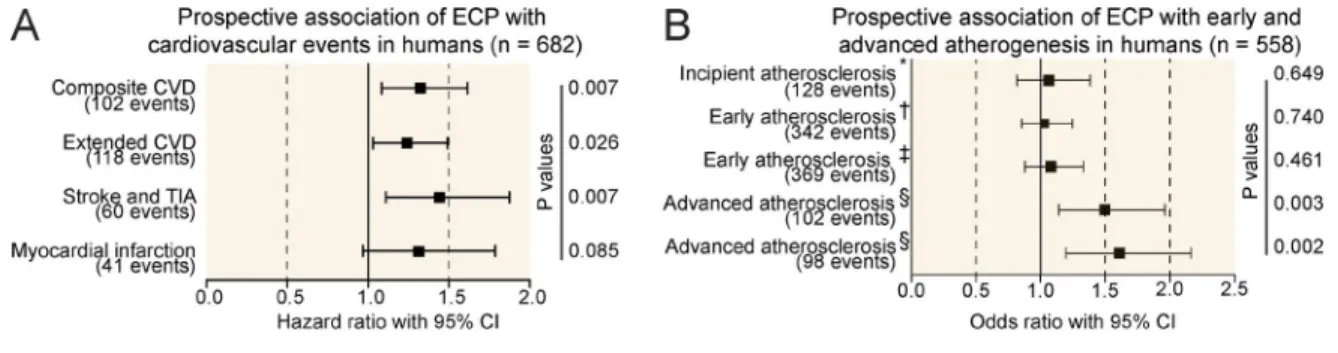
elevation of eosinophil activation markers predicts the onset of cardiovascular events
To elucidate mechanisms that trigger thrombotic events during cardiovascular disease (CVD) in humans, we made use of the prospective population-based Bruneck Study (Stege-mann et al., 2014). Population characteristics are summarized in Table S1. This study allows unique monitoring of sclerosis progression and differentiates between early athero-sclerosis, advanced atherosclerotic lesions, and the occurrence of thrombotic events such as stroke and myocardial infarction. In contrast to early atherosclerosis, which primarily reflects a chronic inflammatory intima thickening, both develop-ment of advanced atherosclerotic lesions and especially the
occurrence of thrombotic events are primarily associated with factors reflecting enhanced plaque vulnerability and a procoagulant state (Willeit et al., 2000). Therefore, we sought to identify novel serologic markers that specifically pre-dict the occurrence of advanced atherosclerotic lesions and thrombotic events. A proteomic proximity ligation assay un-expectedly identified an increased plasma level of ECP as a highly significant predictor of new-onset atherothrombotic events and development of advanced atherosclerotic lesions (Fig. 1, A and B). In contrast, ECP levels were unrelated to early plaque development and growth and showed only weak correlations with age and other vascular risk factors (Fig. 1, B–D). These findings were robust to multivariable adjustment and stable in sensitivity analyses (Fig. 1, C and D). Because plasma ECP levels serve as a potential marker for eosinophil activation, these data were suggestive of a contribution of eo-sinophils to thrombus formation and vessel occlusion.
eosinophils promote intravascular thrombin formation and thrombus growth
Immunofluorescence microscopy of experimentally induced thrombi indeed confirmed enrichment of eosinophils, which accumulated at the boarder of platelet-rich areas throughout the thrombus (Fig. 2, A and B; and Fig. S1, A–E). To deter-mine whether eosinophils actively contributed to the forma-tion of an intravascular thrombus, we consequently performed an injury-induced venous thrombosis model of the inferior venae cava (IVC) in two different eosinophil-deficient mouse strains, namely ΔdblGATA1 mice (Yu et al., 2002) and PHIL mice (Lee et al., 2004), as well as in their WT littermates. Absence of eosinophils resulted in a dramatically attenuated thrombus formation (Fig. 2, C and D), which supported a functional contribution of these cells. Also, WT mice that had received an eosinophil-depleting SiglecF anti-body showed a significantly decreased thrombotic potential (Fig. 2 E and Fig. S1 F).
mice. Whereas an absence of eosinophils did not impair the formation of a first platelet-rich aggregate at the site of in-jury, it provoked a rapid dissolution of this yet unstable ini-tial thrombus (Fig. 2 G and Videos 1–4). In line with these findings, we observed an exacerbated blood loss in eosino-phil-deficient ΔdblGATA1 mice after a medium-scale injury (15-mm tail cut; Fig. 2 H), indicative of a defective coagu-lation-dependent secondary hemostasis. Platelet-dependent primary hemostasis after small-scale injury (3-mm tail cut) was unaffected by the absence of eosinophils (Fig. S2 D). Platelet depletion, in turn, unmasked the defective hemostasis in ΔdblGATA1 mice after the small-scale injury, which was depicted by a dramatically augmented blood loss (Fig. S2, E and F) and, thus, demonstrated that platelets and eosinophils performed synergistic roles during thrombosis and hemosta-sis. Notably, absence of eosinophils did not impair thrombus development in the IVC upon flow restriction (Fig. S2 G), suggesting that this leukocyte subset specifically mediated the hemostatic and thrombotic response upon vascular injury but did not contribute to stasis-induced thrombus formation.
eosinophils autonomously generate thrombin in a tF-dependent manner
Together, our data revealed a key role of eosinophils during injury-induced thrombin generation and blood coagulation in vivo. Consequently, we determined whether these cells were able to autonomously generate thrombin in the absence of exogenous procoagulant phospholipids or recombinant TF. We found that in vitro–differentiated mouse eosinophils as well as isolated human eosinophils indeed exerted a dramatic endogenous thrombin generation potential, which was fur-ther augmented by prestimulation of eosinophils with ADP or collagen (Fig. 3, A and B). Thrombin generation was preceded by formation of FXa and followed by the appearance of fibrin clots (Fig. 3, C and D; and Fig. S3 A). These data indicated the involvement of a TF-dependent pathway and suggested that eosinophils were equipped with all the necessary key compo-nents for the initiation of plasmatic coagulation, which would include an endogenous provision of TF and procoagulant
phospholipids. We confirmed expression of TF by eosinophils, which were additionally able to expose this glycoprotein on their surface upon ADP stimulation (Fig. 3, E and F; and Fig. S3 B). Addition of a blocking antibody against TF abrogated eosinophil-induced thrombin generation, confirming the functional involvement of this glycoprotein (Fig. 3 G).
eosinophils actively provide a procoagulant phospholipid surface that supports tF-mediated thrombin generation
The thrombin generation potential of activated eosino-phils was clearly superior to other TF-expressing cells, such as LPS-stimulated macrophages (Fig. S3 C). These findings suggested that additional factors allowed eosinophils to effi-ciently promote coagulation. As the TF-mediated generation of thrombin is considered to depend on the presence of a pro-coagulant phospholipid surface (Nemerson, 1968; Mackman, 2004), we determined whether eosinophils underwent spe-cific changes within their plasma membrane that would sup-port plasmatic coagulation. The aminophospholipids PS and PE can facilitate thrombin generation but are usually hidden within the inner leaflet of the plasma membrane and, thus, are unavailable for an interaction with plasmatic coagulation factors (Thomas et al., 2010; Yang et al., 2012). Measurement of annexin V surface binding, a surrogate marker for the ex-posure of aminophospholipids (Clark et al., 2013), revealed a rapid increase in the annexin V positivity of eosinophils in response to ADP, indicating that these cells actively changed the polarity of their plasma membrane (Fig. 4 A). Exposure of PE and PS species was additionally confirmed using mass spectrometry (Fig. 4 B). However, masking of surface ami-nophospholipids by annexin V blocked the ADP-induced thrombin generation in a dose-dependent manner (Fig. 4 C), confirming an essential role of these events during eosino-phil-induced coagulation.
In platelets, translocation of PS and PE was shown to involve the activity of Ca2+-sensitive Cl− channels of the
TMEM (transmembrane protein) gene family, which were suggested to additionally control cationic currents and seem to either possess or induce scramblase activity at the plasma
membrane (Suzuki et al., 2010; Tian et al., 2012; Yang et al., 2012; Kunzelmann et al., 2014). Also, eosinophils expressed
several TMEM family members (Fig. S3 D), and the Ca2+
ionophore A23187 rapidly triggered exposure of the ami-nophospholipids PE and PS in eosinophils (Fig. 4 D and Fig. S3 E). Intracellular chelation of Ca2+ by BAP TA/AM or
ad-dition of tannic acid, a nonselective inhibitor of the TMEM family of calcium-activated Cl− channels, in turn, interfered
with the ADP-induced binding of annexin V to their sur-face (Fig. 4 E). In line with these findings, ADP stimula-tion of eosinophils triggered a rapid and transient increase in cytosolic Ca2+ levels in a Ca2+-free environment, which
could be boosted by the addition of extracellular calcium (Fig. 4 F). Prior depletion of intracellular calcium stores by thapsigargin, an inhibitor of the endoplasmic reticulum Ca2+
ATPase, blocked the initial ADP-induced increase in
cyto-solic Ca2+ (Fig. 4 G). These data were suggestive of an
ini-tial ADP-induced Ca2+ release from intracellular stores and
a subsequent activation of a store-operated calcium entry through the plasma membrane (Bergmeier et al., 2013) that triggered exposure of procoagulant aminophospholipids to immediately support TF-dependent formation of the pro-thrombin complex (Fig. 4 H).
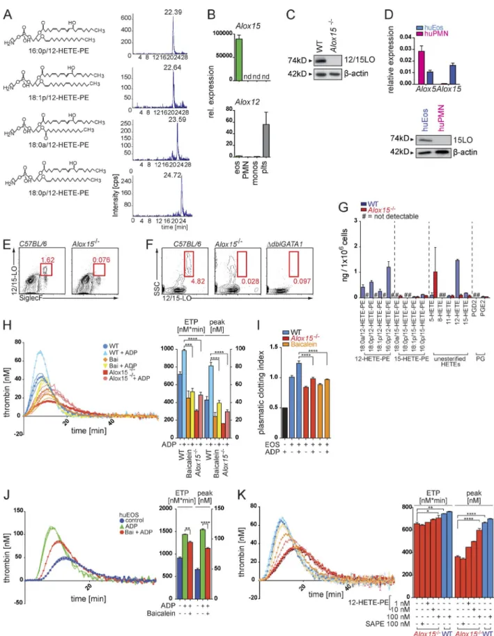
12/15-lo–mediated formation of procoagulant oxidized phospholipids essentially contributes to the thrombin-generation potential of eosinophils
PE oxidation products (12–hydroxyeicosatetraenoic acid–PEs [12-HETE-PEs]; Fig. 5 A). As oxidation of phospholipids had been suggested to increase their procoagulant potential (Thomas et al., 2010), we sought to determine a potential role of PE oxidation and the different oxidized PE species during eosinophil-mediated thrombin generation. 12-LO (Alox12) and 12/15-LO (Alox15) are the two major enzymes exert-ing 12-LO activity in mammals (Kuhn et al., 2015) and thus
eosinophils were the only cells expressing Alox15 within the peripheral blood and the vasculature. 12/15-LO expression was accordingly absent in blood leukocytes of 12/15-LO– deficient (Alox15−/−) and eosinophil-deficient ΔdblGATA1
mice (Fig. 5, E and F). Mass spectrometry showed that Alox15−/− eosinophils indeed lacked the identified 12-HETE-PEs, thus confirming 12/15-LO as the single enzymatic source of these oxidized aminophospholipids in eosinophils (Fig. 5 G). Figure 4. ca2+-dependent exposure of aminophospholipids by eosinophils promote thrombin generation. (A) Flow cytometry analysis of the bind-ing of annexin V (AxV) to aminophospholipids on the surface of restbind-ing or ADP-stimulated mouse eosinophils. Histograms show representative annexin V stainings, and bar graphs show mean geometric fluorescence intensities (gMFI). (B) LC/MS/MS-based quantification of the exposure of the aminophos-pholipids PE and PS in mouse eosinophils in response to ADP stimulation. (C, left) Calibrated thrombin generation assays with resting or ADP-stimulated mouse eosinophils in the presence of annexin V. (Right) Bar graphs show endogenous thrombin potential (ETP; nM*min) and peak of thrombin generation (peak; nM). (D) Flow cytometry analysis of annexin V binding on mouse eosinophils over time in the presence of calcium ionophore A23187, ADP, or vehicle. (E) Flow cytometry analysis of annexin V binding on mouse eosinophils in the presence of tannic acid (TA) or intracellular Ca2+-chelator BAP TA/AM. Bar graphs show geometric mean fluorescence intensity. (F) Flow cytometry–based analysis of intracellular Ca2+ signaling, indicated by Fluo3/FuraRed ratio, over time in a Ca2+-free environment. Where indicated (arrow and Ca2+), CaCl
In accordance with a major contribution of 12/15-LO– mediated membrane oxidation to the thrombin-formation activity of eosinophils, we observed a severely reduced abil-ity of Alox15−/− eosinophils to generate thrombin and FXa as well as to induce fibrin clots (Fig. 5, H and I; and Fig. S3 G). The 12/15-LO inhibitor baicalein potently interfered with the procoagulant activity of mouse and human eosino-phils as well (Fig. 5, H–J), showing that the Alox15-mediated control of plasma coagulation by eosinophils represented an evolutionary conserved mechanism and potential target for pharmacologic intervention during thrombotic disease. Of note, 12/15-LO–deficient eosinophils showed no alterations in their eosinophil maturation markers (Fig. S3, H and I). Phospholipase A2 (PLA2)–dependent cleavage of 12/15-LO products from membrane phospholipids was not involved in the 12/15-LO–mediated thrombin generation activity of eo-sinophils, as inhibition of PLA2 activity did not reduce the procoagulant activity of eosinophils (Fig. S3 J).
We subsequently measured thrombin generation in the presence of liposomes containing different PE species to substitute for 12/15-LO activity. Addition of liposomes with 12-HETE-PE immediately restored the thrombin-generation potential of Alox15−/− eosinophils, whereas identical lipo-somes with nonoxidized PEs were ineffective (Fig. 5 K), supporting the concept of a 12/15-LO–mediated control of thrombin generation that was dependent on generation and provision of 12-HETE-PEs.
12/15-lo expression in eosinophils promotes thrombotic disease and supports physiological hemostasis
To determine the in vivo relevance of 12/15-LO–mediated membrane oxidation by eosinophils during thrombotic dis-ease, we performed an injury-induced thrombosis model in Alox15−/− mice and their WT littermates. In line with our previous data, absence of 12/15-LO resulted in reduced TAT complex formation (Fig. 6 A) and an impaired thrombus for-mation (Fig. 6 B). Alox15−/− mice accordingly displayed a defective hemostatic response (Fig. 6 C), although these
an-imals showed no alterations in the function and number of platelets or in plasmatic coagulation factors (Fig. S4, A–C). We subsequently confirmed a reduced thrombotic potential of mice carrying a conditional deletion of Alox15 in eosinophils as well as in animals that had received baicalein, demonstrat-ing a key role of the identified pathway durdemonstrat-ing coagula-tion in vivo and highlighting its potential as a therapeutic target (Fig. 6, D and E).
dIscussIon
Until recently, eosinophils were solely envisaged as driving force of parasite defense and as mediators of allergic disease (Jacobsen et al., 2012). However, an increasing amount of data suggests a broader and rather homeostatic role of these cells during the resolution of inflammation and regulation of tis-sue repair (Rothenberg and Hogan, 2006; Lee et al., 2010; Heredia et al., 2013; Tani et al., 2014). More recently, eosin-ophils were also described to accumulate in human thrombi (Riegger et al., 2016) and reported to express TF (Moosbauer et al., 2007). Moreover, activated eosinophils were shown to tether to immobilized platelets under shear flow (McCarty et al., 2003), a mechanism which might explain their homing to the freshly formed and yet instable thrombus. Clinical data show that hypereosinophilic patients indeed suffer from an increased incidence of atypical thrombotic events (Ames et al., 2010; Todd et al., 2014). Although these findings have indi-cated a potential role of this leukocyte subset during thrombus formation, functional evidence that supports a direct contri-bution of eosinophils to thrombosis or hemostasis as well as insights into involved molecular pathways have been lacking so far. Our current data show that eosinophils critically con-tribute to thrombin formation and plasmatic coagulation, a process that is often regarded as the most primitive form of immune response to tissue injury and infection (Engelmann and Massberg, 2013). Other leukocyte subsets have been pre-viously implicated as potential sources of TF and might like-wise contribute to thrombus formation in certain settings (von Brühl et al., 2012; Engelmann and Massberg, 2013). However,
eosinophils (eos; side scatterhiCD11b+Siglec-F+), and platelets (plts) after FACS. Expression was normalized to Atcb expression. rel., relative. (C) Western blot of 12/15-LO protein (74 kD) expression in WT and Alox15−/−mouse eosinophils. (D) Quantitative RT-PCR analysis of Alox5 and Alox15 mRNA (top) and
West-ern blot analysis of 15-LO protein (bottom) expression in human neutrophils (huPMN) and human eosinophils (huEos) isolated by Ficoll density gradient centrifugation and magnetic cell separation. mRNA expression was normalized to Actb. (E) Flow cytometry analysis of 12/15-LO in eosinophils (side scat-terhiSiglecF+) isolated from peripheral blood of WT and Alox15−/−mice. (F) Flow cytometry analysis of the side scatter (SSC)hi12/15-LO+ population in blood from WT, Alox15−/−, and ΔdblGATA1 mice. (G) LC/MS/MS-based quantification of different esterified 12- and 15-HETE-PE, nonesterified 5-, 8-, 11-, 12-, and
15-HETE species, and prostaglandins D2 and E2 (PGD2 and PGE2) in WT (blue) and Alox15−/−(red) mouse eosinophils. Levels are presented as nanograms
per 106 cells. (H, left) Calibrated thrombin generation curve of mouse WT eosinophils, eosinophils treated with baicalein (Bai), and Alox15−/−eosinophils.
(Right) Bar graphs show endogenous thrombin potential (ETP; nM*min) and peak of thrombin generation (peak; nM). (I) Plasmatic clotting time experiments with WT mouse eosinophils (EOS), mouse eosinophils treated with the 12/15-LO inhibitor baicalein, and Alox15−/−mouse eosinophils. Bar graphs display
calculated clotting index. The black bar shows plasma with ADP alone. (J, left) Calibrated thrombin generation assay with lysates generated from human eosinophils in the presence of PRP reagent (see Materials and methods). (Right) Bar graphs show endogenous thrombin potential (nM*min) and peak of thrombin generation (nM). (K, left) Calibrated thrombin generation assays with WT and Alox15−/−mouse eosinophils in the presence of
neutrophils and monocytes in the peripheral blood seem to be unable to provide an enzymatically engineered, procoagulant phospholipid surface, whereas platelets express prothrombotic phospholipids but lack intrinsic TF expression. Thus, eosin-ophils seem to combine the abilities of platelets to oxidize and expose distinct prothrombotic aminophospholipids with the capacity of leukocytes to provide TF within a single cell (Fig. 6 F). Notably, neutrophils were shown to contribute to stasis-induced thrombosis but were fully dispensable during hemostasis after vascular injury (Engelmann and Massberg, 2013). Eosinophils, in turn, seem to primarily mediate the hemostatic and thrombotic response that follows tissue injury, suggesting that different leukocyte subsets fulfill distinct tasks during hemostasis and thrombosis.
On a molecular level, we identify mouse 12/15-LO and its human orthologue 15-LO as key enzymes during the generation of procoagulant phospholipids in eosin-ophils and show that this pathway directly contributes to the initiation of thrombus formation after injury. Although
12/15-LO expression in macrophage-derived foam cells has been previously implicated in the oxidation of low-density lipoprotein (LDL) particles and the formation of athero-sclerotic plaques (Cyrus et al., 2001; Uderhardt and Krönke, 2012), the physiological role of 12/15-LO–mediated lipid oxidation has remained unclear (Kühn and O’Donnell, 2006). Our findings suggest that 12/15-LO–mediated enzy-matic lipid oxidation serves as a key mechanism controlling eosinophil-directed coagulation and physiological hemosta-sis. Thus, our current data provide an explanation for the stringent conservation of this enzymatic pathway during mammalian evolution and simultaneously suggest that eo-sinophils as well as 12/15-LO are promising new targets for an anticoagulant therapy.
MaterIal and MetHods Patient cohorts
Bruneck study population. The Bruneck Study is a prospec-tive population-based survey on the epidemiology and patho-Figure 6. 12/15-lo–induced coagulation contributes to thrombus formation and hemostasis. (A and B) TAT complex formation (n = 6; A) and thrombus formation (n = 4; B) after ferric chloride (FeCl)–induced thrombosis of the IVC in Alox15−/−mice (C57BL/6 background) and WT littermates.
(C) Bleeding assays (15-mm tail cut) with WT (C57BL/6) mice, Alox15−/−mice, and WT mice treated with baicalein (n = 6 each). Bar graphs show relative
genesis of atherosclerosis that started in 1990. The study population was recruited as a sex- and age-stratified random sample of all residents age 40 to 79 living in Bruneck (n = 4,739), Northern Italy. Detailed follow up examinations in-cluding high-resolution ultrasound of the carotid vessels were performed every 5 yr. The present analysis focuses on the evaluation in 2000 (n = 682 with complete data including carotid ultrasound), which served as the baseline for this anal-ysis, and on the 10-yr follow-up period between 2000 and 2010. Follow-up for clinical endpoints was 100% complete (n = 682, mean and median follow up time 8.6 and 10 yr), whereas follow up ultrasound examinations in 2005 and 2010 were available in 558 men and women (i.e., 91.9% of survi-vors or 81.8% overall). The study protocol was approved by the Ethics Committees of Verona and Bolzano, and all partic-ipants gave their written informed consent be-fore entering the study.
All risk factors were assessed by means of validated stan-dard procedures described previously (Kiechl et al., 2002, 2013; Stegemann et al., 2014). In brief, body mass index was calculated as weight divided by height squared (kg/m2).
Hy-pertension was defined as blood pressure ≥140/90 mm Hg
(mean of three independent readings obtained with a stan-dard mercury sphygmomanometer after at least 10 min of rest) or the use of antihypertensive drugs. Lifetime smoking was assessed as pack-years. Diabetes was defined based on American Diabetes Association criteria.
The composite CVD endpoint was composed of isch-emic stroke, medical record–confirmed transient ischisch-emic at-tack (TIA), myocardial infarction, and vascular death. A total of 102 individuals experienced primary outcome events. The extended composite endpoint additionally considered revas-cularization procedures, which increased the number of in-dividuals affected to 118. Myocardial infarction was deemed confirmed when World Health Organization criteria for defi-nite disease status were met. Ischemic stroke and TIA were classified according to the criteria of the National Survey of Stroke. A TIA was considered only if the diagnosis could be made with high accuracy (medical record–confirmed TIA). All revascularization procedures (angioplasty and surgery) were carefully recorded. Ascertainment of events or procedures did not rely on hospital discharge codes or the patient’s self-re-port but on a careful review of medical records provided by the general practitioners, death certificates, Bruneck Hospital files, and the extensive clinical and laboratory examinations performed as part of the study protocols. Major advantages of the Bruneck Study are that virtually all subjects living in the Bruneck area were referred to the local hospital and that the network existing between the local hospital and the general practitioners allowed retrieval of practically all medical infor-mation on persons living in the area. We also collected detailed information on the date, causes, and circumstances of death for all study subjects who did not survive the entire follow-up period by consulting death certificates, all medical records ever compiled on study subjects, and autopsy reports in the rare
event of unexpected death. We were able to ascertain 100% of deaths and reliably classify them as vascular deaths, cancer deaths, or deaths from other causes (primary cause of death). Vascular mortality included deaths from ischemic stroke, myo-cardial infarction, rupture of aortic aneurysms, and sudden car-diac deaths. The experienced researcher who categorized all deaths and cardiovascular endpoints was unaware of laboratory data. There was no loss of follow-up for clinical endpoints.
assessment of atherosclerosis. At each study visit, participants underwent bilateral carotid duplex sonography using a 10-MHz transducer and a 5-MHz Doppler. All subjects were examined in supine position. The scanning protocol involved four segments of the right and left carotid artery: proximal common carotid artery (15–30 mm proximal to the carotid bulb), distal common carotid artery (<15 mm proximal to the carotid bulb), proximal internal carotid artery (carotid bulb and the initial 10 mm of the vessel), and distal internal carotid artery (>10 mm above the flow divider). A plaque was de-fined as a focal structure encroaching into the arterial lumen with a thickening of the vessel wall of at least 0.5 mm relative to surrounding segments. The maximum axial diameter of plaques (in millimeters) was assessed on the near and far walls at each of the eight vessel segments. The atherosclerosis sum-mation score was calculated by summing all diameters (intra- observer coefficient of variation, 13.5%; n = 100).
statistical methodology. We tested the hypothesis that base-line ECP level is associated with new-onset CVD and ad-vanced atherosclerosis (2000–2010), both of which rely on atherothrombosis, but not with early atherosclerosis driven by inflammation and standard vascular risk factors. Cox propor-tional hazard models with progressive adjustment were used to analyze time-to-event data on the primary CVD endpoint (ischemic strokes, medical record–confirmed TIAs, myocar-dial infarctions, and vascular deaths), extended CVD endpoint (plus revascularization procedures), and individual disease endpoints (stroke and TIA or myocardial infarction; Fig. 1 A). Subjects who suffered a CVD event were censored with re-spect to subsequent follow-up as were participant who died from nonvascular causes. We detected no departure from the proportional hazards assumption by inspecting Schoenfeld re-siduals and checking the parallelism of log–log survival plots. Associations between ECP and the various measures of ath-erosclerosis were tested by means of logistic regression analy-sis (Fig. 1 B) because the quinquennial examinations in the Bruneck Study provide reliable information on plaque devel-opment/progression in this time interval but no time-to-event information.
Base models were adjusted for age, sex, and either prior CVD (CVD endpoints) or baseline atherosclerosis (ultrasound endpoints). Multivariable analyses were additionally adjusted for hypertension, smoking (pack-years), diabetes, loge
-trans-formed C-reactive protein, body-mass index, and LDL and high-density lipoprotein (HDL) cholesterol. We modeled ECP as a continuous variable and calculated hazard and odds ratios for a 1-SD unit–higher level of ECP.
In subsidiary analyses, we excluded subjects with prior CVD, eosinophil fractions >5%, or platelet inhibitor therapy. All p-values were two sided, and an α level of 0.05 was used. Analyses were conducted using SPSS and R 3.2.2 (R Foun-dation for Statistical Computing).
Proteomic serum analysis. Blood samples were drawn in the year 2000 after an overnight fast and 12 h of abstinence from smoking and immediately frozen and stored at −70°C (with-out any thawing-freezing cycle). Laboratory parameters were measured by standard assays, and a blood differential was per-formed using an automated analyzer (Kiechl et al., 2002, 2013; Stegemann et al., 2014). Eosinophil cationic protein (ECP) was measured in plasma samples collected in the year 2000 as part of a novel proteomics chip (Proximity Extension Assay; 4 Proseek Multiplex CVD I96 × 96; Olink Bioscience) as described previously (Assarsson et al., 2014).
animals
Animal experiments were approved by the government of Mittelfranken, the Mayo Clinic, and the Ludwig Maximil-ian University, Munich. 12/15-LO–deficient (Alox15−/−;
C57BL/6 background), ΔdblGata1 (BALB/c background),
and PHIL mice were described previously (Yu et al., 2002; Lee et al., 2004). ΔdblGata1mice carry a deletion of a high-affinity
GATA-binding site in the GATA-1 promoter (ΔdblGATA-1
mice), whereas PHIL mice express a diphtheria toxin A trans-gene that is driven by a fragment of the eosinophil peroxidase promoter. Both mouse strains show a selective and complete absence of the eosinophilic lineage. To achieve an eosin-ophil-specific deletion of Alox15, we crossed an EPX-Cre mouse (Doyle et al., 2013) with mice carrying floxed Alox15 alleles (both C57BL/6 background; Cole et al., 2012). Ex-periments were performed at an age of 8–10 wk. Whereas
experiments with ΔdblGATA-1 mice were performed in
hemizygous mutant males and WT male littermates, the ex-periments with other mouse strains were performed with an equal gender distribution of mutant and WT mice. Animal experiments were performed by a blinded investigator.
cell culture
Eosinophils were generated from bone marrow isolated from 8-wk-old mice as previously described (Dyer et al., 2008) with minor modifications. In brief, bone marrow was incu-bated in RPMI medium (Gibco) containing 20% heat-in-activated feral calf serum (FCS), 25 mM Hepes, 100 IU/ml penicillin (Gibco), 10 µg/ml streptomycin (Gibco), 2 mM glutamine (Gibco), 1× nonessential amino acids (NEAA; Sigma-Aldrich), 1 mM sodium pyruvate (Sigma-Aldrich),
50 µM β-mercaptoethanol (Gibco), 100 ng/ml mFLT3L
(PeproTech), and 100 ng/ml mouse stem cell factor (Pepro-Tech) for 4 d, followed by 10-d differentiation with 10 ng/ml IL-5 (PeproTech). Half of the medium was changed every other day. Maturation was monitored by flow cytometry of SiglecF and CCR3 expression. Fully matured eosinophils were used after 14 d of total culture. For thrombin generation assays, RAW 264.7 mouse macrophages were treated with 100 ng/ml LPS in RPMI medium containing 10% FCS for 24 h.
Plasma clotting time
Human standard citrate plasma (Siemens Healthcare) was freed from residual cellular and subcellular particles by ul-tracentrifugation and supplemented with 10% rat plasma (GeneTex). Plasma clotting time with or without addition of eosinophils was automatically recorded after recalcification with 20 µl of star-tem (ROT EM; Tem International GmbH) using a ball coagulometer (BC1; SYCOmed) at 37°C. When indicated, eosinophils were stimulated with 40 µM ADP (Roche) or 20 µg/ml collagen (Roche) for 10 min at 37°C before recalcification. Clotting index was calculated as fol-lows: clotting time [sec.]−1 × 100.
calibrated thrombin and FXa generation
added and incubated for 10 min at 37°C. End concentra-tion of ADP (Roche) was 40 µM. Then, 20 µl of recalcifica-tion solurecalcifica-tion containing a fluorogenic substrate for thrombin (Z-Gly-Gly-Arg) was automatically added. α2
-Macroglobu-lin–thrombin was used separately for calibration according to the manufacturer’s specifications (Thermo Fisher Scien-tific). FXa generation was performed equally but with the fluorogenic substrate Pefafluor FXa (Loxo GMBH) and a filter pair with excitation at 340 nm and emission at 440 nm. When indicated, eosinophils were pretreated with 1 µM baicalein (EMD Millipore) or DMSO control and 40 µg/ ml anti–mouse coagulation factor III/TF antibody (AF3178; R&D Systems) or isotype control (normal goat IgG; AB-108-C; R&D Systems) for 1 h at 37°C before stimulation. For thrombin generation with platelet-rich plasma (PRP) or platelet-poor plasma (PPP), PRP reagent (final concentration 0.5 pM TF; cat. no. TS42.00; Thrombinoscope) or PPP re-agent (final concentration 5 pM TF and 4 mM phospholipids; cat. no. TS30.00; Thrombinoscope) was added according to the manufacturer’s specifications. Data were evaluated using Thrombinoscope 5.0 software.
calcium signaling
500,000 cells were stained with 6 µM Fluo-3 (Invitrogen) and 12 µM FuraRed (Invitrogen) in 200 µl of staining buffer (RPMI; 10 mM Hepes and 2% FCS) for 45 min at 37°C with gentle agitation. Cells were subsequently washed twice in
Ca2+-free Tyrode’s buffer (134 mM NaCl, 12 mM NaHCO
3,
2.9 mM KCl, 0.34 mM Na2HPO4, 1 mM MgCl2, 10 mM
Hepes, 5 mM Hepes, and 0.5% BSA, pH 7.4) and incubated for 20 min at room temperature in the dark. Where indicated, CaCl2 was added to a final concentration of 1 mM. Calcium
signaling experiments were performed at room temperature with a Gallios flow cytometer (Beckman Coulter) record-ing the Fluo-3/FuraRed ratio over time. Data were analyzed using FlowJo software (Tree Star). The reagents used were: BAP TA/AM and thapsigargin (EMD Millipore), tannic acid, and A23187 (Sigma-Aldrich).
antibodies
Rat anti–mouse SiglecF PE (E50-2440; BD), rabbit poly-clonal anti–12-LO (ab23678; Abcam), rabbit anti–mouse TF (EPR8986; Abcam), polyclonal goat anti–mouse coagu-lation factor III/TF antibody (no. AF3178; R&D Systems), goat anti–rabbit Cy5 or Cy3 (Dianova), anti–mouse CD11b PE/Cy7 (M1/70), anti–human CD16 FITC (3G8), anti– mouse Ly6G FITC (1A8), anti–mouse CD41 PE or APC (MWReg30), anti–mouse CD115 PE or APC (AFS98), anti– mouse CD45 APC/Cy7 (30-F11), and anti–mouse CD16/32 (TruStain fcX) were used. Annexin V FITC and DAPI stain-ing were purchased from BioLegend.
Facs and flow cytometry
Bone marrow was isolated from sacrificed WT mice, and blood was drawn from the IVC of anaesthetized mice using
a syringe (23-G needle) containing 10% 0.1 M sodium ci-trate. Red blood cells were gently lysed with ice-cold distilled water. Cells were pelleted and resuspended in buffer (1× PBS containing 0.5% BSA and 2 mM EDTA). After Fc recep-tor blockade, cells were stained with antibodies as indicated. For intracellular staining of 12/15-LO in blood leukocytes, cells were fixed and permeabilized before staining using a intracellular staining kit (00-5523; eBioscience). FACS was performed on a MoFlo XDP cell sorter (Beckman Coulter), and flow cytometry was performed on a Gallios flow cytom-eter (Beckman Coulter). Data were analyzed using FlowJo software. Platelet counts were recorded from citrated whole blood with an Adivia 120 cytometer (Siemens Healthcare).
Immunofluorescence microscopy
For microscopy, thrombi were only partially dissected from the vessels to increase stability during preparation of the his-tological sections. Samples were embedded in Tissue-Tek op-timal cutting temperature compound (Sakura), snap frozen, and stored at −80°C. 4-µm cryosections were fixated and per-meabilized with ice-cold acetone and stored at −20°C until staining. Slides were stained with the indicated antibodies (dilution 1:100) and analyzed on an ECL IPSE Ni-U micro-scope (Nikon). Pictures were processed using NIS-Elements software (BR4.0; Nikon). For quantification of eosinophils in mouse thrombi, we analyzed thrombi of three independent experiments (four slides per individual thrombus).
Isolation of platelets and generation of PrP and PPP
To avoid artificial platelet activation, blood was carefully drawn from the IVC of anaesthetized mice, using a syringe (23-G needle) containing 10% 0.1 M sodium citrate. To iso-late piso-latelets, a same volume of EDTA-Hepes-saline buffer (150 mM NaCl, 1 mM EDTA, and 10 mM Hepes) was added to the citrated blood, followed by two 20-min centrifugations at 50 g. Platelets were pelleted for 5 min at 300 g and resus-pended in HBSA buffer (20 mM Hepes, 100 mM NaCl, and 1 mg/ml BSA) for further analysis. PRP was generated out of citrated plasma by 10-min centrifugation at 50 g and diluted to 140 cells/nl with autologous plasma. PPP was generated by two 10-min centrifugations at 1,500 g.
Isolation of human eosinophils
resuspended in RPMI medium containing 10% FCS and stimulated with ADP or baicalein for 30 min at 37°C. Then, cells were washed and resuspended in HBSA buffer, and cell lysates were generated by three repeated freeze-thaw cycles. Samples were stored at −80°C until further analysis.
Western blotting
Cells were washed twice with PBS and then lysed in radioim-munoprecipitation assay (RIPA) buffer (50 mM Tris, 150 mM NaCl, 1 mM EDTA, 1% Triton X-100, 1% sodium deoxy-cholate, and 0.1% SDS) containing 1% protease/phosphatase inhibitor (P9599; Sigma-Aldrich) and 1 mM PMSF (Active Motif) for 30 min on ice. Aortas were rinsed three times with PBS containing 1,000 IU heparin, cut into little pieces, and digested in serum-free DMEM containing 2 mg/ml colla-genase type 2 (Worthington Biochemical Corporation) for 1 h at 37°C and then lysed in RIPA buffer for 30 min on ice. Protein content was assessed using a BCA Protein Assay kit (Thermo Fisher Scientific) according to the manufactur-er’s specifications. Protein extracts were separated by SDS-PAGE using a 10% SDS-polyacrylamide gel, transferred to a Trans-Blot Nitrocellulose membrane (Bio-Rad Laborato-ries), and immunoblotted overnight in TBS-Tween contain-ing 5% nonfat dry milk with the followcontain-ing antibodies: rabbit polyclonal anti–12-LO (ab23678; dilution 1:1,000; Abcam), mouse coagulation factor III/TF antibody (no. AF3178; di-lution 1:200; R&D Systems), and rabbit anti–mouse β-actin (clone AC-74; dilution 1:1,000; Sigma-Aldrich). As a positive control for TF expression, lysates of LPS-treated RAW 264.7 mouse macrophages were used. As a positive control for 12/15-LO, mouse-resident macrophages were isolated from the peritoneal cavity of C57BL/6 mice by lavage, plated in RPMI medium, and washed thoroughly after 1 h to remove any nonadherent cells. After overnight culture in RPMI me-dium containing 10% FCS, cells were lysed in RIPA buffer and stored at −20°C until further analysis.
real-time Pcr analysis
RNA was isolated with peqGOLD TRIFast (peqlab), and 500 ng of total RNA was reverse transcribed with human leukemia virus reverse transcription using the Gene Amp RNA PCR kit (Applied Biosystems) and oligo deoxythy-midine primers. For quantitative RT-PCR after FACS, leu-kocyte subpopulations were directly sorted into lysis buffer, and RNA isolation was performed with an RNeasy Mini
kit (QIA GEN) including DNA digestion (DNase I; no.
EN0521; Fermentas) according to the manufacturer’s spec-ifications. Quantitative real-time PCR was performed using a LightCycler instrument and SYBR Green I kit (Roche). We normalized the ratio of mRNA expression of the gene of interest to the mRNA expression for housekeeping genes Actb or Gapdh (with comparable results); depicted if not oth-erwise stated are the results for normalization to Actb (2−▵Ct).
For detection of TF mRNA, two different primer pairs were used with comparable results; depicted in the paper are the
results for primer pair 1. Used PCR primer sequences for
mouse mRNA were: Actb forward, 5′-TGT CCA CCT TCC
AGC AGA TGT-3′ and reverse, 5′-AGC TCA GTA ACA GTC
CGC CTA GA-3′; Gapdh forward, 5′-CTA CAC TGA GGA
CCA GGT TGT CT-3′ and reverse, 5′-CAG GAA ATG AGC
TTG ACA AAG TT-3′; Alox5 forward, 5′-ATT GCC ATC
CAG CTC AAC CA-3′ and reverse, 5′-ACT GGA ACG CAC
CCA GAT TT-3′; Alox12 forward, 5′-CGC TGT TGC CAC
CAT GAG AT-3′ and reverse, 5′-ATG AGC TGG GTC CGC
GTTC-3′; Alox15 forward, 5′-CTC TCA AGG CCT GTT
CAG GA-3′ and reverse, 5′-GTC CAT TGT CCC CAG AAC
CT-3′; Ccr3 forward, 5′-ACT GGA CTC ATA AAG GAC TTA
GCA-3′ and reverse, 5′-CCA TGA CCC CAG CTC TTT
GA-3′; Epx forward, 5′-CGC CTG GAT AGC CAG TAT CG-3′
and reverse, 5′-ATG GAA TCC TGC CGG TTC AG-3′;
Si-glecf forward, 5′-TCA GCC CTG AAA GTA GCA GC-3′ and
reverse, 5′-TTT GGG TGT CTG GGA CTG TG-3′; F3
(co-agulation factor III; TF) no. 1 forward, 5′-AGG ATG TAC
CTG GGC CTAT-3′ and reverse, 5′-GGC TGT CCA AGG
TTT GTG TC-3′; and F3 (coagulation factor III; TF) no. 2
forward, 5′-GAA ACT GGA AAA ACA AGT GCT TCTT-3′
and reverse, 5′-CCA GGT CAC ATC CTT CAC GAT-3′. Used
PCR primer sequences for human mRNA were: Alox15
for-ward, 5′-GTG TCC ACT GGG GCC TCG CT-3′ and reverse,
5′-GCG GCC CCA GAT ACT CCG GTA-3′; Alox5 forward,
5′-GAC GTT CAC GGC CGA GGT GG-3′ and reverse, 5′
-AGC TGG CCG AAG TTG ACC GC-3′; Actb forward, 5′
-AGA AAA TCT GGC ACC ACA CC-3′ and reverse, 5′-TAG
CAC AGC CTG GAT AGC AA-3′; and Gapdh forward, 5′
-TGA -TGA CAT CAA GAA GGT GGT GAAG-3′ and reverse,
5′-TCC TTG GAG GCC ATG TGG GCC AT-3′.
lipid extraction
10 ng 1,2-dimyristoyl-PE (DMPE) and 5 ng 15-HETE-d8 were added to each sample before extraction as an internal standard. Hydroperoxides were reduced to the corresponding alcohol by adding 1 mM SnCl2 for 10 min at room
tempera-ture. Lipids were extracted by adding 1 M acetic acid and 2-propanol hexane (2:20:30, vol/vol) to the sample at a ratio of 2.5 ml of solvent to 1 ml of sample by vortexing and then adding 2.5 ml of hexane. After vortexing and centrifugation at 1,500 rpm for 5 min, lipids were recovered in the upper hexane layer. Then, the samples were reextracted by the addi-tion of an equal volume of hexane followed by further vor-texing and centrifugation. The combined hexane layers were then dried under vacuum and analyzed for HETE-PEs and free eicosanoids using liquid chromatography tandem mass spectrometry (LC/MS/MS).
Hete-Pe quantitation using lc/Ms/Ms
with a flow rate of 200 µl/min. Lipids were monitored using multiple reaction–monitoring mode. Transitions monitored were for parent ions of m/z 738.6, 764.6, 766.6, and 782.6 [M−H]− fragmenting to daughter ions with m/z 219.2 (15-HETE), 115.1 (5-(15-HETE), or 179.1 (12-HETE). Standard curves were generated using internal standards (DMPE) and different synthetic primary standards. Products were quanti-fied by LC/MS/MS electrospray ionization on an Applied Biosystems 4000 Q-Trap system. Acquisition of product ion spectra was triggered during elution of ions of interest, with the instrument operating in ion trap mode.
Free eicosanoid quantitation using lc/Ms/Ms
Lipids were separated on a C18 Spherisorb ODS2 column (150 × 4.6 mm; 5 µm particle; Waters Ltd) using a gradient of 50–90% B over 30 min, followed by 5 min at 90% B (A, water/ acetonitrile/acetic acid, 75:25:0.1; B, methanol/acetonitrile/ acetic acid, 60:40:0.1) with a flow rate of 1 ml/min. Eicosa-noid species were monitored with specific parent to daughter ion transitions in negative ion mode ([M−H]−) for HETEs (m/z 319.2) at 115 (5-HETE), 179.1 (12-HETE), 219 (15-HETE), 155 (8-(15-HETE), and 167 (11-HETE). 15-HETE-d8 was monitored at m/z 327 to 226. Products were identified and quantified using primary standards, and internal standard runs in parallel under the same method conditions.
Quantification of externalized aminophospholipids
Externalization of PE and PS species in eosinophils was measured according as previously described (Thomas et al., 2014). In brief, cultured mouse eosinophils (4 × 10⁶ per ml) were stimulated with
ADP (40 µM) or the calcium ionophore A23187 (10 µM) and treated with EZ-link NHS-biotin or EZ-link sulfo-NHS-biotin (Thermo Fisher Scientific) for measuring total cellular lipids and external aminophospholipids, respectively, by LC/MS/MS.
Generation of liposomes
Liposomes used in the thrombin generation experiments contained 20% of the indicated oxidized or unoxidized phos-pholipid species and 75% PAPC and 5% PAPS as carrier lip-ids. Phospholipids were solved in methanol or chloroform and kept under a layer of argon on −80°C until usage.
Phospholipids used were: PAPS (110670; Avanti Polar Lipids, Inc.), 1-hexadecanoyl-2-(5Z,8Z,11Z,14Z-eicosa-tetraenoyl)-sn-glycero-3-phospho-l-serine PAPE (110638; Avanti Polar Lipids, Inc.), 1-hexadecanoyl-2-(5Z,8Z,11Z,14Z- eicosatetraenoyl)-sn-glycero-3-phosphoethanolamine PAPC (850459C; Avanti Polar Lipids, Inc.), and 1-hexade-canoyl-2-(5Z,8Z,11Z,14Z-eicosatetraenoyl)-sn-glycero-3- phosphocholine 12-HETE-PE. Lipids were generated as pre-viously described (Morgan et al., 2010).
Indicated phospholipids were added to PAPC/PAPS car-rier lipids before solvent was evaporated under a gentle stream of Argon. Phospholipids were resuspended in HBSA buffer and generously vortexed. Then, liposomes were prepared by 10 freeze-thaw cycles and kept on ice before performing the assay.
tail-bleeding assays
Two different tail-bleeding assays were performed as previ-ously described (Liu et al., 2012; Rossaint et al., 2013) with minor modifications. In brief, mice were anaesthetized with ketamine (100 mg/kg body weight) and xylazine (20 mg/kg body weight) and placed under a heating lamp to maintain a constant body temperature of 37°C. A 3-mm (ca. 1–1.2-mm diameter) or 15-mm (ca. 2.2–2.5-mm diameter) piece of the tail tip was cut off with a sharp scalpel. The distal 2.5 cm of the bleeding tail was immediately dipped into a 50-ml tube filled with prewarmed 0.9% saline, and the time until cessation of blood flow for >5 s was recorded. If not otherwise stated, bleeding was monitored for 20 min in total to assess rebleed-ings caused by lacking thrombus stability. The amount of lost blood was assessed by comparison of body weight (including tail tip) before and after bleeding (relative weight loss). Addi-tionally, the collected blood was lysed, and OD575nm was
re-corded, indicating the hemoglobin content of the lost blood.
Injury-related venous thrombosis
Thrombosis was induced as described previously (Wang et al., 2006) with minor modifications. In brief, mice (20–25 g body weight) were anaesthetized with ketamine (100 mg/kg body weight) and xylazine (20 mg/kg body weight) and placed under a heating lamp to maintain a constant body tempera-ture of 37°C. A ventral midline incision was performed, and the intestines were gently put aside. The IVC was laid free carefully, and a filter paper (1 × 2 × 4 mm) soaked with 4% aqueous ferric chloride solution was placed on top of the ves-sel. After 3-min incubation, the filter paper was removed, and the peritoneal cavity was thoroughly rinsed with prewarmed 0.9% saline. After another 30 min, mice were sacrificed, blood was taken by cardiac puncture, and the vena cava contain-ing the thrombus was removed. The clot was dissected free from the vessel and prepared under a microscope for further analysis. Wet thrombus weight was measured using a precision balance (Sartorius R16P) after removal of excess water.
Injury-related arterial thrombosis and intravital microscopy
Mouse platelets were isolated from whole blood, labeled with 5-carboxy-flourescein diacetate succinimidyl ester, and ad-justed to a final concentration of 150 × 106 platelets/250 µl. Mice were anesthetized (Medetomidin, 0.5 mg/kg body weight; Midazolam, 5 mg/kg body weight; and Fentanyl, 0.05 mg/kg body weight), and the platelet suspension was injected i.v. via a jugular vein catheter. The contralateral carotid artery was prepared under a dissecting microscope (Stemi 2000-CS; ZEI SS), and a filter paper (1 × 2 × 4 mm) presoaked in 10% FeCl3 solution was placed on top of the vessel. After
Hamamatsu Photonics). For image acquisition and analysis, a computer (Dell) with Cell^R software (Olympus) was used.
IVc flow restriction model
The IVC flow restriction model was previously described in detail (von Brühl et al., 2012). In brief, a median laparotomy was performed, and the IVC was exposed by atraumatic sur-gery. We positioned a space holder (FloppyR II Guide Wire 0.014 in [0.36 mm]; Guidant Corporation) on the outside of the vessel and placed a permanent narrowing ligature (8.0 monofil polypropylene filament, Premilene; Braun) exactly below the left renal vein. Subsequently, the wire was removed to avoid complete vessel occlusion. Side branches were not ligated or manipulated. Flow velocity was determined imme-diately after the flow restriction (Cap-Image 7.1). Because we wanted to rule out endothelial injury as a trigger for venous thrombosis, all mice with bleedings or any injury of the IVC during surgery were excluded from further analysis. There was no difference in the exclusion rate across the different experimental groups. The median laparotomy was immedi-ately sutured by a 7.0 polypropylene suture (Ethicon). For weight measurement, the vessel was excised just below the renal veins and proximal to the confluence of the common iliac veins. After the restriction procedure, the blood flow ve-locity was reduced by ∼80% (Fig. 1 B). The shear stress was 0.144 dyne/cm2 ± 0.02 SEM before the flow restriction and
0.072 dyne/cm2 ± 0.017 SEM after the procedure in the IVC
close to the site of ligation. Sham experiments consisted of preparation of the IVC and placement of the filament under the vessel without ligation.
depletion of platelets and eosinophils and baicalein treatment
For depletion of eosinophils, 20 µg of purified rat anti– mouse SiglecF (E50-2440; BD) or isotype rat IgG2a (BD) in 100 µl of sterile PBS was injected into the lateral tail vein. Experiments were performed 6 h after injection. For depletion of platelets, anti–platelet antibody (6A6-IgG2a; 0.2 µg/g body weight) or isotope IgG2a was injected into the lateral tail vein (both antibodies were provided by F. Nimmerjahn, Friedrich-Alexander-University Er-langen-Nürnberg, Erlangen, Germany). Experiments were performed 1 h after injection. Depletion of platelets and eosinophils was evaluated by an Advia hemocytometer or Gallios flow cytometer (Beckman Coulter), respectively, in separate experiments after the indicated time to avoid any interference with the in vivo assays by the vascular injury. For inhibition of 12/15-LO in vivo, 10 µg/g body weight baicalein or DMSO control in 200 µl PBS was injected in-traperitoneal two times within 24 h. Experiments were per-formed 1 h after last injection.
elI sas
Mouse blood was drawn into syringes containing 10% 0.1 M citrate and immediately put on ice. Plasma was
ob-tained by 10-min centrifugation (3,000 g) and stored at
−80°C until analysis. The following ELI SA kits were used: Mouse TAT Complex ELI SA kit (EMT1020-1; Assaypro) and Human ECP ELI SA kit (SK00128-01; Aviscera Bio-science). ELI SAs were performed according to the manu-facturer’s specifications.
statistics
Data are shown as means ± SEM. Group mean values from in vivo experiments were compared by unpaired, two-tailed Student’s t test, and in vitro experiments were analyzed using one-way ANO VA (Bonferroni correction for multiple com-parison). If not otherwise indicated, the data shown are rep-resentative of at least three experiments producing similar results. *, P < 0.05; **, P < 0.01; ***, P < 0.001.
online supplemental material
Table S1 shows a summary of the variables of the study co-hort and includes additional data on the distribution of eo-sinophils within thrombi. Fig. S1 shows immunofluorescence imaging data of mouse thrombi. Fig. S2 includes data on the parameters of platelet function and plasmatic coagulation in
ΔdblGATA1 mice. Fig. S3 includes data on the used gating
strategies, 12/15-LO expression in the vascular wall, and the procoagulatory potential of eosinophils. Fig. S4 shows addi-tional data about parameters of platelet function and plas-matic coagulation in Alox15−/− mice.
acknoWledGMents
We thank Alexandra Klej and Cornelia Stoll for excellent technical assistance and David Slatter for help during coagulation assays and generation of liposomes. 6A6 antibody was a kind gift from Falk Nimmerjahn.
This study was supported the Deutsche Forschungsgemeinschaft (grant CRC1181 to G. Krönke and G. Schett as well as grant ZA428/8-1 to A. Zarbock), the Else-Kröner Fresenius Stiftung (2013_A274 to G. Krönke), the European Union Euro-pean Research Council (grant StG 640087–SOS to G. Krönke), the National Institutes of Health (grant HL065228 to J.J. Lee.), as well as the ELAN-fond of the University Erlangen-Nuremberg (grant 12-09-24-1-Uderhardt to S. Uderhardt). M. Mayr is a senior fellow of the British Heart Foundation. The study was supported by the Na-tional Institute for Health Research Biomedical Research Center based at Guy’s and St Thomas’ National Health Service Foundation Trust and King’s College London in partnership with King’s College Hospital. Funding from the Wellcome Trust (grant 094143/Z/10/Z) and British Heart Foundation (grant RG/12/11/29815) program grants is gratefully acknowledged (V.B. O’Donnell and P.W. Collins). S. Kiechl, J. Willeit, P. Santer, and M. Mayr are supported by an excellence initiative (Competence Centers for Excellent Technologies) of the Austrian Research Promotion Agency Forschungs-förderungsgesellschaft Research Center of Excellence in Vascular Ageing, Tyrol (K-Project no. 843536) funded by the BMV IT, BMW FW, and Wirtschaftsagentur Wien and Standortagentur Tirol.
The authors declare no competing financial interests.
N. Mackman, S. Massberg, M. Rauh, K.R. Zellner, J.L. Nadler, and G. Schett helped to design the study and provided important technical input. J.J. Lee and V.B. O’Donnell helped to design the study and wrote the manuscript. G. Krönke designed the study and wrote the manuscript. All authors read and commented on the manuscript.
Submitted: 11 July 2016 Revised: 12 February 2017 Accepted: 19 April 2017
reFerences
Ames, P.R., M. Margaglione, S. Mackie, and J.D. Alves. 2010. Eosinophilia and thrombophilia in churg strauss syndrome: a clinical and pathogenetic overview. Clin. Appl. Thromb. Hemost. 16:628–636. http ://dx .doi .org /10 .1177 /1076029609348647
Assarsson, E., M. Lundberg, G. Holmquist, J. Björkesten, S.B. Thorsen, D. Ekman, A. Eriksson, E. Rennel Dickens, S. Ohlsson, G. Edfeldt, et al. 2014. Homogenous 96-plex PEA immunoassay exhibiting high sensitivity, specificity, and excellent scalability. PLoS One. 9:e95192. http ://dx .doi .org /10 .1371 /journal .pone .0095192
Bergmeier, W., C. Weidinger, I. Zee, and S. Feske. 2013. Emerging roles of store-operated Ca2+ entry through STIM and ORAI proteins in
immunity, hemostasis and cancer. Channels (Austin). 7:379–391. http :// dx .doi .org /10 .4161 /chan .24302
Bouchard, B.A., K.G. Mann, and S. Butenas. 2010. No evidence for tissue factor on platelets. Blood. 116:854–855. http ://dx .doi .org /10 .1182 / blood -2010 -05 -285627
Clark, S.R., C.P. Thomas, V.J. Hammond, M. Aldrovandi, G.W. Wilkinson, K.W. Hart, R.C. Murphy, P.W. Collins, and V.B. O’Donnell. 2013. Characterization of platelet aminophospholipid externalization reveals fatty acids as molecular determinants that regulate coagulation. Proc. Natl. Acad. Sci. USA. 110:5875–5880. http ://dx .doi .org /10 .1073 /pnas .1222419110
Cole, B.K., M.A. Morris, W.J. Grzesik, K.A. Leone, and J.L. Nadler. 2012. Adipose tissue-specific deletion of 12/15-lipoxygenase protects mice from the consequences of a high-fat diet. Mediators Inflamm. 2012:851798. http ://dx .doi .org /10 .1155 /2012 /851798
Cyrus, T., D. Praticò, L. Zhao, J.L. Witztum, D.J. Rader, J. Rokach, G.A. FitzGerald, and C.D. Funk. 2001. Absence of 12/15-lipoxygenase expression decreases lipid peroxidation and atherogenesis in apolipoprotein e-deficient mice. Circulation. 103:2277–2282. http ://dx .doi .org /10 .1161 /01 .CIR .103 .18 .2277
Doyle, A.D., E.A. Jacobsen, S.I. Ochkur, L. Willetts, K. Shim, J. Neely, J. Kloeber, W.E. Lesuer, R.S. Pero, P. Lacy, et al. 2013. Homologous recombination into the eosinophil peroxidase locus generates a strain of mice expressing Cre recombinase exclusively in eosinophils. J. Leukoc. Biol. 94:17–24. http ://dx .doi .org /10 .1189 /jlb .0213089
Dyer, K.D., J.M. Moser, M. Czapiga, S.J. Siegel, C.M. Percopo, and H.F. Rosenberg. 2008. Functionally competent eosinophils differentiated ex vivo in high purity from normal mouse bone marrow. J. Immunol.
181:4004–4009. http ://dx .doi .org /10 .4049 /jimmunol .181 .6 .4004 Engelmann, B., and S. Massberg. 2013. Thrombosis as an intravascular effector
of innate immunity. Nat. Rev. Immunol. 13:34–45. http ://dx .doi .org /10 .1038 /nri3345
Flaumenhaft, R. 2014. Thrombus formation reimagined. Blood. 124:1697– 1698. http ://dx .doi .org /10 .1182 /blood -2014 -06 -579656
Heredia, J.E., L. Mukundan, F.M. Chen, A.A. Mueller, R.C. Deo, R.M. Locksley, T.A. Rando, and A. Chawla. 2013. Type 2 innate signals stimulate fibro/adipogenic progenitors to facilitate muscle regeneration.
Cell. 153:376–388. http ://dx .doi .org /10 .1016 /j .cell .2013 .02 .053
Jacobsen, E.A., R.A. Helmers, J.J. Lee, and N.A. Lee. 2012. The expanding role(s) of eosinophils in health and disease. Blood. 120:3882–3890. http ://dx .doi .org /10 .1182 /blood -2012 -06 -330845
Kiechl, S., and J. Willeit. 1999a. The natural course of atherosclerosis. Part I: incidence and progression. Arterioscler. Thromb. Vasc. Biol. 19:1484–1490. http ://dx .doi .org /10 .1161 /01 .ATV .19 .6 .1484
Kiechl, S., and J. Willeit. Bruneck Study Group. 1999b. The natural course of atherosclerosis. Part II: vascular remodeling. Arterioscler. Thromb. Vasc. Biol.
19:1491–1498. http ://dx .doi .org /10 .1161 /01 .ATV .19 .6 .1491
Kiechl, S., E. Lorenz, M. Reindl, C.J. Wiedermann, F. Oberhollenzer, E. Bonora, J. Willeit, and D.A. Schwartz. 2002. Toll-like receptor 4 polymorphisms and atherogenesis. N. Engl. J. Med. 347:185–192. http :// dx .doi .org /10 .1056 /NEJMoa012673
Kiechl, S., G. Schett, J. Schwaiger, K. Seppi, P. Eder, G. Egger, P. Santer, A. Mayr, Q. Xu, and J. Willeit. 2007. Soluble receptor activator of nuclear factor-κB ligand and risk for cardiovascular disease. Circulation. 116:385– 391. http ://dx .doi .org /10 .1161 /CIR CUL ATI ONA HA .106 .686774 Kiechl, S., J. Wittmann, A. Giaccari, M. Knoflach, P. Willeit, A. Bozec, A.R.
Moschen, G. Muscogiuri, G.P. Sorice, T. Kireva, et al. 2013. Blockade of receptor activator of nuclear factor-κB (RAN KL) signaling improves hepatic insulin resistance and prevents development of diabetes mellitus.
Nat. Med. 19:358–363. http ://dx .doi .org /10 .1038 /nm .3084
Kuhn, H., S. Banthiya, and K. van Leyen. 2015. Mammalian lipoxygenases and their biological relevance. Biochim. Biophys. Acta. 1851:308–330. http ://dx .doi .org /10 .1016 /j .bbalip .2014 .10 .002
Kühn, H., and V.B. O’Donnell. 2006. Inflammation and immune regulation by 12/15-lipoxygenases. Prog. Lipid Res. 45:334–356. http ://dx .doi .org /10 .1016 /j .plipres .2006 .02 .003
Kunzelmann, K., B. Nilius, G. Owsianik, R. Schreiber, J. Ousingsawat, L. Sirianant, P. Wanitchakool, E.M. Bevers, and J.W. Heemskerk. 2014. Molecular functions of anoctamin 6 (TMEM16F): a chloride channel, cation channel, or phospholipid scramblase? Pflugers Arch. 466:407–414. http ://dx .doi .org /10 .1007 /s00424 -013 -1305 -1
Lee, J.J., D. Dimina, M.P. Macias, S.I. Ochkur, M.P. McGarry, K.R. O’Neill, C. Protheroe, R. Pero, T. Nguyen, S.A. Cormier, et al. 2004. Defining a link with asthma in mice congenitally deficient in eosinophils. Science.
305:1773–1776. http ://dx .doi .org /10 .1126 /science .1099472
Lee, J.J., E.A. Jacobsen, M.P. McGarry, R.P. Schleimer, and N.A. Lee. 2010. Eosinophils in health and disease: the LIAR hypothesis. Clin. Exp. Allergy.
40:563–575. http ://dx .doi .org /10 .1111 /j .1365 -2222 .2010 .03484 .x Liu, Y., N.L. Jennings, A.M. Dart, and X.J. Du. 2012. Standardizing a simpler,
more sensitive and accurate tail bleeding assay in mice. World J. Exp. Med.
2:30–36. http ://dx .doi .org /10 .5493 /wjem .v2 .i2 .30
Mackman, N. 2004. Role of tissue factor in hemostasis, thrombosis, and vascular development. Arterioscler. Thromb. Vasc. Biol. 24:1015–1022. http ://dx .doi .org /10 .1161 /01 .ATV .0000130465 .23430 .74
McCarty, O.J., N. Tien, B.S. Bochner, and K. Konstantopoulos. 2003. Exogenous eosinophil activation converts PSGL-1-dependent binding to CD18-dependent stable adhesion to platelets in shear flow. Am. J. Physiol. Cell Physiol. 284:C1223–C1234. http ://dx .doi .org /10 .1152 / ajpcell .00403 .2002
Moosbauer, C., E. Morgenstern, S.L. Cuvelier, D. Manukyan, K. Bidzhekov, S. Albrecht, P. Lohse, K.D. Patel, and B. Engelmann. 2007. Eosinophils are a major intravascular location for tissue factor storage and exposure. Blood.
109:995–1002. http ://dx .doi .org /10 .1182 /blood -2006 -02 -004945 Morgan, A.H., V.J. Hammond, L. Morgan, C.P. Thomas, K.A. Tallman, Y.R.
Nemerson, Y. 1968. The phospholipid requirement of tissue factor in blood coagulation. J. Clin. Invest. 47:72–80. http ://dx .doi .org /10 .1172 / JCI105716
O’Donnell, V.B., R.C. Murphy, and S.P. Watson. 2014. Platelet lipidomics: modern day perspective on lipid discovery and characterization in platelets. Circ. Res. 114:1185–1203. http ://dx .doi .org /10 .1161 /CIR CRE SAHA .114 .301597
Riegger, J., R.A. Byrne, M. Joner, S. Chandraratne, A.H. Gershlick, J.M. Ten Berg, T. Adriaenssens, G. Guagliumi, T.C. Godschalk, F.J. Neumann, et al. Prevention of Late Stent Thrombosis by an Interdisciplinary Global European Effort (PRE STI GE) Investigators. 2016. Histopathological evaluation of thrombus in patients presenting with stent thrombosis. A multicenter European study: a report of the prevention of late stent thrombosis by an interdisciplinary global European effort consortium.
Eur. Heart J. 37:1538–1549. http ://dx .doi .org /10 .1093 /eurheartj / ehv419
Rossaint, J., D. Vestweber, and A. Zarbock. 2013. GDF-15 prevents platelet integrin activation and thrombus formation. J. Thromb. Haemost. 11:335– 344. http ://dx .doi .org /10 .1111 /jth .12100
Rothenberg, M.E., and S.P. Hogan. 2006. The eosinophil. Annu. Rev. Immunol. 24:147–174. http ://dx .doi .org /10 .1146 /annurev .immunol .24 .021605 .090720
Stegemann, C., R. Pechlaner, P. Willeit, S.R. Langley, M. Mangino, U. Mayr, C. Menni, A. Moayyeri, P. Santer, G. Rungger, et al. 2014. Lipidomics profiling and risk of cardiovascular disease in the prospective population-based Bruneck study. Circulation. 129:1821–1831. http ://dx .doi .org /10 .1161 /CIR CUL ATI ONA HA .113 .002500
Stevens, G., M. Mascarenhas, and C. Mathers. 2009. Global health risks: progress and challenges. Bull. World Health Organ. 87:646. http ://dx .doi .org /10 .2471 /BLT .09 .070565
Suzuki, J., M. Umeda, P.J. Sims, and S. Nagata. 2010. Calcium-dependent phospholipid scrambling by TMEM16F. Nature. 468:834–838. http ://dx .doi .org /10 .1038 /nature09583
Tani, Y., Y. Isobe, Y. Imoto, E. Segi-Nishida, Y. Sugimoto, H. Arai, and M. Arita. 2014. Eosinophils control the resolution of inflammation and draining lymph node hypertrophy through the proresolving mediators and CXCL13 pathway in mice. FAS EB J. 28:4036–4043. http ://dx .doi .org /10 .1096 /fj .14 -251132
Thomas, C.P., L.T. Morgan, B.H. Maskrey, R.C. Murphy, H. Kühn, S.L. Hazen, A.H. Goodall, H.A. Hamali, P.W. Collins, and V.B. O’Donnell. 2010. Phospholipid-esterified eicosanoids are generated in agonist-activated human platelets and enhance tissue factor-dependent thrombin
generation. J. Biol. Chem. 285:6891–6903. http ://dx .doi .org /10 .1074 / jbc .M109 .078428
Thomas, C.P., S.R. Clark, V.J. Hammond, M. Aldrovandi, P.W. Collins, and V.B. O’Donnell. 2014. Identification and quantification of aminophospholipid molecular species on the surface of apoptotic and activated cells. Nat. Protoc. 9:51–63. http ://dx .doi .org /10 .1038 /nprot .2013 .163
Tian, Y., R. Schreiber, and K. Kunzelmann. 2012. Anoctamins are a family of Ca2+-activated Cl− channels. J. Cell Sci. 125:4991–4998. http ://dx .doi .org /10 .1242 /jcs .109553
Todd, S., C. Hemmaway, and Z. Nagy. 2014. Catastrophic thrombosis in idiopathic hypereosinophilic syndrome. Br. J. Haematol. 165:425. http :// dx .doi .org /10 .1111 /bjh .12729
Uderhardt, S., and G. Krönke. 2012. 12/15-lipoxygenase during the regulation of inflammation, immunity, and self-tolerance. J. Mol. Med. (Berl.). 90:1247–1256. http ://dx .doi .org /10 .1007 /s00109 -012 -0954 -4 von Brühl, M.L., K. Stark, A. Steinhart, S. Chandraratne, I. Konrad, M. Lorenz,
A. Khandoga, A. Tirniceriu, R. Coletti, M. Köllnberger, et al. 2012. Monocytes, neutrophils, and platelets cooperate to initiate and propagate venous thrombosis in mice in vivo. J. Exp. Med. 209:819–835. http ://dx .doi .org /10 .1084 /jem .20112322
Wang, X., P.L. Smith, M.Y. Hsu, M.L. Ogletree, and W.A. Schumacher. 2006. Murine model of ferric chloride-induced vena cava thrombosis: evidence for effect of potato carboxypeptidase inhibitor. J. Thromb. Haemost. 4:403–410. http ://dx .doi .org /10 .1111 /j .1538 -7836 .2006 .01703 .x
Willeit, J., S. Kiechl, F. Oberhollenzer, G. Rungger, G. Egger, E. Bonora, M. Mitterer, and M. Muggeo. 2000. Distinct risk profiles of early and advanced atherosclerosis: prospective results from the Bruneck Study.
Arterioscler. Thromb. Vasc. Biol. 20:529–537. http ://dx .doi .org /10 .1161 /01 .ATV .20 .2 .529
Wolberg, A.S. 2007. Thrombin generation and fibrin clot structure. Blood Rev.
21:131–142. http ://dx .doi .org /10 .1016 /j .blre .2006 .11 .001
Yang, H., A. Kim, T. David, D. Palmer, T. Jin, J. Tien, F. Huang, T. Cheng, S.R. Coughlin, Y.N. Jan, and L.Y. Jan. 2012. TMEM16F forms a Ca2+
-activated cation channel required for lipid scrambling in platelets during blood coagulation. Cell. 151:111–122. http ://dx .doi .org /10 .1016 /j .cell .2012 .07 .036