DELETION OF CHDH (CHOLINE DEHYDROGENASE) IN MICE DOES NOT ALTER BRAIN MITOSIS AND APOPTOSIS
Sai Lao
A thesis submitted to the faculty of the University of North Carolina at Chapel Hill in partial fulfillment of the requirements for the degree of Master of Science in the Department of
Nutrition, Gillings School of Global Public Health.
Chapel Hill 2013
Approved by:
ii Abstract
Sai Lao: Deletion of Chdh (Choline dehydrogenase) in mice does not alter brain mitosis and apoptosis
(Under the direction of Dr. Steven H. Zeisel)
iii
Acknowledgements
iv
Table of Contents
List of Tables ________________________________________________________________ vi List of Figures _______________________________________________________________ vii List of Abbreviations and Symbols_______________________________________________ viii Chapter I – Background _________________________________________________________1 Introduction _____________________________________________________________1 Choline and Health _______________________________________________________2 Choline Metabolism ______________________________________________________4 Choline and Epigenetics ___________________________________________________5 Hypothesis______________________________________________________________7
Chapter II – Journal Manuscript ___________________________________________________8 Introduction _____________________________________________________________8 Methods________________________________________________________________9 Results ________________________________________________________________13 Discussion _____________________________________________________________15 Figures________________________________________________________________17 Figure 1 _________________________________________________________17 Figure 2 _________________________________________________________18 Figure 3 _________________________________________________________19 Supplemental Data ______________________________________________________20
v
Choline, Mitosis, and Apoptosis ____________________________________________23 Chdh Expression ________________________________________________________25 Limitations ____________________________________________________________25 Future Studies __________________________________________________________26 Proposed Study 1 _________________________________________________26 Proposed Study 2 _________________________________________________27 Proposed Study 3 _________________________________________________27
vi List of Tables
Supplemental Table 1. Difference between KO and WT for each
vii List of Figures
viii
List of Abbreviations
AI Adequate Intake
BADH Betaine Aldehyde Dehydrogenase BHMT Betaine-homocysteine Methyltransferase CDKN3 Cyclin-dependent Kinase Inhibitor 3 cDNA Complementary Deoxyribonucleic Acid CH3 Methyl group
Chdh encode Choline Dehydgrogenase
Chdh-/- Choline Dehydrogenase Knockout Mice Chdh+/+ Choline Dehydrogenase Wildtype Mice CP-A Caudoputamen-Amygdala
CPK Creatine Phosphokinase
DAPI 4’,6-diamidino-2-phenylindole Dihydrochloride DNA Deoxyribonucleic Acid
E17 Embryonic Day 17
IACUC Institutional Animal Care and Use Committee IHC Immunohistochemistry
IOM Institute of Medicine
ix PBS Phosphate Buffered Saline
PBS-T Phosphate Buffered Saline with Tween-20 PC Phosphatidylcholine
PE Phosphatidylethanolamine
PEMT Phophatidylethanolamine N-methyltransferase
qRT-PCR Quantitative Reverse Transcription Polymerase Chain Reaction RNA Ribonucleic Acid
SAH S-adenosylhomocysteine SAM S-adenosylmethionine
SNPs Single nucleotide polymorphisms
Chapter 1 - Background
Introduction
Choline is an essential nutrient required for the proper function of nearly every cell, and is especially important for the developing fetus. It is a critical component of cellular membranes, neurotransmission, and methylation. In humans, the need for choline is highly individualized as functional single nucleotide polymorphisms (SNPs) in genes within the choline synthesis and metabolism pathways may alter dietary requirements [1, 2]. Previous human and rodent studies have focused on modulating maternal choline intake during pregnancy and the effect on fetal brain development [3-7]. However, the deletion of a key gene in choline metabolism, choline dehydrogenase (Chdh), in the fetus may alter brain development regardless of maternal diet and genotype.
S-2
adenosylmethionine (SAM). Dietary choline may reduce SAM pool and subsequently reduce availability of methylation reactions which influence gene transcription.
As aforementioned, choline may influence brain development in numerous ways since it is critical to multiple processes. In this section, the importance of choline to general health, choline metabolism, and the epigenetic influence of choline will be discussed.
Choline and Health
In 1998, the Institute of Medicine (IOM) officially designated choline as an essential nutrient. Adequate intake (AI) for choline as established by the IOM, Food and Nutrition Board is 550 milligram/day for adult males and 425 milligrams/day for adult females. AI for pregnant and lactating women is 450 mg/day and 550 mg/day, respectively [12, 13]. Choline deficiency has been linked to several adverse conditions, such as non-alcoholic fatty liver (NAFLD) [14], skeletal muscle damage [15], neural tube defects [16], and increased breast cancer risk [12]. These conditions can be linked to the complex roles that choline plays in important metabolic pathways and as a key element in cellular integrity. The need for choline starts prenatally and continues throughout life.
3
in the hormonal regulation (estrogen response element) of PEMT may alter the endogenous synthesis of choline and thus require increased dietary choline, even for premenopausal women [17]. Phosphatidylcholine is an essential phospholipid for mammals and is necessary for synthesis of very-low-density lipoprotein (VLDL). Triglycerides are packaged and secreted from the liver in VLDLs for storage in adipose tissues or utilization [18]. Clearly, choline deficiency results in decreased PC synthesis resulting in decreased ability for triglyceride export and subsequently impair liver function. If choline deficiency persists NAFLD, cirrhosis, and hepatic carcinoma may develop [14].
Choline deficiency in humans has also been found to increase skeletal muscle damage as marked by increased serum creatine phosphokinase (CPK) and more fragile myoctyes, which is a result of muscle cell PC depletion [19]. Furthermore, muscle mitochondria contain a choline transporter, SLC44A1, which shows reduced mRNA and protein expression with choline deficiency. Similar to hepatocytes, choline deficient muscle cells accumulate fat [20] which may result from perturbed mitochondrial function and lipid oxidation [14].
Neural tube defects risks were found to be the lowest in women whose intake of choline was above the 75th percentile. Choline metabolism intersects with folate metabolism in the pathway for methyl-group donation (S-adenosylmethionine formation) and it is suggested that this may be the route by which choline affects neural tube closure [16].
4 Choline Metabolism
Choline is a quaternary amine that can be derived from dietary sources or synthesized de novo from PE catalyzed by PEMT to form PC and then catabolism of PC by phospholipases and lysophospholipases. Foods rich in choline are eggs, milk, and animal meat. Approximately 95% of choline in animals is in the form of PC with the remaining as free choline, phosphocholine, CDP-choline, and acetylcholine. Choline metabolism occurs mainly in the liver and kidney and directed into three main routes: betaine, PC, or acetylcholine synthesis [18].
Acetylcholine, an important neurotransmitter in learning and memory function [21], is synthesized from choline and acetate via choline acetyltransferase, and acetylcholine esterase catalyzes the reaction in the reverse direction [18]; thus, allowing for choline generation from this neurotransmitter when needed.
As previously mentioned, choline is mainly directed towards the formation of PC via the Kennedy Pathway [22]. Choline kinase phosphorylates choline to phosphocholine which is then converted to CDP-choline by CTP:phosphocholine cytidylyltransferase. In the final step of PC synthesized by CDP-choline:1,2-diacylglycerol cholinephosphotransferase. Phosphatidylcholine can then be incorporated into lipoproteins, membranes, sphingomyelin, phosphatidylserine, and bile among others.
5
Homocysteine and betaine are precursors for methionine synthesis which is catalyzed by betaine-homocysteine methyltransferase (BHMT) [23]. SAM is generated from methionine by methionine adenosyltransferase (MAT) and can be utilized in transmethylation reactions [24]. After donating its methyl group, SAM is converted to S-adenosylhomocysteine (SAH). Eventually, SAH results in homocysteine and betaine is required for remethylation of homocysteine.
Endogenous choline synthesis requires PE and three transmethylation reactions catalyzed by PEMT to form PC. Consequently, transmethylation reactions utilize SAM and the problem becomes increasingly apparent when choline deficiency occurs – reduced ability to produce betaine resulting in reduced SAM pool leading to reduced methylation reactions that control expression of important genes and decreased endogenous synthesis of choline, PC, betaine, and acetylcholine. Thus, the need for choline is increased during pregnancy due to requirements for proper function of maternal organs and for proper fetal development.
During pregnancy, choline is actively transported across the placenta to the fetus [25-27]. Maternal choline intake during pregnancy has been shown to affect the developing fetus. Dietary choline deficiency during pregnancy leads to impaired memory function [28], alter fetal brain angiogenesis [29], decreased progenitor cell proliferation and increased apoptosis in the fetal brain[4], and epigenetic alterations in the brain [5, 30]. Thus, the demand for choline is particularly high during this critical developmental period.
Choline and Epigenetics
6
modifications are heritable mitotically and/or meiotically and include DNA methylation and chromatin modifications. DNA methylation involves addition of a methyl group (CH3) to
cytosine, adenine, or guanine. Methylation of cytosine at CpG sites and CpG islands, CG dinucleotide rich regions, to from 5-methylcytosine are most widely studied due to their implications in carcinogenesis. If the CpG site is within the promoter of a gene, DNA methylation may alter the binding of transcription factors and thus prevent proper expression or inhibition of the gene. DNA stability and repair is also affected by methylation. Another type of epigenetic modification that was recently discovered is hydroxymethylation, which is the replacement of hydrogen by a hydroxymethyl group at the 5-position of cytosine. Hydroxymethylation is thought to be part of the demethylation process; therefore, methylation is potentially a dynamic and reversible process [31-37].
7 Hypothesis
Choline has complex and varied roles at different stages of development for multiple tissues and dietary need is different for each individual based on genetic disposition. Utilization of a mouse model allows close examination of the role of choline in multiple organs during developmental periods. With the evidence presented above, a mouse model in which Chdh has been deleted will be used to determine the effect of this gene on neural progenitor cell proliferation in fetal samples. We hypothesized that deletion of a key gene in one-carbon metabolism and important pathway in methyl-group donation will result in aberrant methylation patterns influencing gene transcription and ultimately organ development and function.
Chapter 2 – Journal Manuscript
Introduction
Choline, a water-soluble essential nutrient found in foods such as eggs and animal meat, is involved in many biological processes including neurotransmitter and membrane biosynthesis and methyl-group donation [12]. In order to be used as a methyl-donor, choline must be oxidized to form betaine in a 2-step pathway catalyzed by choline dehydrogenase (encoded by Chdh) and betaine aldehyde dehydrogenase (BADH). In humans, there are functional single nucleotide polymorphisms (SNPs) in CHDH that decrease the formation of betaine from choline [23, 38].
9
subsequent studies, using laser-capture micro-dissection (LCM), we harvested neural progenitor cells from the prime germinal zone of the dentate gyrus (hippocampus) of fetuses from dams fed a low choline diet and observed decreased gene-specific DNA methylation of the gene (Cdkn3) that encodes for Kap, correlating with increased expression of this protein [5]. Maternal dietary choline also modulated methylation of histones in fetal brain [43]. These epigenetic effects of a low choline diet provided a plausible explanation for the relationship between dietary choline and neural progenitor cell proliferation and apoptosis.
In order to test this hypothesis, we created a mouse in which Chdh was deleted [23]. This mouse cannot form betaine from choline [23] and therefore cannot use choline as a methyl-donor. We hypothesized that the knockout mouse would have changes in fetal brain that were similar to those seen when dams were fed a low choline diet - diminished neurogenesis and increased neural progenitor cell apoptosis in the hippocampus. We report that this was not the case - neurogenesis and neural progenitor cell apoptosis in the hippocampus were not affected by Chdh gene deletion.
Methods
Animal models and tissue collection
All procedures were reviewed and approved by the Institutional Animal Care and Use Committee (IACUC) of the University of North Carolina at Chapel Hill (Chapel Hill, NC, USA) and at the David H. Murdock Research Institute (Kannapolis, NC, USA).
10
129 backgrounds were timed-mated. Throughout pregnancy, mice had ad libitum access to a defined diet (AIN-76A) containing 1.1 g/kg choline chloride (Dyets Inc., Bethlehem, PA, USA) and water. Dams were anesthetized with isoflurane, and fetuses collected on day E17 of gestation. Typical litter size for each dam is 6-8 offspring. Animal genotypes and sex of fetal samples were determined as previously described [23]. Fetal brains were collected as described previously [4], fixed in 4% paraformaldehyde overnight, paraffin embedded and then sectioned at 5 µm.
For gene expression experiments, adult male Chdh wildtype mice were anesthetized with isoflurane and liver and brain was extracted and immediately snap-frozen in liquid nitrogen. Methods for liver and brain collection from fetuses has been previously described [4]. Samples were stored at -80°C until analysis.
Chdh mRNA expression
Choline dehydrogenase mRNA expression was analyzed to determine if the gene is expressed in the fetal brain and its relative expression level to adult tissues. RNeasy Protect Mini Kit (Cat. No. 74124, Qiagen, Venlo, Netherlands) was used following manufacturer’s protocol with to extract RNA from frozen liver and brain tissues of Chdh fetal and adult mice. Complementary DNA (cDNA) synthesis was synthesized with Applied Biosystems High Capacity cDNA Reverse Transcription Kit (Cat. No. 4368814, NY, USA) following manufacturer’s protocol in 20 uL reaction volume with an Eppendorf Mastercycler epGradient
11
and 20X mGapdh assay (Mm99999915_g1, Applied Biosystems, NY, USA). Quantitative RT-PCR was performed on an Eppendorf Realplex2 Mastercycler epGradient S running Eppendorf Mastercycler ep realplex 2.2 (Hamburg, Germany). Gene expression data was analyzed by the comparative CT method (2-∆∆CT)[44] with data normalized to Gapdh and then normalized to adult expression values.
Immunohistochemistry
Mitosis and apoptosis were assessed in at least three alternating 5 µm sections of the hippocampus from each of 4-6 fetal brains. Both hemispheres were analyzed in each section.
To assess mitosis in the fetal hippocampus, coronal sections of the embryonic day 17 (E17) fetal brain were probed for phosphorylated histone H3, a core protein of the nucleosome and marker for mitosis. The immunohistochemistry protocol used has been previously described [4]. Rabbit anti-H3Ser10ph (Cat. No. 06-570, Millipore, MA, USA) was used as the primary antibody. Goat anti-rabbit Cy3 conjugated secondary antibody (Cat. No. AP132C, Millipore, MA, USA) and 4’,6-diamidino-2-phenylindole dihydrochloride (DAPI) (Cat. No. 32670,
Sigma-Aldrich, MO, USA) were used to fluorescently label the primary antibody and all nucleated cells, respectively. Images were acquired at the University of North Carolina at Chapel Hill Microscopy Services Laboratory (Chapel Hill, NC, USA) using an Olympus BX61 (Tokyo, Japan) upright fluorescence microscope controlled by Improvision Volocity (PerkinElmer, Waltham, MA, USA) acquisition software on an Apple iMAC under OS X Version 10.6.8 (Cupertino, CA, USA).
12
transferase dUTP nick end labeling (TUNEL)-based assay, enzymatically labels the free 3’-OH end on single and double DNA strand breaks for fluorescent detection. Manufacturer’s protocol was followed with several modifications; xylene washes and sample rehydration was followed according to mitosis protocol as previously described [4]. Sections were incubated in 1x PBS-T overnight at 4°C following the stop/wash buffer step. Following the anti-dioxigenin conjugate incubation, samples were rinsed in PBS-T, then incubated in a DAPI working buffer protected from light, washed with PBS-T, mounted in 80% tris-buffered glycerol (pH 7.0) with No. 1 glass coverslip, and sealed with commercial nail polish. Images were acquired at the David H. Murdock Research Institute Microscopy Facility (Kannapolis, NC, USA) using a Zeiss Axio Imager.M1 (Oberkochen, Germany) and Zeiss AxioVision Relesase 4.8.1 (11-2009) (Oberkochen, Germany).
Image analysis
13
genotype of the samples when counting the mitosis- and apoptosis-positive cells and measuring ventricle length.
Statistics
For Chdh expression differences between adult and E17 fetal samples, mitosis data, apoptosis data, and global DNA methylation data, Student’s t-Test (α = 0.05) assuming equal variances (Excel 2010, Microsoft, Redmond, WA, USA) was performed to determine significance between groups in each experiment. P-values less than 0.05 were considered statistically significant for all statistical analyses.
Results
Chdh expression
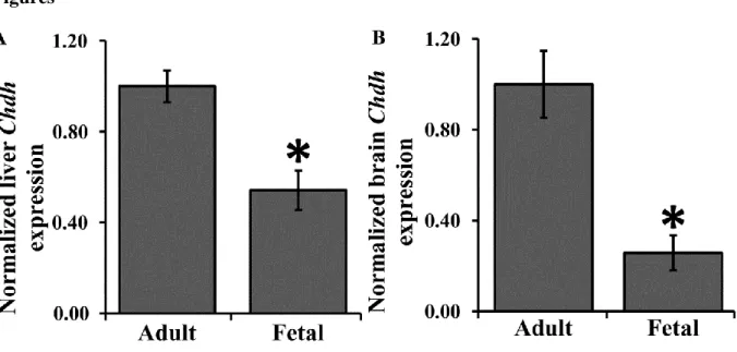
Chdh expression in fetal liver (Figure 1A) and brain (Figure 1B) of male wildtype mice was significantly lower than that of the same tissues in adult male wildtype mice. No Chdh was expressed in knockout mice. Results here verify that Chdh mRNA expression is present in fetal liver and brain.
Mitosis
14
neocortical region, Chdh+/+ (17.0 ± 2.6 H3ser10ph positive cells; n = 4) and Chdh-/- (13.1 ± 2.1 H3ser10ph positive cells; n = 6) (Figure 2C). P-value was greater than 0.05 for both analyses.
Ventricular length was measured and was not different between genotypes (hippocampal ventricular length Chdh+/+ 1295 ± 67 µm, n =4; Chdh-/- 1439 ± 92 µm, n =4; neocortical ventricular length Chdh+/+ 1487 ± 51 µm, n =4; Chdh-/- 1561 ± 93 µm, n =4; Figure 2D).
Sample size (n) for mitosis, apoptosis, and global DNA methylation analysis represents the number pregnant dams from which fetal samples were collected. Several Chdh+/+ and Chdh -/-samples were collected from the same dam.
Apoptosis
TUNEL analysis of apoptosis indicated cell death throughout the brain. There was no difference in numbers of TUNEL-positive cells in any of the 3 regions studied ((hippocampus, neocortex, or caudoputamen-amygdala; (Figure 3A-C). In the hippocampus region, Chdh+/+ and Chdh-/- had 5.2 ± 0.4 (n = 6) vs. 4.9 ± 0.9 (n = 6) apoptotic cells, respectively. In the neocortex region, Chdh+/+ and Chdh-/- had 9.3 ± 0.8 (n = 6) vs. 9.4 ± 1.7 (n = 6) apoptotic cells, respectively. In the caudoputamen and amygdala (CP-A) region, Chdh+/+ and Chdh-/- had 4.3 ± 0.8 (n = 6) vs. 5.5 ± 0.7 (n = 6) apoptotic cells, respectively. P-value was greater than 0.05 for all three analyses.
15
For mitosis and apoptosis data, Means and 95% confidence intervals for KO minus WT were computed for each measure and region from the mitosis and apoptosis data (Supplemental Table 1).
Discussion
Deletion of Chdh in mice did not significantly alter the founder population of progenitor cells during brain development when compared to Chdh wildtype in brain on day E17. In addition, in wildtype mice, we found that expression of Chdh in brain and liver was much lower in fetuses than in adult mice.
As discussed earlier, a potential epigenetic mechanism for the effects of choline on fetal brain neurogenesis and apoptosis has been suggested [5, 9, 43]. Because choline must be converted into betaine before it can serve as a methyl-donor [48], we expected that the effect of deleting this gene on fetal brain development would be similar to the effect of feeding a low choline diet to dams – decreased mitosis and increased apoptosis of neural progenitor cells in the developing E17 hippocampus. In future studies we plan to measure epigenetic marks on genes regulating cell cycle (i.e., Cdkn3) and histones with increased sample size and expect to see no difference by fetal genotype.
The Chdh mouse does not make betaine, and therefore accumulates more choline and the phosphorylated products of choline, including phosphatidylcholine, in tissues [23]. If the
16
apoptosis of neural progenitor cells in the developing E17 hippocampus. This was not the case. Analysis of Cdkn3 methylation and KAP in Chdh-/- fetal samples will provide a direct
comparison against the choline deficient model.
Several functional SNPs in the human CHDH gene have been identified that may alter choline metabolism [1, 53]. Studying a knockout mouse model provides insight into the
importance of the gene in multiple body systems, particularly the fetal brain in the present study. The current study has limitations. First, the mice model used is on a mixed 129/C57BL6
background. One study found inter-strain differences in mouse liver pathology when placed on a choline- and folate-deficient diet; C57BL/6J and 129S1/SvImJ were among the mouse strains used in the study [54]. Thus, it is possible that the mixed background of the mouse model may potentially mask differences. Secondly, we did not directly measure DNA and histone
methylation and therefore cannot be sure that deletion of Chdh does not alter epigenetic marks, especially on genes critical to cell cycle regulation. Finally, we use mothers that were
17 Figures
Figure 1. Chdh Expression
18 Figure 2. Mitosis in Chdh+/+ and Chdh-/- fetal brains
19 Figure 3. Apoptosis in Chdh+/+ and Chdh-/- fetal brains
20 Supplemental data
Supplemental Table 1. Difference between KO and WT for each measure along with the 95%
confidence interval
Measure Region P-value Mean 95% CI of KO-WT
Mitosis Hippocampus 0.92 0.4 (-8.4, 9.1)
Mitosis Neocortex 0.27 -3.9 (-11,5 , 3.7)
Mitosis Hippocampus Length 0.29 90.5 (-92.6 , 273.5)
Mitosis Neocortex Length 0.56 46.9 (-131.6, 225.4)
Apoptosis Hippocampus 0.93 -0.1 ( -2.3, 2.1)
Apoptosis Neocortex 0.73 0.7 ( -3.7, 5.1)
Apoptosis CP-A 0.22 1.5 ( -1.0, 4.0)
Apoptosis Hippocampus Length 0.47 89.3 (-177.6, 356.2)
Apoptosis Neocortex Length 0.90 15.3 (-256.5, 287.0)
Means and 95% confidence intervals for KO minus WT were computed for each measure and region from the mitosis and apoptosis data.
Region-specific Chdh expression in adult brain
Animal model and sample collection
21 Methods
RNA was extracted from frozen brain regions with RNeasy Protect Mini Kit (50) (Cat. No. 74124, Qiagen, Venlo, Netherlands) following manufacturer’s protocol. Complementary DNA was synthesized with Bio-Rad iScript™ Reverse Transcription Supermix (Cat. No. 170-8840, Hercules, CA, USA) following manufacturer’s protocol using an Eppendorf Mastercycler
epGradient (Hamburg, Germany) machine. Gene expression was assessed using quantitative real-time PCR (q-PCR). Materials used were Bio-Rad SsoFast™ Probes Supermix (Cat. No. 172-5230, Hercules, CA, USA), Applied Biosystems 20X mChdh assay (Mm00549261_m1), and 20X mGapdh assay (Mm99999915_g1, Applied Biosystems, NY, USA). Quantitative RT-PCR was performed on an Eppendorf Realplex2 Mastercycler epGradient S running Eppendorf Mastercycler ep realplex 2.2 (Hamburg, Germany). The whole brain samples were used to generate a standard curve with each qRT-PCR.
Data was normalized to Gapdh housekeeping gene for each brain region of each individual specimen were calculated following the Relative Standard Curve Method in the Applied Biosystems Guide to Performing Relative Quantitation of Gene Expression Using Real-Time Quantitative PCR1. Region-specific Chdh expression, statistical analysis was performed with REST 2009 software [46, 47]. A repeated measures ANOVA was used to compare the means of all six regions simultaneously and test for an overall difference. No significance was detected (P = 0.18).
Discussion
Since Chdh deletion resulted in no differences in fetal mitosis apoptosis we further questioned the role of choline in the more mature, complex adult brain. Neurogenesis begins
1
22
prenatally and continues throughout the adult life with limited neuron formation in the dentate gyrus postnatally [49]. Chdh deletion could prove to be more influential in adult neurogenesis because of the increased Chdh expression and activity in adult brain samples. Differential Chdh expression in different brain regions in the adult animal potentially illustrates the importance of betaine to the function to those areas. To determine if Chdh shows region-specific expression in the brain, adult brains of C57BL/6J mice (n = 4) approximately three months old were harvested and analyzed. ANOVA analysis showed no overall statistical difference across all six regions, but there seems to be a trend with lower Chdh expression in the dentate gyrus. This is pilot data and requires increased sample size for any definitive conclusions.
Supplemental Figure 1. Region-specific Chdh expression in adult mice brain
Chapter 3 – Conclusions
Conclusions
The role of choline as an essential nutrient has only been recently identified, and it has been recognized that the recommended AI for choline is different for different populations. The need for choline is modulated by a variety of factors, such as age, gender, pregnancy, and genetic SNPs. Research has shown that choline is important for prenatal development, but the role of fetal choline metabolism has yet to be established. More importantly, does deletion of a key gene in choline metabolism, Chdh, in the fetus effect measurements of prenatal brain development even if the mother is supplied adequate amounts of dietary choline?
In this study we focused on identifying changes in progenitor cell proliferation between Chdh+/+ and Chdh-/- fetuses harvested at E17 from Chdh+/- mating pairs with prenatal diets adequate in choline. We did not observe any significant changes in markers of mitosis and apoptosis between the wildtype and knockout Chdh fetal genotypes. In this section, we focus on more in-depth discussion of the study results, limitations, and suggestions for future studies.
Choline, Mitosis, and Apoptosis
24
nucleosome and critical for chromatin condensation maintenance during mitosis. This IHC method also enables visualization of different stages of mitosis based on cell shape and color intesnity. Mitotic cell counts along the hippocampal and neocortical ventricular zone of the fetal brain did not reveal any significant differences in progenitor cell proliferation between Chdh+/+ and Chdh-/- fetuses.
Apoptosis, as measured by fluorescent TUNEL IHC, also revealed no difference between number of apoptotic cells in Chdh+/+ and Chdh-/- fetuses. The TUNEL assay enzymatically labels fragmented ends of single and double stranded DNA allowing for fluorescent identification. In the beginning stages of apoptosis, the cell shrink, chromatin condense, and DNA fragmentation occurs. This is followed by membrane blebbing and nucleus fragmentation. In the final stages of apoptosis, the cell fragments in to apoptotic bodies and subsequently consumed by phagocytes[55]. TUNEL IHC is able to capture apoptosis at these different stages, resulting in clusters (early apoptosis) of fluorescent fragments or isolated fragments as they break off from the intact cell (late apoptosis).
25
supply affect the availability of SAM, the universal methyl donor, leading to aberrant methylation patterns subsequently affect progenitor cell proliferation. Alterations in brain development during critical periods may greatly affect brain function in later life.
Chdh Expression
Fetal mouse brains showed significantly reduced Chdh expression compared to adults. A study in a rat model showed that Chdh activity in the liver of the progeny does not substantially increased until well after birth and weaning. Clow et al. (2008) study demonstrated that liver Chdh activity steadily increased after birth and plateaued around 35-42 days post-birth. Kidney Chdh also gradually increased after birth. Choline dehydrogenase activity was not measured in the brain [57]. Thus, it can be speculated that much of the choline and betaine found in fetal tissues is a result of maternal diet and not endogenous synthesis by the fetus.
Limitations
Aside from the limitations presented in the discussion section of Chapter 2, there are several others to note. First, the effect of maternal diet was not assessed. The fetus relies heavily on the mother for its nutrition, and thus fetal development may reflect the mother’s ability to
26
choline dehydrogenase and epigenetic reprogramming. Last, we only analyzed progenitor cell proliferation in the fetal brain. The brain consists of multiple cell types (i.e., astrocytes, ogliodendrocytes) that contribute to brain development. Astrocytes are the most abundant cell type in the mammalian brain and contribute to structure and formation of the blood-brain barrier. Additionally, astrocytogenesis is influenced by epigenetic mechanisms [58]. It is also hypothesized that astrocytes regulate energy supply for neurons, and thus survival [59].
Future Studies
Results from the current study generate several questions that could prompt further studies. Here I briefly propose future studies to address these questions, particularly the role of methylation and epigenetic regulation on progenitor cell proliferation.
Proposed Study 1
27 Proposed Study 2
Epigenetic regulation has been implicated in choline deficient models and this proposed study will determine if differential DNA methylation is present in Chdh-/- fetuses compared to Chdh+/+ fetuses. Chdh+/- mating pairs will be maintained on diet AIN-76A with 1.1g/kg choline chloride to determine the role of the Chdh gene on DNA methylation. Fetal brains harvested from Chdh+/+ and Chdh-/- will be fixed, sectioned at 5 µm, and mounted on special membrane slides for laser-capture micro-dissection (LCM) of Ammon’s horn ventricular and subventricular zones and the dentate gyrus for bisulfite modification and pyrosequencing. One gene of interest in this study is Cdkn3, an important regulatory gene in mitosis, particularly the G1/S-phase transition[60]. DNA methylation changes between fetal genotypes will validate (or counter) results seen in choline deficiency during pregnancy. Global DNA methylation and Kap protein expression will be determine by immunohistochemistry. Methods for these analyses have been previously described [5].
Results here combined with those in the current study and Proposed Study 1 will help to elucidate the impact of fetal Chdh deletion on brain development.
Proposed Study 3
Maternal diet has been shown to influence mitosis, apoptosis, and DNA methylation. To elucidate the interaction of the gene Chdh and diet, it would be of interest to assess progenitor cell proliferation, apoptosis, and methylation in E17 brains of Chdh-/- fetuses from Chdh+/- dams under normal and low dietary choline during pregnancy. Methylation pattern of genes that
28
29 References
1. da Costa, K.A., et al., Common genetic polymorphisms affect the human requirement for the nutrient choline. FASEB J, 2006. 20(9): p. 1336-44.
2. Zeisel, S.H., Gene response elements, genetic polymorphisms and epigenetics influence the human dietary requirement for choline. IUBMB Life, 2007. 59(6): p. 380-7.
3. Cheatham, C.L., et al., Phosphatidylcholine supplementation in pregnant women consuming moderate-choline diets does not enhance infant cognitive function: a randomized, double-blind, placebo-controlled trial. Am J Clin Nutr, 2012. 96(6): p. 1465-72.
4. Craciunescu, C.N., et al., Choline availability during embryonic development alters progenitor cell mitosis in developing mouse hippocampus. J Nutr, 2003. 133(11): p. 3614-8.
5. Niculescu, M.D., C.N. Craciunescu, and S.H. Zeisel, Dietary choline deficiency alters global and gene-specific DNA methylation in the developing hippocampus of mouse fetal brains. FASEB J, 2006. 20(1): p. 43-9.
6. Signore, C., et al., Choline concentrations in human maternal and cord blood and intelligence at 5 y of age. Am J Clin Nutr, 2008. 87(4): p. 896-902.
7. Wu, B.T., et al., Early second trimester maternal plasma choline and betaine are related to measures of early cognitive development in term infants. PLoS One, 2012. 7(8): p. e43448.
8. Meck, W.H., R.A. Smith, and C.L. Williams, Pre- and postnatal choline supplementation produces long-term facilitation of spatial memory. Dev Psychobiol, 1988. 21(4): p. 339-53.
9. Niculescu, M.D., Y. Yamamuro, and S.H. Zeisel, Choline availability modulates human neuroblastoma cell proliferation and alters the methylation of the promoter region of the cyclin-dependent kinase inhibitor 3 gene. J Neurochem, 2004. 89(5): p. 1252-9.
10. Meck, W.H. and C.L. Williams, Simultaneous temporal processing is sensitive to prenatal choline availability in mature and aged rats. Neuroreport, 1997. 8(14): p. 3045-51.
11. Zeisel, S.H. and M.D. Niculescu, Perinatal choline influences brain structure and function. Nutr Rev, 2006. 64(4): p. 197-203.
30
13. Yates, A.A., S.A. Schlicker, and C.W. Suitor, Dietary Reference Intakes: the new basis for recommendations for calcium and related nutrients, B vitamins, and choline. J Am Diet Assoc, 1998. 98(6): p. 699-706.
14. Corbin, K.D. and S.H. Zeisel, Choline metabolism provides novel insights into nonalcoholic fatty liver disease and its progression. Curr Opin Gastroenterol, 2012. 28(2): p. 159-65.
15. Fischer, L.M., et al., Sex and menopausal status influence human dietary requirements for the nutrient choline. Am J Clin Nutr, 2007. 85(5): p. 1275-85.
16. Shaw, G.M., et al., Periconceptional dietary intake of choline and betaine and neural tube defects in offspring. Am J Epidemiol, 2004. 160(2): p. 102-9.
17. Resseguie, M.E., et al., Aberrant estrogen regulation of PEMT results in choline deficiency-associated liver dysfunction. J Biol Chem, 2011. 286(2): p. 1649-58.
18. Li, Z. and D.E. Vance, Phosphatidylcholine and choline homeostasis. J Lipid Res, 2008. 49(6): p. 1187-94.
19. da Costa, K.A., et al., Elevated serum creatine phosphokinase in choline-deficient
humans: mechanistic studies in C2C12 mouse myoblasts. Am J Clin Nutr, 2004. 80(1): p. 163-70.
20. Michel, V., R.K. Singh, and M. Bakovic, The impact of choline availability on muscle lipid metabolism. Food Funct, 2011. 2(1): p. 53-62.
21. Hasselmo, M.E., The role of acetylcholine in learning and memory. Curr Opin Neurobiol, 2006. 16(6): p. 710-5.
22. Gibellini, F. and T.K. Smith, The Kennedy pathway--De novo synthesis of
phosphatidylethanolamine and phosphatidylcholine. IUBMB Life, 2010. 62(6): p. 414-28.
23. Johnson, A.R., et al., Deletion of murine choline dehydrogenase results in diminished sperm motility. FASEB J, 2010. 24(8): p. 2752-61.
24. Mato, J.M., et al., S-adenosylmethionine synthesis: molecular mechanisms and clinical implications. Pharmacol Ther, 1997. 73(3): p. 265-80.
25. Garner, S.C., et al., Characterization of choline metabolism and secretion by human placental trophoblasts in culture. Biochim Biophys Acta, 1993. 1168(3): p. 358-64. 26. Sweiry, J.H. and D.L. Yudilevich, Characterization of choline transport at maternal and
31
27. Molloy, A.M., et al., Choline and homocysteine interrelations in umbilical cord and maternal plasma at delivery. Am J Clin Nutr, 2005. 82(4): p. 836-42.
28. Meck, W.H. and C.L. Williams, Metabolic imprinting of choline by its availability during gestation: implications for memory and attentional processing across the lifespan.
Neurosci Biobehav Rev, 2003. 27(4): p. 385-99.
29. Mehedint, M.G., C.N. Craciunescu, and S.H. Zeisel, Maternal dietary choline deficiency alters angiogenesis in fetal mouse hippocampus. Proc Natl Acad Sci U S A, 2010. 107(29): p. 12834-9.
30. Pogribny, I.P., et al., Epigenetic alterations in the brains of Fisher 344 rats induced by long-term administration of folate/methyl-deficient diet. Brain Res, 2008. 1237: p. 25-34. 31. Dupont, C., D.R. Armant, and C.A. Brenner, Epigenetics: definition, mechanisms and
clinical perspective. Semin Reprod Med, 2009. 27(5): p. 351-7.
32. Weinhold, B., Epigenetics: the science of change. Environ Health Perspect, 2006. 114(3): p. A160-7.
33. Berger, S.L., et al., An operational definition of epigenetics. Genes Dev, 2009. 23(7): p. 781-3.
34. Das, P.M. and R. Singal, DNA methylation and cancer. J Clin Oncol, 2004. 22(22): p. 4632-42.
35. Robertson, K.D. and P.A. Jones, DNA methylation: past, present and future directions. Carcinogenesis, 2000. 21(3): p. 461-7.
36. Guibert, S. and M. Weber, Functions of DNA methylation and hydroxymethylation in mammalian development. Curr Top Dev Biol, 2013. 104: p. 47-83.
37. Salbert, G. and M. Weber, Tracking genomic hydroxymethylation by the base. Nat Methods, 2012. 9(1): p. 45-6.
38. Zeisel, S.H., Dietary choline deficiency causes DNA strand breaks and alters epigenetic marks on DNA and histones. Mutat Res, 2012. 733(1-2): p. 34-8.
39. Zeisel, S.H., et al., Effect of choline deficiency on S-adenosylmethionine and methionine concentrations in rat liver. Biochem J, 1989. 259(3): p. 725-9.
32
41. Albright, C.D., et al., Maternal dietary choline availability alters mitosis, apoptosis and the localization of TOAD-64 protein in the developing fetal rat septum. Brain Res 1999. 115(2): p. 123-9.
42. Meck, W.H., R.A. Smith, and C.L. Williams, Pre- and postnatal choline supplementation produces long-term facilitation of spatial memory. Dev. Psychobiol., 1988. 21(4): p. 339-353.
43. Mehedint, M.G., et al., Choline deficiency alters global histone methylation and
epigenetic marking at the Re1 site of the calbindin 1 gene. FASEB J, 2010. 24(1): p. 184-95.
44. Livak, K.J. and T.D. Schmittgen, Analysis of relative gene expression data using real-time quantitative PCR and the 2(-Delta Delta C(T)) Method. Methods, 2001. 25(4): p. 402-8.
45. Schindelin, J., et al., Fiji: an open-source platform for biological-image analysis. Nat Methods, 2012. 9(7): p. 676-82.
46. Pfaffl, M.W., A new mathematical model for relative quantification in real-time RT-PCR. Nucleic Acids Res, 2001. 29(9): p. e45.
47. Pfaffl, M.W., G.W. Horgan, and L. Dempfle, Relative expression software tool (REST) for group-wise comparison and statistical analysis of relative expression results in real-time PCR. Nucleic Acids Res, 2002. 30(9): p. e36.
48. Zeisel, S.H., Choline: critical role during fetal development and dietary requirements in adults. Annu Rev Nutr, 2006. 26: p. 229-50.
49. Stiles, J. and T.L. Jernigan, The basics of brain development. Neuropsychol Rev, 2010. 20(4): p. 327-48.
50. Timmann, D. and I. Daum, Cerebellar contributions to cognitive functions: a progress report after two decades of research. Cerebellum, 2007. 6(3): p. 159-62.
51. Schmahmann, J.D. and D. Caplan, Cognition, emotion and the cerebellum. Brain, 2006. 129(Pt 2): p. 290-2.
52. Rochefort, C., J.M. Lefort, and L. Rondi-Reig, The cerebellum: a new key structure in the navigation system. Front Neural Circuits, 2013. 7: p. 35.
33
54. Tryndyak, V.P., et al., Plasma microRNAs are sensitive indicators of inter-strain differences in the severity of liver injury induced in mice by a choline- and folate-deficient diet. Toxicol Appl Pharmacol, 2012. 262(1): p. 52-9.
55. Guerin, M.B., et al., Retinal ganglion cells: dying to survive. Int J Dev Biol, 2006. 50(8): p. 665-74.
56. Friesen, R.W., et al., Relationship of dimethylglycine, choline, and betaine with oxoproline in plasma of pregnant women and their newborn infants. J Nutr, 2007. 137(12): p. 2641-6.
57. Clow, K.A., et al., Elevated tissue betaine contents in developing rats are due to dietary betaine, not to synthesis. J Nutr, 2008. 138(9): p. 1641-6.
58. Namihira, M. and K. Nakashima, Mechanisms of astrocytogenesis in the mammalian brain. Curr Opin Neurobiol, 2013.
59. Stobart, J.L. and C.M. Anderson, Multifunctional role of astrocytes as gatekeepers of neuronal energy supply. Front Cell Neurosci, 2013. 7: p. 38.