Mammalian Period represses and de-represses
transcription by displacing CLOCK
–
BMAL1 from
promoters in a Cryptochrome-dependent manner
Yi-Ying Chioua,1, Yanyan Yanga,1, Naim Rashidb,c,1, Rui Yea, Christopher P. Selbya, and Aziz Sancara,c,2
aDepartment of Biochemistry and Biophysics, University of North Carolina School of Medicine, Chapel Hill, NC 27599;bDepartment of Biostatistics, University of North Carolina, Chapel Hill, NC 27599; andcLineberger Comprehensive Cancer Center, University of North Carolina, Chapel Hill, NC 27599
Contributed by Aziz Sancar, August 17, 2016 (sent for review July 15, 2016; reviewed by Carl Hirschie Johnson and Andrew C. Liu)
The mammalian circadian clock is based on a transcription-translation feedback loop (TTFL) consolidated by secondary loops. In the primary TTFL, the circadian locomotor output cycles kaput (CLOCK)–brain and muscle Arnt-like protein-1 (BMAL1) heterodimer acts as the transcrip-tional activator, and Cryptochrome (CRY) and Period (PER) proteins function as repressors. PER represses by displacing CLOCK–BMAL1 from promoters in a CRY-dependent manner. Interestingly, genes with complex promoters may either be repressed or de-repressed by PER, depending on the particular promoter regulatory elements. Here, using mouse cell lines with defined knockout mutations in clock genes, RNA-seq, ChIP-seq, and reporter gene assays coupled with measurements of DNA–protein interactions in nuclear extracts, we elucidate the dual functions of PER as repressor and de-repressor in a context-dependent manner.
circadian
|
Period|
transcription|
activator|
DNA bindingT
he circadian clock is the molecular system that confers a∼24-h periodicity to many biological processes. In mammals, CLOCK, BMAL1, Cryptochrome (CRY), and Period (PER) proteins and their paralogs are essential for generating rhythmicity (1–4). Rhyth-micity is the product of a transcription-translation feedback loop (TTFL): The circadian locomotor output cycles kaput (CLOCK)– brain and muscle Arnt-like protein-1 (BMAL1) heterodimer binds to E-boxes in the promoters ofCry1, Cry2,Per1, andPer2genes and activates their transcription. The CRY and PER proteins, after a time lag, enter the nucleus and inhibit their own transcription (core clock circuit) (5–9), as well as transcription of other clock-controlled genes to maintain rhythmicity at the cellular and organismal levels. This core circuit is consolidated and stabilized by the nuclear re-ceptors NR1D1 and NR1D2 (REV-ERBα/β), which bind to the retinoic acid response elements (RREs) in some of the clock gene promoters (10–14). Although the canonical model has provided a useful conceptual framework, it has been insufficient in constructing a mechanistic model for the clock. This is in part due to the multiple interactions among clock proteins and to the lack of an in vitro sys-tem for analyzing the clock using purified proteins and their target promoter elements along with an appropriate readout.To simplify the analysis of clock protein functions, previously we reported the construction of cell lines that lack CRYs, PERs, and both CRYs and PERs and that express ectopic CRY or PER that can be targeted to the nucleus in a controllable manner (15). Using this system initially, we analyzed the canonical clock model by restricting our experiments to theNr1d1and D-site albumin promoter binding protein (Dbp) genes, which are controlled solely by the binding of CLOCK–BMAL1 to E-box regulatory elements. These experiments confirmed the repressor functions of CRY and PER but also revealed some features of their mechanisms: CRY alone can bind to the CLOCK–BMAL1–E-box complex and inhibit the transcription of cognate genes by forming a stable CRY–CLOCK–BMAL1 ternary complex in the promoter: “blocking-type” repression. In contrast, PER represses CLOCK–BMAL1–E-box-mediated transcription by removing the CLOCK–BMAL1 complex from the E-box in a CRY-dependent manner:“displacement-type”repression (15). Although
these findings clarified the roles of PERs and CRYs in regulation of simple promoters exclusively controlled by E-box binding of transcription factors, they did not address the issue of the control of key clock genes with multiple cis-elements.
Here, we report the construction of cell lines with clock gene knockout mutations and expression systems to analyze the mecha-nisms of control of more complex promoters by PER2 protein. We show that the removal of CLOCK–BMAL1 from promoters affects gene expression in three different ways, depending on the type of regulatory elements present in the target promoters. First, in the case of a simple promoter controlled exclusively by an E-box, such as in theNr1d1gene, CLOCK–BMAL1 removal represses transcription. Second, in genes such asBmal1, which is controlled exclusively by an RRE, CLOCK–BMAL1 removal from theNr1d1promoter indirectly activates transcription of Bmal1by down-regulation of NR1D1/2, which represses by binding to the RRE in the Bmal1 promoter. Third, in the case of theCry1gene, removal of CLOCK–BMAL1 facilitates transactivation by other transcription factors that bind to theCry1promoter independently of NR1D1/2.
Results
Experimental System to Analyze the Role of PER in the TTFL.In Fig. 1 we present a highly idealized model for the mammalian circadian clock: The CLOCK–BMAL1 heterodimer activates transcription of
Cry1/2,Per1/2, andNr1d1/2by binding to E-box (CACGTG/T) se-quences in the promoters of these genes. The CRY and PER
Significance
The mammalian circadian clock is controlled by a transcription-translation feedback loop consisting of transcriptional activators circadian locomotor output cycles kaput (CLOCK)–brain and mus-cle Arnt-like protein-1 (BMAL1), which function as a complex at E/E’-box elements, and repressors Cryptochrome 1 (CRY1)/CRY2 and PER1/PER2. CRYs repress upon binding as CRY–CLOCK–
BMAL1–E-box complexes. Period proteins (PERs) repress by re-moving the heterotrimeric complexes from the E-box. We report here that in theCry1promoter, the CRY1–CLOCK–BMAL1–E-box complex represses a transcriptional activator acting in cis, and removal of the heterotrimeric complex by PER2 de-represses the transcriptional activator. ChIP-seq and RNA-seq experi-ments identified other genes also de-repressed by PER2. These data clarify the role of PER2 and reveal the level of complexity in regulation ofCry1and other circadian-controlled genes.
Author contributions: Y.-Y.C., R.Y., and A.S. designed research; Y.-Y.C., Y.Y., and R.Y. performed research; Y.-Y.C., Y.Y., and N.R. analyzed data; and C.P.S. and A.S. wrote the paper.
Reviewers: C.H.J., Vanderbilt University; and A.C.L., University of Memphis.
The authors declare no conflict of interest.
1Y.-Y.C., Y.Y., and N.R. contributed equally to this work.
2To whom correspondence should be addressed. Email: [email protected].
proteins inhibit transcriptional activation by CLOCK–BMAL1 to close the feedback loop, CRY by binding to the CLOCK– BMAL1–E-box and interfering with transactivation, and PER by causing the displacement of CLOCK–BMAL1 from E-boxes in a CRY-dependent manner. This primary circuit is consolidated by a stabilizing loop: TheNr1d1/2nuclear receptor genes are also con-trolled by CLOCK–BMAL1, and NR1D1/2 proteins in turn repress
Bmal1andCry1transcription by binding to the RRE elements in the promoters of these genes (12–14, 16, 17). Some observations, such as the repressor activity of CLOCK–BMAL1 (18, 19) and the apparent transcriptional up-regulation by PER (20–23), do not readily fit into this model and require further experimentation.
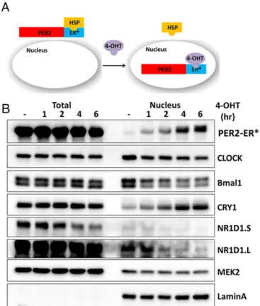
Because there is no in vitro system to analyze the roles of different clock proteins, individually or in various combinations, on transcrip-tion of cognate genes, we developed cell lines with various clock gene knockout mutations (Table S1) and with the controllable PER2 nu-clear entry system shown in Fig. 2Afor carrying out“in cellulo bio-chemistry”for better insight into the clock mechanism. These cell lines were isolated from knockout mouse strains or from mouse embryonic fibroblasts by using transcription activator-like effector nuclease (TALEN) and CRISPR technologies. The cell lines also ectopically express PER2-ER*, which is composed of PER2 protein fused to a mutant form of the ligand-binding domain of the es-trogen receptor (ER*). PER2-ER* is trapped in the cytoplasm, and it enters the nucleus upon 4-hydroxytamoxifen (4-OHT) treatment. Thus, the effect of PER2 on gene expression can be analyzed in a controllable manner as shown in Fig. 2B. As previously reported (15), PER2 and CRY1 enter the nucleus together in this system.
PER2 as a Repressor and Antirepressor.We addressed the regulatory roles of PER using a PER2-ER* ectopic expression system in cells with various clock gene knockout mutations. We analyzed tran-scription of three genes selected for their different promoter complexities, and reported differences in their regulation by PER2 (9–13). The plots in Fig. 3Ashow the levels of mRNA expressed
from theNr1d1,Bmal1, andCry1genes inPer1/2−/−cells following 0–6 h of treatment with 4-OHT, which induces translocation of PER2-ER* to the nucleus. The Nr1d1 promoter is controlled solely by an E-box, theBmal1promoter is controlled solely by the RRE element, and theCry1promoter is controlled by an E-box, an RRE element, and a D-box (14). As expected (15), the data in Fig. 3A (PER2-ER* enters the nucleus of Per1/2−/−cells) show that PER2 inhibits transcription of Nr1d1 (by removing CLOCK– BMAL1 from the E-box; Fig. 3A,Left, red); however, PER2 also induces transcription ofBmal1andCry1(Fig. 3A,MiddleandRight, red). Because PER2 inhibits the expression ofNr1d1and because both Bmal1andCry1 have RRE elements in their promoters to which NR1D1/2 binds (12–14, 16, 24), it has been suggested that PER2 de-represses genes by down-regulating the NR1D1 repressor. To test this prediction, we investigated the effect ofNr1d1/2 dele-tions on PER2-ER*–mediated up-regulation of Bmal1 and Cry1
transcription. In the absence of NR1D1/2, there is no up-regulation ofBmal1transcription by PER2-ER* (Fig. 3A,Middle, blue), sup-porting the view that PER2 up-regulatesBmal1by down-regulating NR1D1/2. In contrast, and surprisingly, the NR1D1/2 deletions have no effect on PER2-mediated up-regulation ofCry1(Fig. 3A,
Right, blue). This agrees with the observation that NR1D1/2 is dispensable for the rhythmic expression ofCry1(12) and indicates Fig. 1. TTFL model for the mammalian circadian clock. In the TTFL model,
CLOCK–BMAL1 heterodimers bind to the E-boxes of Cry1/2,Per1/2, and
Nr1d1/2promoters and activate the transcription of these genes. Increased CRY protein then inhibits transcription through binding to the CLOCK– BMAL1 complex. PER protein inhibits transcription in a CRY-dependent manner by reducing CLOCK–BMAL1 binding to the E-box. This core circuit (solid lines) is stabilized by a secondary feedback loop (dashed lines) in which CLOCK–BMAL1-controlled NR1D1/2 inhibits the transcription ofBmal1and
Cry1through binding to the RRE of their promoters.
Fig. 2. Cell-based system for study of mammalian clock regulation. (A) Sche-matic of the experimental system. PER2 fused to a mutant form of the estrogen receptor ligand-binding domain (ER*) is constrained to the cytosol when bound to endogenous heat-shock protein (HSP). Addition of 4-OHT to the cell culture displaces the HSP, and PER2-ER* then enters the nucleus. (B) Immunoblot analysis of clock proteins upon 4-OHT addition. Total and nuclear amounts of clock and control proteins (MEK2 and LaminA, which is a nuclear protein) up to 6 h fol-lowing addition of 4-OHT toPer1/2−/−;PER2-ER* cells are shown. The blot shows that the total NR1D1 protein level (and thus nuclear NR1D1) decreases following incubation with 4-OHT. An increased level of nuclear CRY1 but not total CRY1 is associated with the increased nuclear PER2. This supports the idea that CRY and PER enter the nucleus together and that CRY-dependent PER2 activity is through the formation of a CRY–PER complex. NR1D1.s and NR1D1.L are short and long exposures of the same blot.
BIO
CHEMISTRY
PNAS
that PER2 up-regulatesCry1 transcription by a mechanism inde-pendent of NR1D1/2.
PER2 Repressor and Antirepressor Activities Are BMAL1- and CRY-Dependent. Based on indirect evidence, it has been previously reported that PER2 can up-regulate gene transcription by interacting with another transcription factor (25) or by removing CRY from CLOCK–BMAL1 (20). We have shown that PER2 represses tran-scription by removing the CLOCK–BMAL1 complex from cognate promoters in a CRY-dependent manner (15). We wished to know if the transcriptional up-regulation effect of PER was also mediated through removal of the CLOCK–BMAL1–CRY complex. To this end, we tested the effect of PER2-ER* nuclear entry on transcription of the three sentinel genes in cells lacking BMAL1. In theseBmal1
knockout cells, endogenous PER2 is undetectable (4), and thus any effect that might be seen upon 4-OHT addition can be ascribed to PER2-ER*. The results (Fig. 3A, orange) show that in the absence of BMAL1 there is no repression or de-repression of these genes upon nuclear entry of PER2-ER*. This excluded the possibility that PER2 facilitates a transcriptional activator in a BMAL1-independent manner to activateCry1transcription. To examine whether PER2-induced up-regulation is dependent on CRY proteins, we tested the effect of PER2-ER* on expression of all three sentinel genes in cells lacking CRY1/2. As seen in Fig. 3A, the effect of PER2 on all these three genes (red) is abolished in the Cry1/2knockout background
(green), indicating that PER2 exerts its up-regulation activity only through a CRY-mediated mechanism as it does its repressor activity.
Gene Up-Regulation by PER2 Is Associated with Removal of CLOCK–
BMAL1. We then used ChIP to directly examine the binding of BMAL1 to the sentinel gene promoters in response to PER2. In Fig. 3B,Middle, there is no BMAL1 binding to theBmal1promoter, as it lacks an E-box (1, 2, 4). Fig. 3B,Leftshows that, as expected, nuclear entry of PER2 following 4-OHT treatment causes displacement of BMAL1 from the (PER2-repressed)Nr1d1promoter (red), and this displacement is NR1D1/2-independent (blue) but CRY-dependent (green). Importantly, the positive regulation of the (PER2-activated)
Cry1promoter shown in Fig. 3A, Right coincides with NR1D1/2-independent but CRY-dependent displacement of BMAL1 (Fig. 3B,Right). Taken together, these data indicate that BMAL1 and CRY are required for both PER-mediated transcriptional repres-sion and up-regulation in a context-dependent manner. Indeed, the idea of repression by CRY in a CLOCK–BMAL1-dependent manner was suggested in a previous study, although there was no direct experimental evidence (19). We suggest that in theCry1
promoter, CLOCK–BMAL1 binding to an E-box represses another activator, which is the predominant transcription activator for this gene. Here, we show that the PER2-mediated up-regulation ofCry1
gene transcription is accompanied with the removal of the CLOCK– BMAL1–CRY complex from theCry1promoter and thus present Fig. 3. Effect of PER2 nuclear entry on transcription ofNr1d1,Bmal1, andCry1inPer1/2 ,Per1/2 ;Nr1d1/2 ,Per1/2 ;Cry1/2 , andBmal1 cells. (A) Nuclear entry of PER2-ER* following addition of 4-OHT (red) repressesNr1d1transcription (Left) but inducesBmal1(Middle) andCry1(Right) transcription analyzed by RT-quantitative PCR. The effect of PER2 on these three genes is CRY-dependent (green). NR1D1/2 is required for PER2-inducedBmal1transcription but is not required for the repression ofNr1d1or for the induction ofCry1transcription (blue). InBmal1−/−cells (orange), nuclear entry of PER2-ER* has no effect onNr1d1,Bmal1, or
direct evidence for these counterintuitive roles of CLOCK–BMAL1 in gene repression and PER2 (with CRY) in gene de-repression.
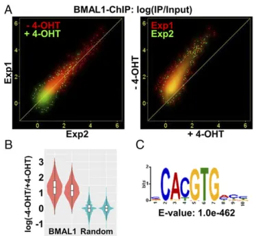
Genome-Wide Effect of PER on Regulation and Promoter Binding by the CLOCK–BMAL1 Complex.The model that emerges from the results presented here as well as our previous studies is that PER displaces CLOCK–BMAL1 from cognate gene promoters and in doing so, depending on the complement of regulatory elements in the target promoter, it either inhibits or up-regulates (de-represses) transcrip-tion in a CRY-dependent manner (15). Because there are several thousands of CLOCK–BMAL1 binding sites in the genome (26, 27), we wished to find out whether PER-mediated CLOCK–BMAL1 removal and both the repression and de-repression functions of PER are observed on a global scale. To this end, we performed ChIP-seq experiments using an anti-BMAL1 antibody to probe CLOCK– BMAL1 binding sites and RNA-seq experiments to analyze RNA levels before and after nuclear entry of PER2 inPer1/2−/−;Nr1d1/2−/−; PER2-ER* cells so as to avoid complications arising from second-order clock-controlled genes regulated by NR1D1/2 (such asBmal1). ChIP-seq experiments were conducted in duplicate with cells not treated with 4-OHT. Specific BMAL1 binding sites were identified based on 10–15 million input and precipitated DNA reads (Materials and Methods). There were 4,789 and 8,615 binding sites in the two experiments and 4,740 common binding sites seen in both experi-ments. Induction of PER2 with 4-OHT reduced the number of common BMAL1 binding sites to 483, and PER2 induction pro-duced no novel BMAL1 binding sites. Thus, PER2 eliminated de-tectable binding of BMAL1 to most of the regulatory regions to which it binds. To further assess BMAL1 binding, we analyzed reads mapped to the 4,740 common BMAL1 binding sites as relative BMAL1 binding strength, expressed as“IP/input.”We observed that relative BMAL1 binding strength was negatively associated with PER2 induction and was highly correlated between the two inde-pendent experiments (Fig. 4A,Left). BMAL1 binding was stronger without 4-OHT than with 4-OHT on almost all regions (Fig. 4A,
Right). To determine whether the observed decrease in BMAL1 binding was due to experiment-specific technical factors such as se-quencing depth, we compared binding differences from these BMAL1 binding sites to reads randomly selected from the genome. Relative to the BMAL1 binding sites, there is no difference of BMAL1 binding between without 4-OHT and with 4-OHT in the random areas (Fig. 4B). Results of motif analysis of the BMAL1 binding sites showed high enrichment of the E-box sequence (Fig. 4C), which further supports the finding that PER2 removes CLOCK–BMAL1 from the E-box sequence of the promoters.
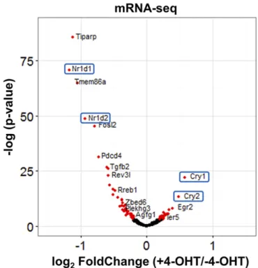
To analyze the effect of PER2 on CLOCK–BMAL1-controlled gene expression, we first defined CLOCK–BMAL1-controlled genes as genes with transcription start sites within ±5 kb from BMAL1 binding sites. Based on our ChIP-seq data, 3,317 genes were selected for the analysis. As apparent from the volcano plot in Fig. 5, nuclear entry of PER2-ER* changes the expression pattern, with 53 transcripts going down, 28 going up, and most remaining unchanged (Table S2). Interestingly,Cry1andCry2are increased andNr1d1andNr1d2are decreased when PER2 enters the nucleus. To ascertain the quality of the data from BMAL1 ChIP-seq and RNA-seq, in Fig. 6 we focus on
Cry1and Nr1d1 loci, on chromosomes 10 and 11, respectively: In agreement with the RT-quantitative PCR and ChIP-quantitative PCR data in Fig. 3, nuclear entry of PER2 up-regulatesCry1expression and represses theNr1d1transcript level and both are associated with a decrease in BMAL1 binding on the promoter. Similarly, increased
Cry2,Dec1(Bhlhe40), and decreasedNr1d2transcription (Table S2) are confirmed by RT-quantitative PCR (Fig. S1).
Decreased CLOCK–BMAL1 Binding on the Cry1 Promoter Increases
Cry1Transcription.Finally, the experiments described so far were carried out with 4-OHT–induced nuclear entry of PER2-ER*, and it could be argued that in this model system, the observed removal of CLOCK–BMAL1 from promoters and the accompanying
up-regulation ofCry1might be due to some unique feature of our system and that the CLOCK–BMAL1 removal from the E-box upon PER2-ER* nuclear entry andCry1up-regulation might not be physiologically relevant. To address this issue, we conducted reporter gene assays using a 1.5 kbp promoter fragment ofCry1, which can sustain rhythmic transcription (14). This fragment pos-sesses two D-box and two E-box sequences that partially overlap, as shown in Fig. 7A. To characterize the effect of CLOCK–BMAL1 binding onCry1transcription, we first compared the reporter ac-tivity of Cry1 promoters with either wild-type (WT) or mutant E-box (mutE) sequences (Fig. 7A). In a preliminary control exper-iment to ascertain the specificity of CLOCK–BMAL1 binding to the E-box in theCry1promoter, the two 1.5 kbp promoter fragments, end-labeled with biotin, were incubated with NIH 3T3 cell-free extract. The fragments were pulled down and the bound BMAL1 was visualized in the Western blot shown in Fig. 7B. BMAL1 bound to the WT promoter but only weakly to the mutant promoter.
Reporter assays were then performed following cotransfection of theCry1promoter/reporter constructs with and without PER2 and/or CRY1-expressing plasmids, which is a commonly used circadian experimental system. Fig. 7C, lane 1 shows that in the absence of exogenous PER2 or CRY1, there was much more expression from the mutE (green) than the WT (red) promoter. This increase is consistent with strong transcription from another element on the 1.5 kbp fragment that is inhibited by CLOCK– BMAL1–CRY1 binding to the WT E-box. Expression of PER2 led to substantially increased transcription from the WT promoter fragment (lanes 2 and 3). Apparently the basal level of CRY1/2 in these cells is sufficient to enable the removal of CLOCK–BMAL1 Fig. 4. Effect of PER2 nuclear entry on BMAL1 promoter binding by BMAL1– ChIP-Seq inPer1/2−/−;Nr1d1/2−/−cells. (A) Strength of BMAL1 binding (ChIP/Input) to 4,740 common binding sites is shown as a scatter plot. (Left) Results with (green) and without (red) 4-OHT show high correlation between the two ex-periments. The results obtained with 4-OHT are closest to the origin, indicating weak or no binding of BMAL1 following PER2 induction. (Right) The strength of BMAL1 binding in the condition with (xaxis) and without (yaxis) 4-OHT is plotted. The distributions largely overlap in the two experiments and are above the line representingy=x, indicating reduced BMAL1 binding following PER2 induction. (B) Distribution of relative strength of BMAL1 binding–4-OHT/+4-OHT was plotted by violin and box plot. Stronger binding of BMAL1 without 4-OHT was detected on the BMAL1 binding sites (red) but not with reads randomly selected from the genome (blue). (C) Result from motif analysis of BMAL1 binding sites shows strong enrichment of the E-box sequence.
BIO
CHEMISTRY
PNAS
by exogenous PER2 so as to reveal strong transcription. A modest increase in transcription of the mutE promoter by exogenous PER2 is consistent with the removal of residual CLOCK–BMAL1 bound weakly to the mutant promoter (green, lane 1 vs. lanes 2 and 3). Comparing lane 4 versus lane 1 (red) shows that exogenous CRY1 inhibited transcription mediated by CLOCK–BMAL1. Increasing the amount of exogenous PER2, in the presence of exogenous CRY1 (lanes 5–8), resulted in increased transcription, as PER2 removed CLOCK–BMAL1 from the E-boxes in a CRY1-dependent manner. These data from cellular oscillators support our conclusion that up-regulation ofCry1by PER2 is not simply due to removal of CRY1 to facilitate CLOCK–BMAL1-mediated transcription but in fact is due to the removal of the entire CRY1–CLOCK–BMAL1
“repressor complex”to facilitate the activation of transcription by another sequence element in the 1.5 kbpCry1promoter region.
It has been shown thatCry1 transcription could be regulated through the D-box in the promoter (14). It is possible that removal of CLOCK–BMAL1 from the E-boxes by PER facilitates the transcription ofCry1through the nearby and overlapping D-boxes (Fig. 7A). We considered that D-box binding proteins, such as DBP, TEF, and HLF, would activate the transcription ofCry1when not blocked by the binding of CLOCK–BMAL1–CRY to the E-boxes. To test this, we measured the reporter activity of aCry1promoter with mutated D-box and E-box (mutDE, Fig. 7A). As seen with the mutE promoter, mutDE promoter activity in the absence of exog-enous PER2 or CRY1 was much higher than WT (lane 1, blue) and furthermore was very similar to mutE in all conditions tested. Thus, the increase inCry1transcription mediated by PER as a result of CLOCK–BMAL1 dissociation is not by removal of a repressor complex interfering with D-box–mediated activation. It is most likely that another transcriptional activator with a binding site in the 1.5 kbp Cry1promoter fragment is responsible for increased
Cry1expression following removal of CLOCK–BMAL1. For pur-poses of discussion, this possible transcription factor will be referred
to as factor T, which binds to a postulated T element in theCry1
promoter (Fig. 8).
Discussion
The mammalian molecular circadian clock consists of a TTFL generated by four genes (core clock genes) and their paralogs. Research over the past two decades has supported the TTFL model and identified a secondary loop involving nuclear recep-torsNr1d1/2. Furthermore, identification of kinases (28, 29) and ubiquitin ligases (30, 31) that modulate the activity and stability of the core clock proteins has provided insights into the mech-anisms that are necessary to establish a daily rhythm of high amplitude, precise period, and flexibility for phase resetting in response to stimuli.
Despite these important developments, the negative arm of the TTFL model has remained poorly defined. In particular, the re-spective roles of CRY and PER in repression have been unclear. There are several reasons for this uncertainty. First, the two pro-teins interact strongly and aid in one another’s stability and nuclear entry (32–34). Second, removal of one affects the posttranslational modification and stability of the other. Also, although CRY has been shown to be capable of repressing target genes on its own, there is no convincing evidence that PER affects the TTFL in the absence of CRY (15). Moreover, depending on the clock gene tested, PER has been reported to function as either a repressor (for
Nr1d1) or an up-regulator (forCry1andBmal1).
To examine the clock model, we have developed an“in cellulo biochemical”system consisting of mouse cell lines with knockout mutations in one or more of the core clock genes and that have a controllable delivery system for CRY or PER proteins (15). In Fig. 5. Effect of PER2 nuclear entry on transcriptome by mRNA-Seq inPer1/
2−/−;Nr1d1/2−/−cells. Effect of PER2 nuclear entry on transcription of indi-vidual genes is shown by Volcano plot with thexaxis representing level of difference (+4-OHT vs.–4-OHT) and they axis representing the level of statistical significance. Genes with a statistically significant difference be-tween with and without 4-OHT are shown in red.
Fig. 6. Visualization of BMAL1–ChIP-seq and RNA-seq onNr1d1(Top) andCry1
(Bottom) gene loci. (Top) An 11 kbp region of chromosome 11 containing the
Nr1d1gene. (Bottom) A 57 kbp region of chromosome 10 containing theCry1
this study, using the PER2 delivery system, we have obtained RT-quantitative PCR, RNA-seq, ChIP, and ChIP-seq data that have enabled us to revise the consensus mammalian clock model so as to reconcile various views regarding the roles of CRY and PER in the negative arm of the TTFL and eliminate the“internal inconsistencies”of the conventional model (3).
We previously reported that PER causes the removal of CLOCK–BMAL1 from the cognate promoters in a CRY-dependent manner and that it inhibits the transcription of certain genes with simple promoters (promoters with only an E-box control element) by this mechanism. In this study, we investigated the effect of PER2 on transcription of genes with more complex promoters. In aggregate, our work shows that PER2 exerts three types of effect on clock gene transcription, depending upon the type of promoter. Furthermore, we show that all of these effects are CRY-dependent, and moreover our data reconcile some previous seemingly contradictory reports on PER function in the circadian clock. We explain the three different effects of PER2 on circadian gene expression as follows:
i) Nr1d1(repressed by PER2): This gene is predominantly regu-lated by an E-box. Binding of CRY1 (or CRY2) to the CLOCK– BMAL1–E-box complex represses transcription. When PER2 is abundant enough, it removes the CRY1–CLOCK–BMAL1 complex from the promoter as part of the dual repression mechanism of CRY and PER (15).
ii) Bmal1 (de-repressed by PER2): This gene is predominantly regulated by NR1D1/NR1D2 nuclear receptors; CRY1/CRY2 de-repress its transcription. We show that de-repression is
through the effects of CRY1/2 and PER1/2 on NR1D1/ NR1D2 expression. When CRY and PER are at high enough concentrations, they inhibit NR1D1/NR1D2 expression through the E-box and thus down-regulate the repressors of
Bmal1causing up-regulation ofBmal1transcription. Knockout of Nr1d1/Nr1d2eliminates the effect of CRYs and PERs on
Bmal1transcription, which is mediated by NR1D1/NR1D2.
iii) Cry1(de-repressed by PER2): This gene has several transcrip-tional elements, three of which have been studied previously— D-box (day element), E-box (morning element), and RRE (evening element)—which are the targets of transcription fac-tors DBP, CLOCK–BMAL1, and NR1D1/NR1D2, respectively (14). Up-regulation (de-repression) of the Cry1promoter by PER2 seen in our experiments is not consistent with the ac-cepted role of PER2 as a repressor. Kondratov et al. (19) and Liu et al. (12) observed related, unexpected results, namely the up-regulation ofCry1inBmal1knockout cells. Kondratov et al. (19) suggested that with some genes, such asCry1, there is a high“basal”level of transcription that is not substantially in-creased by CLOCK–BMAL1 binding to the promoter, and binding of CRY1 to the CLOCK–BMAL1 complex inhibits both CLOCK–BMAL1 activated and basal transcription. In this vein, our findings are consistent with the existence of an additional transcriptional activator (factor T) that binds to a regulatory element (T element) in theCry1promoter (12) and Fig. 7. Decreased CLOCK–BMAL1 binding increases reporter gene
expres-sion driven from theCry1promoter. (A) Sequences of theCry1promoter region including WT, mutant E-boxes, and D-boxes are shown. Nucleotides conserved in the E-box and D-box sequences are colored in green and blue, respectively. Nucleotides mutated in mutE (E-box mutation) and mutDE (D-box and E-box mutation) are colored in red. (B) CLOCK–BMAL1 binding affinity toCry1promoters with WT or mutE sequences. BMAL1 was pulled down from nuclear extract from NIH 3T3 cells by immobilized biotin–DNA and analyzed by Western blot. (C) Reporter gene assay of theCry1 pro-moters with the WT or mutant promoter elements is shown. Reporter signals were first normalized to the signals from cotransfected control plasmid and then were normalized to the condition withoutCry1andPer2plasmids. Note that reporter gene expression driven by the mutantCry1promoter is higher than expression driven by the WTCry1promoter. SEs are plotted inC.
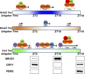
Fig. 8. Model of PER-mediated regulation ofNr1d1,Bmal1, andCry1genes. This model represents transcription and regulation patterns of the Nr1d1,
Bmal1, andCry1genes individually as a function of the circadian time. Gradient-colored bars represent transcription ofNr1d1 (blue),Bmal1(orange), and
Cry1(green) genes based on nascent RNA-seq data from mouse liver (27). Above the bars are the proposed status of promoter occupation and tran-scription. Spacing of the T element, E-box, and RRE in this representation of theCry1promoter is for illustrative purposes only. The RRE element is in intron 1 ofCry1∼23 kbp downstream from the E-box. At the bottom of the model are the levels of NR1D1, CRY1, and PER2 in mouse liver nuclei. The levels of these and additional proteins from different times were cropped from a single image (Fig. S2). ForNr1d1, which is controlled solely by an E-box, transcription is inhibited by either binding of CRY on CLOCK–BMAL1 or removal of CRY1–CLOCK–BMAL1 from the E-box by PER. ForBmal1, which is controlled solely by an RRE, transcription is inversely correlated with the NR1D1 level. ForCry1, which is controlled by an E-box, RRE, and a putative factor T, binding of either the CRY–CLOCK–BMAL1 complex on the E-box or NR1D1 on the RRE represses the transcription ofCry1from another element (T). PER eliminates both pathways of inhibition by removing CLOCK–BMAL1 from the E-box.
BIO
CHEMISTRY
PNAS
is inhibited by the CRY1–CLOCK–BMAL1 complex. In this scenario, removal of CRY1-CLOCK-BMAL1 from the Cry1
promoter by PER2 restores transcription via factor T.
Regarding the elevatedCry1level in theBmal1knockout cells, Liu et al. (12) hypothesized that the absence of BMAL1 led to less NR1D1/NR1D2, which normally binds to the RRE element and represses transcription ofCry1. In fact, in their experiments, overexpression of NR1D1 in Bmal1 knockout cells did inhibit
Cry1expression. Thus, their interpretation of BMAL1 indirectly inhibitingCry1expression through NR1D1/NR1D2 appears ap-propriate to their defined experimental system, and it is reasonable to expect that NR1D1/NR1D2 influence Cry1expression in vivo. However, our cell-based (Fig. 3) and reporter assay (Fig. 7) data show that PER2-mediated expression of Cry1is independent of NR1D1 (our reporter lacked the NR1D1 binding site). These seemingly conflicting results are reconciled and incorporated in the revised clock model shown in Fig. 8, which also shows levels of NR1D1, CRY1, and PER2 proteins in mouse liver at various cir-cadian times. At ZT10, NR1D1 is high and it likely repressesCry1
by binding to the RRE element, whereas at ZT18 NR1D1 is low, and this likely contributes to initiateCry1expression. Also, at ZT18, PER2 is high and PER2 at this time likely helps initiate Cry1
transcription by removing the inhibitory CRY1–CLOCK–BMAL1 complex. Thus, in this model, PER2 has dual roles in regulation of
Cry1. First, through displacement of CRY–CLOCK–BMAL1 from theNr1d1/Nr1d2promoters, it represses NR1D1/NR1D2 and thus indirectly activatesCry1expression. Second, through displacement of CRY–CLOCK–BMAL1 from theCry1promoter, it directly de-repressesCry1expression.
We note that our findings contradict a recent report (20) that claimed that PER2 up-regulatesCry1transcription by removing CRY1 from the CRY1–CLOCK–BMAL1 complex at theCry1
E-box and at the E-boxes of other genes regulated by CLOCK– BMAL1. Here, we confirm our earlier finding (15) that in fact PER2 nuclear entry causes the removal of CLOCK–BMAL1 from theCry1promoter in a CRY-dependent manner while at the same time up-regulatingCry1transcription.
Because the D-box in theCry1promoter has been shown to play a role inCry1regulation, we considered transcription factors (DBP, TEF, and HLF) that bind to the D-box as potentialCry1
transcriptional activators when PER2 removes the CRY–CLOCK– BMAL1 complex from theCry1promoter. We tested this possibility by using reporter gene assays with a 1.5 kbp fragment carrying the
Cry1 promoter including the D-box and E-box elements. As expected, in this system, CRY1 repressed and PER2 together with CRY1 de-repressed transcription ofCry1. MutE and double mutant E-box/D-box constructs, with greatly reduced CLOCK–BMAL binding, demonstrated reduced transcription stimulation following PER2 induction. Thus, DBP cannot be factor T whose activity is revealed by dissociation of the CRY1–CLOCK–BMAL1 complex. Additional studies are needed to identify the postulated T element and T factor that acts in a tonic manner (12) to regulate Cry1
transcription and whose effect is rendered circadian in the context of the entire circadian ensemble.
Materials and Methods
Cells and Antibodies. Cell lines (Table S1) were maintained in DMEM supplemented with 10% (vol/vol) FBS in 5% (vol/vol) CO2at 37 °C. Blasticidin (5μg/mL) was present during cell culture and absent during experiments. To induce PER2-ER* nuclear entry, 4-hydoxytamoxifen was added to the medium to 1μM when cells were∼90% confluent.
TheBmal1−/−,Per1/2−/−, andCry1/2−/−;Per1/2−/−mouse embryonic fibro-blasts have been described (15, 32). ThePer1/2−/−;Nr1d1/2−/-mouse embry-onic fibroblast cell line was made by CRISPR technology using LentiCRISPRv2 (35) obtained from Addgene to mutate theNr1d1/2alleles inPer1/2−/− fi-broblasts. Single colonies lacking NR1D1/2 proteins were isolated and screened by Western blot, and mutational inactivation was confirmed by genomic DNA sequencing. Mutated sequences in the isolated clone (PN-P2ER)
are shown inFig. S3. Lack of NR1D1/2 protein and movement of PER2-ER* to the nucleus are shown inFig. S4.
DNA containing mPER2-ER* was subcloned from pBABE-puro-mPER2-ER* into plasmid pWZL-blast to create pWZL-blast-mPER2-ER*. Cells stably expressing P-P2ER, PN-P2ER, and B-P2ER were made by retrovirus infection using pWZL-blast-mPER2-ER* (15).
Anti-mCRY1 (IgM-type monoclonal) antibodies were described previously (18). Anti-CLOCK (Bethyl Laboratories), anti-BMAL1 (Bethyl Laboratories), anti-CRY2 (Bethyl Laboratories), anti-PER2 (Alpha Diagnostic International), anti–Rev-Erbαα(NR1D1) (Cell Signaling Technology), anti–Rev-Erbβ(NR1D2) (Santa Cruz Biotechnology), anti-MEK2 (BD Biosciences), and anti-Lamin A/C (EMD Millipore) antibodies were obtained from commercial sources.
ChIP and mRNA Real-Time PCR.ChIP and mRNA real-time PCR were performed as previous described with minimal modifications (15). Protein G Dynabeads (Thermo Fisher Scientific) were used for purification of immune complexes, and eluted DNA was purified using phenol-chloroform extraction and eth-anol precipitation. Real-time PCR assays were performed using an ABI 7500 system (Applied Biosystems) using primers shown inTables S3andS4.
Real-time PCR assays measured expression of RNA following addition of 4-OHT. For each gene, in each cell line, the values for RNA expressed after 4-OHT addition were normalized to give the percent expression relative to the level expressed at zero time. It should be noted that at zero time, the RNA levels of some clock RNAs varied between cell lines, compared with expression of control RNA (GAPDH). Thus, in Fig. 3A,Right, although all cell lines showed a similar level ofCry1RNA expression at zero time, inBmal1−/−cells,
Cry1RNA was expressed constitutively at a high level.
Library Generation and Next Generation Sequencing.
ChIP-seq.DNA libraries of input or BMAL1-ChIP fromPer1/2−/−;Nr1d1/2−/−;
PER2-ER* cells with or without 4-OHT treatment (4 h) were made using ThruPLEX DNA-seq Kit (RUBICON GENOMICS). Libraries were sequenced using Illumina HiSEq.2000(1×50). Two independent experiments were performed. The number of reads obtained from each sample was around 15–20 million.
RNA-seq.Total RNA was extracted from PN-P2ER cells with or without 4-OHT treatment (4 h) using TRIzol RNA extraction (Thermo Fisher Scientific). After phase separation, RNA was purified using the PureLink RNA Mini Kit (Thermo Fisher Scientific). Libraries were made using TruSeq stranded mRNA preparation kit (Illumina) and sequenced using Illumina HiSEq.2000(1×50×2 lanes). The number of reads obtained from each sample was around 80∼90 million.
Next Generation Sequencing Data Analysis.The sequences were trimmed to remove adaptor sequences using BBDuk tool (bbmap/35.82) (36) using pa-rameters“ktrim=r k=23 hdist=1 minlen=50.”
ChIP-Seq analysis.Sequencing reads from each experiment were mapped to the mouse genome (mm10) using Bowtie2/2.2.8 (37) using default parameters. Low-quality reads were filtered using Samtools/1.3 (38) with option“-q 10.” Enrichment regions (peaks) relative to the matching input control for each experiment were determined using Callpeaks function in MACS/2015–04-20 (39) with options“-f BAM -g mm -B -q 0.01.”Given the peak regions called in each experiment, we next determined the common peak regions using In-tersect function in Bedtools/2.25.0 (40). From two experiments, 4,740 common peak regions were determined in the condition without 4-OHT treatment, and 483 regions were determined in the condition with 4-OHT treatment. Because the regions determined in the condition with 4-OHT treatment were included in the regions determined in the condition without 4-OHT, we used these 4,740 regions for further quantitative analysis. To verify the specificity of our data, we performed motif analysis of these regions by MEME-ChIP (41) and the result showed high enrichment of the E-box sequence (CACGTG). To compare with random regions from the genome, we generated random regions with the same length distribution using the ShuffleBed function in Bedtools/2.25.0.
To quantify the relative binding strength of BMAL1, we first tabulated the number of unique mapping reads per region using BamtoBed and Intersect functions (with option“-c”) in Bedtools/2.25.0. Then, the numbers were nor-malized to number of reads per 10 million reads. We then determined the relative peak binding strength, defined as (number of reads in ChIP+c)/ (number of reads in Input+c) in these regions. The constant“c”was used to smooth fluctuations in fold change due to very small denominator values, shrinking the fold change toward 1 for small counts. We chose c=5 in this case as a conservative measure.
featureCounts (42) with option“-T 4 -t exon.”Gene expression analysis was performed using Deseq2 (43). To analyze CLOCK–BMAL1 controlled genes, the transcription start sites of annotated genes were compared with the BMAL1 binding sites (extended to 10 kb) identified in our ChIP-seq using Intersect functions in Bedtools/2.25.0. Data of annotated genes with a transcription start site within 5 kb from a BMAL1 binding site were selected from the Deseq2 analysis and were shown by Volcano blot.
Reporter Gene Assay and Biotin-DNA Pull Down.The mouseCry1promoter (1.5 kb from–1208 to+328) was amplified by PCR from genomic DNA of PN-P2ER* cells using the primer sequences provided in a previous publication (14). The amplified DNA fragment was cloned into pGL4.16 plasmid (Promega) using NheI and XhoI. Two E-boxes identified in this region (AACGTG, CACGTG) were mutated to AAGCTG and CAGCTG (mutE). D-boxes were mutated using the published sequence (14) and plasmid mutE as template to generate the mCry1 promoter with mutDE. Mutagenesis was performed by Q5 mutagenesis kit (NEB). Reporter gene assay was performed by transfection of pGL4.16 (250 ng), pBind (50 ng), pcDNA3-mCry1 (50 ng), pcDNA3-mPER2
(100, 200, 400, and 800 ng), and pcDNA4-myc-his (to final 1,150 ng) into NIH 3T3 cells in 24-well plates using Lipofetamine 3000 (Thermo Fisher Scientific). After 24 h, reporter gene expression was analyzed using the Dual-Luciferase Reporter Assay System (Promega).
For biotin–DNA pull-down analysis, the 1.5 kbp NheI–XhoI WT and mu-tantCry1promoter fragments were first purified after NheI and EcoRV (next to XhoI) digestion of the reporter plasmids. Biotin-11-dUTP was incorpo-rated into the DNA fragments by fill-in of the 5′-overhangs of the NheI-digested sites using exo−Klenow (NEB). DNA was purified by microSpin G50 column (GE Healthcare Life Sciences) and phenol-chloroform/ethanol pre-cipitation to remove residual Biotin-dUTP. Binding of CLOCK–BMAL1 was performed in 30μL reactions with 600 ng of poly-dI:C, 30μg of NIH 3T3 nuclear extract, 0.33 M urea, 0.33% Nonidet P-40, 100 mM NaCl, and 300 fmol biotin–DNA. After 30 min incubation on ice, 20μL of each reaction was mixed with 5μL Dynabeads M280 streptavidin (Thermo Fisher Scientific) and incubated for another 30 min with rotation. After washing with TE buffer, BMAL1 was eluted with SDS sample buffer and detected by Western blot.
1. Partch CL, Green CB, Takahashi JS (2014) Molecular architecture of the mammalian circadian clock.Trends Cell Biol24(2):90–99.
2. Hardin PE, Panda S (2013) Circadian timekeeping and output mechanisms in animals. Curr Opin Neurobiol23(5):724–731.
3. Levi F, Schibler U (2007) Circadian rhythms: Mechanisms and therapeutic implications. Annu Rev Pharmacol Toxicol47:593–628.
4. Reppert SM, Weaver DR (2002) Coordination of circadian timing in mammals.Nature 418(6901):935–941.
5. Vitaterna MH, et al. (1999) Differential regulation of mammalian period genes and circadian rhythmicity by cryptochromes 1 and 2.Proc Natl Acad Sci USA96(21): 12114–12119.
6. Shearman LP, et al. (2000) Interacting molecular loops in the mammalian circadian clock.Science288(5468):1013–1019.
7. Kume K, et al. (1999) mCRY1 and mCRY2 are essential components of the negative limb of the circadian clock feedback loop.Cell98(2):193–205.
8. Zheng B, et al. (2001) Nonredundant roles of the mPer1 and mPer2 genes in the mammalian circadian clock.Cell105(5):683–694.
9. Zheng B, et al. (1999) The mPer2 gene encodes a functional component of the mammalian circadian clock.Nature400(6740):169–173.
10. Bugge A, et al. (2012) Rev-erbαand Rev-erbβcoordinately protect the circadian clock and normal metabolic function.Genes Dev26(7):657–667.
11. Cho H, et al. (2012) Regulation of circadian behaviour and metabolism by REV-ERB-α
and REV-ERB-β.Nature485(7396):123–127.
12. Liu AC, et al. (2008) Redundant function of REV-ERBalpha and beta and non-essential role for Bmal1 cycling in transcriptional regulation of intracellular circadian rhythms. PLoS Genet4(2):e1000023.
13. Preitner N, et al. (2002) The orphan nuclear receptor REV-ERBalpha controls circadian transcription within the positive limb of the mammalian circadian oscillator.Cell 110(2):251–260.
14. Ukai-Tadenuma M, et al. (2011) Delay in feedback repression by cryptochrome 1 is required for circadian clock function.Cell144(2):268–281.
15. Ye R, et al. (2014) Dual modes of CLOCK:BMAL1 inhibition mediated by Cryptochrome and Period proteins in the mammalian circadian clock.Genes Dev28(18):1989–1998. 16. Etchegaray JP, Lee C, Wade PA, Reppert SM (2003) Rhythmic histone acetylation underlies transcription in the mammalian circadian clock.Nature421(6919):177–182. 17. Xu H, et al. (2015) Cryptochrome 1 regulates the circadian clock through dynamic
interactions with the BMAL1 C terminus.Nat Struct Mol Biol22(6):476–484. 18. Nguyen KD, et al. (2013) Circadian gene Bmal1 regulates diurnal oscillations of
Ly6C(hi) inflammatory monocytes.Science341(6153):1483–1488.
19. Kondratov RV, Shamanna RK, Kondratova AA, Gorbacheva VY, Antoch MP (2006) Dual role of the CLOCK/BMAL1 circadian complex in transcriptional regulation.FASEB J20(3):530–532.
20. Akashi M, et al. (2014) A positive role for PERIOD in mammalian circadian gene ex-pression.Cell Reports7(4):1056–1064.
21. Chappuis S, et al. (2013) Role of the circadian clock gene Per2 in adaptation to cold temperature.Mol Metab2(3):184–193.
22. Hampp G, et al. (2008) Regulation of monoamine oxidase A by circadian-clock com-ponents implies clock influence on mood.Curr Biol18(9):678–683.
23. Kaasik K, Lee CC (2004) Reciprocal regulation of haem biosynthesis and the circadian clock in mammals.Nature430(6998):467–471.
24. Ueda HR, et al. (2005) System-level identification of transcriptional circuits underlying mammalian circadian clocks.Nat Genet37(2):187–192.
25. Schmutz I, Ripperger JA, Baeriswyl-Aebischer S, Albrecht U (2010) The mammalian clock component PERIOD2 coordinates circadian output by interaction with nuclear receptors.Genes Dev24(4):345–357.
26. Koike N, et al. (2012) Transcriptional architecture and chromatin landscape of the core circadian clock in mammals.Science338(6105):349–354.
27. Menet JS, Rodriguez J, Abruzzi KC, Rosbash M (2012) Nascent-Seq reveals novel features of mouse circadian transcriptional regulation.eLife1:e00011.
28. Vielhaber E, Eide E, Rivers A, Gao ZH, Virshup DM (2000) Nuclear entry of the circa-dian regulator mPER1 is controlled by mammalian casein kinase I epsilon.Mol Cell Biol20(13):4888–4899.
29. Eide EJ, Vielhaber EL, Hinz WA, Virshup DM (2002) The circadian regulatory proteins BMAL1 and cryptochromes are substrates of casein kinase Iepsilon.J Biol Chem 277(19):17248–17254.
30. Siepka SM, et al. (2007) Circadian mutant Overtime reveals F-box protein FBXL3 regulation of cryptochrome and period gene expression.Cell129(5):1011–1023. 31. Yoo SH, et al. (2013) Competing E3 ubiquitin ligases govern circadian periodicity by
degradation of CRY in nucleus and cytoplasm.Cell152(5):1091–1105.
32. Ye R, Selby CP, Ozturk N, Annayev Y, Sancar A (2011) Biochemical analysis of the ca-nonical model for the mammalian circadian clock.J Biol Chem286(29):25891–25902. 33. Nangle SN, et al. (2014) Molecular assembly of the period-cryptochrome circadian
transcriptional repressor complex.eLife3:e03674.
34. Lee C, Etchegaray JP, Cagampang FR, Loudon AS, Reppert SM (2001) Posttranslational mechanisms regulate the mammalian circadian clock.Cell107(7):855–867. 35. Sanjana NE, Shalem O, Zhang F (2014) Improved vectors and genome-wide libraries
for CRISPR screening.Nat Methods11(8):783–784.
36. Bushnell B (2014)BBMap. Available at https://sourceforge.net/projects/bbmap/. 37. Langmead B, Salzberg SL (2012) Fast gapped-read alignment with Bowtie 2.Nat
Methods9(4):357–359.
38. Li H, et al.; 1000 Genome Project Data Processing Subgroup (2009) The Sequence Alignment/Map format and SAMtools.Bioinformatics25(16):2078–2079.
39. Zhang Y, et al. (2008) Model-based analysis of ChIP-Seq (MACS).Genome Biol9(9): R137.
40. Quinlan AR, Hall IM (2010) BEDTools: A flexible suite of utilities for comparing ge-nomic features.Bioinformatics26(6):841–842.
41. Ma W, Noble WS, Bailey TL (2014) Motif-based analysis of large nucleotide data sets using MEME-ChIP.Nat Protoc9(6):1428–1450.
42. Liao Y, Smyth GK, Shi W (2014) featureCounts: An efficient general purpose program for assigning sequence reads to genomic features.Bioinformatics30(7):923–930. 43. Love MI, Huber W, Anders S (2014) Moderated estimation of fold change and
dis-persion for RNA-seq data with DESeq2.Genome Biol15(12):550.
BIO
CHEMISTRY
PNAS