Deep red fluoride dots-in-nanoparticles for high
color quality micro white light-emitting diodes
D
OANT
HIT
UYET,
1V
UT
HIH
ONGQ
UAN,
1,2B
ARTOSZB
ONDZIOR,
2P
RZEMYSŁAWJ
ACEKD
ERE ´N,
2,6R
AVIT
EJAV
ELPULA,
3H
IEUP
HAMT
RUNGN
GUYEN,
3,7L
UUA
NHT
UYEN,
4N
GUYENQ
UANGH
UNG,
5 ANDH
OANG-D
UYN
GUYEN1,81Institute of Chemical Technology, Vietnam Academy of Science and Technology, Ho Chi Minh City 700000, Vietnam
2Optical Spectroscopy Department, W Trzebiatowski Institute of Low Temperature and Structural Research of the Polish Academy of Sciences, Wroc≥aw, Poland
3Helen and John C. Hartmann Department of Electrical and Computer Engineering, New Jersey Institute of Technology Newark, New Jersey 07102, USA
4Center for Nuclear Technology, Vietnam Atomic Energy Institute, Ho Chi Minh City 700000, Vietnam 5Institute of Fundamental and Applied Sciences, Duy Tan University, Ho Chi Minh City 700000, Vietnam 6[email protected]
7[email protected] 8[email protected]
Abstract: In this study, a novel nanostructure of fluoride red emitting phosphor is synthesized via soft templates. K2SiF6:Mn4+nanocrystals in the range of 3-5 nm diameter are found inside the porous K2SiF6:Mn4+nanoparticle hosts, forming unique dots-in-nanoparticles (d-NPs) structures with controlled optical properties. The porous K2SiF6:Mn4+d-NPs exhibit a sharp and deep red emission with an excellent quantum yield of⇠95.9%, and ultra-high color purity with the
corresponding x and y in the CIE chromaticity coordinates are 0.7102 and 0.2870, respectively. Moreover, this nanophosphor possesses good thermal stability in range of 300 K–500 K, under light excitation of 455 nm. The K2SiF6:Mn4+d-NPs are covered onto a surface of 100⇥100 µm2blue-yellow InxGa1 xN nanowire light-emitting diode (LED) to make warm white LEDs (WLEDs). The fabricated WLEDs present an excellent color rendering index of⇠95.4 and a
low correlated color temperature of⇠3649 K. Porous K2SiF6:Mn4+d-NPs are suggested as a
potential red component for high color quality micro WLED applications.
© 2020 Optical Society of America under the terms of theOSA Open Access Publishing Agreement
1. Introduction
Nanostructures with dimensions comparable with the wavelength of light are particularly suited for enhancing light–matter interactions [1,2]. Interest to develop high quantum yield (QY) nanostructure emitters is considerable to extend light-emitting diodes (LED) usage to other types of applications, such as wearable displays, curved or flexible television screens, biomedical devices and future lighting sources [3–5]. A new class of 3D LEDs based on nitride nanorod/nanowire structure grown on Si fabricated via a top-down approach has demonstrated remarkable improvement in emission intensity compared with quantum well LED [6–8]. Phosphor-free InxGa1 xN nanowire structural light-emitters in millimeter to micrometer sizes have been studied to produce full-color light in smart display applications [9–11]. Micro-sized white LEDs (µWLEDs) with wavelengths tunable from blue to red by controlling the In composition in the device’s active regions have been fabricated via plasma-assisted molecular-beam epitaxy (MBE) technique [9]. TheµWLEDs with circular shape and a diameter of 30–100µm have been reported with stable and strong white light emission. Moreover, such nanostructuredµWLEDs exhibit a high color rendering index (CRI) of 94 [10]. However, quantum efficiency (QE) degradation
#400848 https://doi.org/10.1364/OE.400848
and device reliability are the most concerns in high In content based InxGa1 xN (green and red emissions) LEDs. On the other hand, phosphor-converted warm white LEDs (pc-WLEDs) have been intensively investigated [12,13]. However, the cost-effective and high-color quality pc-µWLEDs with a hybrid structure of blue-yellow InGaN/AlGaN nanowire LED combining with red-emitting nanomaterials have not been studied yet.
Hexafluoride red-emitting phosphors doped with Mn4+have elicited significant attention
because of low cost and their excellent optical properties [14,15]. K2LiGaF6:Mn4+nanophosphor with irregular aggregated particles measuring 100–200 nm has been prepared via a cation exchange method [16]. K2NaGaF6:Mn4+synthesized using a two-step coprecipitation method presents a sphere-like structure with a diameter within the range of 150–250 nm [17]. These nanophosphors exhibit high red-emission intensity with QY⇠61%, good thermal quenching
behavior in the range of 300 K–425 K, under 465–467 nm light excitation. The pc-WLEDs with a low CCT of⇠3700 K–4200 K and a high CRI of⇠80–89 were obtained using the fluoride
nanophosphors as red component in thin-film blue LED and YAG:Ce yellow phosphor [16–17]. However, beside nano-size, deep red emission and high quantum yield of fluoride phosphor should be considered for high color qualityµWLED applications. Recently, microporous nanosized crystals have shown emerging applications in the fields of semiconductor industry, optical materials, chemical sensors and medicine due to the specificity of their structures and related physicochemical properties [18]. A porous structure has been investigated to enhance the QY of luminescent materials as dye encapsulated into microporous polymer [19] and fluorescent SiC with a porous surface [20]. K2SiF6:Mn4+(KSFM) material is one of the most efficient red emitting phosphors [14,21,22], however, nanostructure of KSFM has not been studied up to this presence. In this regard, we have reported porous K2SiF6:Mn4+dots-in-nanoparticles (d-NPs) which are prepared based on emulsion templates. The porous KSFM d-NPs have shown a sharp and deep red emission with excellent quantum yield, ultra-high color purity and good thermal stability upon blue light illumination. For cost-effective and high-color quality, the µWLEDs fabricated using blue and yellow InxGa1 xN nanowire LEDs and porous KSFM red d-NPs present excellent CRI and low CCT. Moreover, nanostructured pc-WLEDs with highly stable white light emission have also been demonstrated.
2. Experimental section
2.1. Porous KSFM nanophosphor synthetic procedures
All chemicals were of standard grade and used as received without further purification. Porous KSFM d-NPs were synthesized through a feasible emulsification route. A mixture of KMnO4.H2O (0.09 0.15 g), 3.2 g of KF.2H2O and 0.02 g of Tween80 (Tw) surfactant was dissolved in 20 mL of hydrofluoric acid (HF) 40% solution and cooled to 3°C (Water phase A). Oil phase composed of 2.0 mL of tetraethyl orthosilicate (TEOS), 2.0 g of oleic acid (OA) and 5.0 mL of diethyl ether. A 0.015 g of NaBH4 was well dispersed in 5.0 mL of isopropanol under ultrasonic assistant (Water phase B). The mixture of Oil phase and Water phase B was added to Water phase A under vigorous stirring until the purple solution turned into yellow. The obtained powder was rinsed with HF 20% solution and then with isopropanol several times. Lastly, the powder was dried at 50°C in oven. The samples prepared using Tw and OA are abbreviated as KSFM-TwOA. The host K2SiF6and KSFM (at 0.13 g KMnO4) samples prepared without using surfactants are abbreviated as KSF and KSFM-WS, respectively.
2.2. Structural and optical characterizations
(ICP-MS 7700x, Agilent Technologies). A scanning electron microscope (SEM, JEOL, JSM-6700F) and high-resolution transmission electron microscopy (HRTEM, JEOL, JEM-2100) were used to characterize the morphology of the fluoride materials. Defects of KSFM samples were investigated via the Positron lifetime annihilation (PAL) spectroscopy using a22Na radiation-isotope source (Ortec, USA). The N2adsorption/desorption isotherm of the sample was recorded using Quantachrome NOVA 1000e. The surface area and pore diameter were determined using Barrett-Emmett-Teller (BET) method and Barrett-Joyner-Halenda (BJH) method, respectively. A F-7000 FL Spectrophotometer equipped with a 150 W Xe lamp was used to measure the RT excitation and emission spectra. Decay profiles have been recorded using a McPherson spectrometer with Hamamatsu R928 photomultiplier, digital oscilloscope Tektronix MDO3052 and Ti-sapphire Laser pumped with Nd:YAG pulse laser. For the temperature-dependent measurements, the samples were placed in a small platinum hold with its temperature controlled by a Linkam THMS600 heating/freezing stage (Linkam Scientific Instruments Ltd., UK). Light was radiated by a Hamamatsu R928 photo-multiplier tube. The quantum yield was measured using the Hamamatsu PMA-12 spectrophotometer equipped with an integrating sphere.
2.3. InGaN nanowire pc-WLEDs fabrication
Vertically aligned InGaN/AlGaN core-shell nanowire LED heterostructures were grown on 2 inch n-Si(111) substrates by Veeco Gen II MBE system equipped with a radio frequency plasma-assisted nitrogen source. The epitaxial growth of nanowire structure was first performed with a⇠200 nm GaN:Si nanowire segment, followed by the device active region consisting of
ten InGaN/AlGaN quantum wells. Each quantum well includes a 3 nm InGaN and 3 nm AlGaN barrier. Finally,⇠200 nm GaN:Mg was grown on the top of the active region. The GaN:Si
and GaN:Mg segments were grown at⇠800°C. During the growth of InGaN/AlGaN active
region, the substrate temperature was reduced to 650°C–680°C to enhance In incorporation in the InGaN quantum wells. After being grown by MBE, the nanowire LEDs were fabricated using standard fabrication processes which can be found in our previous studies [9–11,23]. The pc-µWLEDs were fabricated by covering an isopropanol solution of 6% KSFM d-NPs (µWLED6) and 10% KSFM d-NPs (µWLED10) onto the 100⇥100µm2InGaN nanowire LEDs surface via a
MS-B100 spin coater. The electroluminescence (EL) of theµLED devices was collected by an optical fiber and analyzed using an USB2000 Ocean Optics spectrometer.
3. Results and discussion
3.1. Structural and morphological characterization
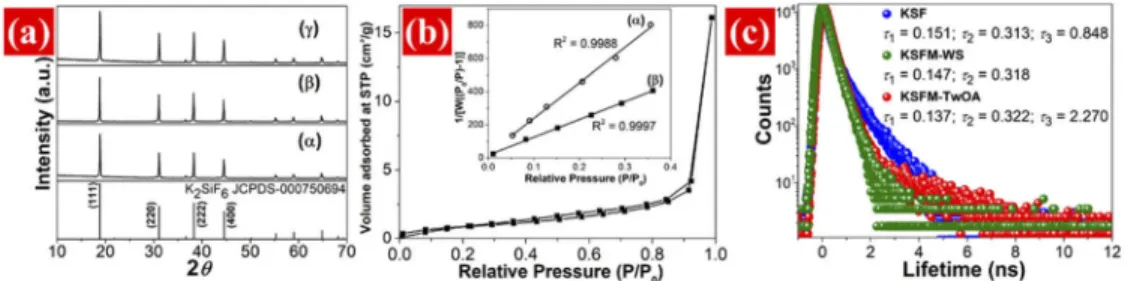
Fig. 1.SEM images of (a) KSFM-WS and (b) KSFM-TwOA; (c) TEM and (d, e, f) HRTEM images of KSFM-TwOA.
The X-ray diffraction (XRD) patterns of the prepared samples are shown in Fig.2(a). All the diffraction peaks can be indexed to the space groupFm-3mof cubic K2SiF6 (JCPDS No. 00075-0694). The main peaks are located at 18.89°, 31.09°, 38.31°and 44.55°, respectively cor-responding to the (111), (220), (222) and (400) reflections. Shape of the N2adsorption/desorption isotherm presents micro-mesoporous structure [27] of KSFM d-NPs with average pore volume of approximately 0.026 cm3/g, and average pore diameter of⇠12.09 Å (Fig.2(b)). The multipoint BET plots of KSFM-TwOA and KSFM-WS are shown in inserted picture of Fig.2(b). The surface area SBET⇠3.182 m2/g and⇠1.612 m2/g were calculated for KSFM-TwOA d-NPs and
KSFM-WS, respectively.
Fig. 2. (a) XRD patterns of (↵) KSF, ( ) KSFM-WS and ( ) KSFM-TwOA; (b) N2
adsorption/desorption isotherm of KSFM-TwOA. Inserted picture shows the multi-point
BET plots of (↵) KSFM-WS and ( ) KSFM-TwOA; (c) PAL spectra of the KSF, KSFM-WS
and KSFM-TwOA.
The PAL spectra of the KSF, KSFM-WS and KSFM-TwOA samples are presented in Fig.2(c). The results of positron lifetimes (⌧1,⌧2and⌧3) indicate the existence of defects in the prepared samples. Two long positron lifetime components (⌧2and⌧3) are observed in the PAL spectra of the KSF host. The lifetime component⌧2=⇠0.313 ns, agrees well with the corresponding theoretical prediction for Si4+mono-vacancy in O/F compounds (⌧=⇠0.3 ns) due to the formation
of a tetrahedral structure [28,29]. The⌧3=⇠0.848 ns is predicted for K++di-vacancy due to the
absence of two K+ions (⌧=⇠0.411 ns for K+vacancy [29]) in the KSF lattice. However, the⌧
is not observed in the Mn4+-doped sample as KSFM-WS. The ionic radius of Mn4+(r⇠0.53 Å,
CN=6 [30]) is much smaller than that of K++di-vacancy (⇠3.28 Å due torK+⇠1.64 Å, CN=12
[30]). Thus, some Mn4+ions may enter the K++di-vacancy site. The longer⌧
3value⇠2.27 ns is found for the KSFM-TwOA due to its porous structure [31]. The very short⌧3(<1.0 ns) values of KSF and KSFM-WS implied that these materials do not have a porous structure [32]. This result indicates the critical role of surfactants in the porous structure formation of the KSFM d-NPs. Short positron lifetimes (⌧1) corresponding to the bulk structure defects of KSFM-WS
(⇠0.147 ns) and KSFM-TwOA (⇠0.137 ns) are lower than that of KSF (⇠0.151 ns), indicating the
location of Mn4+in Si4+vacancy. Given that the electron density of Mn is considerably higher
than that of Si, positron annihilation probability is increased in tetrahedral structures, decreasing the⌧1value. By contrast, the charge of Mn4+is higher than that of K+, and thus, electrons on the
surface of neighbourhood Si4+vacancy are pulled towards Mn4+, slightly increasing⌧2. The
shortest⌧1value of KSFM-TwOA indicates that the porous nanosized crystals can reduce the
bulk defect density, and thus, may improve electron mobility and photon emission. Moreover, the porous structure acting as roughened surface may reduce the total internal reflections, resulting in enhancement of light emission efficiency [33]. Hence, the porous KSFM d-NPs exhibit an excellent quantum yield of⇠95.9% which is much higher than that of KSFM-WS (without a
porous structure)⇠57.7%, and close to that of the commercial KSFM phosphor (with 35µm in
size)⇠95.6% [34].
3.2. Photoluminescence properties
The photoluminescence excitation (PLE) and photoluminescence (PL) spectra of the KSFM-WS and KSFM-TwOA (at 0.13 g KMnO4), were measured at room temperature (RT). The emission at 631 nm was monitored. The excitation bands of the Mn4+assigned to the allowed4A
2!4T1 and4A2!4T2transitions are located at the 300–500 nm region with the maxima at 355 nm and 455 nm, respectively (Fig.3(a)). The PL spectra consist of seven narrow lines in the range of 600–650 nm are due to spin-forbidden2Eg!4A2gtransitions. The emission peaks are situated at 597.8, 608.6, 613.1, 619.2, 630.6, 634.8 and 647.4 nm due to transitions of the⌫3(t1u),⌫4 (t1u),⌫6(t2u), zero phonon line (ZPL),⌫6(t2u),⌫4(t1u) andv3(t1u) vibronic modes, respectively [14,15] (Fig.3(b)). The FWHM of two main PL peaks at 630.6 nm and 634.8 nm of KSFM-TwOA are⇠2.70 nm and⇠2.45 nm, respectively (Fig.3(c)). Importantly, the FWHM of 2.70 nm of
the emission at 630.6 nm is narrower than that of the reported KSFM phosphors (10 50µm)
⇠8.4 nm [35], enabling a high color quality nanophosphor for display applications. Moreover,
the dimension of the dots and pores in porous KSFM d-NPs is much smaller than the wavelength of red emissions resulting in the light diffraction phenomenon and light extraction improvement, as depicted in Fig.3(d). Therefore, the emission intensity of KSFM-TwOA is remarkably higher than that of KSFM-WS.
The PL spectra of the KSFM-TwOA preapred at different KMnO4amounts (0.09 0.15 g) are presented in Fig.3(e). Under 455 nm light excitation, the PL intensity of KSFM-TwOA initially increased with an increase in KMnO4content, reached its maximum value when KMnO4 content was 0.13 g (⇠2.60%mol of Mn4+ion incorporated in KSF) and then decreased upon
further increase of dopant content because of concentration quenching. The RT luminescent decay characteristics of the emitting state2Egin the KSFM-WS and KSFM-TwOA are shown in Fig.3(f). PL decay time was determined based on a single exponential fit. The emission lifetime of the KSFM-TwOA with 0.11 g KMnO4(⇠1.40%mol Mn) is 14.7 ms. For the samples
with 0.13 g KMnO4(⇠2.60%mol Mn) and 0.15 g KMnO4(⇠2.86%mol Mn), the mean decay
Fig. 3.(a) PLE and (b) PL spectra of KSFM-WS and KSFM-TwOA; (c) The deconvolution and FWHM calculation of emission bands at 630.6 nm and 634.8 nm; (d) The digital images
of the KSFM-WS and porous KSFM d-NPs in isopropanol under UV light ( ex=365 nm)
excitation; (e) PL spectra of KSFM-TwOA with different KMnO4contents: (↵) 0.09 g, ( )
0.11 g, ( ) 0.13 g and ( ) 0.15 g; (f) PL decay curves of KSFM-TwOA with: ( ) 0.11 g, ( )
0.13 g and ( ) 0.15 g KMnO4, and ( ) KSFM-WS at 289 K.
The temperature-dependent emission spectra of KSFM-TwOA under 455 nm light excitation are presented in Figs.4(a) and4(b). When temperature was increased from 77 K to 573 K, all the emission lines became broader and slight red shifted because of the increase in photon absorption and the enhanced vibration transition coupling associated with the vibration modes of the MnF62 octahedron. The integrated intensity of anti-Stokes emission lines gradually increased in temperature range of 77 K–473 K, whereas that of Stokes emission lines slight decreased, and then the both degrated with further increasing temperature (>473 K) (Fig.4(c)). The temperature-dependent behavior of the relative PL intensity (IPL(T K)/IPL(298 K)⇥100) within
the temperature range of 298 K–573 K is shown in Fig.4(d). The relative emission intensity at 473 K was over the 100% level which is attributed by the combination of the emission lines broadening and the increase in intensity of the anti-Stokes emission lines. At 498 K, the emission intensity dropped to⇠90% of the initial PL intensity, and the quenching temperatureT1/2 reached to 530 K; this thermal stability is better than that of the other fluoride nanophosphor (⇠71.9 88.8% at 423 K [16,17]) and the widely used YAG:Ce phosphor (70% at 423 K [38]).
Non-radiative transition probability increased with temperature, and the relative PL intensity exhibited thermal quenching, which can be fitted byIT/I0=[1+Dexp( Ea/kT)] 1, whereI0is the initial emission intensity,ITis the intensity at temperature T,Eais the activation energy,
Dis a constant and k is the Boltzmann’s constant. The thermal activation energyEa⇠1.16 eV obtained for the porous KSFM d-NPs is similar to that of the reported KSFM microphosphors (0.81 1.07 eV [22]).
Fig. 4. Temperature-dependent emission spectra of KSFM-TwOA in temperature range of (a) 77 K–298 K and (b) 298 K–573 K. (c) The integrated intensity of anti-Stokes and Stokes emission lines. (d) The relative red PL intensity as a function of temperature (77 K–573 K). The solid line represents the fitting result with the expressionIT/I0=[1+D
exp( Ea/kBT)] 1. (e) CIE chromaticity coordinates (x, y) and (f) chromaticity shift from
298 K to 498 K of KSFM-TwOA.
from 298 K to 498 K, the CIE coordinates slightly shifted in the red region from (0.7102, 0.2870) to (0.6955, 0.3015). In addition, an ultra-high color purity value of the porous KSFM d-NPs obtained from the CIE coordinates using Eq. (1) is 99% at room temperature. Color purity is 98% and 96% at 373 K and 473 K, respectively, indicating that it remained above 96% within the working temperature range for WLED [40].
colour purity= q
(x xi)2+(y yi)2
q
(xd xi)2+(yd yi)2
⇥100%, (1)
where (x, y) is the CIE coordinates of the sample, (xi, yi)=(0.3333, 0.3333) is the coordinates of white light, and (xd, yd) is the coordinates of the dominant wavelength of the light source.
The chromaticity shift was also calculated from the (x, y) CIE parameters using Eq. (2):
E= q
(u0t u00)2+(v0t v00)2+(w0t w00)2, (2)
whereu0=4x/(3–2x+12y),v0=9y/(3–2x+12y) andw0=1–u0–v0are the chromaticity coordinates in uniform color space 1976, and x and y are the chromaticity coordinates in CIE 1931. The 0 and t subscripts denote room temperature and high temperature parameters, respectively. The calculated CIE shifts of KSFM-TwOA within the temperature range of 298 K to 498 K are plotted in Fig.4(f). The chromaticity shift was 9.6⇥10 3at 423 K, 26.0⇥10 3at 473 K and
43.2⇥10 3at 498 K. Within the working temperature range for a pc-WLED, i.e., 298 K to 398 K,
the porous KSFM d-NPs exhibited a chromaticity shift of 5.6⇥10 3, which is significantly lower
than that of the high efficient K3AlF6:Mn4+red phosphor (⇠17.5⇥10 3) [41] and close to that of commercially available pc-WLED (typically close to 7.0⇥10 3) [40]. This result indicates
3.3. Application to microWLEDs
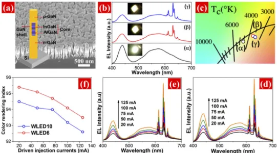
An SEM image of InGaN/AlGaN nanowires on silicon substrate taken under 45 degree titled angle is presented in Fig.5(a). It shows that the nanowires are relatively uniform in the diameter and length. The structure of a single InGaN/AlGaN nanowire heterostructures is schematically illustrated in inserted picture of Fig.5(a). The EL spectrum of the InxGa1 xN nanowire LED presents cool white light via mixing yellow and blue emissions (Fig.5(b(↵))) with a CRI⇠75.9
and CCT⇠4890 K. The sharp emission lines of Mn4+in the K2SiF6lattice were observed in the
EL spectra of theµWLED6 andµWLED10, as shown in Figs.5(b( )) and 5(b( )), respectively. The excellent CRI⇠95.4 forµWLED6 and CRI⇠94.5 forµWLED10 were obtained under a
driving current of 20 mA; these values are better than that for phosphor-free nanowire white LEDs (with CRI in the range of⇠90 94) [10,42]. The bright cool white-light emission of the
nanowire LED and warm white-light emission ofµWLED6 andµWLED10 are shown in the inserted pictures of Fig.5(b). The chromaticity coordinates (0.3570, 0.3510) of blue-yellow nanowire LED, (0.3924, 0.3703) ofµWLED6 and (0.3999, 0.3674) ofµWLED10 at 20 mA are close to the black body locus in the CIE 1931 color spaces, as shown in Fig.5(c).
Fig. 5.(a) A 45°tilted SEM image of the nanowires on silicon wafer. Inserted image shows the schematic structure of an InGaN/AlGaN core-shell single nanowire LED; (b) EL spectra
and (c) Expanded CIE chromaticity diagram of (↵) the blue-yellow InGaN nanowire LED,
( )µWLED6 and ( )µWLED10 measured under a driving current of 20 mA at RT. Inserted
pictures show bright white-light emissions from the fabricatedµWLEDs; EL spectra of (d)
µWLED6 and (e)µWLED10 under different driven injection currents from 20 125 mA; (f)
CRI ofµWLED6 (red circle) andµWLED10 (blue circle) under different driven injection
currents 20 125 mA.
The EL characteristics of µWLED6 and µWLED10 are presented in Figs. 5(d) and5(e), respectively. In bothµWLEDs, the EL intensities were enhanced gradually upon increasing current from 20 mA to 125 mA. No significant change occurred in the shapes of the EL spectra, indicating the good lighting color stability of the fabricated devices with a CCT of
⇠[3641 K 3704 K] and⇠[3362 K 3432 K] forµWLED6 andµWLED10, respectively. The slight
based thin-film WLEDs [43]. Such a distinct property is related to the significantly reduced Quantum confined Stark effect and reduce heating effect in nanowire based LED structures compared to the thin-film counterparts [11,23]. The excellent QY, ultra-high color purity, thermal stability, and package results indicate that porous KSFM red d-NPs is a promising candidate for improving the color reproducibility ofµWLEDs for practical applications.
4. Summary
In conclusion, porous K2SiF6:Mn4+ dots-in-nanoparticles are synthesized via a convenient
emulsification method using Tween80 and oleic acid surfactants. The prepared KSFM possess a deep red emission with narrow spectral linewidth of⇠2.7 nm and high QY of⇠95.9% under
455 nm light excitation. Furthermore, the porous KSFM d-NPs exhibit an ultra-high color purity of⇠99% at 298 K and low thermal quenching with the relative luminescent intensity of⇠100% at
473 K. A remarkable improvement in CRI (⇠95.4) and in CCT (⇠3649 K) of the InGaN nanowire µWLEDs fabricated using porous KSFM d-NPs is observed. Moreover, the nanowireµWLEDs exhibited minimal change in CCT and CRI with increasing drive currents from 20 mA to 125 mA. The present study demonstrates that porous KSFM nanomaterial is a promising red component for the cost effectiveµWLEDs, which are highly expected for the next-generation display technology.
Funding
Vietnam National Foundation for Science and Technology Development (NAFOSTED) (103.03-2017.312); The Bilateral Scientific Cooperation between the Vietnam Academy of Science and Technology and the Polish Academy of Sciences (QTPL01.03/18-19).
Disclosures
The authors declare no conflicts of interest.
References
1. I. Malkiel, M. Mrejen, A. Nagler, U. Arieli, L. Wolf, and H. Suchowski, “Plasmonic nanostructure design and characterization via deep learning,”Light: Sci. Appl.7, 60 (2018).
2. Y. Wu, K. Zhang, and B. Yang, “Ordered hybrid micro/nanostructures and their optical applications,”Adv. Opt.
Mater.7(7), 1800980 (2019).
3. E. Virey, “Are microLEDs really the next display revolution?”J. Inf. Disp.34(3), 22–27 (2018).
4. M. K. Choi, J. Yang, T. Hyeon, and D.-H. Kim, “Flexible quantum dot light-emitting diodes for next-generation displays,” npj Flex,”npj Flex Electron2(1), 10 (2018).
5. Y. Sun, Y. Jiang, X. W. Sun, S. Zhang, and S. Chen, “Beyond OLED: Efficient quantum dot light-emitting diodes for display and lighting application,”Chem. Rec.19(8), 1729–1752 (2019).
6. J. Bai, Q. Wang, and T. Wang, “Characterization of InGaN-based nanorod light emitting diodes with different indium compositions,”J. Appl. Phys.111(11), 113103 (2012).
7. M. Hartensveld, G. Ouin, C. Liu, and J. Zhang, “Effect of KOH passivation for top-down fabricated InGaN nanowire light emitting diodes,”J. Appl. Phys.126(18), 183102 (2019).
8. L. Qiming, W. Karl R, C. Mary H, L. Stephen R, K. Daniel D, F. Jeffery J, C. Karen C, F. Saeed, M. Zetian, and W. George T, “Optical performance of top-down fabricated InGaN/GaN nanorod light emitting diode arrays,”Opt.
Express19(25), 25528–25534 (2011).
9. M. Rajan Philip, D. D. Choudhary, M. Djavid, M. N. Bhuyian, T. H. Q. Bui, D. Misra, A. Khreishah, J. Piao, H. D. Nguyen, K. Q. Le, and H. P. T. Nguyen, “Fabrication of phosphor-free III-nitride nanowire light-emitting diodes on metal substrates for flexible photonics,”ACS Omega2(9), 5708–5714 (2017).
10. H. Q. T. Bui, V. Ravi Teja, J. Barsha, O. H. Aref, H. D. Nguyen, T. R. Lenka, and H. P. T. Nguyen, “Full-color InGaN/AlGaN nanowire micro light-emitting diodes grown by molecular beam epitaxy: A promising candidate for next generation micro displays,”Micromachines10(8), 492 (2019).
11. J. Barsha, R. T. Velpula, H. Q. T. Bui, H. D. Nguyen, T. R. Lenka, T. K. Nguyen, and H. P. T. Nguyen, “High performance electron blocking layer-free InGaN/GaN nanowire white-light-emitting diodes,”Opt. Express28(1), 665–675 (2020).
13. Y. N. Ahn, K. D. Kim, G. Anoop, G. S. Kim, and J. S. Yoo, “Design of highly efficient phosphor-converted white light-emitting diodes with color rendering indices (R1 - R15)>/=95 for artificial lighting,”Sci. Rep.9(1), 16848 (2019).
14. H. D. Nguyen and R. S. Liu, “Narrow-band red-emitting Mn4+-doped hexafluoride phosphors: synthesis, opto-electronic properties, and applications in white light-emitting diodes,”J. Mater. Chem. C4(46), 10759–10775 (2016).
15. S. Adachi, “Review—Mn4+vs Cr3+: A comparative study as activator ions in red and deep red-emitting phosphors,”
ECS J. Solid State Sci. Technol.9(2), 026003 (2020).
16. Y. Zhu, J. Yu, Y. Liu, M. G. Brik, L. Huang, T. Xuan, and J. Wang, “Photoluminescence properties of a novel red fluoride K2LiGaF6:Mn4+nanophosphor,”RSC Adv.7(49), 30588–30593 (2017).
17. C. Jiang, M. G. Brik, L. Li, L. Li, J. Peng, J. Wu, M. S. Molokeev, K.-L. Wong, and M. Peng, “The electronic and optical properties of a narrow-band red-emitting nanophosphor K2NaGaF6:Mn4+for warm white light-emitting diodes,”J. Mater. Chem. C6(12), 3016–3025 (2018).
18. S. Mintova, M. Jaber, and V. Valtchev, “Nanosized microporous crystals: emerging applications,”Chem. Soc. Rev.
44(20), 7207–7233 (2015).
19. P. Pallavi, S. Bandyopadhyay, J. Louis, A. Deshmukh, and A. Patra, “A soluble conjugated porous organic polymer: efficient white light emission in solution, nanoparticles, gel and transparent thin film,”Chem. Commun.53(7), 1257–1260 (2017).
20. W. Lu, Y. Ou, E. M. Fiordaliso, Y. Iwasa, V. Jokubavicius, M. Syvajarvi, S. Kamiyama, P. M. Petersen, and H. Ou, “White light emission from fluorescent SiC with porous surface,”Sci. Rep.7(1), 9798 (2017).
21. H. D. Nguyen, C. C. Lin, and R. S. Liu, “Waterproof alkyl phosphate coated fluoride phosphors for optoelectronic materials,”Angew. Chem.54(37), 10862–10866 (2015).
22. H. F. Sijbom, R. Verstraete, J. J. Joos, D. Poelman, and P. F. Smet, “K2SiF6:Mn4+as a red phosphor for displays and warm-white LEDs: a review of properties and perspectives,”Opt. Mater. Express7(9), 3332–3365 (2017). 23. H. P. Nguyen, S. Zhang, K. Cui, X. Han, S. Fathololoumi, M. Couillard, G. A. Botton, and Z. Mi, “p-Type modulation
doped InGaN/GaN dot-in-a-wire white-light-emitting diodes monolithically grown on Si(111),”Nano Lett.11(5), 1919–1924 (2011).
24. B. Jostrom, J. Bergenstahl, and B. Ronberg, “A method for the preparation of submicron particles of sparingly water-soluble drugs by precipitation in oil-in-water emulsions. II: Influence of the emulsifier, the solvent and the drug substance,”J. Pharm. Sci.82(6), 584–589 (1993).
25. P. Selvaraj, A. Roy, H. Ullah, P. Sujatha Devi, A. A. Tahir, T. K. Mallick, and S. Sundaram, “Soft-template synthesis of high surface area mesoporous titanium dioxide for dye-sensitized solar cells,”Int. J. Energy Res.43(1), 523–534 (2018).
26. J. Wang, A. Shi, D. Agyei, and Q. Wang, “Formulation of water-in-oil-in-water (W/O/W) emulsions containing trans-resveratrol,”RSC Adv.7(57), 35917–35927 (2017).
27. M. Thommes, “Physical adsorption characterization of nanoporous materials,”Chem. Ing. Tech.82(7), 1059–1073 (2010).
28. S. Dannefaer, T. Bretagnon, and D. Kerr, “Vacancy-type defects in crystalline and amorphous SiO2,”J. Appl. Phys.
74(2), 884–890 (1993).
29. J. M. Campillo Robles, E. Ogando, and F. Plazaola, “Positron lifetime calculation for the elements of the periodic table,”J. Phys.: Condens. Matter19(17), 176222 (2007).
30. R. D. Shannon, “Revised effective ionic radii and systematic studies of interatomic distances in halides and chalcogenides,”Acta Cryst. A32(5), 751–767 (1976).
31. A. T. Le, Q. H. Nguyen, C. L. Cuong, D. D. Khiem, P. T. Phuc, L. L. Nguyen, N. T. N. Hue, P. T. Hue, and D. V. Phuc, “Simultaneous existence of defects and mesopores in nanosized ZSM-5 zeolite studied by positron annihilation and X-ray diffraction spectroscopies,”J. Appl. Phys.121(8), 084303 (2017).
32. S. J. Tao, “Positronium annihilation in molecular substances,”J. Chem. Phys.56(11), 5499–5510 (1972). 33. H.-D. Nguyen, H. P. T. Nguyen, J.-J. Lee, and S.-I. Mho, “Preparing nano-hole arrays by using porous anodic
aluminum oxide nano-structural masks for the enhanced emission from InGaN/GaN blue light-emitting diodes,”Adv.
Nat. Sci: Nanosci. Nanotechnol.3(4), 045018 (2012).
34. Z. Hou, X. Tang, X. Luo, T. Zhou, L. Zhang, and R.-J. Xie, “A green synthetic route to the highly efficient K2SiF6:Mn4+narrow-band red phosphor for warm white light-emitting diodes,”J. Mater. Chem. C6(11), 2741–2746 (2018).
35. J. H. Oh, H. Kang, Y. J. Eo, H. K. Park, and Y. R. Do, “Synthesis of narrow-band red-emitting K2SiF6:Mn4+ phosphors for a deep red monochromatic LED and ultrahigh color quality warm-white LEDs,”J. Mater. Chem. C
3(3), 607–615 (2015).
36. C. Long, H. Yoshihito, S. Shu, and J. Donglin, “Light-harvesting conjugated microporous polymers: rapid and highly efficient flow of light energy with a porous polyphenylene framework as antenna,”J. Am. Chem. Soc.132(19), 6742–6748 (2010).
37. O. Hai, Z. zhang, Q. Ren, X. Wu, Q. Zhang, B. Zeng, and D. Li, “The preparation and functional studies of the porous long afterglow luminescent materials,”Dyes Pigm.156, 160–166 (2018).
39. H. Zhang, X. Lu, J. Zhao, Q. Xin, J. Lin, and X. Gao, “Preparing of green-emitting CdZnSe ternary nanocrystals with narrow emission spectrum,”Superlattices Microstruct.113, 394–400 (2018).
40. N. Nadarajah, Y. Gu, L. Jayasinghe, J.P. Freyssinier, and Y. Zhu, “Long-term performance of white LEDs and systems,” (Lighting Research Center, 2007).
41. E. Song, J. Wang, J. Shi, T. Deng, S. Ye, M. Peng, J. Wang, L. Wondraczek, and Q. Zhang, “Highly efficient and thermally stable K3AlF6:Mn4+as a red phosphor for ultra-high-performance warm white light-emitting diodes,”
ACS Appl. Mater. Interfaces9(10), 8805–8812 (2017).
42. Y. C. Li, L.-B. Chang, H.-J. Chen, C.-Y. Yen, K.-W. Pan, B.-R. Huang, W.-Y. Kuo, L. Chow, D. Zhou, and E. Popko, “Phosphor-free InGaN white light emitting diodes using flip-chip technology,”Materials10(4), 432 (2017). 43. S. Ye, F. Xiao, Y. X. Pan, Y. Y. Ma, and Q. Y. Zhang, “Phosphors in phosphor-converted white light-emitting diodes:
Recent advances in materials, techniques and properties,”Mater. Sci. Eng. R Rep.71(1), 1–34 (2010).