Chapter 14
© McGraw-Hill Education.
14-2
Overview of Host Defense
Mechanisms (1 of 2)
• Innate, natural defenses: present at birth,
provide nonspecific resistance to infection
• Adaptive immunities: specific, must be
Overview of Host Defense
Mechanisms (2 of 2)
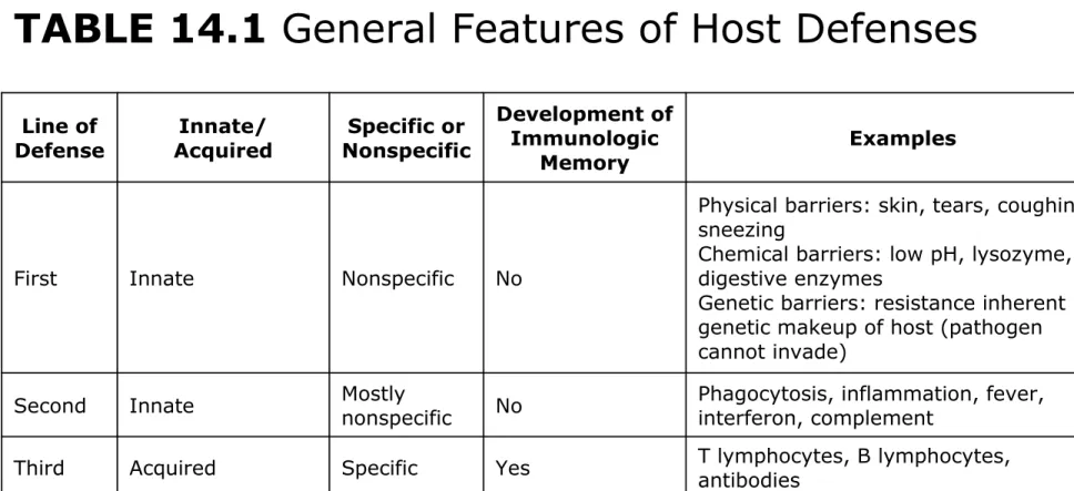
TABLE 14.1 General Features of Host Defenses
Line of
Defense AcquiredInnate/ NonspecificSpecific or
Development of Immunologic
Memory Examples
First Innate Nonspecific No
Physical barriers: skin, tears, coughing, sneezing
Chemical barriers: low pH, lysozyme, digestive enzymes
Genetic barriers: resistance inherent in genetic makeup of host (pathogen cannot invade)
Second Innate Mostly nonspecific No Phagocytosis, inflammation, fever, interferon, complement
© McGraw-Hill Education.
14-4
Defense Mechanisms of the Host
• To protect the body against pathogens, the
immune system relies on a multilevel network of physical barriers, immunologically active cells, and a variety of chemicals
– First line of defense – any barrier that blocks invasion at the portal of entry – nonspecific
– Second line of defense – protective cells and fluids; inflammation and phagocytosis – nonspecific
– Third line of defense – acquired with exposure to foreign substance; produces protective antibodies and creates memory cells – specific
• The lines of defense do not work separately, most
1st Line of Defense: Barriers at the
Portal of Entry
•
A number of defenses are a normal part of
the body’s anatomy and physiology
•
These are inborn (innate), nonspecific
defenses
•
They can be divided into three categories:
1. Physical or anatomical barriers at the body surface
2. Chemical defenses
3. Genetic resistance to infection
© McGraw-Hill Education.
14-6
Physical or Anatomical Barriers
(1 of 4)
• Built-in defenses in skin
– Outermost layer of skin with epithelial cells
cemented together, and impregnated with keratin
– Flushing effect of sweat
• Mucous membranes
– Coating of digestive, genitourinary, and respiratory tracts
– Blinking and tear production
– Flow of saliva
© McGraw-Hill Education.
14-8
Physical or Anatomical Barriers
(3 of 4)
• Mucous membranes
– Mucous coat impedes attachment and entry of bacteria
Physical or Anatomical Barriers
(4 of 4)
© McGraw-Hill Education.
14-10
Nonspecific Chemical Defenses
(1 of 2)
• From skin and mucous membranes
– Sebaceous secretions
– Antimicrobial secretions from specialized glands (meibomian glands)
• Other defenses in tears, saliva and skin:
– Lysozyme, an enzyme that hydrolyzes the cell wall of bacteria
– Defensins, peptides that lyse bacteria and fungi
– High lactic acid and electrolyte concentration in sweat
Nonspecific Chemical Defenses
(2 of 2)
• Hydrochloric acid in stomach
• Digestive juices and bile of intestines
• Semen contains an antimicrobial chemical
© McGraw-Hill Education.
14-12
Genetic Defenses
•
Some hosts are genetically immune to the
diseases of other hosts
– “Humans can’t acquire distemper from cats,
and cats can’t get mumps from humans”
•
Viruses have great specificity for their host
receptors
•
Some genetic differences in susceptibility exist
for other pathogens, including differences
within members of the same species
– Humans carrying a gene or genes for sickle-cell
Structure and Function of the Organs
of Defense and Immunity (1 of 3)
•
Immunology
: study of the body’s 2
ndand 3
rdlines of defense
•
Primary functions of a healthy immune
system:
1. Surveillance of the body
2. Recognition and differentiation of normal versus foreign material
© McGraw-Hill Education.
14-14
Structure and Function of the Organs
of Defense and Immunity (2 of 3)
•
White blood cells (leukocytes) – innate
capacity to recognize and differentiate any
foreign material
– Nonself – foreign material
– Self – normal cells of the body
•
Pathogen-associated patterns (PAMPs) –
molecules shared by microorganisms
Structure and Function of the Organs
of Defense and Immunity (3 of 3)
•
All this is mainly carried our by
white blood
cells (leukocytes)
– innate capacity to
recognize and differentiate any foreign
material
– Nonself – foreign material
© McGraw-Hill Education.
14-16
How do White Blood Cells Work?
• Pathogen-associated patterns (PAMPs) – molecules shared by microorganisms
Components and Connections of the
Immune System
• The immune system is a large, complex, and
diffuse network of cells and fluids that penetrate into every organ and tissue
• Body compartments that participate in immune
function:
1. Reticuloendothelial system (RES) 2. Extracellular fluid (ECF)
3. Bloodstream
4. Lymphatic system
• For effective immune responsiveness, the activities
© McGraw-Hill Education.
14-18
© McGraw-Hill Education.
14-20
Origin, Composition, and Functions
of Blood (1 of 2)
• Whole blood: plasma and formed elements
(blood cells)
– Serum: liquid portion of blood after a clot has formed (minus clotting factors)
Origin, Composition, and Functions
of Blood (2 of 2)
© McGraw-Hill Education.
14-22
A Survey of Blood Cells
•
Hemopoiesis
(or
hematopoiesis
)–
production of blood cells
•
Stem cells – undifferentiated cells, precursor
of new blood cells
•
Primary cell lines:
– Platelets (thrombocytes)
– Red blood cells (RBCs)
– Leukocytes or white blood cells (WBCs),
responsible for immune function
Granulocytes: lobed nucleus
© McGraw-Hill Education.
14-24
Granulocytes (1 of 4)
•
Neutrophils
– 55-90% - lobed nuclei with
lavender granules; phagocytes
•
Eosinophils
– 1-3% - orange granules and
bilobed nucleus; destroy eukaryotic
pathogens
•
Basophils
– 0.5% - constricted nuclei, dark
blue granules; release potent chemical
mediators
– Mast cells: nonmotile elements bound to
Granulocytes (2 of 4)
© McGraw-Hill Education.
14-26
Granulocytes (3 of 4)
Granulocytes (4 of 4)
© McGraw-Hill Education.
14-28
Agranulocytes (1 of 3)
• Lymphocytes – 20-35%, specific immune
response
– B (humoral immunity): activated B cells produce antibodies
– T cells (cell-mediated immunity): activated T cells modulate immune functions and kill foreign cells
• Monocytes, macrophages – 3-7% - largest of
WBCs, kidney-shaped nucleus; phagocytic
– Macrophages: final differentiation of monocytes
Agranulocytes (2 of 3)
© McGraw-Hill Education.
14-30
Agranulocytes (3 of 3)
Leukocytes (1 of 2)
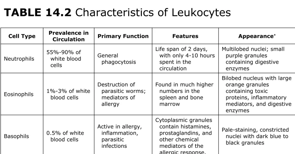
TABLE 14.2 Characteristics of Leukocytes
Cell Type Prevalence in Circulation Primary Function Features Appearance*
Neutrophils 55%-90% of white blood cells
General
phagocytosis
Life span of 2 days, with only 4-10 hours spent in the
circulation
Multilobed nuclei; small purple granules
containing digestive enzymes
Eosinophils 1%-3% of white blood cells
Destruction of parasitic worms; mediators of allergy
Found in much higher numbers in the spleen and bone marrow
Bilobed nucleus with large orange granules
containing toxic
proteins, inflammatory mediators, and digestive enzymes
Basophils 0.5% of white blood cells
Active in allergy, inflammation, parasitic infections Cytoplasmic granules contain histamines, prostaglandins, and other chemical mediators of the allergic response.
© McGraw-Hill Education.
14-32
Leukocytes (2 of 2)
The “TABLE 14.2” continues on this slide.
Cell Type Prevalence in Circulation Primary Function Features Appearance*
Monocytes 3%-7% of white blood cells Phagocytosis, followed by final differentiation into macrophages and dendritic cells
Monocytes also secrete several chemicals that moderate the
functions of the immune system.
Largest WBC; nuclei large, ovoid, and often indented-no cytoplasmic
granules visible using a light microscope
Lymphocytes 20%-35% of white blood cells
Specific (acquired) immunity
Two types of
lymphocytes exist. T cells are responsible for cell-mediated immunity, whereas B cells are responsible for humoral immunity.
Erythrocytes and Platelet Lines
•
Erythrocytes
: develop from bone marrow
stem cells, lose nucleus, simple biconcave
sacs of hemoglobin
© McGraw-Hill Education.
14-34
Lymphatic System (1 of 2)
•
Provides an auxiliary route for return of
extracellular fluid to the circulatory system
•
Acts as a drain-off system for the
inflammatory response
© McGraw-Hill Education.
14-36
Lymphatic Fluid
•
Lymph
is a plasma-like liquid carried by
lymphatic circulation
•
Formed when blood components move out of
blood vessels into extracellular spaces
•
Made up of water, dissolved salts, 2-5%
proteins
Lymphatic Vessels (1 of 2)
•
Lymphatic capillaries
permeate all parts of
the body except the CNS, bone, placenta, and
thymus
•
Thin walls easily permeated by extracellular
fluid which is then moved through contraction
of skeletal muscles
•
Functions to return lymph to circulation; flow
is one-direction – toward the heart –
© McGraw-Hill Education.
14-38
Classification of Lymphoid Organs
and Tissues (1 of 2)
•
Primary organs
– Sites of lymphocytic origin and maturation
Thymus gland
Bone marrow
•
Secondary organs and tissues
– Circulatory-based locations
Lymph nodes
Classification of Lymphoid Organs
and Tissues (2 of 2)
– Collections of cells distributed throughout skin
and mucous membranes
MALT—mucosal-associated lymphoid tissue
SALT—skin-associated lymphoid tissue
© McGraw-Hill Education.
14-40
Lymphoid Organs (1 of 2)
• Thymus
– High growth and activity until puberty, then begins to shrink
– Site of T-cell maturation
• Lymph nodes
– Small, encapsulated, bean-shaped organs along lymphatic channels and large blood vessels of the thoracic and abdominal cavities
• Spleen
– Nestled below the diaphragm and left of the stomach
– Structurally similar to lymph node; filters
© McGraw-Hill Education.
14-42
Second-Line Defenses: Inflammation
•
Mechanisms that play important roles in host
defenses:
– Recognition
– Inflammation
– Phagocytosis
– Interferon
– Complement
Inflammatory Response (1 of 2)
• Reaction to any traumatic event in the tissues
that attempts to restore homeostasis
• Helps to clear away invading microbes and
cellular debris left by immune reactions
Redness – increased circulation and vasodilation in
response to chemical mediators
Warmth – heat given off by the increased blood
flow
Swelling – increased fluid in the tissue as blood
vessels dilate – edema; WBC’s, microbes, debris, and fluid collect to form pus; prevents spread of infection
© McGraw-Hill Education.
14-44
Major Inflammatory Events (1 of 4)
a) Injury/Immediate Reactions: Blood vessels
© McGraw-Hill Education.
14-46
Major Inflammatory Events (2 of 4)
b) Vascular Reactions: Nearby blood vessels dilate;
increased blood flow; increased vascular
Major Inflammatory Events (3 of 4)
c) Edema and Pus Formation: Collection of fluid;
© McGraw-Hill Education.
14-48
Major Inflammatory Events (4 of 4)
d) Resolution/Scar Formation: Macrophages
© McGraw-Hill Education.
14-50
Fever: An Adjunct to Inflammation
(1 of 2)
•
Initiated by circulating
pyrogens
which reset
the hypothalamus to increase body
temperature; signals muscles to increase
heat production and vasoconstriction
– Exogenous pyrogens – products of infectious agents; endotoxin
– Endogenous pyrogens – liberated by
monocytes, neutrophils, and macrophages
Fever: An Adjunct to Inflammation
(2 of 2)
•
Benefits of fever:
– Inhibits multiplication of temperature-sensitive
microorganisms
– Impedes nutrition of bacteria by reducing the
available iron
– Increases metabolism and stimulates immune
© McGraw-Hill Education.
14-52
Second-Line of Defenses:
Phagocytosis (1 of 2)
•
General activities of phagocytes:
1. To survey tissue compartments and discover microbes, particulate matter, and dead or
injured cells
2. To ingest and eliminate these materials
Second-Line of Defenses:
Phagocytosis (2 of 2)
•
Major categories of phagocytes:
– Neutrophils – general-purpose; react early to
bacteria and other foreign materials, and to damaged tissue
– Eosinophils – attracted to sites of parasitic
infections and antigen-antibody reactions
– Macrophages – derived from monocytes;
scavenge and process foreign substances to prepare them for reactions with B and T
© McGraw-Hill Education.
14-54
Development of Monocytes and
Macrophages (1 of 2)
•
After leaving the bloodstream and entering
the tissue, monocytes are transformed by
inflammatory mediators into macrophages or
dendritic cells
© McGraw-Hill Education.
14-56
Phagocytic Recognition
• Protein receptors within cell membrane of
macrophages, called Toll-like receptors (TLRs)
• Detect foreign molecules and signal the
Mechanisms of Phagocytosis (1 of 2)
•
Chemotaxis, binding and ingestion
– Phagocytes migrate to inflammation following
a gradient of stimulant products
– Using TLRs they bind pathogen-associated
molecular pattern (PAMPs) receptors
•
Phagolysosome formation
– Phagocyte extends pseudopods that enclose
pathogen in a vacuole called a phagosome
– In a short time, lysosomes with antimicrobial
substances fuse with the phagosome to form
© McGraw-Hill Education.
14-58
Mechanisms of Phagocytosis (2 of 2)
•
Destruction and elimination
– Oxygen-dependent system (respiratory
burst)
– Liberation of lactic acid, lysozyme, and nitric
oxide
– Undigestible debris are released from the
Sequential Events in Phagocytosis
© McGraw-Hill Education.
14-60
Interferon (1 of 2)
•
Small protein produced by certain WBCs and
tissue cells
•
Three major types of IFNs:
– Interferon alpha – product of lymphocytes and macrophages
– Interferon beta – product of fibroblasts and epithelial cells
– Interferon gamma – product of T cells
Interferon (2 of 2)
•
All three types bind to cell surfaces and
induce expression of antiviral proteins and
inhibit expression of cancer genes
•
IFNs alpha and beta stimulate phagocytes
© McGraw-Hill Education.
14-62
Complement
•
Consists of 26 blood proteins that work in
concert to destroy bacteria and viruses
•
Complement proteins are activated by
cleavage (cascade reaction)
•
Pathways
– Classical – activated by the presence of
antibody bound to microorganism
– Lectin pathway – nonspecific reaction of a host
serum protein that binds mannan
– Alternative – begins when complement
© McGraw-Hill Education.
14-64
4 Stages in the Complement
Cascade
© McGraw-Hill Education.
14-66
Membrane Attack Complex (MAC)
Overview of the Major Host Defenses
(1 of 3)
The figure of “Classification of Host Defenses.”
•
HOST DEFENSES
– Innate, nonspecific
First line of defense
o Physical barriers
o Chemical barriers
o Genetic barriers
The first line of defense is a surface protection
© McGraw-Hill Education.
14-68
Overview of the Major Host Defenses
(2 of 3)
The figure of “Classification of Host Defenses” continues on this slide.
Second line of defense
o Inflammatory response
o Interferons
o Phagocytosis
o Complement
The second line of defense is a cellular and
chemical system that comes immediately into play if infectious agents make it past the surface
defenses. Examples include phagocytes that
Overview of the Major Host Defenses
(3 of 3)
The figure of “Classification of Host Defenses” continues on this slide.
– Acquired, specific (See chapter 15)
Third line of defense
o B and T lymphocytes, antibodies, cytotoxicity
The third line of defense includes specific host
defenses that must be developed uniquely for each microbe through the action of specialized white
blood cells. This form of immunity is marked by its activity toward specific pathogens and
© McGraw-Hill Education. All rights reserved. Authorized only for instructor use in the classroom. No reproduction or further distribution permitted without the prior written consent of McGraw-Hill Education.
3-70 14-70