THE EFFECTS OF PROLONGED SITTING ON ARTERIAL STIFFNESS AND CIRCULATING ANGIOGENIC CELLS IN HEALTHY ADULTS
William S. Evans
A thesis submitted to the faculty at the University of North Carolina at Chapel Hill in partial fulfillment of the requirements for the degree of Masters of Arts in the Department of Exercise
and Sports Science (Exercise Physiology) in the College of Arts and Sciences.
Chapel Hill 2018
Approved by:
iii ABSTRACT
William S. Evans: The Effects of Prolonged Sitting on Arterial Stiffness and Circulating Angiogenic Cells in Healthy Adults
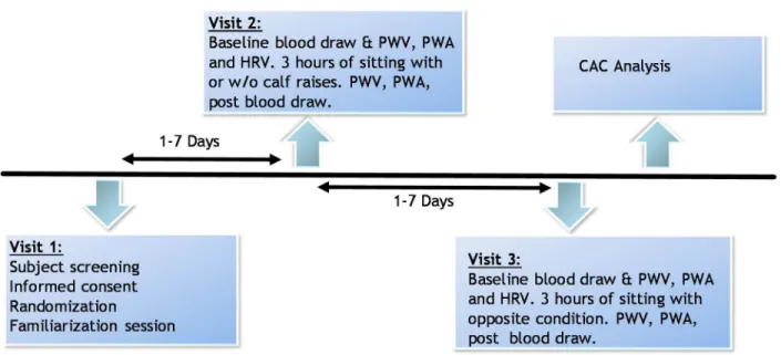
To assess the effects of prolonged sitting on measures of arterial stiffness (AS) and circulating angiogenic cells (CAC), healthy but sedentary subjects completed 3 hours of sitting in a randomized crossover design with 10 calf raises every ten minutes (CALF) or a control
iv
ACKNOWLEDGEMENTS
vi
TABLE OF CONTENTS
LIST OF ABBREVIATIONS ... viii
CHAPTER I ... 1
Introduction ... 1
Research Questions ... 4
Research Hypothesis ... 4
Assumptions... 5
Delimitations ... 5
Limitations ... 5
Significance of Study ... 5
Chapter II ... 7
Literature Review ... 7
Section 1: Mechanisms of Inactivity, Sitting, and Vascular physiology ... 7
1.1 Animal Models... 7
1.2 Human Models ... 9
1.3 Prolonged Sitting... 10
Section 2. AS and Inactivity ... 12
Section 3. AS and Endothelial Dysfunction ... 13
Section 4. Progenitor Cells and eNOS/NO ... 16
Section 5. Physical Activity and CACs ... 19
Chapter III ... 22
Methods ... 22
Subjects ... 22
Study Design and Pilot Testing ... 22
Familiarization ... 24
vii
Arterial Stiffness ... 25
HRV ... 26
Peripheral Blood Mononuclear Cells (PBMC) Isolation and Immunofluorescence Labeling ... 26
CD34+Isolation and Gene Analysis ... 27
Power Calculation ... 28
Statistical Analyses ... 28
Chapter IV ... 29
Results ... 29
Participants Information ... 29
Arterial Stiffness ... 29
HRV ... 30
Blood Pressure ... 30
CD34+ CAC ... 30
CD34+ CAC mRNA expression ... 31
Chapter V ... 36
Discussion ... 36
Arterial Stiffness ... 36
Circulating Angiogenic Cells... 38
Calf Raises ... 40
Sitting Recommendations ... 40
Limitations ... 41
Conclusions ... 42
Clinical Implications ... 42
Appendices ... 46
Appendix A ... 46
Appendix B ... 50
Appendix C ... 52
Appendix D ... 56
Appendix E ... 61
Appendix F ... 69
viii
LIST OF ABBREVIATIONS
AIx – Augmentation Index
APL – Applied Physiology Laboratory AS – Arterial Stiffness
BF – Blood Flow CFU – Colony Forming Unit CAC –Circulating Angiogenic Cells
CAD – Coronary Artery Disease CD – Cluster of Differentiation
cDBP – Central Diastolic Blood Pressure cSBP – Central Systolic Blood Pressure CVD – Cardiovascular Disease
EC – Endothelial Cell
eNOS – Endothelial Nitric Oxide Synthase EPC – Endothelial Progenitor Cell
FMD – Flow Mediated Dilation HRV – Heart Rate Variability HSC – Hematopoietic Stem Cells MAP – Mean Arterial Pressure NIRS – Near Infrared Spectroscopy NO – Nitric Oxide
iii PI3K/Akt – Phosphoinositide 3-kinase/Akt pathway PWA – Pulse Wave Analysis
PWV – Aortic Pulse Wave Velocity
RMSSD – Root Mean Square of Standard Deviations ROS – Reactive Oxidative Stress
SCI – Spinal Cord Injury
1 CHAPTER I Introduction
Emerging evidence suggests that sedentary behavior, including prolonged sitting, increases the risk for cardiovascular disease (CVD), the leading cause of death globally [1-3]. While chronic associations are known, the underlying mechanism are unclear. Evidence from acute bouts of sitting suggest that prolonged sitting decreases blood flow and shear stress (SS) resulting in local endothelial dysfunction of the lower extremity, but these changes may be rescued by intermittent physical activity, and the systemic effects remain unclear [4-6].
Optimally, assessing central changes in arterial stiffness (AS) such as aortic pulse wave velocity (PWV) and pulse wave analysis (PWA) would aid in understanding the adverse effects of sitting and the increased burden on the heart. While endothelial dysfunction increases during sitting, evidence also suggests that endothelial repair is attenuated in response to 10 days of physical inactivity as indicated by CFU-Hill counts[7]. CACs have paracrine function and contribute to repair of damaged vessels. However, the effects of acute inactivity on CAC number and function are not known. Therefore, to better understand functional changes in stiffness and the effects of sitting on vascular repair, PWV, PWA, and CAC count and function need to be examined before and after acute bouts of inactivity. Additionally, strategies to increase blood flow and shear stress need to be included to 1) provide additional mechanistic insight and 2) to develop other
2
PWV is the gold standard approach for measuring aortic AS [8]. The aorta is directly proximal to the heart and its elasticity is responsible for dampening the speed and amplitude of retrograde pressure waves that increase the heart’s workload during systole [9]. Indeed, aortic PWV has particular relevance to CVD, with a 1 m/s increase in PWV corresponding to a 14% and 15% increase in total CV events and CV mortality respectively, independent of traditional risk factors [10]. Pulse wave analysis (PWA) is also commonly used in junction with PWV to assess arterial wave reflection and central blood pressure, which has been shown as a better predictor of CVD than brachial blood pressure [10, 11]. Together these measures provide valuable information relevant to cardiac burden and serve as clinically relevant markers of arterial stiffness.
Acutely, endothelial dysfunction and AS may occur in response to decreased blood flow and shear stress. Indeed, it has been repeatedly shown that one bout of sitting reduces the NO dependent measure FMD in the lower leg suggesting endothelial dysfunction while local heating, exercise, and fidgeting have been shown to restore vascular health via increased shear stress [6, 12, 13]. However, these changes only represent local changes in vascular health and do not provide central insight unlike PWV and augmentation index (AIx). Though changes in PWV and AIx are generally chronic and observed with aging, evidence does show a functional stiffening in response to NO antagonists [14]. In light of this evidence, simple perturbations such as fidgeting or greater stimuli such as calf raises should be generalizable to changes in central measures of AS such as PWV and AIx.
3
CD34+ hematopoietic stem cells that were initially termed endothelial progenitor cells. In
response to increased blood flow and shear stress, CACs increase expression of endothelial nitric oxide (eNOS) and NO production which mediates function of important angiogenic cytokines such as VEGF[18]. Additionally, shear stress dose dependently increases surface protein
expression of VEGFR-2, VE-cadherin and Tie2 which are important for angiogenic capabilities and maintaining a restrictive endothelial barrier[19]. Furthermore, shear stress has been shown to augment homing and engraftment of CACs via increased signaling of CXCR4/JAK-2 pathway while also increasing expressing of CXCR4 in-vitro[19]. By affecting the secretion of VEGF and CXCR-4, shear stress may impact the homing, engraftment and angiogenic properties of CACs on damaged endothelium. Therefore, assessment of VEGF and CXCR-4 as gene targets on CACs is important, as they are likely impacted by decreased shear stress.
Though blood flow and shear stress affect CACs, little is known regarding the effects of physical inactivity on CACs. Ten days of reduced physical inactivity reduced colony forming unit counts but did not affect circulating counts of CACs [7] but the acute effects of inactivity have yet to be determined. On the other hand, it is generally accepted that CAC counts and function increase with acute exercise [20-23]. If acute inactivity decreases CAC function, it begs the question if simple lower extremity muscular contractions (i.e. calf raises) may increase shear stress and CAC activity to offset the effects of sitting. Given their role in repair, regulation by blood flow and shear stress, and the paucity of evidence, it is important to establish the effects of sitting on both the quantity and quality of CACs.
4
variables that yield clinical and mechanistic relevance. Therefore, the purpose of this study is to understand the balance between damage and repair during prolonged sitting through assessing 1) changes in PWV and PWA, 2) CAC counts and gene expression and 3) if intermittent calf raises during prolonged sitting attenuates these changes. If prolonged sitting does increase AS and calf raises can prevent these changes, current public health recommendations and guidelines can be updated to prevent the deleterious effects of sitting.
Research Questions
1. Does PWV and PWA increase with 3 hours of prolonged sitting?
2. Do CD34+ CAC counts decrease following 3 hours of prolonged sitting? 3. Does gene expression of CACs change following 3 hours of prolonged sitting? 4. Do intermittent calf raises prevent negative changes in AS or CACs?
Research Hypothesis
1. A 3-hour bout of prolonged sitting will increase PWV and AIx from PWA from pre to post testing.
2. A 3-hour bout of prolonged sitting will decrease CAC counts.
3. A 3-hour bout of prolonged sitting will decrease number of CD34+ CACs and expression of VEGF and CXCR-4.
5 Assumptions
1. Subjects followed pre-assessment guidelines.
2. Pre-assessment guidelines were adequate to control for baseline changes in outcomes 3. All subjects answered the medical history questionnaire and physical activity readiness
questionnaire truthfully. Delimitations
1. When able, women’s menstrual cycles were recorded and controlled.
2. Repeated measures design was used to control for condition specific variability. 3. All subjects had similar dietary intake prior to and during testing.
4. All subjects were between the ages of 18-35. Limitations
1. Blood flow induced shear stress was not directly measured during testing. 2. Magnetic isolation of CD34 CACs did not result in 100% purity.
Significance of Study
6
7 Chapter II Literature Review
This review is divided into the following sections: 1) Mechanisms of Inactivity, Sitting, and Vascular Physiology 2) AS and Inactivity 3) AS and Endothelial Dysfunction 4) Progenitor Cells and eNOS/NO 5) Physical Activity and CACs.
Section 1: Mechanisms of Inactivity, Sitting, and Vascular physiology
The effects of inactivity are well documented in current literature. Beginning with the London bus study, study of physical activity and inactivity, has progressed [24]. Through this evidence, sedentary behavior is now considered a risk factor for the development of
cardiovascular disease (CVD) according to the American College of Sports Medicine [25]. However, the precise mechanisms governing these changes are not concrete, and recent evidence suggests the type of inactivity seems to matter.
1.1 Animal Models
8
decrease in blood flow due to inactivity and the absence of gravity result in a decrease in the expression of eNOS mRNA as well as protein expression, suggesting a decreased ability to produce NO [26]. This model also showed a decrease in the ability to vasodilate in response to acetylcholine and differing flow patterns, functionally supporting the hypothesis that the absence of flow decreases NO availability and sensitivity. Further testing these mechanisms, studies used acetylcholine and NO, which induces vasodilation, and inhibitors of NO, thus decreasing
vasodilation, on arteries from hind-limb unloaded rats. Similar to previous studies, these findings indicated a decreased vasodilatory response to acetylcholine in hindlimb unloaded rats [27]. Furthermore, administration of eNOS and NO inhibitors reduced acetylcholine vasodilation by 40% in control rats, but completely inhibited vasodilation in hindlimb unloaded rats [28].
Together, these findings showed attenuated endothelium-dependent vasodilation with a particular emphasis on the NO pathways, thus providing evidence that shear stress modulates NO pathways and potentially vascular health. In addition to changes in vasodilatory function, vasoconstriction is also inhibited in response to hind-limb unloading [29]. In response to vasoconstrictors such as norepinephrine and KCl after hindlimb unloading, gastrocnemius arteries showed decreased ability to vasoconstrict and maintain myogenic tone at high pressures, indicating a loss of vascular sensitivity and strength. These studies also found that the changes in vasodilation and vasoconstriction may be a result of decreased thickness of smooth muscles and subsequently loss of motor strength.
9
response to acetylcholine than no-flow. Again, administration of NOS inhibitors abolished vasodilation. Together, these findings suggest that blood flow induced transmural pressure and shear stress are important for the maintenance of endothelial function, which depends on release of eNOS and NO and subsequently sensitivity to NO.
1.2 Human Models
In human models of decreased activity, spinal cord injury patients (SCI) and bed rest have been used. These studies compared SCI and healthy men for differences in endothelial function via flow mediated dilation (FMD), a test of endothelial dependent response to shear stress, and in response to nitroglycerin spray, a test of endothelial independent sensitivity to NO [31]. SCI showed a significantly enhanced FMD response in the superficial femoral artery, but no significant differences were found in the brachial artery. When correcting for the amount of shear rate stimulus (FMD is dose dependent), there was no significant difference between SFA arteries of SCI and control, but control had significantly greater increase in brachial artery FMD. The response to nitroglycerin spray was not significantly different between the conditions, suggesting that these patients had preserved vascular function, even in inactive legs. Similar to this study, 52 days of deconditioning by bed rest resulted in an increased sensitivity to nitric oxide in the SFA as measured by dilation in response to nitroglycerin and FMD [32]. This study did find a reduction in the diameter of conduit arteries. Further studies assessing 60 days of bed rest found decreases in plasma volume, increases in isovolumic relaxation time and myocardial performance index, suggesting a decrease in cardiac function [33]. These studies found decreases in anterior tibial artery intimal-medial thickness and a trend towards decreased orthostatic
10
significant difference in response to sublingual nitroglycerin spray, and a significant increase in FMD of the anterior tibial artery. Although these seem different, it was proposed that the decrease in smooth muscle cross sectional area would increase the diffusion of NO into to the muscle to induce vasodilation, while the NO-sGC signal transduction cascade in smooth muscle (tested via nitroglycerin spray) was unlikely, as there is no systemic change in endothelium-independent NO dilation. Although these studies report somewhat surprising findings regarding the mechanisms behind changes in changes in endothelial function with inactivity, evidence remains that SCI and inactivity increases risk for CVD, suggesting confounding variables such as changes in smooth muscle volume may partially explain these findings.
Another possible explanation of these findings was proposed by a study evaluating autonomic nervous system function [34]. In this study, heart rate variability (HRV) and blood pressure variability were monitored in SCI and CON. Using spectral analysis of R to R interval, cross-spectral analyses of low-frequency spectra, this study showed that SCI had reduced sympathetic drive to the heart and vasculature, increased baroreflex delta in cervical SCI and reduced cardiac vagal tone in thoracic SCI subjects. Together, these findings were used to show that autonomic nervous system dysfunction may partially explain changes in cardiovascular health.
1.3 Prolonged Sitting
11
of the time to almost all of the time were progressively more likely to die from cardiovascular disease even after adjustment for other variables such as smoking, age, sex, and body mass index. This study reported hazard ratios ranging from 1.00 when active to 1.54 when sitting almost all of the time [35]. Although this study clearly established the connection between sitting and cardiovascular disease. Sitting also significantly increases the risk of all-cause mortality (HR, 1.220), diabetes (HR, 1.910), cardiovascular disease incidents (HR, 1.143), and cancer incidence (HR, 1.130) independent of exercise [36].
12
however, walking resulted in no significant declines in these measures. These findings illustrated that macrovascular function was clearly impaired during sitting and was likely regulating by changes in blood flow and shear stress.
To test changes in microvascular function, as the level of artery can differ in functional responses to shear stress, a similar design was used. Subjects again sat for 6 hours while measures of FMD and reactive hyperemia (a measure of microvascular reactivity) in the popliteal artery and BA were collected [5, 39]. This study found that sitting resulted in
significant decreases in popliteal artery FMD and reactive hyperemia; conversely, BA FMD was unchanged after sitting while microvascular function was impaired. Additionally, walking post sitting restored measures of FMD and reactive hyperemia.
Several follow-up studies by the same condition used only 3 hours of sitting while using local heating or fidgeting one leg to assess the role of blood flow and shear stress [6, 13]. This condition found that local heating and fidgeting restored measures of blood flow, shear stress, and FMD, while the control leg showed significant decreases in FMD. These findings were consistent with hypotheses that sitting induced leg endothelial dysfunction via reductions in blood flow and shear stress, and that simple interventions could be used to mitigate these findings.
Section 2. AS and Inactivity
13
endothelial dysfunction, such as decreased bioavailability of NO, and AS exist. Thus, this relationship may partially explain the underlying mechanisms by which sitting may result in AS and subsequently CVD.
AS is a measure of arterial compliance, aortic pulse wave velocity is the gold standard for measuring arterial compliance [8]. A recent study assessed aortic pulse wave velocity (PWV) in 2835 subjects who were apparently healthy [40]. Hazard ratios for development of coronary heart disease for subjects in the second and third tertile were 1.72 and 2.45, respectively. Additionally, these hazard ratios did not significantly differ after adjustment for cardiovascular risk factors. This study poses a strong prognostic value of PWV.
Additionally, a meta analyses including over 15,000 subjects found that increased PWV resulted in an increased risk clinical events including all-cause mortality, cardiovascular events, and cancer. [10]This increased in a step wise fashion. These studies established a clear
relationship between increased PWV and disease.
Prolonged sitting and AS increases the risk for developing CVD and other non-communicable disease. A study of 1241 Brazilian adults 30 years of age showed a positive association between sedentary time measured by accelerometry and PWV [1]. Furthermore, a negative association was seen between individuals engaging in more PA and PWV. In summary, these findings indicate that AS could be altered by physical inactivity or vis versa.
Section 3. AS and Endothelial Dysfunction
14
As aforementioned, endothelial dysfunction is characterized by a decrease in the release of NO. Decreases in the bioavailability of NO can be observed in response to decreases in blood flow and shear stress. Studies have assessed the role of NO and shear stress on AS, and the two appear to be interrelated[30, 41]. For example, Kinlay et al. injected various concentrations of Nitroglycerin, an exogenous form of NO, and L-NMMA an inhibitor of NO and measured changes in PWV[42]. L-NNMA resulted in increased measures of PWV, while nitroglycerin had the opposite effects. Another study administered concentrations of acetylcholine and glyceryl trinitrate, both characterized as NO donors, and observed significant decreases in PWV[43]. Furthermore, this study illustrated that co-injection of acetylcholine and L-NMMA, an NO inhibitor, resulted in no significant difference in elasticity. Of note, these changes occurred independent of changes in mean arterial pressure (MAP). This is significant given the fact that increases in vasoconstriction would increase MAP thus increasing PWV.
15
In a similar design, this was tested. L-NMMA was introduced with norepinephrine and dobutamine to help control for potential changes in stiffness as a result of pressure [44].
Norepinephrine increases pressure through an increases peripheral resistance, but dobutamine is a positive inotrope that vasodilates smooth muscle. Despite their differing mechanism of action, both of these drugs increase MAP. In this study, increases in PWV and AIx were observed during injection of L-NMMA, suggesting that inhibition of NO increases stiffness. However, these effects were similar to those observed during injection of norepinephrine and dobutamine. Therefore, this study suggested that changes in stiffness were not actually a result of compliance, but instead a result of changes in MAP.
Notably, elasticity in relation to NO production can change as a function of exercise. A rat study using 8-weeks of aerobic exercise illustrated increases in aortic rings vasorelaxation [45]. This study dissected thoracic aortic rings and tested NO production along with aortic tension in response to methacholine, an endothelium dependent agonist of NO. This study established that trained rats had increased production of NO and increased maximum relaxation, suggesting again that NO’s effect on vasomotor tone of smooth muscles can be modified by physical activity and is partially dependent on NO.
However, these studies primarily assessed changes in stiffness using either drug
16
blood pools in the legs and systemic flow decreases [4, 38]. Interestingly, some studies have proposed that stiffness may actually increase endothelial dysfunction, suggesting a cyclic relationship [46]. Although local changes have clearly been assessed, it is unclear whether central measures of elastic arteries such as the aorta decrease.
Section 4. Progenitor Cells and eNOS/NO
Endothelial Progenitor Cells (EPC) are a bone marrow derived stem-cell capable with angiogenic capabilities. EPC are now more broadly recognized as circulating angiogenic cells (CAC). Similar to conventional endothelial cells (EC), this cell type produces and is regulated by shear stress, eNOS and NO. Although much debate surrounds their specific function, origin, and phenotyping, these cells are important for repair and generation of endothelial cells, and counts and assays are clinically relevant in regard to the development of CVD and CV related events.
17
However, this cell population has some drawbacks which have been extensively covered in a review by Yoder 2013. [48] Briefly, EPCs characterized by various clinical are most likely HSCs at various stages of development during their isolation. Additionally, these cells may also be monocytes and macrophages that release proangiogenic cytokines, thus contributing to the angiogenic properties. Therefore, these cells are more recently termed Circulating Angiogenic Cells (CAC) to describe a heterogenous cell population labeled with traditional markers, such as different combinations of CD34, VEGFR2, and CD133, capable of promoting angiogenesis and neovascularization either directly or indirectly.
Specifically, the use of mouse models where the SFA is cut, thus inhibiting blood flow, have been used to test their neovascular capabilities. [49] Asahara et al. also included a plethora of other tests utilizing a mouse model and staining for EPCs to show that EPCs appear to recruit to capillaries among skeletal myocytes after ischemic damage post myocardial infarction [16]. Additionally, it was shown that EPC help reestablish blood flow after cutting of the SFA.
18
hind-limb ischemia model. Bone marrow transplantation did not have these effects suggesting that eNOS plays a role in proper recruitment and development of EPCs during hematopoiesis. Furthermore, CACs migration is regulated by eNOS/NO. In healthy subjects, the ability of CACs to migrate in response to VEGF was impaired by NOS inhibitors, while addition of NO donors resulted in increased movement and migration in response to VEGF[55]. Additionally, CAD patients showed no expression of eNOS in comparison to healthy participants, and CACs from CAD patients showed impaired response to VEGF that was restored when NO donors were administered. Patients with CAD also showed decreased measures of NO dependent FMD and concentration of plasma NO. Together these findings showed that CACs function is dependent on proper expression of eNOS and subsequently NO. Furthermore, patients with CAD have impaired CAC responses and eNOS expression suggesting that absence of NO may not only increase development of atherosclerosis but may also impair repair of endothelial cells. These results are not surprising given the association between presence of CVD and decreased count and migratory ability of CACs [56].
19 Section 5. Physical Activity and CACs
Multiple relationships exist between inactivity, endothelial dysfunction and disease. Additionally, similar links exist between CACs, endothelial dysfunction and disease. Therefore, some evidence exists relating changes in CACs to both physical activity and inactivity.
CACs do respond to physical activity. Volunteer subjects (n=22) performed exercise to max intensity, and pre and post blood draws were compared. The markers used in this study were VE-Cadherin and CD133. The number of CACs and EPCs increased immediately post exercise by over 2.5-fold in CACs and 4-fold in EPCs. However, secretion of angiogenic cytokines VEGF, GM-CSF, HGH, and G-CDF were not significantly different in pre to post measures from CACs.
However, a more recent study using a more comprehensive anti-body panel did not see as robust changes. This study was designed to assess the kinetics of CAC and EPC, so multiple time points during exercise were examined [58]. CD34+ CACs were significantly increased at 20 minutes and 40 minutes of training compared to pre-test measures. Additionally, HSPCs and HSCs labeled as CD34+/CD45low and CD34+/CD45low/CD38-, respectively, were
significantly increased at 20 minutes during exercise compared to 60 minutes post exercise, suggesting a biphasic response. This data indicated slightly different results most likely due to the use of different markers, and different intensities.
20
suggesting that training may reduce sensitivity of CACs to stress. Lastly, EPC counts were significantly greater post training in CAD patients. These findings suggest that repeated bouts of exercise may initially increase measures of CAC to induce repair, which is subsequently
reflected by changes in FMD and improved functionality of endothelial cells.
In addition to the effects of exercise training, studies assessing the effects of training status and detraining on behavior of CACs has provided insight into their responses [60]. Comparisons between highly active endurance trained men and healthy low-active men showed no significant difference between baseline values of CACs or EPC. However, following 10 days of detraining, highly active men showed significant decreases in CACs, and a positive
correlation between the change EPCs and reactive hyperemia. Additionally, the percent change in EPCs and EPC senescence was correlated with changes in total oxidative capacity. These results introduced new and interesting perspectives with regards to inactivity and the possible underlying mechanism, i.e. oxidative stress and nitric oxide.
A follow-up study using very similar methods further assessed the role of NO and
oxidative stress[20]. This study utilized a similar design in apparently healthy men and evaluated changes in CAC counts, CFU-CACs, intracellular NO, related gene expression of NO and oxidative stress. This study found significant decreases in the number of CFU CACs along with production of intracellular NO. However, this study did not observe any significant decreases in genes related to NO or oxidative stress. Additionally, this study did not observe any significant differences in CD34+ CACs. In light of previous findings, this study suggested that changes in CACs is population dependent.
21
In these studies, apparently healthy active, inactive, and endurance trained subjects were assessed. Again, no differences in redox status was found in any group across either cell type. Media from CD34+ and CD34-/CD31+ CACs of endurance trained individuals and inactive individuals’ cells was added to human umbilical vein endothelial cells. In inactive participants, endothelial cells produced vessels that were shorter and less complex. Furthermore, the changes in tube formation was attributed to two specific proteins and their respective ratios.
These findings establish relationships between CACs and physical activity. Although the role of NO and other proteins appears insignificant, it is likely that NO still plays a role in their function with activity. These studies have been observed either due to training status or
22 Chapter III
Methods Subjects
Five males and 15 females, ages 18-35 were recruited. All subjects were sedentary, according to the American College of Sports Medicine Guidelines. Exclusion criteria included: any known cardio-metabolic disorders, pregnant women, smoking, physically active more than 150 minutes of moderate physical activity per week, and any medication known to affect
cardiovascular health. Because fluctuations in estrogen or testosterone can affect cardiovascular measures, women were tested within 1-10 days of their menstrual cycle. The study was approved by the University of North Carolina at Chapel Hill’s institutional review board. Subjects signed an informed consent form prior to participate in the study.
Study Design and Pilot Testing
23
Because calf raises have not been previously shown to restore leg vascular function, pilot testing was performed. To verify that calf raises were sufficient to increase blood flow and shear stress, a one hour sitting protocol with single leg calf raises was administered. Subjects rested in the supine position for 20 minutes and were then seated for one hour. During sitting subjects performed 10 calf raises on one leg every 10 minutes. An ultrasound probe was placed on the femoral artery to measure blood flow, antegrade shear stress, retrograde shear stress and
oscillating shear stress on both legs. Measurements were collected for five minutes at 5, 15, 25, 35, 45, and 55 minutes on each leg.
24 Familiarization
Subjects reported to the laboratory to review documentation and to sign the informed consent. Participants were placed in a chair and chair height was selected to place feet flat on ground and thigh to calf angle at approximately 90 degrees to standardize within and between trials. During this time, participants practiced several sets of 10 calf raises. After reviewing pre-test guidelines, subjects were given a supplement bar (Pure Protein, Bohemia, NY, USA) to consume 2 hours before their first visit to prevent risk of hypoglycemia.
Visit 1 & 2
Subjects arrived after an overnight fast between 0600 and 1000 and one to seven days following the familiarization visit. After reviewing pretest guidelines, participants were fitted for an accelerometer (wActiSleep +; ActiGraph LLC, Fort Walton Beach, FL) on their right ankle to covary for spontaneous movement, and a 3-lead electrocardiogram for HRV analysis (HRV Add-On For LabChart, PowerLab 4/26; ADInstruments, Colorado Springs, CO, USA). Subjects then rested in the supine position for 20 minutes for accurate resting baseline measures. PWV and PWA were collected in triplicate followed by a venous blood draw.
25
After 180 minutes, subjects were carefully moved back to the supine position and rested for 20 minutes followed by measures of PWV, PWA and a final blood draw (36 mL). Blood could not be collected for pre to post CAC comparison on two subjects, and there was insufficient blood volume from four subjects to complete CAC gene expression.
Arterial Stiffness
During rest, a blood pressure cuff was placed on the non-dominant leg for calculation of PWV and on the non-dominant arm for PWA. To determine PWV, a path length from the carotid artery to the femoral artery is needed. Path length for PWV was measured by palpating for the carotid artery and marking the neck. The carotid notch was identified superior to the meeting of the clavicles and sternum. A cuff was then placed on the non-dominant leg so the top of cuff aligned near the femoral artery. Distance from the carotid artery to sternal notch, the top of cuff to sternal notch and from the top of cuff to femoral crease was recorded and entered into
Sphygmocor software for PWV calculation. The top edge of the cuff was marked to ensure identical placement during post sitting measures. A tonometer was placed on the neck, and PWV was collected in triplicate when wave form and quality control were recognized by the
Sphygmocor.
26 HRV
HRV was collected to give indices of the autonomic nervous system, as it has been shown to affect vascular parameters [34, 63]. For analysis, 5-minute of ECG data was pooled and ectopic beats were excluded. Spectral analysis of the R-R interval was used to assess contribution of the parasympathetic nervous system. The root mean square of the standard deviation of R to R intervals was also recorded as an indication of autonomic nervous system function.
Peripheral Blood Mononuclear Cells (PBMC) Isolation and Immunofluorescence Labeling Immediately following the final draw, peripheral bound mononuclear cells (PBMCs) were isolated using SepMateTM-50 (Stemcell, Vancouver, BC Canada) as specified by the manufacture. CAC cell phenotyping was determined using direct immunofluorescence labeling of cell surface markers with mouse anti-human monoclonal antibodies. Cells were stained with Live-Dead Zombie Dye (Green) and incubated at 4°C for 15 minutes in the dark. Cells were then incubated with pre-titrated antibodies [CD3 (APC-Cy7); CD45 (PerCP-Cy5.5); VEGFR-2 (PE); CD34 (BV421); CD31 (APC); Biolegend, San Diego, CA, USA)] and FCr blocker (Miltenyl Biotec, Auburn, CA, USA) in a total volume of 100µL of cell staining buffer (Biolegend, San Diego, CA, USA). Cells were washed to remove excess antibody and fixed in 2%
27 CD34+Isolation and Gene Analysis
Remaining PBMCs were resuspended at a density of 2*108 cells / mL (RoboSep Buffer, Stemcell, Vancouver, BC Canada). Anti-CD34 antibodies were added at a concentration of 100µL/mL and incubated at room temperature for 15 minutes. After incubation, magnetic particles were added at a concentration of 50µL/mL and incubated for 10 more minutes at room temperature. Cells were then placed in the EasySep™ Magnet (Stemcell, Vancouver, BC Canada) and incubated for five minutes at RT. The negative fraction was decanted and the remaining positive fraction was washed with 1mL of RoboSep Buffer and topped up to 2.5mL, and this was repeated four more times. Pilot testing was peformed prior to starting the
experiments to determine the optimal balance between sufficient cell yield and purity (refer to appendix?). Following washes, cells were resuspended in 1 mL of TRIzol (Invitrogen, Carlsbad, CA, USA) and placed in -80°C freezer until further analysis.
Gene analysis was performed according to previously published results [64]. Briefly, RNA was quantified using a spectrophotometer (BioTek H1 Synergy Hybrid Reader, BioTek Instruments Inc.) and reverse transcriptase was used to generate cDNA (Life Technologies, Grand Island, N.Y., USA). Quantitative RT-PCR was completed using a Applied BioSystems 7300 Real-Time PCR System. Purchased from IDT (Coralville, Iowa, USA), primers were optimized for concentrations producing efficacy of >90%. Reactions were performed in duplicate on a 96-well plate and contained iTaq Universal Probes Supermix (Bio-rad, Hercules, Calif., USA), respective primer probe, and the cDNA template. The PCR conditions used were as follows: 95 °C for 3 min, followed by 50 cycles of 95 °C for 15 s, and 60 °C for 45 s.
28
values are presented as 2−ΔCT, where ΔCT is the CT of the target gene minus GAPDH control for each condition
Power Calculation
Sample size calculations were based on the primary central vascular health outcome, aortic pulse wave velocity (PWV). While the effects of prolonged sitting on central vascular health have not been investigated, previous studies have reported that prolonged sitting reduces leg vascular health (endothelial function) between 57-80%[6]. For the current study, we opted to sample based on a conservative change score of 1 m/s. We also opted to use a conservative typical error of 1 m/s. Using magnitude-based inference, to estimate the sample size required to detect the smallest detrimental (or beneficial) effect in a cross-over study, with the maximum chances of a type 1 and 2 error set at 5% (i.e. very unlikely), approximately twelve participants were required. Oversampling was performed to account for potential incomplete cases and the exploratory nature of sitting on CAC number and function.
Statistical Analyses
29 4.0 4.5 5.0 5.5 6.0 6.5 7.0 Pre Post P W V ( m /s ) CON CALF Chapter IV Results Participants Information
Seventy percent of the recruited participants were women and 21.7 (2.9) years old. The average BMI was 25.7 (5.3) kg/m2.
Arterial Stiffness
There was no significant interaction for PWV or PWA. PWV significantly increased from pre to post [0.30 m/s (0.46), p<0.01 ES=0.532] following sitting (Figure 2A). During sitting AIx significantly decreased for both conditions from 10 to 90 and stayed depressed at 180 [-9.64 % (11.78), p<0.01] and [-8.26 % (13.91), p=0.01] (Figure 2B). In the supine position, AIx from PWA significantly decreased from pre to post in both conditions [-9.14 % (11.09), p<0.001].
a
b b
# a) -15 -10 -5 0 5 10 15 20 25 30
10 min 90 min 170 min
30 HRV
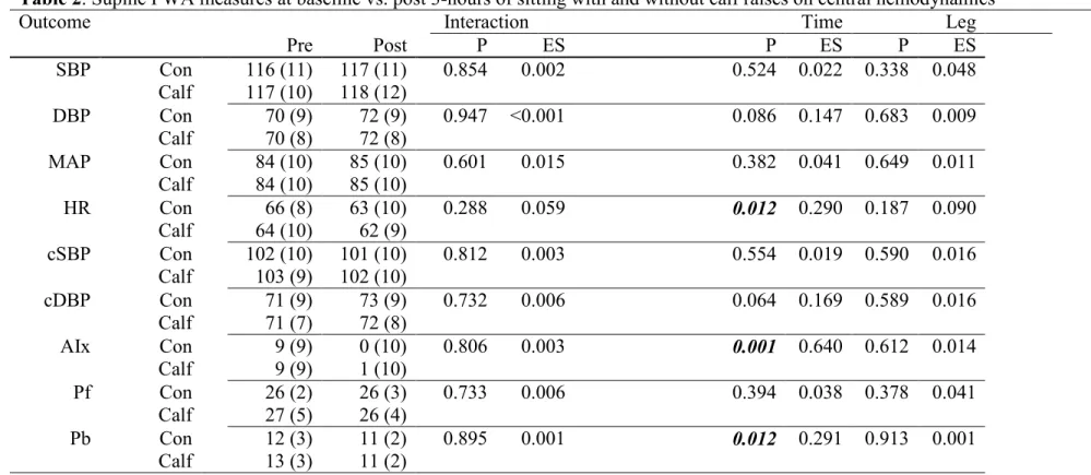
Heart rate significantly decreased from pre to post in the supine position (Table 2. p=0.012) but did not significantly change during sitting (Table 3). RMSSD used to calculate HRV
significantly increased during sitting from 10 to 180 minutes [10.3 (25.19), p=0.014 ES=0.236]. The low frequency (LF) range of HRV tended to change across time (p=0.056) with a trend for LF to increase from 10 to 90 minutes [7.4 (12.18), p=0.057] and the high frequency range (HF) of HRV significantly decreased during sitting (p<0.035) (Table 4)
Blood Pressure
SBP, cSBP, cDBP, DBP, and MAP were not significantly different in either condition during or after sitting (Table 2 and 3).
CD34+ CAC
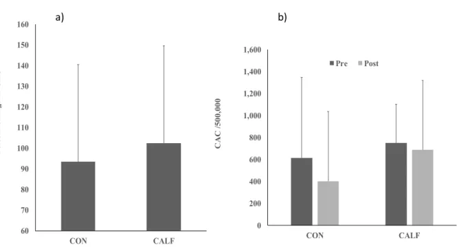
There was no significant interaction for CACs as a percentage of the parent population, and CACs per 500,000 events, nor was there a significant main effect of time or condition. (Figure 3).
Figure 2. Changes in a) PWV from before (pre) and after (post) 3 hours of sitting. PWV
significantly increases following sitting in both conditions(p<0.05). b) AIx before and after sitting in CON and CALF. AIx significantly decreases from 10 minutes to 90 minutes and stays depressed at 170 minutes in both conditions (p<0.05). # represents a significant effect of
31 CD34+ CAC mRNA expression
Expression of the angiogenic cytokine VEGF and the chemokine receptor CXCR4 relative to GADP expression did not significantly change between conditions or across time for immunomagnetically separated CD34+ CACs (Figure 4).
b) a) 0 200 400 600 800 1,000 1,200 1,400 1,600 CON CALF C A C / 5 0 0 ,0 0 0 Pre Post 60 70 80 90 100 110 120 130 140 150 160 CON CALF P e rc e n t c h a n g e i n C A C
Figure 3. Graph illustrating a) Percent change in CACs in CON vs. CALF and b) changes in CAC counts/500,000 from pre to post in CON and CALF. There were no significant
32
Figure 4. Gene expression of A) the angiogenic cytokine VEGF and B) the chemokine receptor CXCR4 did not change with sitting or not change with sitting or calf raises. mRNA is expressed without units relative to GADPH expression. There were no significant group, time or interaction effects. Error bars are represented as SD. VEGF vascular endothelial growth factor, CXCR4 CXC chemokine receptor-4, CAC circulating angiogenic cells, CON control group, CALF calf raise group, SD standard deviation.
0.0 0.2 0.4 0.6 0.8 1.0 1.2 1.4 1.6 1.8 2.0 CON CALF C X C R 4 m R N A ( re la ti v e u n it
s) Pre Post
0.0E+00 5.0E-03 1.0E-02 1.5E-02 2.0E-02 CON CALF V E G F m R N A ( re la ti v e u n it
s) Pre Post
33
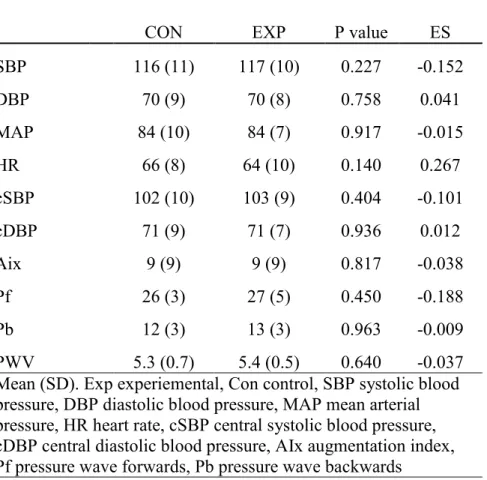
Table 1. Baseline differences between hemodynamic measures in young healthy men and women
CON EXP P value ES
SBP 116 (11) 117 (10) 0.227 -0.152
DBP 70 (9) 70 (8) 0.758 0.041
MAP 84 (10) 84 (7) 0.917 -0.015
HR 66 (8) 64 (10) 0.140 0.267
cSBP 102 (10) 103 (9) 0.404 -0.101
cDBP 71 (9) 71 (7) 0.936 0.012
Aix 9 (9) 9 (9) 0.817 -0.038
Pf 26 (3) 27 (5) 0.450 -0.188
Pb 12 (3) 13 (3) 0.963 -0.009
34
Table 2. Supine PWA measures at baseline vs. post 3-hours of sitting with and without calf raises on central hemodynamics
Outcome Interaction Time Leg
Pre Post P ES P ES P ES
SBP Con 116 (11) 117 (11) 0.854 0.002 0.524 0.022 0.338 0.048
Calf 117 (10) 118 (12)
DBP Con 70 (9) 72 (9) 0.947 <0.001 0.086 0.147 0.683 0.009
Calf 70 (8) 72 (8)
MAP Con 84 (10) 85 (10) 0.601 0.015 0.382 0.041 0.649 0.011
Calf 84 (10) 85 (10)
HR Con 66 (8) 63 (10) 0.288 0.059 0.012 0.290 0.187 0.090
Calf 64 (10) 62 (9)
cSBP Con 102 (10) 101 (10) 0.812 0.003 0.554 0.019 0.590 0.016
Calf 103 (9) 102 (10)
cDBP Con 71 (9) 73 (9) 0.732 0.006 0.064 0.169 0.589 0.016
Calf 71 (7) 72 (8)
AIx Con 9 (9) 0 (10) 0.806 0.003 0.001 0.640 0.612 0.014
Calf 9 (9) 1 (10)
Pf Con 26 (2) 26 (3) 0.733 0.006 0.394 0.038 0.378 0.041
Calf 27 (5) 26 (4)
Pb Con 12 (3) 11 (2) 0.895 0.001 0.012 0.291 0.913 0.001
Calf 13 (3) 11 (2)
Mean (SD), SBP systolic blood pressure, DBP diastolic blood pressure, MAP mean arterial pressure, HR heart rate, cSBP central systolic blood pressure, cDBP central diastolic blood pressure, AIx augmentation index, Pf pressure wave forwards, Pb pressure wave backwards
3
35
Table 3. PWA and HRV throughout a 3-hour bout of sitting with or without calf raises
Measure Time (min) Interaction Time Condition
10 90 170 P ES P ES P ES
SBP Con 110 (11) 110 (10) 111 (11) 0.531 0.068 0.725 0.035 0.668 0.010
Calf 111 (11) 110 (11) 110 (10)
DBP Con 69 (8) 69 (8) 70 (8) 0.381 0.102 0.111 0.217 0.349 0.046
Calf 67(6) 69 (7) 69 (7)
MAP Con 82 (9) 82 (8) 82 (9) 0.109 0.219 0.115 0.214 0.401 0.037
Calf 67 (6) 69 (7) 69 (7)
HR Con 73 (10) 75 (11) 74 (11) 0.535 0.067 0.294 0.127 0.569 0.017
Calf 72 (10) 74 (11) 74 (13)
cSBP Con 98 (10) 97 (9) 98 (11) 1.000 0.076 0.241 0.114 0.864 0.001
Calf 98 (10) 96 (9) 98 (8)
cDBP Con 70 (8) 71 (8) 71 (8) 0.398 0.097 0.507 0.105 0.188 0.089
Calf 67 (7) 70 (8) 67 (16)
Pf Con 25 (4) 25 (5) 23 (4) 0.405 0.102 0.086 0.242 0.631 0.012
Calf 26 (6) 24 (4) 24 (4)
Pb Con 13 (2) 11 (2) 11 (2) 0.870 0.025 <0.001 0.495 0.771 0.005
Calf 13 (4) 11 (3) 11 (2)
HF Con 28 (18) 19 (15) 18 (13 0.142 1.030 0.035 0.170 0.768 0.005
Calf 21 (14) 16 (13) 21 (15)
LF Con 36 (16) 42 (22) 37 (15) 0.756 0.015 0.056 0.148 0.269 0.269
Calf 27 (12) 36 (14) 32 (12)
Mean (SD), Con control SBP systolic blood pressure, DBP diastolic blood pressure, MAP mean arterial pressure, HR heart rate, cSBP central systolic blood pressure, cDBP central diastolic blood pressure, AIx augmentation index, Pf pressure wave forwards, Pb pressure wave backwards
3
36 Chapter V Discussion
This study shows for the first time that an acute bout of 3 hours of sitting increases aortic AS and burden on the heart as indicated by increases in PWV. Despite increases in PWV, AIx decreased during sitting while RMSSD from HRV increased. Contrary to our hypothesis, sitting did not affect absolute or relative CAC counts. Additionally, intermittent calf raises were
insufficient to prevent the changes in AS observed with acute inactivity and did not affect CACs. Cumulatively, these findings suggest: 1) a complex central hemodynamic response with different responses in AS versus arterial wave reflection, 2) that CD34+ CAC mobilization is not altered during 3 hours of sitting, and 3) that calf raises every 10 minutes do not appear to be a strong enough stimulus to alter central hemodynamics or circulating factors, contrary to previous work showing alterations at the local level.
Arterial Stiffness
This is the first study to our knowledge measuring the effects of an acute bout of prolonged sitting on PWV. We observed that PWV increased by 0.35m/s following 3 hours of sitting, which was statistically significant but falls below the 1.0 m/s that has been deemed clinically significant, at least chronically [10]. The chronic effects sitting are well demonstrated on CV risk and mortality, but their effects on AS are less prevalent. The best available
37
increases PWV along with acute increases in PWV during one bout of sitting implies that increased burden on the heart may contribute to increased risk of CVD. The aorta is the most proximal elastic tube that the heart “feels” during each beat. Therefore, repeated bouts of sitting resulting in increased aortic AS suggest repeated burden on the heart.
Despite the increase in PWV, we found ~9% decrease in AIx during sitting which is indicative of decreased arterial wave reflection and also AS. The conflicting results of PWV and PWA may be a result of different theory and methodology behind PWV and PWA, which must be discussed to determine potential explanatory mechanisms. The expert consensus document on AS suggests that the methods of PWV and PWA incorporate differing levels of the arterial tree to different degrees [8]. PWV is a direct measurement of aortic stiffness and is considered the gold standard that corresponds to the accepted propagative model of the arterial system and is highly dependent on the elasticity of the aorta. Meanwhile, PWA is an indirect measure of AS calculated from forward and reflected pressure waves. The origin of these reflected pressure waves is a result of forward and reflected waves that incorporate many conduit arteries. These conduit arteries have greater smooth muscle content and vasomotor tone, which does impact AIx independently of PWV [66]. As such, the nervous system provides as one potential mediator. There was a trend for increasing RMSSD during sitting which suggests an increase in
38
the decreased AIx may resonate from increased vasodilation which may be occurring as a result of decreased sympathetic outflow during sitting. Lastly, a 10 bpm increase in heart rate has been demonstrated to decrease absolute AIx by 4%, but there were no significant changes in HR during sitting thus eliminating this possibility [67]. In summary, these findings suggest that sitting promotes a complex central hemodynamic response that incorporates many heterogeneous regions of the arterial tree that are variably affected during sitting and potentially regulated by the ANS.
Circulating Angiogenic Cells
Moderate and vigorous intensity exercise mobilize CD34+ CACs in an intensity dependent manner, but the precise mechanisms are not clear [68] [69]. We hypothesized that increased shear stress during exercise was one potential mediator, the decrease in shear stress during sitting would elicit the opposite effect [23, 68, 69]. Interestingly, despite very different populations, baseline CAC counts are similar (after unit conversion) to reports in young active males with VO2max >50ml/kg/min in which counts were ~250 CD34+ events/1mL [58].
However, CAC mobilization do not appear to be affected by prolonged sitting as there was no change in absolute or relative CD34+ CAC counts. This was contrary to our hypothesis that sitting would decrease the release of CACs. Because this is the first study to our knowledge to assess the effects of an acute bout of sitting on CACs, comparisons to previous work are
39
While exercise increases CAC counts and this change may be due to increases in shear stress, the decreases associated with sitting were likely not as large of a magnitude, albeit in the opposite direction. Blood flow and shear stress changes with exercise are sustained throughout the entirety of the bout. In our pilot data, calf raises did elicit considerable increased in shear stress, but these changes were transient and returned to resting values rapidly (within 1-2 minutes). Additionally, sitting decrease blood flow in the legs, but the same has not been found systemically. It is likely that the changes in systemic flow after sitting or during calf raises were not strong enough to alter CAC mobilization[5]. However, activity or health status could be affecting baseline counts as others have reported a relationship between risk of CVD and decreasing CAC counts [70]. Therefore, the possibility of low CAC counts to begin with may have overshadowed the potential decrease observed from sitting.
40 Calf Raises
Previous evidence suggests that simple perturbations during sitting such as walking, fidgeting or standing can improve endothelial health in the popliteal or superficial femoral artery [12, 13, 38], however, these studies did not report changes in the arm, which is the generally accepted measure of endothelial health. In this study, we attempted to address this gap by
utilizing regular muscular contractions (e.g. calf raises) to activate the muscle pump and increase venous return while examining clinically relevant measures of vascular health, albeit at a
different segment of the arterial tree. Data suggests that despite the decrease in blood pooling, calf raises do not acutely affect aortic AS. Despite evidence demonstrating the benefits of
fidgeting and walking on leg endothelial function, similar perturbations such as calf raises do not alter AS during sitting. This may be as a result of the fact that FMD is highly dependent on shear stress whereas, AS is only partially dependent on it and this varies depending on the location within the arterial tree [74]. Additionally, the changes from fidgeting and brief exercise were only seen in the legs and not the arms, which may mean that the change in blood flow and shear stress are not sufficient to affect the arterial tree overall, rather the legs are may be more sensitive to small increases in shear stress and that only monitoring the legs may lead to misleading
conclusions. Here we show that while fidgeting may be sufficient to prevent decreases in leg health, such simple perturbations appear to be inadequate for inducing changes in variables related to overall CV health.
Sitting Recommendations
41
guidelines recommending 150 minutes of moderate to vigorous physical activity should also be recommended to prevent other consequences of sedentary lifestyles [75]. Lastly, preliminary evidence on the acute effects of sitting suggest that interventions such as fidgeting, local heating, and intermittent exercise can prevent the detrimental changes in local blood flow associated with sitting, but these changes may not be generalized to systemic vascular health such as PWV.
Limitations
42 Conclusions
Prolonged sitting is still an emerging risk factor for the development of CVD. However, the precise mechanism explaining the effects of sitting remained in question, with many
clinically relevant targets still unexamined. Here we show that sitting in may increase central burden on the heart, and that calf raises do not appear to clinically alter these changes.
Furthermore, sitting does not induce changes in CAC counts or function. Because sitting confers increased risk of mortality, these findings suggest that increased burden on the heart due to increased aortic AS may be a contributing factor. Cumulatively, these findings suggest that sitting produces a detrimental vascular response from a systemic but not stem cell level that intermittent calf raises do not seem to effect, and that the nervous system likely plays a compensatory mechanistic role.
Clinical Implications
Practically, these results add to the body of evidence regarding the effects of sitting. In terms of lifestyle interventions, ACSM guidelines of 150 minutes of moderate to vigorous physical activity are recommended for optimal health benefits; if these can’t be met current at least 60-75 minutes of moderate to vigorous activity can prevent the increased risks associated with sitting [62, 75]. Interventions while sitting lack longitudinal clinical evidence, but
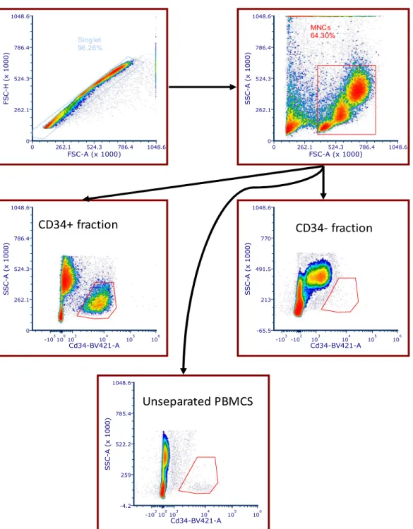
43 Figure S1. Gating Strategy and CD34 Purity
Live_Dead-FITC-A C o u n t
-103102 104 105 106 0 3115 6229 9344 12458 97.18%
FSC-A (x 1000)
F S C -H ( x 1 0 0 0 )
0 262.1 524.3 786.4 1048.6 0 262.1 524.3 786.4 1048.6 Singlets 88.34%
FSC-A (x 1000)
S S C -A ( x 1 0 0 0 )
0 262.1 524.3 786.4 1048.6 -65.5 213 491.5 770 1048.6 MNC 51.94%
FSC-A (x 1000)
S S C -A ( x 1 0 0 0 )
0 262.1 524.3 786.4 1048.6 -65.5
213 491.5 770 1048.6
Lymph ocyte gate 83.16% CD34-BV421-A S S C -A ( x 1 0 0 0 )
44 B)
Fig S1. A) Gating strategy for CACs and purity of magnetic separation. Live cells were gated first. Singlets were gated, followed by all mononuclear cells and a lymphocyte gate to capture
CD34+ PBMCs. B) Flow cytometry data collected from magnetic isolation and subsequent staining with anti-CD34+ antibodies. Data represents positive fraction, negative fraction, and unseparated PBMCs from the same participant.
FSC-A (x 1000)
F S C -H ( x 1 0 0 0 )
0 262.1 524.3 786.4 1048.6 0 262.1 524.3 786.4 1048.6 Sing let 96.26%
FSC-A (x 1000)
S S C -A ( x 1 0 0 0 )
0 262.1 524.3 786.4 1048.6 0 262.1 524.3 786.4 1048.6 MNCs 64.30% Cd34-BV421-A S S C -A ( x 1 0 0 0 )
-103100103 104 105 106 0 262.1 524.3 786.4 1048.6 Cd34-BV421-A S S C -A ( x 1 0 0 0 )
-103-102 103 104 105 106 -65.5 213 491.5 770 1048.6 Cd34-BV421-A S S C -A ( x 1 0 0 0 )
-103100103 104 105 106 -4.2
259 522.2 785.4 1048.6
CD34+ fraction CD34- fraction
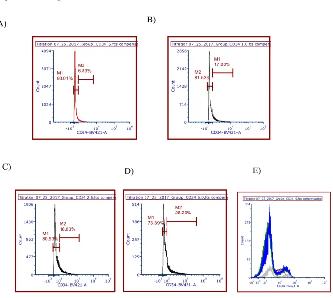
45 Figure S2. Example titration data for CD34
Fig S2. Example titration data for CD34. Concentration with greatest signal to noise ratio was selected as optimal concentration. A-D) Samples were prepared using step wise increasing concentrations of anti-body stains from 0.5 to 1.0 to 2.5 to 5.0 µL. E) Overlaid histograms with 0.5 in plum, 1.0 in green, 2.5 in blue, and 5.0 in gray.
Titration 07_25_2017_Group_CD34 .5.fcs compensated
CD34-BV421-A C o u n t
-103 104 105 106 0 1024 2047 3071 4094 M2 6.83% M1 93.01%
Titration 07_25_2017_Group_CD34 1.0.fcs compensated
CD34- BV421-A C o u n t
-103 104 105 106 0 714 1428 2142 2856 M2 81.53% M1 17.80%
Titration 07_25_2017_Group_CD34 2.5.fcs compensated
CD34-BV421-A C o u n t
-103 103 104 105 106 0 477 953 1430 1906 M2 18.83% M1 80.93%
Titration 07_25_2017_Group_CD34 5.0.fcs compensated
CD34-BV421-A C o u n t
-103103 104 105 106 0 129 257 386 514 M2 26.29% M1 73.39%
Titration 07_25_2017_Group_CD34 .5.fcs compensated
CD34-BV421-A C o u n t
-102100102 103 104 105 106 0 91 182 273 364 A) C)
D) E)
46 Appendices
Appendix A
Department of Exercise and Sport Science Medical History Questionnaire Screening
Subject:__________________________ Telephone:______________
Address:________________________________________________________________ Email:___________________________________ Age:______________________ Patient History
1. How would you describe your general health at present? YES NO 2. Excellent______ Good_______ Fair______ Poor______
3. Do you have any health problems at the present time? _____ _____ 4. If yes, please describe:
5. Have you ever been told you have heart trouble? _____ _____ 6. If yes, please describe:
7. Do you ever get pain in your chest? _____ _____
8. Do you ever feel light-headed or have you ever fainted? _____ _____ 9. If yes, please describe:
47
12. Have you ever had difficulty breathing at rest or with exertion? _____ _____ 13. If yes, please describe:
14. Have you ever been treated for infectious mononucleosis, hepatitis, pneumonia, or another infectious disease during the past year? _____ _____ 15. If yes, name the disease:
16. Have you ever been treated for or told you might have diabetes? _____ _____ 17. Have you ever been treated for low blood sugar? _____ _____ 18. Have you ever experienced heat stroke or heat exhaustion? _____ _____ 19. If yes, when?
20. Are you now taking any pills, medications, or supplements? _____ _____ 21. If yes, please list:
22. Have you had any recent (within 1 year) difficulties with your:
a. Feet _____ _____
b. Legs _____ _____
c. Back _____ _____
Menstrual Cycle
23. What was the start date of your most recent menstrual cycle? __________ Family History
48
a. Sudden death _____ _____
b. Cardiac disease _____ _____
c. Marfan’s syndrome _____ _____
Bone and Joint History
25. Have you ever been treated for Osgood-Schlatter’s disease? _____ _____ 26. Have you ever had any injury to your neck involving nerves or
vertebrae? _____ _____
27. Do you experience pain in your back? _____ _____
28. Have you ever had an injury to your back? _____ _____ 29. If yes, did you seek the advice of a doctor? _____ _____ 30. Have you ever been told that you injured the ligaments or cartilage of either knee
joint? _____
_____
31. Do you think you have a trick knee? _____ _____ 32. Do you have a pin, screw, or plate anywhere in your body as the result of bone or joint surgery that presently limits your physical capacity? _____ ____
33. If yes, indicate where:
Activity History
34. During your early childhood (to age 12) would you say you were:
49
Very active ____ Quite active____ Moderately active____ Seldom active____ 36. Did you participate in:
Intramural high school sports? _____ _____
Community sponsored sports? _____ _____
Varsity high school sports? _____ _____
Active family recreation? _____ _____
37. Since leaving high school, how active have you been?
Very active ____ Quite active____ Active____ Inactive____
38. Have you previous participated in strength training _____ _____ 39. Do you participate in any moderate to vigorous activity at present? _____ _____ 40. If yes, please list:
Activity Frequency Duration Intensity
41. Whom shall we notify in case of emergency? Name:
Phone: (Home) (Work)
50 Appendix B
52 Appendix C University of North Carolina at Chapel Hill
Consent for Storing Biological Specimens Without Identifying Information
_______________________________________________________________________ Consent Form Version Date: ______________
IRB Study # 16-3051
Title of Study: Effects of Prolonged Sitting on Cerebral Perfusion and Executive Function Principal Investigator: Quentin Willey
Principal Investigator Department: Exercise and Sport Science Principal Investigator Phone number: (919) 962-0396
Principal Investigator Email Address: [email protected] Co-Investigators: Erik Hanson, Claudio Battaglini, William Evans
Faculty Advisor: Lee Stoner
Faculty Advisor Contact Information: (919) 962-0534
___________________________________________________________________________ What are some general things you should know about research?
Research is designed to gain scientific information that may help other people in the future. You may not receive any direct benefit from participating. There also may be risks.
You may refuse to take part in research. If you are a patient with an illness, you do not have to be in research in order to receive treatment.
Details are discussed below. It is important that you understand this information so that you can make an informed choice. You will be given a copy of this consent form. You should ask the researchers named above, or staff members who may assist them, any questions you have about this study at any time.
What is the purpose of this specimen repository or “biobank?”
53
The purpose of this particular repository or biobank is to better understand the mechanisms altering vascular repair during sitting. This will include blood plasma or whole blood. Future research may be assessment of mRNA with sitting to assess potential changes in protein development during sitting.
How will the specimens be collected?
• Provide specific details about how the specimen will be collected, OR
• if specimen already exists from previous clinical sources or research studies, inform subjects.
What will happen to the specimens?
Address specific areas about how the sample will be used and stored:
• Provide a clear description of the operation of the specimen repository
• Where will the specimen be stored?
• When will the specimens be destroyed?
• Inform subjects of conditions under which data and specimens will be released to other investigators.
What are Genome Wide Association Studies (GWAS)?
Delete if this does not apply to your study and you know data will never be submitted to GWAS The National Institutes of Health (NIH) has established a national database that will hold
information from many individuals across the country, including medical information and genetic information. Your blood and tissues contain genes which are made of DNA that is unique to you. Access to this national database will be controlled and limited to other researchers.
What are the possible benefits to you?
Benefits to you are unlikely. Studies that use specimens from this repository may provide additional information that will be helpful in understanding the effects of prolonged sitting. What are the possible risks or discomforts involved with the use of your specimens? Describe immediate and long-term social, physical, and psychological risks/discomforts related to the specimen collection and storage. Address all risks that are applicable.
• Unknown risk: Subjects should be informed that there may be risks that at this time are unknown.
• Physical risks: If new samples are being collected include the physical risk associated with the sample collection for research purposes.
• In addition, use the following.
54
Will there be any cost to you for storage of the specimens?
There will be no cost to you for the storage and use of the specimens for research purposes. Will you receive anything for the use of your specimens?
You will not be receiving compensation for taking part in this study.
Who owns the specimens?
Insert any contract, grant or agreement language related to specimen ownership or modify the following boilerplate.
Any blood, body fluids, or tissue specimens obtained for this purpose become the exclusive property of the University of North Carolina at Chapel Hill. This organization may retain, preserve or dispose of these specimens and may use these specimens for research that may result in commercial applications. There are no plans to compensate you for any future commercial use of these specimens.
How will information about you be protected?
The researchers will not have any identifying information about you so there is no risk to your privacy.
Will you receive study results of future research involving your specimens?
Most research with your specimens is not expected to yield new information that would be meaningful to share with you personally. In this case that would be impossible, because the researchers have no information that identifies you.
Can you withdraw the specimen from this repository?
You may not withdraw your specimen in the future because there are no identifiers on the specimen and the researchers will not know which specimen is yours.
What will happen if you are injured by this research? Omit this section if the specimens have already been collected.
All research involves a chance that something bad might happen to you. This may include the risk of personal injury. In spite of all safety measures, you might develop a reaction or injury from having your specimen collected. If such problems occur, the researchers will help you get medical care, but any costs for the medical care will be billed to you and/or your insurance company. The University of North Carolina at Chapel Hill has not set aside funds to pay you for any such reactions or injuries, or for the related medical care. However, by signing this form, you do not give up any of your legal rights.
Who is sponsoring this research?