EARLY OVARIAN CANCER
Cover Page
The handle
http://hdl.handle.net/1887/29822
holds various files of this Leiden University
dissertation.
Author
: Timmers, Petra Jeanette
Title
: Early ovarian cancer
EARLY OVARIAN CANCER
The research described in this thesis was performed at the European Organisation for Research and Treatment of Cancer in Brussels, Belgium.
ISBN/EAN: 978-94-91955-03-7 Author: Petra Timmers Cover design: Els Kruit
Printed by: Pasmans, Den Haag
© Petra J. Timmers, 2014
All rights reserved. No part of this thesis may be reproduced or transmitted, in any form or by any means, without prior permission of the author.
www.fsc.org
MIX
Paper from responsible sources
EARLY OVARIAN CANCER
Proefschrift
ter verkrijging van de graad van Doctor aan de Universiteit Leiden,
op gezag van Rector Magnificus prof. mr. C.J.J.M. Stolker, volgens besluit van het College voor Promoties
te verdedigen op donderdag 16 oktober 2014
klokke 15.00 uur
door
Petra Jeanette Timmers
PROMOTIECOMMISSIE
Promotores Prof. dr. J.B.M.Z. Trimbos
Prof. dr. I. Vergote
Overige leden Prof. dr. G.J. Fleuren
Prof. dr. ir. J.J.M. van der Hoeven Prof. dr. G.G. Kenter
Contents
CHAPTER 1 9
General Introduction
CHAPTER 2 27
Impact of adjuvant chemotherapy and surgical staging in early-stage ovarian carcinoma: European Organisation for Research and Treatment of Cancer– Adjuvant ChemoTherapy in Ovarian Neoplasm trial
J Natl Cancer Inst 2003; 95(2): 113–125
CHAPTER 3
Chemotherapy for early ovarian cancer 53
Curr Opin Obstet Gynecol 2004; 16(1): 43-48
CHAPTER 4 65
Clear cell carcinoma compared to serous adenocarcinoma in early ovarian cancer: same prognosis in a large randomized trial
Int J Gynecol Cancer 2009; 19(1): 89-93
CHAPTER 5 81
Understanding the problem of inadequately staging early ovarian cancer Eur J Cancer 2010; 46(5): 880-884
CHAPTER 6 93
Lymph node sampling and taking of blind biopsies are important elements of the surgical staging of early ovarian cancer
CHAPTER 7 105 Prognostic value of Ic substages in invasive epithelial ovarian carcinoma
Submitted
CHAPTER 8 117
Surgical staging and treatment of early ovarian cancer: long-term analysis from a randomized trial
J Natl Cancer Instit 2010; 102(13): 982-987
CHAPTER 9 131
General Discussion
CHAPTER 10 143
Summary
CHAPTER 11 149
Nederlandse Samenvatting
Abbreviations 159
Curriculum Vitae 161
Publications 163
Authors and affiliations 165
Chapter
1
General Introduction 11
1
EPIDEMIOLOGY
Ovarian cancer is the second most common gynecologic malignancy with an incidence of about 15 cases per 100,000 women in Western countries [1-3] and is ranked the seventh leading cause of cancer-related death in women worldwide [4,5]. The high mortality rate is due partly to the fact that most ovarian cancers are diagnosed at an advanced stage of the disease. Early ovarian cancer accounts for approximately one-third of all newly diagnosed ovarian carcinomas [6]. The early-stage disease is defined by the International Federation of Gynecology and Obstetrics (FIGO) as stage I and IIa and is histologically confined to the internal gonads in the small pelvis [7]. Worldwide around 68,000 new cases of early ovarian carcinoma will be diagnosed annually [5]. The cumulative risk (age 0-64) for ovarian cancer is 0.5 [5]. The incidence of early-stage ovarian cancer in the Netherlands is approximately 370 patients per year. The median age at diagnosis is 57 year with around 30% less than 50 year [8].
CLASSIFICATION
12 Chapter 1
PATHOGENESIS AND HIS TOPATHOLOGY
Several theories exist on the pathogenesis of epithelial ovarian cancer. While it is widely believed that the epithelial component of the ovary gives rise to the common epithelial ovarian carcinomas [17], it is not clear whether these cancers originate from a single-cell layer of surface epithelium or in architectural aberrations of the surface epithelium. These include surface epithelial-lined clefts and cortical inclusions cysts, thought to result from post-ovulatory wound repair, tissue remodeling associated with pregnancy or aging, para-ovarian adhesions, or simply the dynamic interaction between surface epithelium and underlying stroma [18-20]. A study of Pothuri et al. supports a model in which ovarian cancers frequently arise within epithelial inclusion cysts, but not the surface epithelium per se, and that carcinoma may be preceded by a dysplastic precursor lesion [21]. Another histopathology-based theory holds that epithelial ovarian cancer may arise in components of the secondary Müllerian system, located within or adjacent to the ovary [22]. The histologic subtypes of epithelial ovarian cancer include serous, mucinous, endometrioid, clear cell, mixed type and undifferentiated tumors. A new model for the pathogenesis of ovarian cancer is proposed by Kurman and coworkers [23]. In this model ovarian tumors are divided into two broad groups designated Type I and Type II. Type I tumors are slow growing, generally confined to the ovary at diagnosis and develop from well esthablised precursor lesions (borderline tumors). Type I tumors included low-grade micropapillary serous carcinoma, mucinous, endometrioid and clear cell carcinoma. They are genetically stable tumors and are characterized by mutations
in a number of different genes including KRAS, BRAF, PTEN, and beta-catenin. Type II
tumors, like the high grade serous carcinomas, are rapidly growing, highly aggressive neoplasms for which well defined precursor lesions have not yet been described. This group of tumors have a high level of instability and are characterized by mutation of TP53. Some other studies suggested that type II ovarian carcinomas are perhaps not ovarian cancers at all, but rather originate in the fallopian tube [24,25].
General Introduction 13
1
type of tumor, which has been shown to be poorly reproducible between pathologistsin several studies [29-31].
Silverberg proposed a new grading system modeled on the Nottingham system of breast cancer grading and designed to be applied to all invasive epithelial carcinomas of the ovary, including clear cell tumors [32].
SURGICAL S TAGING
Major advances in the understanding of the natural history of early ovarian cancer occurred in the 1970s and 1980s when some authors defined the incidence of occult disease in the omentum, paracolic gutters, diaphragm, in the peritoneal washings and the lymph nodes [33-36].
Piver et al. report that microscopic metastases in the abdominal cavity at different sites like the right diaphragm (11%), the omentum (3%) and malignant cells in peritoneal washings (33%) were found in patients with presumed early-stage ovarian cancer [33]. Another route of metastasis is via the lymphatic channels. In 1974 Knapp and Friedmann reported at first their experience with aortic lymph node metastases in patients with early ovarian cancer [37]. In the decades thereafter other studies have extended our knowledge about the routes and incidence of lymphatic spread to the pelvic and aortic lymph nodes. The anatomy of ovarian lymph drainage is complex. It has been stated that the drainage trunks leaving the subovarian plexus take a cephaled course toward the aortic nodes via the infundibulopelvic ligament [38]. Another lymphatic channel courses from the hilus of the ovary within the folds of the broad ligament to drain into the obturator, external, and common iliac nodes which are interconnected by a great variety of anastomoses [39]. Lymphatic vessels also enter and travel along the round ligament to reach the inguinal region [40]. Involvement of pelvic nodes has been reported to occur in 8-15% [33,41] and of para-aortic nodes in 5-24% of patients with stage I disease [41,42].
14 Chapter 1
These findings have led to a better understanding of the spread of early ovarian cancer within the peritoneal cavity and the need for a comprehensive surgical staging of these patients. Complete surgical staging consists of abdominal hysterectomy with bilateral salpingo-oophorectomy, infracolic omentectomy, sampling of pelvic and para-aortic lymph nodes, careful inspection of the whole abdominal cavity, taking of blind peritoneal biopsies and biopsies of any suspect lesions and of adhesions adjacent to the tumor and peritoneal washings. The stage of ovarian cancer is defined as the extent of the disease at the time of diagnosis. This can only be determined by exploratory surgery and meticulous evaluation of all areas of disease dissemination. The surgical procedures and the requirements for optimal intraperitoneal surgical staging of cancers apparently confined to the pelvis are well established by the FIGO [46]. Different guidelines for surgical staging of ovarian cancer were defined like the European guidelines of staging of ovarian cancer (EGSOG), the European Organisation for Research and Treatment of Cancer (EORTC) guidelines and the Gynecologic Oncology Group (GOG) guidelines [47,48]. EGSOG guidelines require lymph node sampling among the external and common iliac vessels and along the aorta and vena cava, specifically between the inferior mesenteric artery and the level of the left renal vein. The GOG guidelines recommend also excision of the distal half of the obturator fatpad anterior to the obturator nerve and prescribe dissection from the area between the inferior mesenteric artery and the left renal vein only in case of palpably suspicious nodes [47] .
In a study of Petru et al., 55% of lymph node metastases in presumed early-stage ovarian cancer were less than 2 mm in diameter [49]. Similar findings have been found by Wu et al., 33% of clinically nonsuspicious nodes harbored metastases of epithelial ovarian cancer [36].
The result of surgical staging in ovarian cancer is a redistribution of stages. Several studies have shown that a substantial number of patients initially believed to have disease confined to the ovaries will be upstaged. Young et al. [34] reported on a group of 100 patients of whom 62 agreed to be restaged after being referred for treatment of stage I or II ovarian cancer. Of these patients almost one third (31%) were upstaged, with the final stage in most becoming stage III. Comparable studies by Soper, Hellewa and Buchsbaum reported similar results [50-52].
General Introduction 15
1
and general surgeons 32%. These findings are also supported by others [53-56].Five-year survival and disease-free survival, respectively, for stage I-II ovarian cancer patients surgically staged by a gynecologic oncologist were 83% ± 7% and 76% ± 8%, compared to 59% ± 11% (P < 0.05) and 39% ± 11% (P < 0.03) for the group operated upon by a non -oncologist [57]. In a study of Vernooij et al. among patients with FIGO stage I-IIa disease, risk of ovarian cancer-specific mortality was 30% and 42% lower after treatment in semi-specialized and specialized hospitals, respectively, as compared to general hospitals [58]. In another study the level of specialization and the volume of hospitals and the number of gynecologists were strongly related to the proportion of adequately staged patients [59]. Comprehensive surgical staging is essential for prognostic determination and treatment planning for patients with apparent early-stage ovarian cancer as it defines a subset of patients that do not require adjuvant treatment in order to reduce the risks of late complications of chemotherapy as well as the morbidity and costs caused by such therapy [60].
TREATMENT MODALITIES
In the past many randomized trials have enrolled patients with early ovarian cancer in order to evaluate the value of adjuvant therapies like external radiotherapy,
intraperitoneal installation of radionuclides such as gold-198 (198Au) or phosphorus-32
(32P), single alkylating agents or platinum-based single or combination chemotherapy.
Some of these studies were of low quality because of the omission of a control arm, inclusion of borderline tumors and incomplete surgical staging [61-67]. It was stated that: “the inclusion of inaccurately staged, incompletely evaluated patients in trials attempting to test the potential value, if any, of adjuvant treatment will be difficult to interpret at best and misleading at worst” [68].
Meta-analyses performed by Winter-Roach et al. [69] for those trials with complete surgical staging procedures comparing adjuvant chemotherapy (AC) versus radiotherapy showed no significant difference between the effects of AC and radiotherapy on overall survival (OS) and disease-free survival (DFS). The main analysis of OS showed an HR of 0.85 (95% CI 0.62 to 1.17) and the DFS showed an HR of 0.94 with a 95% CI of
0.56 to 1.59. In the subgroups, AC versus 32P, AC versus whole abdominal radiation
(WAR) or platinum-based AC versus 32P, radiotherapy showed no statistical advantage
for any modality. Cisplatin containing regimens are preferable to radiotherapy and
16 Chapter 1
Most randomized trials compared two or three different treatment arms and almost all had a very low power because of the small number of patients or too few events. Furthermore, the efficacy of AC cannot be firmly established without an untreated observation arm.
In a study of Young et al. [70], 81 patients with grade 1 or 2 stage Ia or Ib (FIGO 1973) ovarian cancer were randomly assigned to receive 12 cycles of orally administered adjuvant melphalan and 81 no adjuvant treatment. No significant difference in overall survival (OS, 94% versus 98%) or disease-free survival (DFS, 91% versus 98%) was found. Bolis et al. [71] showed in 85 FIGO stage Ia or Ib, grade 2 or 3 patients a significant DFS advantage in the cisplatin group (83%) compared to the observation arm (65%). However, when the controls were treated with cisplatin at relapse, they had the same overall 5-year survival as the group receiving cisplatin treatment, as an adjuvant modality following initial surgery: 82% and 88% respectively. This result suggests that eight of the ten women in the cisplatin arm had been overtreated. If survival after relapse is compared, the patients in the upfront cisplatin group did much worse than patients in the nontreated group. Therefore the authors suggested that salvage treatment was more effective in the observation arm than in the chemotherapy arm, but also in this trial the number of patients was too small to draw too strong conclusions.
The Nordic Cooperative Ovarian Cancer Group performed a randomized study between 1992-1997 in patients with high risk epithelial ovarian cancer (stage I) including 162 eligible patients comparing carboplatin and observation [72]. High risk was defined as grade 2 or 3 tumor, all clear cell and DNA aneuploid tumors, independent of grade. Only 10% of the patients had a complete comprehensive surgical staging. The study was closed prematurely due to poor accrual. The estimated 5-year OS and DFS rates were 86% versus 85% and 70% versus 71% for the adjuvant chemotherapy and control group, respectively.
General Introduction 17
1
PROGNOSIS
The survival rates reported in the literature for patients with early ovarian cancer (EOC) vary, partly due to the differences in completeness of surgical staging and grade of differentiation and inclusion in some series of borderline tumors. The 5-year survival rates range from 76-95% for stage I [4,75-78] and 42-70% for stage II patients [75,78]. Over the past decades the improvement of the relative survival has occurred during the period in which adjuvant chemotherapy has been used in the treatment of EOC together with an improvement of the surgical staging in the same period. Although the overall survival curves are good compared to patients with advanced disease, approximately 10-50% of women with early EOC will experience a recurrence or die as a result of the disease [6,76,79,80]. Because of these long-term figures, major efforts have been made to develop adjuvant therapies, to optimize surgical staging and to identify prognostic factors that can predict patient outcome.
Women with early-stage ovarian cancer have a much better chance of achieving a cure than do women with late-stage disease. This difference makes screening for ovarian cancer, with the hope of detecting it in its presymptomatic state, an attractive concept. Unfortunately, efforts to demonstrate that screening for ovarian cancer in the general population can decrease mortality have been disappointing [81]. No accurate screening test is available but transvaginal sonography and CA 125 determinations can be valuable in selected patients [82] as well as the Risk of Malignancy Index (RMI) [83,84]. The results of the prevalence screen of the UK Collaborative Trial of Ovarian Cancer Screening (UKCTOCS) shows that the sensitivity of the multimodel screening (MMS) with annual CA 125 screening with transvaginal ultrasound scan as a second line test and annual screening with transvaginal ultrasound (USS) is encouraging. Specificity was higher in the MMS group than in the USS group, resulting in lower rates of repeat testing and surgery. The results of ongoing screening are awaited so that the effect of screening on mortality can be determined [85].
18 Chapter 1
PROGNOS TIC FACTORS
Prognostic variables can divide patients into risk groups. It is generally accepted that stage Ia grade 1 tumors with complete surgical staging have a very good prognosis with a 5-year overall survival of 95% permitting fertility sparing surgery [6]. Trimbos et al. described a 100% disease-free 5-year survival in patients with well differentiated early ovarian cancer who had undergone a careful staging procedure [89]. Young found a 98% disease-free survival rate in 38 patients with well-staged, low risk early ovarian cancer [68]. In a study of Vergote et al. none of the 77 patients with well differentiated DNA diploid tumors had relapses [74].
Several prognostic factors for early-stage ovarian carcinoma have been analyzed. Some of them are biological and clinical in nature, but others such as the thoroughness of the staging procedure, the extent of the surgery, and the philosophy of treatment, are defined by human nature [90].
Many clinical and pathological characteristics have been found to correlate with survival in early ovarian carcinoma including stage, histologic type, tumor grade, ascites, age and ploidy [15,70,72,89,91-101]. Furthermore rupture of the tumor, dense adhesions and surgical staging have been indicated as independent prognostic factors in prior reports [89-91,102-105]. In a multivariate analysis of 351 patients with stage I ovarian cancer Zanetta et al. [90] found that the extent of surgical staging was a statistically significant independent prognostic factor for disease-free and overall survival.
The largest retrospective multivariate analysis in stage I epithelial ovarian cancer of Vergote et al. including 1,545 patients, concluded that the most important independent prognostic factors were degree of differentiation followed by rupture before surgery, FIGO substage Ib versus Ia and age [104].
General Introduction 19
1
SCOPE AND OUTLINE OF THE THESIS
In order to evaluate the effect of adjuvant chemotherapy and surgical staging in early ovarian cancer patients, the European Organisation for Research and Treatment of Cancer - Gynecologic Cancer Group (EORTC-GCG) performed the Adjuvant Chemo-Therapy in Ovarian Neoplasm (ACTION) trial (EORTC trial 55904). The ACTION trial was a randomized study on the role of platinum containing adjuvant chemotherapy in early ovarian cancer patients with FIGO stages Ia and Ib (grade II-III) and stages Ic and IIa (grade I-III) and all stages Ia-IIa clear cell carcinoma after surgery. Randomization between platinum containing chemotherapy and no adjuvant treatment took place after surgical staging and patients randomized to receive platinum based chemotherapy were treated within four weeks after surgical treatment for at least four consecutive courses.
Because there still exist a lot of discussion and questions about the treatment and prognosis of patients with early ovarian cancer, we further examined the surgical staging categories and the different subgroups in order to get some answers on these subjects.
In chapter 2 the results of the first analysis of the ACTION trial are described between the adjuvant chemotherapy arm and the no treatment arm (observation arm) on the role of platinum-based chemotherapy on disease-free survival (DFS) and overall survival (OS). Furthermore, analyses were performed in optimally staged patients versus non-optimally staged patients.
A review of the treatment modalities and recent findings in early ovarian cancer patients is given in chapter 3.
The clinical characteristics and response to platinum-based chemotherapy in patients with clear cell carcinoma (CCC) versus serous adenocarcinoma (SAC) randomized in the ACTION trial are described in chapter 4.
Early ovarian cancer patients are often incompletely staged during their initial surgery. In chapter 5 we discuss the possible reasons for inadequately staging early ovarian cancer patients.
20 Chapter 1
REFERENCES
1. Ferlay J, Parkin DM, Steliarova-Foucher E. Es-timates of cancer incidences and mortality in Europe 2008. Eur J Cancer 2010; 46: 765-781. 2. Bray F, Loos AH, Tognazzo S, La VC. Ovarian
cancer in Europe: cross sectional trends in inci-dence of mortality in 28 countries, 1953-2000. Int J Cancer 2005; 113: 977-990.
3. Levi F, Lucchini F, Negri E, Boyle P, La Vecchia C. Cancer mortality in Europe 1995-1999 and an overview of trends since 1960. Int J Cancer 2004; 110: 155-169.
4. Jemal A, Siegel R, Ward E, Hao Y, Xu J, Thun J. Cancer statistics 2009. CA Cancer J Clin 2009; 59: 225-249.
5. Parkin DM, Bray F, Ferlay J, Pisani P. Global cancer statistics. CA Cancer J Clin 2005; 55: 74-108.
6. Young RC. The treatment of early-stage ovarian cancer. Semin Oncol 1995; 22: 76-79. 7. Announcement. Staging announcement: FIGO
cancer committee. Gynecol Oncol 1986; 25: 383-385.
8. Chan JK, Fuh K, Shin JY, et al. The treatment and outcomes of early-stage epithelial ovarian cancer: have we made any progress?. Br J Can-cer 2008; 98: 1191-1196.
9. Jones HW. Recent advances in gynecologic can-cer. Bull Sloane Hosp Wom NY 1965; 11: 33. 10. International Federation of Gynecology and
Obstetrics. Classification and staging of malig-nant tumors in the female pelvis. Acta Obstet Gynecol Scand 1971; 50: 1-7.
11. Annual report on the results of treatment in carcinoma of the uterus, vagina and ovary. Kottmeier HL ed. 1973; 15: 15-18.
12. Vergote I, De Brabander J, Fyles A, et al. Prog-nostic importance of degree of differentiation
and cyst rupture in stage I invasive epithelial ovarian carcinoma. Lancet 2001; 357(9251): 176-182.
13. Paulsen T, Kærn J, Tropé C. Improved 5-year disease-free survival for FIGO stage I epithelial ovarian cancer patients without tumor rupture during surgery. Gynecol Oncol 2011; 122(1): 83-88.
14. Kottmeier HL. Presentation of therapeutic re-sults of treatment in carcinoma of the femeale pelvis: experience of the annual report on the results of treatment in carcinoma of the uterus, vagina and ovary. Gynecol Oncol 1976; 4: 16-18.
15. Pecorelli S. Revised FIGO staging for carcinoma of the vulva, cervix, and endometrium. Int J Gy-naecol Obstet 2009; 104: 103-104.
16. Prat J. FIGO staging for uterine sarcomas. Int J Gynaecol Obstet 2009; 104: 177-178. 17. Seidman JD. Pathology of borderline (low
ma-lignant potential) ovarian tumours. Best Pract Res Clin Obstet Gynaecol 2002; 16: 499-512. 18. Radisavljevic SV. The pathogenesis of ovarian
inclusion cysts and cystomas. Obstet Gynecol 1977; 49: 424-429.
19. Scully RE. Pathology of ovarian cancer precur-sors. J Cell Biochem Suppl 1995; 23: 208-218. 20. Auersperg N, Wong AS, Choi KC, Kang SK,
Leung PC. Ovarian surface epithelium: bio logy, endocrinology, and pathology. Endocr Rev 2001; 22: 255-288.
21. Pothuri B, Leitao MM, Levine DA, et al. Genetic analysis of the early natural history of epithelial ovarian carcinoma. PloS One 2010; 5: e10358. 22. Dubeau L. The cell of origin of epithelial tumors
and the ovarian surface dogma: does the em-peror have no clothes? Gynecol Oncol 1999; 72 (3): 437-442.
In chapter 7 the prognostic value of the FIGO Ic substages including capsule rupturing, ascites containing malignant cells, surface tumor and positive peritoneal fluid in relation to disease-free and overall survival are described.
General Introduction 21
1
23. Kurman RJ, Shih I. Pathogenesis of ovariancancer. Lessons from morphology and molecu-lar biology and their clinical implications. Int J Gynecol Pathol 2008; 27: 151-160.
24. Levanon K, Crum C, Drapkin R. New insights into the pathogenesis of serous ovarian cancer and its clininical impact. J Clin Oncol 2008; 26 (32): 5284-5293.
25. Piek JM, Kenemans P, Verheijen RH. Intra-peritoneal serous adenocarcinoma: a critical appraisal of three hypotheses on its cause. Am J Obstet Gynecol 2004; 191: 18-32.
26. International Federation of Gynecology and Obstetrics. FIGO news. Int J Gynecol Obstet 1971; 9: 178-179.
27. Serov SF, Scully RE, Sobin LH, eds. Histological Typing of Ovarian Tumours. In: International Histological Classifications of Tumours, no 9. Geneva: World Health Organization, 1973: 17-54.
28. Benda JA, Zaino R. GOG pathology manual. Buffalo, NY: Gynecologic Oncology Group, 1994.
29. Hernandez E, Bhagavan BS, Parmley TH, Ro-senshein NB. Interobserver variability in the in-terpretation of epithelial ovarian cancer. Gyne-col OnGyne-col 1984; 17: 117-123.
30. Baak JPA, Langley FA, Talerman A, Delemarre JFM. Interpathologist and intrapathologist dis-agreement in ovarian grading and typing. Anal Quant Cytol Histol 1986; 8: 354-357.
31. Stalsberg H, Abeler V, Blom GP, Bostad L, Skarland E, Westgaard G. Observer variation in histologic clasification of malignant and bor-derline ovarian tumors. Hum Pathol 1988; 19: 130-135.
32. Silverberg SG. Histopathologic grading of ova-rian carcinoma; a review and proposal. Int J Gynecol Pathol 2000; 19: 7-15.
33. Piver MS, Barlow JJ, Lele SB. Incidence of sub-clinical metastasis in stage I and II ovarian car-cinoma. Obstet Gynecol 1978; 52: 100-104. 34. Young RC, Decker DG, Wharton JT, et al.
Sta-ging laparotomy in early ovarian cancer. JAMA 1983; 250(22): 3072-3076.
35. Mangioni C, Bolis G, Molteni P, Belloni C. Indi-cations, advantages, and limits of laparoscopy in ovarian cancer. Gynecol Oncol 1979; 7: 47-55.
36. Wu PC, Qu JY, Lang JH, Huang RL, Tang MY, Lian LJ. Lymph node metastasis of ovarian
can-cer: a preliminary survey of 74 cases of lymp-hadenectomy. Am J Obstet Gynecol 1986; 155: 1103-1108.
37. Knapp R, Friedmann E. Aortic lymphnode me-tastases in early ovarian cancer. Am J Obstet Gynecol 1974; 119: 1013-1017.
38. Benedetti-Panici P, Greggi S, Maneschi F, et al. Anatomical and pathological study of retro-peritoneal nodes in epithelial ovarian cancer. Gynecol Oncol 1993; 51: 150-154.
39. Walter A, Magrina J. Contralateral pelvic and aortic lymph node metastasis in clinical stage I epithelial ovarian cancer. Gynecol Oncol 1999; 74: 128-129.
40. Mujezinoviç F, Takac I. Pelvic lymph node dis-section in early ovarian cancer: success of re-trieval of lymph nodes by individual lymph node groups in respect to pelvic laterality. Eur J Ob-stet Gynecol Reprod Biol 2010; 151(2): 208-211.
41. Burghardt E, Giardi F, Lahousen M, Tamassino K. Patterns of pelvic and paraaortic lymph node involvement in ovarian cancer. Gynecol Oncol 1991; 40:103-106.
42. Musemeci R, Banfi A, Bolis G, et al. Lymph-angiography in patients with ovarian cancer epithelial cancer. Cancer 1977; 40: 1444-1449. 43. Ozols RF, Schwartz PE, Eifel PJ. Ovarian can-cer, fallopian tube carcinoma, and peritoneal carcinoma. In: DeVita VT, Hellman S, Rosen-berg DA, eds. Cancer: principles and practice
oncology. 6th ed. Philadelphia, Pa: Lippincott
Williams & Wilkins, 2001; 1597-1632.
44. Meyers MA. The spread and localization of acute peritoneal effusions. Radiology 1970; 95: 547-554.
45. Meyers MA. Distribution of intra-abdominal malignant seeding; dynamics of flow of ascitic fluid. Am J Roentgenol Radium Ther Nucl Med 1973; 119: 198-206.
46. FIGO Committee on Gynecologic Oncology. FIGO staging classifications and clinical prac-tice guidelines in the management of gyneco-logic cancers. Int J Gynaecol Obstet 2000; 70: 209-262.
47. Trimbos JB, Bolis G. Guidelines for surgical staging of ovarian cancer. Obstet Gynecol Surv 1994; 49: 814-816.
Organi-22 Chapter 1
sation for Research and Treatment of Cancer-Adjuvant ChemoTherapy in Ovarian Neoplasm trial. J Natl Cancer Instit 2003; 95(2): 113-125. 49. Petru E, Lahousen M, Tamussino K, et al. Lymp-hadenectomy in stage I ovarian cancer. Am J Obstet Gynecol 1994; 170: 656-662.
50. Soper JT, Johnson P, Johnson V, Berchuck A, Clarke-Pearson DL. Comprehensive restaging laparotomy in women with apparently early ovarian carcinoma. Obstet Gynecol 1992; 80: 949-953.
51. Helewa ME, Krepart GV, Lotocki R. Staging la-parotomy in early epithelial ovarian carcinoma. Am J Obstet Gynecol 1986; 154: 282-286. 52. Buchsbaum HJ, Brady MF, Delgado G, et al.
Surgical staging of carcinoma of the ovaries. Surg Gynecol Obstet 1989; 169: 226-232. 53. Mc Gowan L, Lesher LP, Norris HJ, Barnett M.
Misstaging of ovarian cancer. Obstet Gynecol 1985; 65: 568-572.
54. Vernooij F, Heintz P, Witteveen E, van der Graaf Y. The outcomes of ovarian cancer treatment are better when provided by gynecologic onco-logist and in specialized hospitals: a systematic review. Gynecol Oncol 2007; 105: 801-812. 55. Engelen MJA, Kos HE, Willemse PH, et al.
Sur-gery by consultant gynecologic oncologists improves survival in patients with ovarian car-cinoma. Cancer 2006; 106: 589-598.
56. Earle CC, Schrag D, Neville BA, et al. Effect of surgeon speciality on processes of care and out-comes for ovarian cancer. J Natl Cancer Instit 2006: 98: 151-154.
57. Mayer AR, Chambers SK, Graves, et al. Ovarian staging: does it require a gynecologic oncolo-gist? Gynecol Oncol 1992; 47: 223-227. 58. Vernooij F, Heintz AP, Witteveen PO, van der
Heiden-van der Loo M, Coebergh JW, van der Graaf Y. Specialized care and survival of ovarian cancer patients in The Netherlands: nationwide cohort study. J Natl Cancer Inst 2008; 100: 399-406.
59. Vernooij F, Heintz AP, Coebergh JW, Massuger LF, Witteveen PO, van der Graaf Y. Specialized and high-volume care leads to better outcomes of ovarian cancer treatment in the Netherlands. Gynecol Oncol 2009; 112: 455-461.
60. Bristow RE. Surgical standards in the manage-ment of ovarian cancer. Curr Opin Oncol 2000; 12: 474-480.
61. Dembo AJ, Bush RS, Beale FA, et al. The
Prin-cess Margaret Hospital study of ovarian cancer: Stages I, II, and asymptomatic III presentations. Cancer Treat Rep 1979; 63: 249-254.
62. Hreshchyshyn MM, Park RC, Blessing JA, et al. The role of adjuvant therapy in Stage I ovarian cancer. Am J Obstet Gynecol 1980; 138: 139-145.
63. Sigurdsson K, Johnsson JE, Tropé C. Carcinoma of the ovary, stages I and II: A prospective ran-domized study of the effects of postoperative chemotherapy and radiotherapy. Ann Chir Gy-naecol 1982; 71: 321-329.
64. Davy M, Stenwig AE, Kjorstad KE, et al. Early-stage ovarian cancer: The effect of adjuvant treatment with a single alkylating agent. Acta Obstet Gynecol Scand 1985; 64: 531-532. 65. Grönroos M, Nieminen U, Kauppila A, et al. A
prospective, randomized, national trial for tre-atment of ovarian cancer: The role of chemo-therapy and external irradiation. Eur J Obstet Gynecol Reprod Biol 1984; 17: 33-42. 66. Sevalda P, Gitsch E, Dittrich C, et al.
Therapeu-tic and prognosTherapeu-tic results of a prospective mul-ticenter ovarian study of FIGO stages I and II. Geburtshilfe Frauenheilkd 1987; 47: 179-185. 67. Redman CW, Mould J, Warwick J, et al. The West
Midlands epithelial ovarian cancer adjuvant chemotherapy trial. Clin Oncol 1993; 5: 1-5. 68. Young RC. Initial therapy for early ovarian
carci-noma. Cancer 1987; 60: 2042-2049.
69. Winter-Roach B, Hooper L, Kitchener H. Syste-matic review of adjuvant therapy for early-stage (epithelial) ovarian cancer. Int J Gynecol Cancer 2003; 13: 395-404.
70. Young RC, Walton LA, Ellenberg SS, et al. Ad-juvant therapy in stage I and stage II epithelial ovarian cancer. Results of two prospective ran-domized trials. N Engl J Med 1990; 322: 1021-1027.
71. Bolis G, Colombo N, Pecorelli S, et al. Adjuvant treatment for early epithelial ovarian cancer: results of two randomised clinical trials com-paring cisplatin to no further treatment or chromic phosphate (32P). G.I.C.O.G.: Gruppo Interregionale Collaborativo in Ginecologia Oncologica. Ann Oncol 1995; 6: 887-893. 72. Tropé C, Kærn J, Hogberg T, et al.
General Introduction 23
1
73. Bell J, Brady MF, Young RC, et al. Randomizedphase III trial of three versus six cycles of ad-juvant carboplatin and paclitaxel in early-stage epithelial ovarian carcinoma: A Gynecologic Oncology Group study. Gynecol Oncol 2006; 102: 432-439.
74. Vergote I, Kærn J, Abeler VM, Pettersen EO, De Vos LN, Tropé CG. Analysis of prognostic factors in stage I epithelial ovarian carcinoma: importance of degree of differentiation and de-oxyribonucleic acid ploidy in predicting relapse. Am J Obstet Gynecol 1993; 169 (1): 40-52. 75. Brun JL, Feyler A, Chêne G, Saurel J, Brun G,
Hocké C. Long-term results and prognostic fac-tors in patients with epithelial ovarian cancer. Gynecol Oncol 2000; 78: 21-27.
76. Heintz AP, Odicino F, Maisonneuve P, et al. Carcinoma of the ovary. Int J Gynaecol Obstet 2006; 95(S1): S161-S192.
77. Pettersson F (ed.). Annual report on the results of treatment in gynecological cancer. Internati-onal Federation of Gynecology and Obstetrics. Int J Gynecol Oncol 1991; 36 (suppl): 1-315. 78. Kawai M, Kikkawa F, Hattori S, Ohta M, Arii
Y, Tomoda Y. Long-term follow-up of patients with epithelial carcinoma of the ovary. Int J Gy-naecol Obstet 1994; 44: 259-266.
79. Nguyen HN, Averette, HE, Hoskins W, et al. Na-tional survey of ovarian carcinoma. VI. Critical assessment of current International Federation of Gynecology and Obstetrics staging system. Cancer 1993; 72: 3007-3011.
80. Thigpen JT. Limited stage ovarian carcinoma. Semin Oncol 1999; 26: 29-33.
81. Hensley ML, Castiel M, Robson ME. Screening for ovarian cancer: what we know, what we need to know. Oncology 2000; 14 (11): 1601-1607. 82. Van Calster B, Timmerman D, Bourne T, et al.
Discrimination between benign and malignant adnexal masses by specialist ultrasound exa-mination versus CA-125. J Natl Cancer Instit 2007; 99: 1706-1714.
83. Enakpene CA, Omigbodun AO, Goecke TW, Odukogbe AT, Beckmann MW. Preoperative evaluation and triage of women with suspicious adnexal masses using risk of malignancy index. J Obstet Gynaecol Res 2009; 35: 131-138. 84. Ulusoy S, Akbayir O, Numanoglu C, Ulusoy N,
Odabas E, Gulkilik A. The risk of malignancy index in discrimination of adnexal masses. Int J Gynaecol Obstet 2007; 96: 186-191.
85. Menon U, Gentry-Maharaj A, Hallett R, et al. Sensitivity and specificity of multimodal and ul-trasound screening for ovarian cancer, and sta-ge distribution of detected cancers: results of the prevalence screen of the UK Collaborative Trial of Ovarian Cancer screening (UKCTOCS). The Lancet Oncology 2009; 10(4): 327-340. 86. Kohn EC, Azad N, Annunziata C, Dhamoon AS,
Whiteley G. Proteomics as a tool for biomarker discovery. Dis Markers 2007; 23: 411-417. 87. Visintin I, Feng Z, Longton G, et al. Diagnostic
markers for early detection of ovarian cancer. Clin Cancer Research 2008; 14: 1065-1072. 88. Boylan KLM, Andesen JD, Anderson LB, Higgins
L, Skubitz APN. Quantitative proteomic analy-sis by iTRAQ® for the identification of candi-date biomarkers in ovarian cancer serum. Pro-teome Science 2010; 8: 31-39.
89. Schueler JA, Cornelisse CJ, Hermans J, Trimbos JB, van der Burg MEL, Fleuren GJ. Prognostic factors in well-differentiated early-stage epithe-lial ovarian cancer. Cancer 1993; 71: 787-795. 90. Zanetta G, Rota S, Chiari S, et al. The accuracy
of staging: an important prognostic determina-tor in stage I ovarian carcinoma. A multivariate analysis. Ann Oncol 1998; 9(10): 1097-1101. 91. Dembo AJ, Davy M, Stenwig AE, Berle EJ, Bush
RS, Kjörstad K. Prognostic factors in patients with stage I epithelial ovarian cancer. Obstet Gynecol 1990; 75: 263-273.
92. Coukos G, Rubin SC. Early ovarian cancer. Curr Treat Options Oncol 2000; 1: 129-137. 93. Einhorn N, Nilsson B, Sjövall K. Factors
influ-encing survival in carcinoma of the ovary. Study from a well defined Swedish population. Can-cer 1985; 55: 2019-2025.
94. Brun JL, Feyler A, Chêne G, Saurel J, Brun G, Hocké C. Long-term results and prognostic fac-tors in patients with epithelial ovarian cancer. Gynecol Oncol 2000; 78: 21-27.
95. Shimada M, Kigawa J, Kanamori Y, et al. Out-come of patients with early ovarian cancer un-dergoing three courses of adjuvant chemothe-rapy following complete surgical staging. Int J Gynecol Cancer 2005; 15: 601-605.
96. Mizuno M, Kikkawa F, Shibata K, et al. Long-term prognosis of stage I ovarian carcinoma. Prognostic importance of intraoperative rup-ture. Oncology 2003; 65: 29-36.
24 Chapter 1
98. Sevalda P, Gitsch E, Dittrich C, et al. Therapeu-tic and prognosTherapeu-tic results of a prospective mul-ticenter ovarian study of FIGO stages I and II. Geburtshilfe Frauenheilkd 1987; 47: 179-185. 99. Bertelsen K, Holund B, Andersen JE, Nielsen K,
Stroyer I, Ladehoff P. Prognostic factors and adjuvant treatment in early epithelial ovarian cancer. Int J Gynecol Cancer 1993; 3: 211-218. 100. Finn CB, Luesley DM, Buxton EJ, et al. Is stage I
epithelial ovarian cancer overtreated both sur-gically and systemically? Results of a five-year cancer registry review. Br J Obstet Gynaecol 1992; 99: 54-58.
101. Ahmed FY, Wiltshaw E, A’Hern RP, et al. Natu-ral history and prognososis of untreated stage I epithelial ovarian carcinoma. J Clin Oncol 1996; 14: 2968-2975.
102. Kodoma S, Tanaka K, Tokunaga A, Sudo N, Takahashi T, Matsui K. Multivariate analysis of prognostic factors in patients with ovarian cancer stage I and II. Int J Gynaecol Obstet 1997; 56: 147-153.
103. Sainz de la Cuesta R, Goff BA, Fuller AF Jr, Nikrui N, Eichhorn JH, Rice LW. Prognostic importance of intraoperative rupture of ma-lignant ovarian epithelial neoplasms. Obstet Gynecol 1994; 84: 1-7.
104. Vergote I, De Brabander J, Fyles A, et al. Prognostic importance of degree of diffe-rentiation and cyst rupture in stage I invasive epithelial ovarian carcinoma. Lancet 2001; 357: 176-182.
105. Webb MJ, Decker DC, Mussey E, Williams TJ. Factor influencing survival in Stage I ova-rian cancer. Am J Obstet Gynecol 1973; 116: 222-228.
Chapter
2
Impact of adjuvant chemotherapy and
surgical staging in early-stage ovarian
carcinoma
European Organisation for Research and Treatment
of Cancer- Adjuvant ChemoTherapy in
Ovarian Neoplasm trial
J. Baptist Trimbos Ignace Vergote Giorgio Bolis Jan B. Vermorken Constantino Mangioni Caterina Madronal Massimo Franchi Saverio Tateo Gerardo Zanetta Giovanna Scarfone Livia Giurgea Petra Timmers Corneel Coens Sergio Pecorelli For the EORTC–ACTION collaborators
28 Chapter 2
ABS TRACT
Background:All randomized trials of adjuvant chemotherapy for early-stage ovarian cancer have lacked the statistical power to show a difference in the effect on survival between adjuvant chemotherapy and no adjuvant chemotherapy.
They have also not taken into account the adequacy of surgical staging. We performed a prospective unblinded, randomized phase III trial to test the efficacy of adjuvant chemotherapy in patients with early-stage ovarian cancer, with emphasis on the extent of surgical staging.
Methods:Between November 1990 and January 2000, 448 patients from 40 centers in nine European countries were randomly assigned to either adjuvant platinum-based chemotherapy (n = 224) or observation (n = 224) following surgery. Endpoints were overall survival and recurrence-free survival, and the analysis was on an intention-to-treat basis. The Kaplan–Meier method was used to perform time-to-event analysis, and the log-rank test was used to compare differences between treatment arms. Statistical tests were two sided.
Results: After a median follow-up of 5.5 years, the difference in overall survival between the two trial arms was not statistically significant (hazard ratio [HR] = 0.69, 95% confidence interval [CI] = 0.44 to 1.08; P = 0.10).
Recurrence-free survival, however, was statistically significantly improved in the adjuvant chemotherapy arm (HR = 0.63, 95% CI = 0.43 to 0.92; P = 0.02). Approximately one-third of patients (n = 151) had been optimally staged and two-one-thirds (n = 297) had not. Among patients in the observation arm, optimal staging was associated with a statistically significant improvement in overall and recurrence-free survival (HR = 2.31 [95% CI = 1.08 to 4.96]; P = 0.03 and HR = 1.82 [95% CI = 1.02 to 3.24] P = 0.04, respectively).
No such association was observed in the chemotherapy arm. In the non-optimally staged patients, adjuvant chemotherapy was associated with statistically significant improvements in overall and recurrence-free survival (HR = 1.75 [95% CI = 1.04 to 2.95]; P = 0.03 and HR = 1.78 [95% CI = 1.15 to 2.77]; P = 0.009, respectively). In the optimally staged patients, no benefit of adjuvant chemotherapy was seen.
Impact of adjuvant chemotherapy and surgical staging 29
2
INTRODUCTION
Ovarian cancer is a common gynecologic malignancy. Approximately 30% of patients with ovarian cancer are diagnosed with early-stage disease, which is localized to the gynecologic organs and has not spread to adjacent structures in the pelvis or the upper abdomen. Nevertheless, 10%–50% of patients who receive surgery for treatment of early-stage ovarian cancer have a recurrence, and these recurrences are often resistant to various forms of salvage treatment [1].
This high recurrence rate has led to attempts to use different forms of adjuvant treatment, but solid scientific proof of the clinical effectiveness of adjuvant treatment is lacking. Not only is the clinical significance of adjuvant treatment unclear, but the definition of which patients are at high risk of recurrence—that is, in potential need of adjuvant treatment—has remained obscure.
Few randomized trials have tried to address the uncertainties that have been created by this ‘act-before-proof’ approach. Young et al. [2] reported a Gynecologic Oncology Group (GOG) study in which patients with stage Ia or Ib and grade I or II ovarian cancer were randomly assigned to either observation or intermittent oral melphalan following surgery. There was no survival difference between the two groups of patients. Although the number of patients in this trial was too small to draw definitive conclusions, the authors advocated not administering any adjuvant treatment following surgery and comprehensive staging in patients with this stage and grade of disease [2].
Recently the results of two randomized European trials that included an observation arm have become available [3,4]. In the Italian study [3], patients with early-stage ovarian cancer were randomly assigned to receive either cisplatin or observation following surgery. Patients in both arms received salvage therapy on recurrence. A statistically significant difference in recurrence- free survival was found in favor of chemotherapy, but no difference in overall survival was demonstrated (overall survival: hazard ratio [HR] = 1.15 [95% confidence interval [CI] = 0.44 to 2.98]; recurrence-free survival: HR = 0.35 [95% CI = 0.14 to 0.89]). The authors suggested that salvage treatment was more effective in the observation arm than in the adjuvant chemotherapy arm and that, although patient numbers were small, these findings support a policy of deferring
chemotherapy until the actual time of recurrence [3]. In the Scandinavian study [4],
30 Chapter 2
the Italian and Scandinavian studies lacked the power to draw definitive conclusions and did not take into account the extent of the surgical staging of their study groups. The quality of surgical staging in ovarian cancer relates to the reliability of the diagnosis of early-stage disease because it has been well documented that approximately 24% of non-optimally staged patients with early-stage ovarian cancer actually harbor occult residual disease in the peritoneal cavity (stage III disease) [5–8].
In 1990 the European Organisation for Research and Treatment of Cancer– Gynaecological Cancer Group (EORTC–GCG) initiated a randomized clinical trial comparing platinum-based adjuvant chemotherapy with no further treatment (i.e., observation) following surgery in patients with early-stage ovarian cancer.
The study, called Adjuvant ChemoTherapy in Ovarian Neoplasm (ACTION), which ran between November 1990 and January 2000, was designed to have more statistical power than previous trials to detect a survival difference and to emphasize the completeness of surgical staging in the analysis of the endpoints of the study. At the same time, the International Collaborative Ovarian Neoplasm Collaborators initiated a similar trial (ICON1), the results of which are also published [9]. We report on the findings of the ACTION trial.
PATIENTS AND METHODS
Patients and Surgery
Impact of adjuvant chemotherapy and surgical staging 31
2
Surgical Staging
Surgical staging had to consist of at least careful inspection and palpation of all peritoneal surfaces, with biopsies of any suspect lesions, such as adhesions adjacent to the ovarian tumor.
However, far more comprehensive staging was strongly advised, including omentectomy; peritoneal washings; blind biopsies from the peritoneum in the pelvis (pouch of Douglas, bladder, pelvic sidewalls), the paracolic gutters, and the right hemidiaphragm; and iliac and peri-aortic lymph node sampling. If all of these staging requirements were met, the staging performance was considered to be optimal. Three other, less comprehensive staging categories were defined: modified, minimal, and inadequate (Table 1). Strict guidelines were also given for the microscopic assessment of histologic cell type and for the assessment of tumor differentiation, according to WHO criteria [10].
Table 1. Requirements for surgical staging following bilateral salpingo-oophorectomy and total abdominal hysterectomy*
Surgical staging category Staging guidelines
Optimal Inspection and palpation of all peritoneal surfaces; biopsies of any suspect
lesions for metastases; peritoneal washing; infracolic omentectomy; (blind) biopsies of right hemidiaphragm, of right and left paracolic gutter, of pelvic sidewalls, of ovarian fossa, of bladder peritoneum, and of cul-de-sac; sampling of iliac and peri-aortic lymph nodes.
Modified Everything between optimal and minimal staging.
Minimal Inspection and palpation of all peritoneal surfaces and the retroperitoneal area;
biopsies of any suspect lesions for metastases; peritoneal washing; infracolic omentectomy.
Inadequate Less than minimal staging but at least careful inspection and palpation of all
peritoneal surfaces and the retroperitoneal area; biopsies of any suspect lesions for metastases.
* Patients with stage Ia disease who wished to preserve fertility were permitted to have only a unilateral salpingo-oophorectomy.
Randomization
Patients were centrally randomly assigned to either the adjuvant chemotherapy arm or the observation arm by a computer program, using a minimization procedure, at the EORTC Data Center in Brussels. Randomization was stratified according to institution, FIGO stage, and grade of tumor differentiation.
Adjuvant Chemotherapy
32 Chapter 2
of a platinum-based regimen following surgery; however, six courses of treatment were recommended.
Single-agent platinum chemotherapy was also allowed as well as combination regimens. In the case of cisplatin, the required dose was 75 mg/m², and for carboplatin the required dose was 350 mg/m². Dose modifications in the case of drug toxicity were given when appropriate. Each center had to define its adjuvant chemotherapy regimen in advance and had to remain with that regimen for the duration of the trial. After surgery, patients in the observation arm were not treated again until recurrence. Tumor recurrence had to be confirmed cytologically or histologically.
Patients in the observation arm who had tumor recurrence were given the same chemo-therapy regimen that their particular center was using in the adjuvant chemochemo-therapy arm.
Statistical Analysis
Analysis of results was on an intention-to-treat basis. The primary endpoint was overall survival, and the secondary endpoint was recurrence-free survival. Time-to-event analyses were based on the Kaplan–Meier method [14] and events were compared using the log-rank test. Prognostic factor analysis used the Cox proportional hazards regression model, after necessary assumptions were met, to determine statistically significant covariates, such as FIGO stage, tumor grade, histologic cell type, completeness of surgical staging, age, tumor marker carcino antigen 125 (CA 125) level and performance status. Differences in relative size of treatment effect between subgroups of staging
performance were tested using a chi-square (χ²) test for interaction.
Impact of adjuvant chemotherapy and surgical staging 33
2
RESULTS
Baseline Characteristics
Between November 1990 and January 2000, a total of 448 patients were accrued to the trial by 40 centers from nine European countries. Analysis is complete through March 26, 2001 (Fig. 1). Table 2 shows the clinical and tumor characteristics of the patients in both trial arms. The majority of patients in the chemotherapy arm received cisplatin combined with cyclophosphamide (102 patients or 47%) or single-agent carboplatin (71 patients or 33%). The various clinical and pathologic risk factors were well balanced between the two arms. Thirteen patients in the observation arm received
EORTC-ACTION trial
448 patients randomized
224 allocated no adjuvant 224 allocated adjuvant
treatment (observation) chemotherapy
(14 patients received adjuvant (13 patients never received
chemotherapy) chemotherapy)
6 patients lost to follow-up 3 patients lost to follow-up
(median follow-up 66 months) (median follow-up 66 months) alive no recurrence 150 (69%) alive no recurrence 175 (79%) alive no recurrence 23 (11%) alive no recurrence 13 (6%) dead no recurrence 8 (4%) dead no recurrence 5 (2%) dead after recurrence 37* (17%) dead after recurrence 26 (12%)
dead from unknown causes 2 (1%)
Figure 1. CONSORT diagram of the trial profile of the European Organisation for Research and Treatment of Cancer (EORTC)–Adjuvant ChemoTherapy In Ovarian Neoplasm (ACTION) trial.
34 Chapter 2
Table 2. Clinical and tumor characteristics in patients with early ovarian cancer (stage I–IIa) by treatment arm*
Observation Adjuvant chemotherapy
Characteristic (N =224) (N =224)
Age, y (median; range) 55 (22–77) 54 (18–84)
Performance status†, n (%)
0 199 (89) 188 (84)
1 21 (9) 34 (15)
2 3 (1) 2 (1)
Missing 1 (1) 0 (0)
FIGO stage‡, n (%)
Ia 76 (33) 79 (35)
Ib 18 (8) 19 (8)
Ic, ovarian surface 28 (13) 22 (10)
Ic, capsule ruptured 52 (23) 64 (29)
Ic, ascites/malignant washing 33 (15) 24 (11)
IIa 15 (7) 16 (7)
Missing 2 (1) 0 (0)
Tumor grade§, n (%)
Well differentiated 28 (12) 26 (12)
Moderately differentiated 114 (51) 114 (50)
Poorly differentiated 78 (35) 78 (35)
Unknown 2 (1) 6 (3)
Missing 2 (1) 0 (0)
Histologic cell type, n (%)
Serous 74 (33) 82 (37)
Mucinous 35 (16) 42 (19)
Endometrioid 72 (32) 48 (21)
Clear-cell 26 (12) 37 (17)
Undifferentiated 5 (2) 3 (1)
Other 9 (4) 7 (3)
Missing 3 (1) 5 (2)
CA 125, n (%)
Normal 55 (24) 73 (33)
Abnormal 116 (52) 0 (40)
Not done 51 (23) 7 (25)
Missing 2 (1) 4 (2)
Surgical staging performance, n (%)
Optimal 75 (34) 76 (34)
Modified 68 (30) 70 (31)
Minimal 60 (27) 54 (24)
Inadequate 19 (9) 24 (11)
Missing 2 (1) 0 (0)
*Missing = patient information was missing.
Impact of adjuvant chemotherapy and surgical staging 35
2
chemotherapy, and 14 patients in the adjuvant chemotherapy arm did not. The reasonsfor these protocol violations were morbidity, disease progression, administrative error, and patient refusal. Follow-up ranged from 3 months to 9 years, with a median follow-up of 5.5 years. Nine patients were lost to follow-follow-up, six in the observation arm and three in the chemotherapy arm.
During the follow-up period 100 recurrences were detected, 60 in the observation arm and 40 in the chemotherapy arm. The incidence of recurrence in the locoregional, extrapelvic, and combined pelvic and extrapelvic sites in the observation and chemotherapy arms was 33%, 47%, and 20% and 35%, 50%, and 15%, respectively (Table 3). Overall, 78 patients died, 45 in the observation arm and 33 in the chemotherapy arm. Sixty-three of the 78 deaths (81%) were due to ovarian cancer; this percentage was similar between the two trial arms. Eight patients in the observation arm died of causes other than ovarian cancer: two of heart failure, three of other malignancies, two of cerebrovascular accident, and one of respiratory failure. Five patients in the chemotherapy arm died of causes other than ovarian cancer: two of heart failure, one of cerebrovascular accident, one of idiopathic thrombocytopenia, and one of pulmonary thromboembolism following a bone fracture. Two patients in the chemotherapy arm died of unknown causes.
Table 3. Site of disease recurrence in patients with early ovarian cancer by treatment arm
Adjuvant chemotherapy Observation Total
Variable (N = 224) (N = 224) (N = 448)
No recurrence, n (%) 184 (82) 164 (73) 348 (78)
Recurrence, n (%) 40 (18) 60 (27) 100 (22)
Pelvic 14 (6) 20 (9) 34 (8)
Extrapelvic 20 (9) 28 (13) 48 (11)
Both (pelvic + extrapelvic) 6 (3) 12 (5) 18 (4)
Survival Data
Kaplan–Meier analysis of overall survival yielded 5-year survival figures in the observation and the adjuvant chemotherapy arms of 78% and 85%, respectively, a difference of 7% (95% CI = –1.08% to 15.72%). The difference in overall survival between the two arms was not statistically significant, as depicted in Fig. 2 (HR = 0.69 (95% CI = 0.44 to
1.08); P = 0.10). The Kaplan–Meier curves for recurrence-free survival in both arms
36 Chapter 2
44
Figure 2.
Kaplan–Meier curves for overall survival in patients with early-stage ovarian cancer. Adjuvant chemotherapy patients (n = 224) (solid line) were those patients who received immediate adjuvant chemotherapy. Observation patients (n = 224) (dotted line) were those patients who were observed until adjuvant chemotherapy was indicated.
The hazard ratio is 1.45 (95% confidence interval [CI] = 0.93 to 2.27, P = 0.10 using the log-rank test). These results translate into 5-year overall survival figures of 78% for patients in the observation arm and 85% for patients in the adjuvant chemotherapy arm, a difference of 7% (95% CI = –1.08% to 15.72%). N = number of patients; O = number of observations (events).
Of the 100 patients who had tumor recurrence, 66 died (66%; 62 deaths were due to ovarian cancer). Among the optimally staged patients, six of the 13 (46%) patients who had tumor recurrence in the observation arm died and nine of the 12 (75%) patients who had tumor recurrence in the chemotherapy arm died. Among the non-optimally staged patients, the percentages were different; 33 of the 47 (70%) patients who had tumor recurrence in the observation arm died, and 18 of the 28 (64%) patients who had tumor recurrence in the chemotherapy arm died.
Figure 2. Kaplan–Meier curves for overall survival in patients with early-stage ovarian cancer. Adjuvant chemotherapy patients (n = 224) (solid line) were those patients who received immediate adjuvant chemotherapy. Observation patients (n = 224) (dotted line) were those patients who were observed until adjuvant chemotherapy was indicated.
The hazard ratio is 1.45 (95% confidence interval [CI] = 0.93 to 2.27, P = 0.10 using the log-rank test). These
results translate into 5-year overall survival figures of 78% for patients in the observation arm and 85% for patients in the adjuvant chemotherapy arm, a difference of 7% (95% CI = –1.08% to 15.72%). N = number of patients; O = number of observations (events).
Of the 100 patients who had tumor recurrence, 66 died (66%; 62 deaths were due to ovarian cancer). Among the optimally staged patients, six of the 13 (46%) patients who had tumor recurrence in the observation arm died and nine of the 12 (75%) patients who had tumor recurrence in the chemotherapy arm died. Among the non-optimally staged patients, the percentages were different; 33 of the 47 (70%) patients who had tumor recurrence in the observation arm died, and 18 of the 28 (64%) patients who had tumor recurrence in the chemotherapy arm died.
Prognostic Factors and Survival
Impact of adjuvant chemotherapy and surgical staging 37
2
CA 125 analysis was performed in too few of the patients to be considered in the multivariable analysis. FIGO stage was not a statistically significant prognostic factor. Staging adequacy and tumor grade were statistically significant prognostic factors for overall survival and recurrence-free survival in the univariate and multivariable analysis. Histologic cell type was a statistically significant prognostic factor only for overall survival in the univariate and multivariable analysis.
45
Figure 3.
Kaplan–Meier curves for recurrence-free survival in patients with early-stage ovarian cancer. Adjuvant chemotherapy patients (n = 224) (solid line) were those patients who received immediate adjuvant chemotherapy.
Observation patients (n = 224) (dotted line) were those patients who were observed until adjuvant chemotherapy was indicated. The hazard ratio is 1.59 (95% confidence interval [CI] = 1.09 to 2.31, P = 0.02 using the log-rank test) in favor of adjuvant chemotherapy. These results translate into 5-year recurrence-free survival figures of 68% for patients in the observation arm and 76% for patients in the adjuvant chemotherapy arm, a difference of 8% (95% CI = 0.88% to 18.04%). N = number of patients; O = number of observations events).
Figure 3. Kaplan–Meier curves for recurrence-free survival in patients with early-stage ovarian cancer. Adjuvant chemotherapy patients (n = 224) (solid line) were those patients who received immediate adjuvant chemotherapy.
Observation patients (n = 224) (dotted line) were those patients who were observed until adjuvant
chemotherapy was indicated. The hazard ratio is 1.59 (95% confidence interval [CI] = 1.09 to 2.31, P = 0.02
using the log-rank test) in favor of adjuvant chemotherapy.
38 Chapter 2
Because staging adequacy was a statistically significant prognostic factor, we investigated survival by different categories of staging (Table 1). Four categories were defined, and the survival curves are shown in Fig. 4. However, for further survival analyses, these categories were dichotomized into just two categories: optimal and non-optimal. This particular dichotomization was done a priori and for reasons of clarity. From a clinical point of view, optimal staging would be easy to define; that is, all staging steps had to be performed. The other staging categories— modified, minimal, and inadequate (regardless of what and how many staging steps were omitted) were regarded as non-optimal.
Of the 448 patients, 151 were optimally staged (observation arm, 75; chemotherapy arm, 76) and 295 were non-optimally staged (observation arm, 147; chemotherapy arm, 148) and in two patients, the staging status was unknown (Table 2). The various baseline characteristics were well balanced among the different staging categories (data not shown). In the observation arm, patients who underwent non-optimal surgical staging had statistically significantly worse overall survival (Fig. 5, A) (HR = 2.31, 95% CI = 1.08 to 4.96; P = 0.03) and recurrence-free survival (Fig. 5, C) (HR = 1.82, 95% CI = 1.02 to 3.24; P = 0.04) than the optimally staged patients. However, no difference in overall or recurrence-free survival was evident in the patients in the adjuvant chemotherapy arm (Fig. 5, B and D).
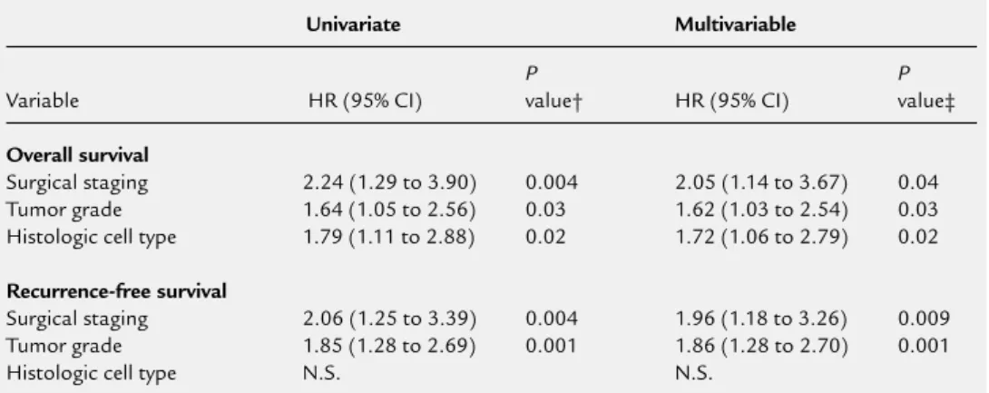
Table 4. Prognostic factors that were identified in the univariate and multivariable analyses*
Univariate Multivariable
P P
Variable HR (95% CI) value† HR (95% CI) value‡
Overall survival
Surgical staging 2.24 (1.29 to 3.90) 0.004 2.05 (1.14 to 3.67) 0.04
Tumor grade 1.64 (1.05 to 2.56) 0.03 1.62 (1.03 to 2.54) 0.03
Histologic cell type 1.79 (1.11 to 2.88) 0.02 1.72 (1.06 to 2.79) 0.02
Recurrence-free survival
Surgical staging 2.06 (1.25 to 3.39) 0.004 1.96 (1.18 to 3.26) 0.009
Tumor grade 1.85 (1.28 to 2.69) 0.001 1.86 (1.28 to 2.70) 0.001
Histologic cell type N.S. N.S.
* HR = hazard ratio; CI = confidence interval. Surgical staging = inadequate versus minimal, modified, and optimal. Tumor grade was in accordance with World Health Organization grading criteria (10). Histologic cell type = mucinous/ endometrioid versus serous, clear-cell, undifferentiated, and other (rare) histology. N.S. = not statistically significant. †P value was determined using the Cox proportional hazards regression model.
Impact of adjuvant chemotherapy and surgical staging 39
2
Extending this subgroup analysis further by looking at the optimal and non-optimal staging groups separately, no difference in overall survival between the observation arm and the chemotherapy arm was found in the optimally staged patients (Fig. 6, A), whereas a statistically significant difference in overall survival between the two arms was demonstrated in the non-optimally staged patients (Fig. 6, B) (HR = 1.75, 95% CI = 1.04 to 2.95; P = 0.03). A similar phenomenon was seen for recurrence-free survival (optimally staged patients: HR =1.14, 95% CI = 0.54 to 2.39; P = 0.7 [Fig. 6, C]; non-optimally staged patients: HR = 1.78, 95% CI = 1.15 to 2.77; P = 0.009 [Fig. 6, D]). However, interactions between treatment effect and the staging subgroups did not reach statistical significance (HR = 2.18, 95% CI . 0.74 to 6.38; P = 0.15; Fig. 7).
46
Figure 4.
Kaplan–Meier curves for overall survival in patients with early-stage ovarian carcinoma by staging type. Optimal staging (n = 151) (solid line), modified staging (n = 138) (solid dotted line), minimal staging (n = 114) (fine dotted line), and inadequate staging (n = 43) (solid/fine dotted line) are in accordance with the staging guidelines presented in Table 1. The hazard ratio is 2.17 (95% confidence interval [CI] = 1.25 to 3.76; P = 0.005 using the log-rank test) in favor of optimal staging. N = number of patients; O = number of observations (events).
Figure 4. Kaplan–Meier curves for overall survival in patients with early-stage ovarian carcinoma by staging type. Optimal staging (n = 151) (solid line), modified staging (n = 138) (solid dotted line), minimal staging (n = 114) (fine dotted line), and inadequate staging (n = 43) (solid/fine dotted line) are in accordance with the staging guidelines presented in Table 1. The hazard ratio is 2.17 (95% confidence interval [CI] = 1.25 to
3.76; P = 0.02 using the log-rank test) in favor of optimal staging. N = number of patients; O = number of
40 Chapter 2
47
Figure 5.
Kaplan–Meier curves for overall and recurrence-free survival in patients with
early-stage ovarian cancer by staging type. Optimal staging (n = 75 in the observation
arm and n = 76 in the chemotherapy arm) (
solid line
) and non-optimal staging (modified,
minimal, inadequate staging categories combined) (n = 147 in the observation arm
and n = 148 in the chemotherapy arm) (
dotted line
) are in accordance with the staging
guidelines presented in Table 1. N = number of patients; O = number of observations
(events). A) Overall survival in the observation arm. The hazard ratio [HR] = 2.31
(95% confidence interval [CI] = 1.06 to 4.96,
P
= 0.03 using the log-rank test) in
favor of optimal staging. B) Overall survival in the adjuvant chemotherapy arm. HR = 1.06
(95% CI = 0.51 to 2.23,
P
= 0.9 using the log-rank test). C) Recurrence-free survival in the
observation arm. HR = 1.82 (95% CI =1.02 to 3.24,
P
= 0.04 using the log-rank test) in favor
of optimal staging. D) Recurrence-free survival in the adjuvant chemotherapy arm. HR 1.17
(95% CI = 0.0.62 to 2.22,
P
= 0.6 using the log-rank test).
Figure 5. Kaplan–Meier curves for overall and recurrence-free survival in patients with early-stage ovarian cancer by staging type.
Optimal staging (n = 75 in the observation arm and n = 76 in the chemotherapy arm) (solid line) and non-optimal staging (modified, minimal, inadequate staging categories combined) (n = 147 in the observation arm and n = 148 in the chemotherapy arm) (dotted line) are in accordance with the staging guidelines presented in Table 1. N = number of patients; O = number of observations (events).
A) Overall survival in the observation arm. The hazard ratio [HR] = 2.31 (95% confidence interval [CI] =
1.06 to 4.96, P = 0.03 using the log-rank test) in favor of optimal staging. B) Overall survival in the adjuvant
chemotherapy arm. HR = 1.06 (95% CI = 0.51 to 2.23, P = 0.9 using the log-rank test).
47
Figure 5.
Kaplan–Meier curves for overall and recurrence-free survival in patients with
early-stage ovarian cancer by staging type. Optimal staging (n = 75 in the observation
arm and n = 76 in the chemotherapy arm) (
solid line
) and non-optimal staging (modified,
minimal, inadequate staging categories combined) (n = 147 in the observation arm
and n = 148 in the chemotherapy arm) (
dotted line
) are in accordance with the staging
guidelines presented in Table 1. N = number of patients; O = number of observations
(events). A) Overall survival in the observation arm. The hazard ratio [HR] = 2.31
(95% confidence interval [CI] = 1.06 to 4.96,
P
= 0.03 using the log-rank test) in
favor of optimal staging. B) Overall survival in the adjuvant chemotherapy arm. HR = 1.06
(95% CI = 0.51 to 2.23,
P
= 0.9 using the log-rank test). C) Recurrence-free survival in the
observation arm. HR = 1.82 (95% CI =1.02 to 3.24,
P
= 0.04 using the log-rank test) in favor
of optimal staging. D) Recurrence-free survival in the adjuvant chemotherapy arm. HR 1.17
(95% CI = 0.0.62 to 2.22,
P
= 0.6 using the log-rank test).
100 90 80 70 60 50 40 30 20 10 0
Overall survival (%)
Overall Logrank test: P = 0.0272
0 1 2 3 4 5 6 7 8 9 10
years from randomization
optimal
non-optimal
O N No. of patients at risk:
___ ___ ____________________________________________________________
8 75 67 57 52 43 30 20 12 4 0 37 147 135 115 96 79 64 49 34 17 3
100 90 80 70 60 50 40 30 20 10 0
Overall survival (%)
Overall Logrank test: P = 0.8750
0 1 2 3 4 5 6 7 8 9 10
years from randomization
optimal non-optimal
O N No. of patients at risk:
___ ___ ____________________________________________________________


![Table 1. Main characteristics and results of two randomized clinical trials on early ovarian cancer [16,17]](https://thumb-us.123doks.com/thumbv2/123dok_us/8175601.2167291/57.722.86.636.139.917/table-characteristics-results-randomized-clinical-trials-ovarian-cancer.webp)

