162
DETECTION OF MELOIDOGYNE INCOGNITA (KOFOLD & WHITE, 1919)
CHITWOOD, 1949 (TYLENCHIDA: MELOIDOGYNIDAE) IN THE FLOWER
PLANTS IN ROMANIA.
RĂDOI (DUMITRU) MARIANA 1*, COSTACHE CLAUDIA 2, TOMA FLORIN 1
1 University of Agronomic Sciences and Veterinary Medicine from Bucharest, Faculty of Horticulture
2 National Phytosanitary Authority , National Phytosanitary Laboratory
*Correspondence author: e-mail: [email protected]
Keywords:Root Knot Nematodes, Hibiscus, Impatiens , morphbiometric and molecular biology.
ABSTRACT
The aim of the study was detection and identification of root knot nematodes infecting the flower crops cultivated in open air and indoor. Studied biological material consist of nematodes was obtained from soil and infested plants using two methods, namely enzymatic digestion for plants samples and Baerman funnel for soil samples.The identification of nematodes has been based on morphobiometrical and molecular characters. It has been identified Meloidogyne incognita (Kofold & White, 1919) Chitwood ,1949 in two samples.
INTRODUCTION
Meloidogyne genus comprises more than 100 nematodes species and the plant hosts may be estimated to reach 3000 (HUNT & HANDOO,2009). These ones are largely spread, presenting a large diversity of species in tropical and subtropical areas, causing significant damages, especially to sustenance crops in the whole world. (LOPES & FERRAZ, 2016).
The species with the largest distribution are: M. incognita, M. javanica, M. arenaria, M. chitwoodi, M. fallax şi M. hapla, representing more than 95% of the species in this genus.The spreading of this species is also improved by the large diversity of plant hosts; the most common species are estimated to be able to contaminate more than 5500 plant species. (TRUDGILL & BLOK, 2001). M. incognita, M. arenaria and M. javanica
species are especially found in tropical areas, while M. hapla, M. chitwoodi and
M. fallax require areas with colder temperatures.
In Romania, the main species of the root knot nematodes (RKN), recorded in the
literature are: Meloidogyne hapla, M. incognita, M. arenaria (Oana T. Moldovan et al., 2007).
Meloidogyne species are sedentary endoparasite nematodes, which induce formation of giant cells inside the roots, out of which these ones feed, until the
complete end of the life cycle
(BALDACCI&CRESP et al., 2015). The nematode may induce the following
alterations under the shape of
biochemical and morphological changes, the abnormal growth of the plant,
symptoms which indicate nutrients
shortages, roots with exteriour knots, forks and other deformations (MOENS et al., 2009). Subsequent to these changes caused by the nematode, the plants availability for food and water decreases.
163 Initially, for the identification of these nematodes, the method of RKN
females perineal pattern areas
observation has been used, this one being a laborious method which requires good taxonomic preparation.
In the last decade, there were developped precise diagnostic protocols at cellular level (SEESAO et al., 2017).
MATERIAL AND METHOD
The analysis of nematodes
extraction from soil and plants, the ones used for morphological and morphometric identification and of molecular biology were carried out within the Phytosanitary National Authority, in the Technical Department of Nematology, respectively Molecular Biology from Voluntary locality, Ilfov County.
In this study, the samples were taken, starting with the autumn of 2018, from more locality, situated in the south of the country. There were analysed 50 soil samples, coming from flower plants cultivated in the field and protected area. Among these, only in two samples coming from a private person from Voluntari locality, Ilfov county, who cultivated Hibiscus rosa sinensis and
Impatiens plants in pots, in greenhouse, there were found nematodes from
Meloidogyne species. The nematodes were extracted both from the soil as from the plant, with the help of Baermann funnel or modification Baermann funnel. The roots were cleaned and washed of the adherent soil, afterwards, being submitted to the process of nematodes extraction by means of the enzyme digestion method. Thus, out of roots, there were extracted not only females and males but also juveniles (J2). All these were used in the following identification. Thus, for males and
juveniles, there were effected
morphometric and morphological
measurements, and for females, it was necessay to cut the perineal area in order to identify the species.
The stage of morphological and morphometric identification was achieved using Zeiss Axio Imager M2 microscope, having also a digital camera, Axiocam 506, with incorporated soft ZEN – 2.6 (blue edition). Identification this species was done using specialized literature (Susan B. Jepson, 1987)
MOLECULAR CHARACTERISATION
The DNA extraction was achieved by performing two extractions from the same sample, having a total of 4 extractions , using one male for each extraction in a population (table 1). One used extraction buffer (worm lysis buffer - WLB) made of (Tris 10 Mm pH = 8.8,
EDTA 1 Mm, Triton X-100, 1%,
proteinasis K 100mg/mL),(Ibrahim et al., 1994).
The extraction was made using a stereomicroscope with a slide, on which one puts an ultrapure drop of water, where the male was put; after eliminating the ultrapure water , the nematode is smashed with a slide, this way, the obtained extract is recovered by means of a pipette, using 10 µl, extraction buffer. Over the achived extract , one adds a tube of 10 µl WLB. It follows the hatching at 60°C for 1h, after at 95°C for 10 min.,(Ibrahim et al., 1994).
The PCR mixture (total volume 25 μL) contained 1x buffer enzyme (with MgCl2)
, 0,5 mM MgCl2, 0.24 μM of each primer
(Finc CTCTGCCCAATGAGCTGTCC and Rinc CTCTGCCCTCACATTAAG - Zijlstra et al., 2000), 1 U Taq DNA polymerase (MP Biomedicals), 0.2 mM dNTPs (MP Biomedicals) and 5 μL DNA extract.
A MasterCycler Pro S (Eppendorf) was used for amplification, and the reaction consisted of a denaturation step at 94°C for 5 min followed by 36 cycles at 94°C for 30 s, 54°C for 30 s, 72°C for 1 min, and a final extension step of 7 min 72°C (Geraldine ANTHOINE, Bucharest, 2004).
164 in a 1% agarose gel. Data analysis was performed using photo documentation system GENi (Syngene) and 100 bp DNA Ladder (GeneRuler, Fermentas) as a molecular size marker.
Table 1 DNA extraction from one male used for the identification of Meloidogyneincognita
Nr. Sp.
Cod
pop. Origin Species
1 1a Ilfov-RO
Impatiens 1
M. incognita
1b Ilfov-RO
Impatiens 1
M. incognita
2 2a Ilfov-RO
Hibiscus rosa sinensis
M. incognita
2b Ilfov-RO
Hibiscus rosa sinensis
M. incognita
RESULTS AND DISCUSSIONS
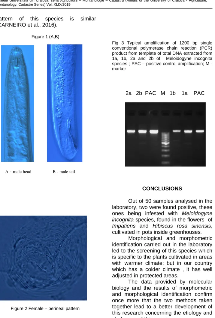
For a correct identification, the male individuals must be looked at , from lateral side. The form of head and stylet
morphology of males are useful
characteristics in identifying some RKN species, such as M. incognita and M. javanica (Figure 1A,B). The males have labial region, which is not “offset” with the labial disk, high and concave as shape, they present transversal anulations and the lateral lips are usually absent. The length of the stylet is comprised between 16 - 25 µm, and the basal knobs offset,
rounded to transversely elongate.
Another differenciation criterion is
represented by the distance from
oesophagus dorsal gland orifice (DGO) to the basis of stylet, which, in this case, is short DGO = 1,5 - 2,9 µm (Table 2).
The size and shape of stylet have also additional taxonomic values for RKN values identification; despite the fact that some RKN species have the same size of the stylet. Identification by using male morphological traits, in the diagnosis of RKN species, by using these techniques, requires deep knowledge of taxonomy (OLIVEIRA et al., 2011).
The females have a pear shape and they do not present protuberances in the posterior side of the body. Stylet has the average length of 14 µm with the basal knobs rounded, offset. Normally, the shape or the perineal pattern of te females was the main technique of identifying the RKN species. The shape and the visual aspect of the whole perienal region, the dorsal arch, the dorsal striae and lateral filds, and the
phasmides are the morphological
characteristics used for the identification. The perineal pattern has an oval to round shape , usually presenting a high dorsal arch, having a square shape with ondulated striae, the lateral fild being absent or poorly demarcated by forked striae (Figure 2).
The juveniles (J2) have the length comprised between 320 – 421 µm with the hemizonid bacward located or adjacent to the excretor pore, tail has the length comprised between 34 – 59 µm and the hialin region with size comprised between 7,5 – 13 µm, having a round shape (PERRY N.R. et al.,2009).
For the sample of Hibiscus rosa sinensis, the morphological and morphometric identification was done, having a control stock of 5 males , unlike
Impatiens test, where we identified only two individuals
This one is a less expensive technique but which requires deep knowledge of taxonomy and time, in order to perform microscopic slides. To achieve this goal, one needs both mature females, namely for the diagnosis, and roots populated with females. (SEESSAO et al., 2016).
This method was used to identify the species of M. incognita, M. javanica, M. arenaria and M. hapla (CHITWOOD, 1949). But because of the discovery of new pathogen species , this technique has proven to be not exactly enough for finding new species.
The wrong identification of M.
enterolobii and M. inornata, as well as of
165
pattern of this species is similar
(CARNEIRO et al., 2016).
Figure 1 (A,B)
A - male head B - male tail
Figure 2 Female – perineal pattern
Fig 3 Typical amplification of 1200 bp single conventional polymerase chain reaction (PCR) product from template of total DNA extracted from 1a, 1b, 2a and 2b of Meloidogyne incognita species ; PAC – positive control amplification; M - marker
2a 2b PAC M 1b 1a PAC
CONCLUSIONS
Out of 50 samples analysed in the laboratory, two were found positive, these ones being infested with Meloidogyne incognita species, found in the flowers of
Impatiens and Hibiscus rosa sinensis, cultivated in pots inside greenhouses.
MorphologicaI and morphometric identification carried out in the laboratory led to the screening of this species which is specific to the plants cultivated in areas with warmer climate; but in our country which has a colder climate , it has well adjusted in protected areas.
166
DGO - the distance from oesophagus dorsal gland orifice to the basis of style
Table 2. Measurements of Meloidogyne incognita species for two host plants; Impatiens şi Hibiscus rosa sinensis
BIBLIOGRAPHY
Adam MAM, Phillips M.S., Block V.C.,
2007, Molecular diagnostic key for
identification of single juveniles of seven common and economically important species
of root-knot nematode (Meloidogyne spp.),
Plant Pathology, 56, 190 – 197.
Carolien Zijlstra, Donkers-Venne Dorine T.H.M. and Fargette Mireille, 2000, Identification of Meloidogyne incognita, M. javanica and M. arenaria using sequence
Hosts plants
Impatiens Hibiscus rosa sinensis
Females n=5 n=6
L 848,4 (788-905) 611,17 (439-722)
A 1,59 (1,58-1,89) 1,86 (1,50-2,20)
Max.body diameter 533,8 (478-612) 328 (291-409)
Neck length 238 (196-314) 226 (175-290 )
Stylet length 14 (13,5-15) 13,88 (12-16,5)
Stylet knob height 2,06 (2-2,4) 2,39 (1,7-3)
Stylet knob width 4,32 (4-4,6) 4,36 (4-4,7)
DGO 3,18 (2,5-3,8) 2,5 (1,2-3,9)
Excretory pore to anterior end 28,33 (20-35) 52,33 (40-65)
Males n=2 n=5
L 1530 (1349-1711) 1183 (916-1476)
A 39,1 (37,47-40,74) 38,61 (33,59-43,41)
C 140,02 (89,93-190,1) 109,93 (83,27-146,14)
Max.body diameter 39 (36-42) 30,86 (23,3-39)
Stylet length 22,9 (21-24,8) 18,68 (15,9-21,3)
Stylet knob height 3,5 (3-4) 3,4 (2,9-4,2)
Stylet knob width 4,7 (4,4-5) 4,94 (3,8-6,5)
DGO 2,7 (2,5-2,9) 2,02 (1,5-2,9)
Excretory pore to anterior end 163 (-) 117,33 (95-147)
Tail length 12 (9-15) 10,82 (10,1-11)
Spicule length(median line) 34,75 (33-36,5) 31,4 (26-35)
Gubernaculum length 7,7 (-) 7 (4-9)
Juveniles n=10 n=10
L 384,7 (320-421) 384 (343-410)
A 27,46 (23,70-32,38) 25,74 (22,77-28,84)
C 8,74 (6,53-11,5) 8,02 (6,23-9,45)
Max.body diameter 14,1 (12,5-16) 15,05 (13-18)
Stylet length 12,86 (11-14) 12,53 (12-14)
Stylet knob height 1,65 (1,05-2,3) 1,47 (1-2,2)
Stylet knob width 2,61 (2-3,4) 2,23 (2-2,6)
DGO 2,22 (1,3-3) 1,93 (1,3-2,5)
Excretory pore to anterior end 72,15 (58-88,5) 71,2 (64-78)
Tail length 45,5 (34-59) 48,45 (42-58)
167 characterised amplified region (SCAR) based PCR assays, Nematology, vol. 2(8), 847 – 853.
Tiago Garcia da Cunha, Visotto L.E., Lopes E.A., Claudio Marcelo Goncalves Oliveira, Pedro Ivo Vieira Good God, 2018, Diagnostic methodes for identification of root – knot nematodes species from Brazil, Ciencia Rural, Santa Maria, v.48: 02, 1 – 11. Weimin Ye, Yongsan Zeng, James Kerns,
2015, Molecular Characterisation and
Diagnosis of Root – Knot Nematodes (Meloidogyne spp.) from Turfgrasses in North Carolina, USA., Plos ONE 10(11), 1 – 16.
Dong K. et al., 2017, Development of PCR primers to identify species of root-knot nematodes Meloidogyne arenaria, M. hapla, M. incognita and M. javanica, Nematropica, v31, n.2., p 271 – 280.
Roland N. Perry, Moens. M, Starr. J.L., 2009
- Root – Knot Nematodes, CAB
International, Cap 3, p 55 – 88.
Kiewnick, S. et al., 2013. Identification of the tropical root – knot nematode species
Meloidogyne incognita, M. javanica and M. arenaria using a multiplex PCR assay. Nematology , v15, p. 891 – 894.
Oliveira C.M.G. et al. Morphological and molecular diagnostic for plant parasitic nematodes working together to get the identification done. Tropical Plant Pathology, v 36, n. 2, p. 65 – 73, 2011.
Susan B. Jepson, 1987, Identification of Root – Knot Nematodes (Meloidogyne species, vol I şi II), CAB International.
Oana T. Moldovan et al., 2007. Lista Faunistică a României , Casa Cărţii de Ştiinţă. Boguleanu GHE., 1988. Fauna Dăunătoare Culturilor Agricole şi Forestiere din România, vol I, Ceres.
Bulletin OEPP/EPPO Bulletin (2016) 46 (2), 190-201.
Lamberti F. and Taylor C.E., 1979, Root-Knot Nematodes (Meloidogyne species) Systematics Biology and Control, Academi Press.