Original Research Article
Evaluation of accommodative function in the dominant and non
dominant eye
Oseleonomhen M. Odigie*, Precious N. Uwagboe, Omawumi P. Okpaghoro
INTRODUCTION
Ocular dominance also known as eyed-ness is the tendency of the visual system to give more preference to the processing of input signals from one eye over the other.1 The dominant eye may capture more attention
readily making that eye more reliant for accurate positional information during binocular viewing.2,3 Hence
the dominant eye plays a controlling role in binocular vision.4 Researches have shown that two third and one
third of the population have dominance in the right and
left eyes respectively.5 Eser et al,found that 70% of males
were right eye dominant and had higher right eye dominance than in female (65%), they concluded that gender appear to be a factor when testing ocular dominance.6 Ocular dominance is divided into three types
these include sighting dominance which is referred to as the preferred eye when looking through a small gap that is created by the overlapping of outstretched arms.7
Sensory-ocular dominance is when the perception of a stimulus in one eye dominates the other in retinal rivalry conditions.8 Sensory ocular dominance can measure
Department of Optometry, University of Benin, Benin-City, Edo state, Nigeria
Received: 25 March 2019
Accepted: 03 May 2019
*Correspondence:
Dr. Oseleonomhen M. Odigie, E-mail: [email protected]
Copyright: © the author(s), publisher and licensee Medip Academy. This is an open-access article distributed under the terms of the Creative Commons Attribution Non-Commercial License, which permits unrestricted non-commercial use, distribution, and reproduction in any medium, provided the original work is properly cited.
ABSTRACT
Background: Ocular dominance is the physiological preference of one eye over the other, hence its input is favoured when there is conflicting information to the two eyes. Accommodation is the mechanism by which the eye changes focus from distant to near images and is produced by a change in the shape of the crystalline lens. The aim of this study was to compare the accommodative amplitude, facility and lag in the dominant and non-dominant eye.
Methods: This cross sectional study was carried out on 80 visually normal subjects. Ocular dominance was determined using hole-in-the- card method. Amplitude of accommodation, accommodative facility and response was measured monocularly and randomly using push up method, ±2.00DS flipper lenses and Nott technique respectively.
Results: Results obtained from the study showed that the right eye was dominant in 62.5% of subjects. The mean (SD) for accommodative amplitude, facility and response (lag) in the dominant eye was 11.08 (2.16) D, 10.00 (1.52) cycles per minute and 0.62 (0.27) respectively. The mean (SD) for accommodative amplitude, facility and lag in the non-dominant eye was 10.98 (2.20) D, 9.86 (1.44) cycles per minute and 0.60 (0.25) D respectively.
Conclusions: It may be inferred that the dominant eye has more accommodative amplitude, facility and lag than the non-dominant eye but this difference was not statistically significant.
Keywords: Accommodative amplitude, Accommodative facility, Ocular dominance
imbalance between the sensory inputs from the two eyes
using binocular rivalry.9-11 Oculo-motor dominance
occurs when there is asymmetry of the vergence eye movements.12,13 Ocular dominance can influence clinical
decision-making process when considering certain ophthalmic refractive and surgical interventions.14
Accommodation can also be defined as the ability of the intraocular lens to increase in altering the eye’s dioptic power and thus enabling light diverging from a near source to be focus upon the retina in order to obtain clear image.15,16 There are various aspect that are used for
assessing the accommodative state of an individual. One of these include accommodative amplitude which is measured from the far point to the near point with maximum exertion. Reduced accommodative amplitude can lead to symptoms such as blur vision, diplopia, eye fatigue, headaches, etc. Accommodative facility measures the speed of the accommodative responsiveness to blur.17
Accommodative response is a change in the power of the crystalline lens driven by blur on the retina. Ibi suggested that the dominant eye plays the primary role in far to near accommodation in binocular viewing.18 Momeni et al, in
their study found the amplitude of accommodation and accommodative facility to be superior in the dominant eye compared with the non-dominant eye.19 Fujimura et
al, reported that under binocular viewing condition the accommodative response in the dominant eye was greater than the non-dominant eye, in contrast to monocular condition between dominant and non-dominant eye where there was no difference in accommodative response between dominant and non-dominant eye.20
Many factors have been attributed to produce a difference in accommodative function among which are anisometropia, antimetropia, glaucoma, amblyopia, retina disorders developmental disorders and trauma.21-23
Hence, the aim of this study is to determine if ocular dominance causes a difference in accommodative function in visually normal adults in our environment.
METHODS
This study was a cross sectional study. Convenience sampling technique was used for the selection of subjects. Eighty subjects consisting of 36 males and 44 females participated in the study. The mean age of the subjects was 21.06±2.94 years. This study was carried out at the Optometry Teaching Clinic, University of Benin, Ugbowo Campus, Benin City, Edo State, Nigeria between April 2018 and November 2018. Informed consent was obtained from all the subjects before they participated in the study. Subjects with best correct visual acuity of 6/
6 or better were included in the study. Subjects
with nystagmus, strabismus, recent history of ocular/head trauma, cataract and corneal pathology, with retinal pathology or currently on systemic medication were excluded from the study.
Description of procedure
A comprehensive eye examination was conducted on the subjects to ensure they meet the inclusion criteria. Retinoscopy was used to determine the subject’s objective refractive errors. Subjective refraction in addition to binocular balancing was to refine the retinoscopic findings.
Ocular dominance test
For ocular dominance, test was carried out using hole in the card method; here the subjects looked at a letter on the distant visual acuity chart through the 3mm hole in the center of the card, The subjects held the card at arm length. The left eye of the subjects was occluded first before occluding the right eye. When the dominant eye was occluded the target was no longer seen through the hole but when the non-dominant eye was occluded, the subject saw the target through the hole with the open dominant eye.
Amplitude of accommodation
Amplitude of accommodation was carried out at 40cm in a normal room illumination, using the push up method. The test object was a near point card and the subject was instructed to read a line above the subject’s best corrected visual acuity at near. The subject was asked to read out the sentence written in the particular paragraph while the near point card was gradually brought towards the subject’ eye until the subject experienced the first sustained blur which is the point where the subject can no longer clear the blur. The distance of the near point card to the subject’s spectacle plane was measured in centimeters three times, the average was recorded and converted to diopter by dividing 100.This procedure was carried out randomly on the dominant and non-dominant eye alike, be it the right or the left eye.
Accommodative facility
Accommodative response
Accommodative response test was done using the NOTT technique. The test was done monocularly and over the subject’s distance prescription. A fixation target was brought 40 cm from the eye. The retinoscopic reflex with the streak oriented vertically was accessed while the subject read the chart, the retinoscope was moved or away from the patient until neutrality, the distance at which neutrality was achieved was recorded and then converted to diopters. Depending on if the retinoscope was brought towards or away from the subject, accommodative lead and accommodative lag was recorded respectively. This procedure was carried out three times and the average was recorded for both dominant and non-dominant eye.
Data obtained from this study was analysed using the statistical package for social Sciences (SPSS) version 22.0. The results were presented in percentages and tables. Paired-t-test was used to test for significant differences in the accommodative amplitude, facility and
response of the dominant and non-dominant eye. Unpaired-t-test was used to test for significant differences in accommodative amplitude, facility and response in males and females.
RESULTS
The right eye was dominant in 62.5% of the subjects while the left was dominant 37.5% (Table 1).
Table 1: Ocular dominance in right and left eye.
Dominant eye Frequency Percentage (%)
Right eye 50 62.5
Left eye 30 37.5
Total 80 100.0
The mean spherical equivalent of refractive power of the dominant eye was 0.35±1.47D respectively while it was 0.36±1.48D for the refractive power of the non-dominant eye (Table 2).
Table 2: Comparing sphere power, cylinder power, and spherical equivalent (SE) between the dominant and non-dominant eye.
Refractive component Dominant mean (SD) Non-dominant Mean (SD) Mean difference (SD) P value
Sphere (D) 0.62 (1.56) 0.61 (1.56) 0.01 (0.13) 0.37
Cylinder (D) -0.53 (0.48) -0.50 (0.48) -0.03 (0.17) 0.11
SE (D) 0.35 (1.47) 0.36 (1.48) -0.00 (0.14) 0.84
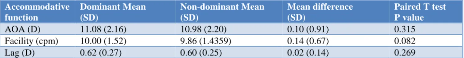
Table 3. Comparing the amplitude of accommodation (AOA), accommodative facility (AF) and accommodative response (lag) between the dominant and non-dominant eye.
Accommodative function
Dominant Mean (SD)
Non-dominant Mean
(SD)
Mean difference (SD)
Paired T test P value
AOA (D) 11.08 (2.16) 10.98 (2.20) 0.10 (0.91) 0.315
Facility (cpm) 10.00 (1.52) 9.86 (1.4359) 0.14 (0.67) 0.082
Lag (D) 0.62 (0.27) 0.60 (0.25) 0.02 (0.14) 0.269
Figure 1:The relationship between the amplitude of accommodation of the dominant eye and the
non-dominant eye.
Figure 2: The relationship between the accommodative facility of the dominant eye and the
The mean difference between accommodative amplitude
in the dominant and non-dominant eye was 0.10±0.91D. The mean difference between accommodative facility in the dominant and non-dominant eye was 0.41±0.67cpm. The mean difference between accommodative response (lag) in the dominant and non-dominant eye was 0.02±0.14D (Table 3). There was a strong and significant relationship between the amplitude of accommodation of the dominant eye and the non-dominant eye. The values are highly correlated (r=0.914, P=0.000) positively i.e. the amplitude of both eyes increase in the same direction. A single stray point was highlighted in the plot above. This indicates a tendency for the non-dominant eye to have greater amplitude of accommodation than the dominant eye (Figure 1). There was a positive and significant (r=0.894, P=0.000) relationship between the accommodative facility in the dominant and non- dominant eye. The points down below indicated instances where the facility in the non-dominant eye was higher than the facility of the dominant eye (Figure 2). There was also a positive and significant (r=0.862, P=0.000) relationship between the accommodative response (lag) of the dominant eye and that of the non-dominant eye. Extreme variations between the two eyes were also seen
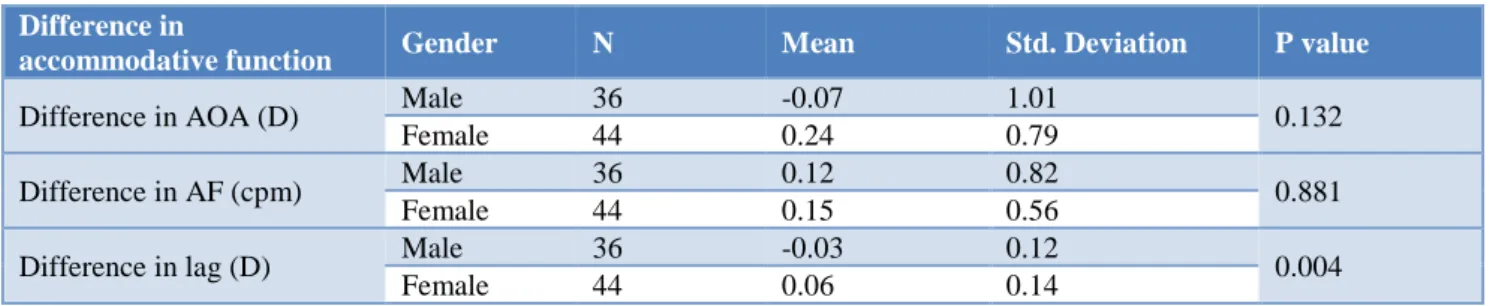
(Figure 3). It was observed that the difference in the accommodative amplitude, facility and lag between the dominant and non-dominant eyes was higher in females relative to the males (Table 4).
Figure 3: The relationship between the accommodative response (lag) of the dominant eye
and that of the non-dominant eye.
Table 4: Gender and the difference in accommodative amplitude, facility and lag between both eyes. Difference in
accommodative function Gender N Mean Std. Deviation P value
Difference in AOA (D) Male 36 -0.07 1.01 0.132
Female 44 0.24 0.79
Difference in AF (cpm) Male 36 0.12 0.82 0.881
Female 44 0.15 0.56
Difference in lag (D) Male 36 -0.03 0.12 0.004
Female 44 0.06 0.14
DISCUSSION
In this study, most of the subjects were right-eye dominant (62.5%) while the left eye dominant subjects accounted for 37.5%. Momeni et al,2013 reported that most subjects (75.7%) were right eye dominant while the rest (24.3%) were left eye dominant.19 Other studies have
also indicated that most persons were right-eye dominant.5,6 There was no significant difference between
the spherical equivalent of the dominant eye and that of the non-dominant eye of our subjects (p>0.05), this indicated that refractive error did not play any role in determining which eye became dominant in the subjects. Similarly, Momeni et al, reported there was no significant difference in refractive error between the dominant and non-dominant eye.19 This was also supported by the study
done by Fujimura et al.20 This similarity may be due to
the fact that both studies utilized the same method (Hole-in-the-card) in the determination of ocular dominance. The subjective refraction was also performed in
monocular viewing conditions. Binocular assessment of refraction in relation to ocular dominance has been proposed by Tsuneyoshi et al, who showed that binocular assessment of refraction is essential for precise refractive therapy after a significant hyperopic shift was observed in binocular refraction (suggestive of the role of ocular dominance in refraction).24 There was a higher value of
accommodative amplitude and facility in the dominant eye compared to the non-dominant eye but this difference was not significant (p>0.05). However Momeni et al, reported a significant difference in accommodative amplitude and facility in dominant and non-dominant eye but stated that these differences might not be clinically significant (<0.05D and <2 cycles per minute).19 Chen et
al, also found significant difference in amplitude of accommodation in the dominant and non-dominant eyes of 18 children with anisohyperopia.25 There was also no
influenced by ocular dominance under binocular viewing
conditions.20 In relation to this, Ibi had conducted a study
on characteristics of dynamic accommodation responses between the dominant and non-dominant eyes in 18 healthy subjects and found in viewing the internal targets near-to-far responses were suppressed, in binocular viewing, the accuracy of accommodative position was increased and the function of dynamic responses was improved.18 It was also found those myopic shifts were
observed in the near position after far-to-near accommodation in the dominant and non-dominant eye.
The accommodative lag was less related in both eyes when compared to accommodative amplitude and facility. The difference in accommodative function between the dominant and non-dominant eyes were higher in females than in males. Studies done byEser et al, and Linke, et al, have found that more of the right eyes of males were dominant than the right eye of females.5,6
There was significant difference in accommodative response in both sexes (p<0.05).
In terms of age, it is expected that the difference between the accommodative functions of both eyes vary less under normal circumstances in order to maintain a balanced binocular vision and this is basically because the study was not on a large age range however large variations with age could be pointers to abnormal conditions.
CONCLUSION
There was no significant difference in the refractive state and accommodative functions (accommodative amplitude, facility and response) between the dominant and non-dominant eye of the subjects. The type of activity performed by a subject should be largely considered when the eye practitioner makes decisions regarding what to prescribe in relation to ocular dominance.
Recommendations
In cases where one eye is overly dominant over the other in both sensory and motor vision, proper investigation should be carried out to determine the underlying factors if not obvious as well as to ensure balance between both eyes where possible. To reduce bias caused by racial and experimental settings and increase generalization to our society, a similar study involving large number of participants with a wide age range should be carried out.
ACKNOWLEDGEMENTS
Authors would like to thank individuals who participated in the study.
Funding: No funding sources Conflict of interest: None declared
Ethical approval: The study was approved by the Institutional Ethics Committee
REFERENCES
1. Feng J, Zheyi C, Hua B, Edgar E, Jun J. Association between ocular sensory dominance and refractive error asymmetry. PLoS. 2015;10(8):e0136222. 2. Yang E, Blake R, McDonald JE. A new interocular
suppression technique for measuring sensory eye dominance. Investigative Ophthalmol Visual Sci. 2010;51(1):588-93.
3. Khan AZ, Crawford JD. Ocular dominance reverses as a function of horizontal gaze angle. Vision Res. 2001;41(14):1743-8.
4. Chia A, Jaurigue A, Gazzard G, Wang Y, Tan D, Stone RA, et al. Ocular dominance, laterality, and refraction in Singaporean children. Investigative Ophthalmol Visual Sci. 2007;48(8):3533-6.
5. Linke SJ, Baviera J, Stenberg J, Richard G, Katz T. Association between ocular dominance and spherical astigmatism, anisometropia, age and sex: analysis of 10,264 myopic individuals. Investigative Ophthalmol Visual Sci. 2011;52(12):9166-73. 6. Eser I, Durrie DS, Schwendeman F, Stahl JE.
Association between ocular dominance and refraction. J Refract Surg. 2008;24(7):685-9. 7. Steinman SB, Steinman BA, Garza RP. Foundations
of binocular vision: a clinical perspective. McGraw- Hill New York; 2000:24-27.
8. Sengpiel F, Blakemore C, Kind PC, Harrad R. Interocular suppression in the visual cortex of strabismic cats. J Neurosci. 1994;14(11):6855-71. 9. Handa T, Mukuno K, Uozato H, Niida T, Shoji N,
Shimizu K. Effects of dominant and non-dominant eyes in binocular rivalry. Optomet Vision Sci. 2006;81(5):377-83.
10. Handa T, Uozato H, Higa R, Nitta M, Kawamorita T, Ishikawa H etc. Quantitative measurement of ocular dominance using binocular rivalry induced by retinometers. J Catar Refract Surg.
2006;32(5):831-6.
11. Ooi TL, He ZJ. Sensory eye dominance. Optometry. 2001;72(3):168-78.
12. Horng JL, Semmlow, JL, Hung GK, et al. Dynamic Asymmetries in disparity convergence eye movements. Vision Res. 1998;38(18):2761-8. 13. Kawata H, Ohtsuka K. Dynamic Asymmetries in
Convergence Eye Movements under Natural Viewing Conditions. Jap J Ophthalmol. 2001;45(5):437-44.
14. Pointer JS. Sighting versus sensory ocular dominance. J Vision. 2012;5(2):52-5.
15. Borish IM. Clinical refraction. 3rd Ed. Professional
press books/ fair child publication. 1970:91-229. 16. Rutstein RP, Eskridge JB. Clinical evaluation of
vertical fixation disparity. Part III. Adaptation to vertical prism. Am J Optomet Physiol Opt. 1985;62(9):585-90.
Accommodative facility in eyes with and without
myopia. Investigative Ophthalmol Visual Sci. 2006;47(11):4725-31.
18. Ibi K. Characteristic of dynamic accommodation responses. Comparison between the dominant and non-dominant eyes. Ophthalmic Physiol Opt. 1997;17(1):44-54.
19. Momeni-Moghaddam H, McAlinden C, Azimi A. Comparing accommodative function between the dominant and non-dominant eye. Graefes Arch Clin Exp Ophthalmol. 2014;252(3):509-14.
20. Fujimura F, Handa T, Kawamorita T, Shoji N. The Effect of Ocular Dominance on Accommodation and Miosis under Binocular Open Viewing Conditions. Open J Ophthalmol. 2017;7(3):158. 21. Eskridge JB, Amos JF, Bartlett JD. Clinical
procedures in optometry. Philadelphia: JB Lippincott; 1991: 687-691.
22. Cheiman M, Wick B. Clinical management of binocular: heterophoric, accommodative and eye
movement disorders, 3rd Ed. Philadelphia:
Lippincott Williams & Wilkins; 2008: 350.
23. Firth AY. Adie syndrome: evidence for refractive error and accommodative asymmetry as the cause of amblyopia. Am J Ophthalmol. 1992;128(1):416-8. 24. Tsuneyoshi Y, Negishi K, Tsubota K. Importance of
accommodation and eye dominance for measuring objective refractions. Am J Ophthalmol. 2017;177:69-76.
25. Chen J, Wang ZZ, Yu XP, Wang YW. Accommodative function in adolescent hyperopic anisometropes. Chin J Optom Ophthalmol. 2009;4(11):254-6.