CELLULAR & MOLECULAR BIOLOGY LETTERS http://www.cmbl.org.pl
Received: 28 May 2007 Volume 13 (2008) pp 92-102 Revised form accepted: 13 July 2007 DOI: 10.2478/s11658-007-0039-5 Published online: 29 October 2007 © 2007 by the University of Wrocław, Poland
# Paper authored by participants of the international conference: XXXIV Winter School of
the Faculty of Biochemistry, Biophysics and Biotechnology of Jagiellonian University, Zakopane, March 7-11, 2007, "The Cell and Its Environment". Publication cost was covered by the organisers of this meeting.
* Author for correspondence; e-mail: [email protected]
Abbreviations used: Cx – connexin, FACS – fluorescence-activated cell sorting
Mini review
THE STAGE-SPECIFIC FUNCTION OF GAP JUNCTIONS DURING
TUMOURIGENESIS #
JAROSŁAW CZYŻ*
Department of Cell Biology, Faculty of Biochemistry, Biophysics and Biotechnology, Jagiellonian University, ul. Gronostajowa 7, 30-378 Kraków,
Poland
Abstract: Tumour development is a process resulting from the disturbance of
various cellular functions including cell proliferation, adhesion and motility. While the role of these cell parameters in tumour promotion and progression has been widely recognized, the mechanisms that influence gap junctional coupling during tumorigenesis remain elusive. Neoplastic cells usually display decreased levels of connexin expression and/or gap junctional coupling. Thus, impaired intercellular communication via gap junctions may facilitate the release of a potentially neoplastic cell from the controlling regime of the surrounding tissue, leading to tumour promotion. However, recent data indicates that metastatic tumour cell lines are often characterized by relatively high levels of connexin expression and gap junctional coupling. This review outlines current knowledge on the role of connexins in tumorigenesis and the possible mechanisms of the interference of gap junctional coupling with the processes of tumour invasion and metastasis.
GAP JUNCTIONS – THEIR STRUCTURE AND BASIC FUNCTIONS IN TISSUE HOMEOSTASIS
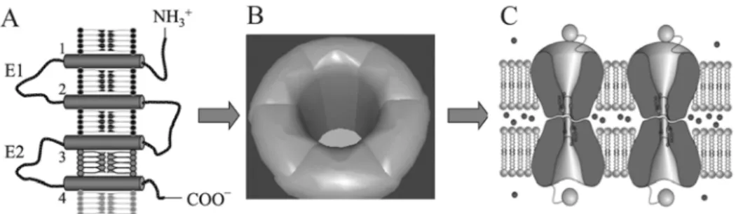
Gap junctions are aqueous intercellular channels which directly link the cytoplasmic compartments of adjacent cells (Fig. 1). These channels are formed upon the docking of two connexons, hexameric hemichannels built of proteins belonging to the connexin family [1]. Gap junctions are present in almost all vertebrate tissues, where they permit the intercellular exchange of small (< 1 kDa) metabolites and second messengers, and thus synchronise cellular functions in tissues [2-4]. Representative examples of the synchronising function of gap junctional coupling in tissues include electrical and metabolic coupling. In the former,gap junctions take part in the intercellular spreading of membrane depolarisation by permitting the rapid cell-to-cell transfer of calcium ions between cardiac muscle cells [1], or by constituting electrical synapses in the nervous system [5]. Metabolic coupling is established when the stimulation of one cell is propagated to a cluster of cells through the spreading of active substances, such as monosaccharides and cyclic nucleotides [2, 6]. While electrical coupling predominantly synchronizes cell functions in excitable tissues, metabolic coupling affects the homeostasis of a spectrum of multicellular systems, including both excitable and non-excitable tissues.
Fig. 1. The structure of gap junctions. Proteins of the connexin family consisting of intramembraneous (1-4), extracellular (E1, E2), and intracellular domains (A), compose hexameres (connexons; B), which give rise to intercellular channels built of connexons contributed by adjacent cells (C). R. Eckert and D. Huelser, private communication, modified.
However, the relative non-specificity of the channel allows the passage of a wide variety of small molecules, and a multitude of signalling systems affect the function of gap junctional channels [11, 12], thus making the mechanistic elucidation of the role of gap junctional coupling in tissue homeostasis difficult. Cx32 knockout mice appeared to be more susceptible to liver carcinogenesis than their wild-type counterparts [13], suggesting that the role of gap junctions during the development of tumours may be of special importance. In this paper, the state of research on the involvement of gap junctions in the processes of tumour arousal and development is outlined.
THE INVOLVEMENT OF GAP JUNCTIONS IN THE REGULATION OF TUMOUR CELL PROLIFERATION
Many reports indicate that natural or provoked deficiency in connexin gene function correlates with increased cellular susceptibility to transformation. In vivo
observations indicating the influence of gap junctions on tumorigenesis are supported by in vitro data demonstrating the down-regulation of connexin expression and gap junction assembly in a wide range of neoplastic cell lines and primary tumours [14-17]. Furthermore, viral transfection was shown to reduce the levels of gap junctional coupling in cell populations, while a forced expression of genes encoding connexins inhibited the proliferation and affected the contact behaviour of transfected tumour cells [16]. Retarded tumorigenicity was described upon connexin transfection of hepatoma SKHep1, breast carcinoma, glioblastoma, PG lung carcinoma, cervical adenocarcinoma HeLa and prostate cancer LNCaP cells, which correlated with the inhibition of their growth in vitro and in vivo, the decrease of their saturation density, i.e. the density attained by cells in culture upon entering the plateau phase [18-20], and the inhibition of anchorage-independent growth [21], i.e. the partial reversion of the tumour phenotype represented by a decrease in the proliferation rate of connexin-transfected tumour cells in semi-solid media [16, 22, 23].
Mechanistic studies demonstrated that the exogenous expression of Cx43 might exert its inhibitory effect on cell proliferation via the inhibition of the expression of S phase kinase-associated protein 2 (skp2), the protein that promotes the ubiquitination of cyclin-dependent kinase inhibitor p27kip. A down-regulation of cyclin D1 in osteosarcoma cells was shown upon Cx43 transfection, which correlated with up-regulation of p27kip [31]. The deletion mutation analyses revealed that the C-terminal domain of Cx43 that did not form gap junctions was still sufficient to inhibit skp2 expression, while coupling inhibitors did not affect the inhibitory effect of Cx43 on skp2 expression, indicating that coupling-independent mechanisms of Cx43 interfere with the cell cycle [30, 31]. It was also recently shown that the phosphorylation of Cx43 on S262 by protein kinase C cancels HEK-293 cell growth inhibition by Cx43 independently of the channel-forming ability [32]. This finding defines a novel, connexin-dependent and gap junctional coupling-independent mechanism for tumour promotion by PKCs. Taking all this into account, it would seem that intercellular diffusion of putative growth inhibitory factors via gap junctions may play an important role during early stages of tumour development in a way complementary to mechanisms including direct, coupling-independent interference of connexin molecules with signalling pathways regulating the cell cycle.
THE EFFECT OF GAP JUNCTIONS ON THE INVASIVE POTENTIAL OF TUMOUR CELLS
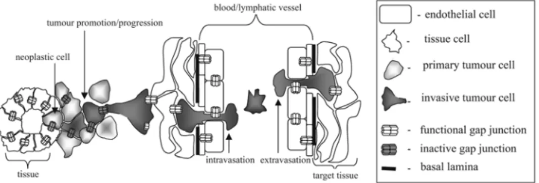
In a simplistic model of tumour development, the potentially neoplastic cell, when released from the controlling regime of the tissue, begins uncontrolled proliferation, thus giving rise to the primary tumour. Gap junctional coupling may take part in this process, as impairment of gap junctional communication may facilitate this process. On the other hand, some data shows that the function of gap junctions may be dependent on the stage of tumorigenesis, i.e. it can differ between neoplastic and metastatic cell populations.
Sequential obligatory steps must be completed after neoplasia (the initial step of tumorigenesis) for the occurrence of metastases. Upon detachment from neighbouring cells and penetration of natural barriers such as the basement membrane and endothelial continuum, neoplastic cells invade blood vessels and/or lymphatic vessels in the process known as intravasation, which enables them to access the circulatory system. After reaching the bloodstream, tumour cells enter the microcirculation and, finally exit the bloodstream by extravasation. They then form metastases via expansive growth in the parenchyma of the target tissue [33]. Thus, a deregulation of the mechanisms determining cell proliferation is followed by the acquisition of the ability of unrestricted migration by tumour cells, a prerequisite of invasion [34].
populations of invasive tumour cells. Kamibayashi et al. [36] demonstrated that even though the expression of Cx26 and Cx43 was reduced in the early stages of mouse skin carcinogenesis, both connexins were expressed on the plasma membranes of cells invading the lymph nodes. Similar observations were made for breast cancer [35], prostate cancer [37, 38] and glioma cell populations [39]. In melanoma cells residing in the basal layer, Cx26 levels remained unchanged, but were significantly up-regulated in the cells invading the dermis. Furthermore, gap junction formation was observed between melanoma cells and mammary adenocarcinoma cells and the endothelium, which could take part in the processes of tumour cell intra- and extravasation [40, 41]. Moreover, transfection of human cervical carcinoma (HeLa) cells with a gene encoding Cx43 significantly increased their in vitro invasive potential [42]. Thus, gap junctions and gap junctional coupling facilitates tumour cell invasion in versatile cellular models, but the specific mechanisms of the correlation between the expression and function of connexins and the metastatic potential of tumour cells remain to be determined.
INTERRELATIONS BETWEEN PROLIFERATION, CELL MOTILITY, AND GAP JUNCTIONAL COUPLING IN INVASIVE AND NON-INVASIVE TUMOUR CELL LINES
As mentioned, the invasive potential of tumour cells in vivo is predominantly based on their ability to penetrate the endothelial barrier, and to spread and proliferate in the adjacent tissue. In order to elucidate how gap junctional coupling facilitates tumour invasion, we have established an experimental approach enabling the quantitative analyses of connexin expression and the function of gap junctions in relation to other cellular traits involved in tumorigenesis, such as cell proliferation and motility [43, 44]. This method exploits pairs of spontaneously aroused tumour cell lines differing in their connexin expression levels and metastatic potential in vivo [37], as well as cell lines established through the stable transfection of connexin-deficient tumour cell lines with gene(s) encoding particular connexins [42, 45, 46]. Cell proliferation and apoptosis was quantified by cell counters and FACS [44], time-lapse video analysis of cell motility provided statistically significant quantitative data on the parameters of tumour cell motility [47, 48], and a calcein spreading assay [49] enabled the quantification of gap junctional coupling in homo- and heterologous cellular systems. Thus, quantitative analyses of tumour cell proliferation, motility and the connexin function of tumour cell lines of common origin differing in terms of connexin expression enabled the in vitro
determination of the interrelations between the cell parameters involved in the process of tumorigenesis.
time-lapse analyses revealed that both cell lines display similar proliferation rates and motility. Since the induction of gap junctional coupling correlates with the increased invasive potential of HeLa Cx43 cells, this data indicates that Cx43-mediated gap junctional coupling directly affects the invasive properties of tumour cells and facilitates the process of metastasis [42, 44]. Similar observations have been made for rat prostate cell lines. Highly metastatic MAT-LyLu cells display significantly higher expressions of Cx43 and coupling levels than non-invasive AT-2 cells, while no significant differences in motile activity on underlying aligned fibroblasts [37] or in growth rate [50] were observed. Thus, cellular systems based on pairs of tumour cell lines differing in the intensity of gap junctional coupling [42, 43] are a suitable tool for analyses of the influence of gap junctions on tumour development in relation to the tumour stage and other cellular traits relevant for tumorigenesis, i.e. cell motility and proliferation.
SUMMARY AND OUTLOOK
Evidence exists that the function of gap junctions affects the development of tumours in a manner dependent on the stage of tumorigenesis. Gap junctions seemingly act as tumour suppressors in the initial stages of this process, while the re-expression of connexins in migrating tumour cells may help these cells to invade surrounding tissue, and intra- and extravasate (Fig. 2). Interestingly, the
Fig. 2. The stage-specific function of gap junctions during tumour development. While the loss of connexin expression/function can initiate and promote tumorigenesis in its initial stages, the re-expression of connexins and/or re-constitution of their function seem to facilitate tumour progression and invasion.
vascular endothelial cells [52, 53]. The expression of connexins in blood and endothelial cells is modulated by pro-inflammatory agents such as LPS and TNF-α[51] and the switch from Cx37/Cx40 to Cx43 expression takes place in endothelial cells during inflammation [54]. Thus, gap junctional coupling between circulating tumour and endothelial cells may represent a functional link between inflammatory processes and the local progression of primary tumours. While the correlation between the intensity of gap junctional coupling and the invasive potential of blood and tumour cells including their penetration through the endothelium seems well documented, the molecular mediators of these processes remain unidentified. Since tumour cells are often characterised by elevated cytoplasmic calcium levels, and gap junctions mediate the intercellular transfer of calcium ions, gap junctional coupling between tumour and endothelial cells may elevate calcium concentrations in the endothelium and induce the retraction of the endothelial layer, thus reducing its barrier function [55]. However, other systems involved in the transmigration of tumour cells can be activated by connexin expression, for example those dependent on metaloproteinases and cell cycle-related regulatory factors.
In summary, connexins should be recognised as potential factors determining the invasive and metastatic potential of tumour cells. They maintain the intercellular communication pathways that malignant cells need in order to overcome tissue barriers and invade the surrounding tissues.
Acknowledgements. The author would like to thank D.F. Huelser and
W. Korohoda for their continuous support and valuable discussions. A part of the described experiments was financially supported by grants from the Polish Ministry of Science and Higher Education (2P04C 125 29 and 2P04C 008 28).
REFERENCES
1. Sohl, G. and Willecke, K. Gap junctions and the connexin protein family.
Cardiovasc. Res. 62 (2004) 228-232.
2. Niessen, H., Harz, H., Bedner, P., Kramer, K. and Willecke, K. Selective permeability of different connexin channels to the second messenger inositol 1,4,5-trisphosphate. J. Cell Sci. 113 (2000) 1365-1372.
3. Plum, A., Hallas, G., Magin, T., Dombrowski, F., Hagendorff, A., Schumacher, B., Wolpert, C., Kim, J., Lamers, W.H., Evert, M., Meda, P., Traub, O. and Willecke, K. Unique and shared functions of different connexins in mice. Curr. Biol. 10 (2000) 1083-1091.
4. Bedner, P., Niessen, H., Odermatt, B., Kretz, M., Willecke, K. and Harz, H. Selective permeability of different connexin channels to the second messenger cyclic AMP. J. Biol. Chem. 281 (2006) 6673-6681.
6. Alexander, D.B. and Goldberg, G.S. Transfer of biologically important molecules between cells through gap junction channels. Curr. Med. Chem.
10 (2003) 2045-2058.
7. White, T.W. and Paul, D.L. Genetic diseases and gene knockouts reveal diverse connexin functions. Annu. Rev. Physiol. 61 (1999) 283-310. 8. Laird, D.W. Life cycle of connexins in health and disease. Biochem. J. 394
(2006) 527-543.
9. Wei, C.J., Xu, X. and Lo, C.W. Connexins and Cell Signaling in Development and Disease. Annu. Rev. Cell Dev. Biol. 20 (2004) 811-838. 10. Nelles, E., Butzler, C., Jung, D., Temme, A., Gabriel, H.D., Dahl, U., Traub, O., Stumpel, F., Jungermann, K., Zielasek, J., Toyka, K.V., Dermietzel, R. and Willecke, K. Defective propagation of signals generated by sympathetic nerve stimulation in the liver of connexin32-deficient mice. Proc. Natl.
Acad. Sci. USA 93 (1996) 9565-9570.
11. Oyamada, M., Oyamada, Y. and Takamatsu, T. Regulation of connexin expression. Biochim. Biophys. Acta 1719 (2005) 6-23.
12. Peracchia, C. Chemical gating of gap junction channels; roles of calcium, pH and calmodulin. Biochim. Biophys. Acta 1662 (2004) 61-80.
13. Luebeck, E.G., Buchmann, A., Schneider, D., Moolgavkar, S.H. and Schwarz, M. Modulation of liver tumorigenesis in Connexin32-deficient mouse. Mutat. Res. 570 (2005) 33-47.
14. Naus, C.C., Bechberger, J.F., Caveney, S. and Wilson, J.X. Expression of gap junction genes in astrocytes and C6 glioma cells. Neurosci. Lett. 126 (1991) 33-36.
15. Jamieson, S., Going, J.J., D'Arcy, R. and George, W.D. Expression of gap junction proteins connexin 26 and connexin 43 in normal human breast and in breast tumours. J. Pathol. 184 (1998) 37-43.
16. Yamasaki, H., Krutovskikh, V., Mesnil, M., Tanaka, T., Zaidan-Dagli, M.L. and Omori, Y. Role of connexin (gap junction) genes in cell growth control and carcinogenesis. C. R. Acad. Sci. III 322 (1999) 151-159.
17. Saunders, M.M., Seraj, M.J., Li, Z., Zhou, Z., Winter, C.R., Welch, D.R. and Donahue, H.J. Breast cancer metastatic potential correlates with a breakdown in homospecific and heterospecific gap junctional intercellular communication. Cancer Res. 61 (2001) 1765-1767.
18. Zhu, D., Caveney, S., Kidder, G.M. and Naus, C.C. Transfection of C6 glioma cells with connexin 43 cDNA: analysis of expression, intercellular coupling, and cell proliferation. Proc. Natl. Acad. Sci. USA 88 (1991) 1883-1887.
19. Hirschi, K.K., Xu, C.E., Tsukamoto, T. and Sager, R. Gap junction genes Cx26 and Cx43 individually suppress the cancer phenotype of human mammary carcinoma cells and restore differentiation potential. Cell Growth
Differ. 7 (1996) 861-870.
communication genes during multistage carcinogenesis. Cancer Detect.
Prev. 23 (1999) 273-279.
21. Goldberg, G.S., Bechberger, J.F., Tajima, Y., Merritt, M., Omori, Y., Gawinowicz, M.A., Narayanan, R., Tan, Y., Sanai, Y., Yamasaki, H., Naus, C.C., Tsuda, H. and Nicholson, B.J. Connexin43 suppresses MFG-E8 while inducing contact growth inhibition of glioma cells. Cancer Res. 60 (2000) 6018-6026.
22. Chen, S.C., Pelletier, D.B., Ao, P. and Boynton, A.L. Connexin43 reverses the phenotype of transformed cells and alters their expression of cyclin/cyclin-dependent kinases. Cell Growth Differ. 6 (1995) 681-690. 23. Fujimoto, E., Satoh, H., Negishi, E., Ueno, K., Nagashima, Y., Hagiwara,
K., Yamasaki, H. and Yano, T. Negative growth control of renal cell carcinoma cell by connexin 32: possible involvement of Her-2. Mol.
Carcinog. 40 (2004) 135-142.
24. Loewenstein, W.R. and Rose, B. The cell-cell channel in the control of growth. Semin. Cell Biol. 3 (1992) 59-79.
25. Rose, B., Mehta, P.P. and Loewenstein, W.R. Gap-junction protein gene suppresses tumorigenicity. Carcinogenesis 14 (1993) 1073-1075.
26. Moorby, C. and Patel, M. Dual functions for connexins: Cx43 regulates growth independently of gap junction formation. Exp. Cell Res. 271 (2001) 238-248.
27. Lesueur, F., Mesnil, M., Delouvee, A., Girault, J. M., Yamasaki, H., Thiery, J.P. and Jouanneau, J. NBT-II carcinoma behaviour is not dependent on cell-cell communication through gap junctions. Biochem. Biophys. Res.
Commun. 294 (2002) 108-115.
28. Qin, H., Shao, Q., Curtis, H., Galipeau, J., Belliveau, D.J., Wang, T., Alaoui-Jamali, M.A. and Laird, D.W. Retroviral delivery of connexin genes to human breast tumor cells inhibits in vivo tumor growth by a mechanism that is independent of significant gap junctional intercellular communication. J. Biol. Chem. 277 (2002) 29132-29138.
29. Alexander, D.B., Ichikawa, H., Bechberger, J.F., Valiunas, V., Ohki, M., Naus, C. C., Kunimoto, T., Tsuda, H., Miller, W.T. and Goldberg, G.S. Normal cells control the growth of neighboring transformed cells independent of gap junctional communication and SRC activity. Cancer Res. 64 (2004) 1347-1358.
30. Zhang, Y.W., Kaneda, M. and Morita, I. The gap junction-independent tumor-suppressing effect of connexin 43. J. Biol. Chem. 278 (2003) 44852-44856.
31. Zhang, Y. W., Nakayama, K., Nakayama, K. and Morita, I. A novel route for connexin 43 to inhibit cell proliferation: negative regulation of S-phase kinase-associated protein (Skp 2). Cancer Res. 63 (2003) 1623-1630. 32. Dang, X., Jeyaraman, M. and Kardami, E. Regulation of
33. Chambers, A.F. The metastatic process: basic research and clinical implications. Oncol. Res. 11 (1999) 161-168.
34. Guo, W. and Giancotti, F.G. Integrin signalling during tumour progression.
Nat. Rev. Mol. Cell Biol. 5 (2004) 816-826.
35. Kanczuga-Koda, L., Sulkowski, S., Lenczewski, A., Koda, M., Wincewicz, A., Baltaziak, M. and Sulkowska, M. Increased expression of connexins 26 and 43 in lymph node metastases of breast cancer. J. Clin. Pathol. 59 (2006) 429-433.
36. Kamibayashi, Y., Oyamada, Y., Mori, M. and Oyamada, M. Aberrant expression of gap junction proteins (connexins) is associated with tumor progression during multistage mouse skin carcinogenesis in vivo.
Carcinogenesis 16 (1995) 1287-1297.
37. Miekus, K., Czernik, M., Sroka, J., Czyz, J. and Madeja, Z. Contact stimulation of prostate cancer cell migration: the role of gap junctional coupling and migration stimulated by heterotypic cell-to-cell contacts in determination of the metastatic phenotype of Dunning rat prostate cancer cells. Biol. Cell 97 (2005) 893-903.
38. Tate, A.W., Lung, T., Radhakrishnan, A., Lim, S.D., Lin, X. and Edlund, M. Changes in gap junctional connexin isoforms during prostate cancer progression. Prostate 66 (2006) 19-31.
39. Zhang, W., Nwagwu, C., Le, D.M., Yong, V.W., Song, H. and Couldwell, W.T. Increased invasive capacity of connexin43-overexpressing malignant glioma cells. J. Neurosurg. 99 (2003) 1039-1046.
40. El Sabban, M.E. and Pauli, B.U. Cytoplasmic dye transfer between metastatic tumor cells and vascular endothelium. J. Cell Biol. 115 (1991) 1375-1382.
41. Ito, A., Katoh, F., Kataoka, T.R., Okada, M., Tsubota, N., Asada, H., Yoshikawa, K., Maeda, S., Kitamura, Y., Yamasaki, H. and Nojima, H. A role for heterologous gap junctions between melanoma and endothelial cells in metastasis. J. Clin. Invest. 105 (2000) 1189-1197.
42. Graeber, S.H. and Hulser, D.F. Connexin transfection induces invasive properties in HeLa cells. Exp. Cell Res. 243 (1998) 142-149.
43. Czyz, J., Irmer, U., Zappe, C., Mauz, M. and Hulser, D.F. Hierarchy of carcinoma cell responses to apigenin: gap junctional coupling versus proliferation. Oncol. Rep. 11 (2004) 739-744.
44. 44. Czyz, J., Madeja, Z., Irmer, U., Korohoda, W. and Hulser, D. F. Flavonoid apigenin inhibits motility and invasiveness of carcinoma cells in vitro. Int. J. Cancer 114 (2005) 12-18.
46. Koffler, L., Roshong, S., Kyu, P.I, Cesen-Cummings, K., Thompson, D.C., Dwyer-Nield, L.D., Rice, P., Mamay, C., Malkinson, A.M. and Ruch, R.J. Growth inhibition in G(1) and altered expression of cyclin D1 and p27(kip-1) after forced connexin expression in lung and liver carcinoma cells. J. Cell
Biochem. 79 (2000) 347-354.
47. Madeja, Z., Szymkiewicz, I., Zaczek, A., Sroka, J., Miekus, K. and Korohoda, W. Contact-activated migration of melanoma B16 and sarcoma XC cells. Biochem. Cell Biol. 79 (2001) 425-440.
48. Sroka, J., Kaminski, R., Michalik, M., Madeja, Z., Przestalski, S. and Korohoda, W. The effect of triethyllead on the motile activity of walker 256 carcinosarcoma cells. Cell. Mol. Biol. Lett. 9 (2004) 15-30.
49. Czyz, J., Irmer, U., Schulz, G., Mindermann, A. and Hulser, D.F. Gap-junctional coupling measured by flow cytometry. Exp. Cell Res. 255 (2000) 40-46.
50. Madeja, Z., Miekus, K., Sroka, J., Djamgoz, M.B. and Korohoda, W. Homotypic cell-cell contacts stimulate the motile activity of rat prostate cancer cells. BJU Int. 88 (2001) 776-786.
51. Eugenin, E.A., Branes, M.C., Berman, J.W. and Saez, J.C. TNF-alpha plus IFN-gamma induce connexin43 expression and formation of gap junctions between human monocytes/macrophages that enhance physiological responses. J. Immunol. 170 (2003) 1320-1328.
52. Van Rijen, H.V., van Kempen, M.J., Postma, S. and Jongsma, H.J. Tumour necrosis factor alpha alters the expression of connexin43, connexin40, and connexin37 in human umbilical vein endothelial cells. Cytokine 10 (1998) 258-264.
53. Van Kempen, M.J. and Jongsma, H.J. Distribution of connexin37, connexin40 and connexin43 in the aorta and coronary artery of several mammals. Histochem. Cell Biol. 112 (1999) 479-486.
54. Kwak, B.R., Mulhaupt, F., Veillard, N., Gros, D.B. and Mach, F. Altered pattern of vascular connexin expression in atherosclerotic plaques.
Arterioscler. Thromb. Vasc. Biol. 22 (2002) 225-230.
55. Lewalle, J.M., Cataldo, D., Bajou, K., Lambert, C.A. and Foidart, J.M. Endothelial cell intracellular Ca2+ concentration is increased upon breast tumor cell contact and mediates tumor cell transendothelial migration. Clin.