ISSN(Online): 2319-8753 ISSN (Print): 2347-6710
I
nternational
J
ournal of
I
nnovative
R
esearch in
S
cience,
E
ngineering and
T
echnology
(A High Impact Factor, Monthly, Peer Reviewed Journal)
Visit: www.ijirset.com
Vol. 7, Issue 3, March 2018
Investigation of Feature Extraction Method
for EEG Signal Processing
Dindar I. Saeed1, Ahmet Cinar 2
Student, Dept. of Computer Engineering, Firat University, Elazig, Turkey 1 Associate Professor. Dr, Dept. of Computer Engineering, Firat University, Elazig, Turkey 2
ABSTRACT: Today, in view of human on the computer in most fields, communication has to be documented between human and computer to farther to be done by keyboard or mouse, like brain computer interface (BCI) technique. since the brain is a very complicated and accurate device, signals emitted from it are different. on that, the signals classed into several types like Electroencephalography (EEG), the cheaper and easiest to use, and from this point, the (EEG) was chosen in this research. to be able to use the (EEG) for BCI technique we need to several steps for signal processing, and the most important step in this process is feature extraction technique, as for feature extraction technique, it can be done by a variety types of methods like Fast Fourier Transform (FFT) and Wavelet Transform (WT). in this paper we explained what is brain computer interface and for what it's important uses and how we can use, and also we have explained (EEG) signals and feature extraction methods uses to (BCI).
KEYWORDS: Brainn computerr interfacee (BCI), Electroencephalography (EEG), Features Extraction, Fast Fourier
Transform (FFT), Wavelet Transform (WT).
I. INTRODUCTION
Human should use many components to control computers such as the traditional input devices (keyboard, mouse, pen… etc.). But not everyone can use them on a daily basis, like people with special needs can't use those devices because physically disabled people can't use their limbs to control such devices especially when they require a mouse or keyboard. In this contemporary there are many techniques helped the human to solve those problems and make direct connection between human or animals and computers like brain computer interface(BCI).
Thanks to the growing capacity of computers over time, and the increased ability to perform information processing and storage, it seems that some ideas classified as "science fiction" will become a reality over time, and some have already become a reality, through our thoughts and our brain, without physically touching them.
A. BRAIN COMPUTER INTERFACE (BCI):
BCI is a new communications technique can use to communicate between human and computers by using human brain waves, in recent years, this technique has a great aim to control external devices. Brain Computer Interface (BCI) has been considered as another communication channel that uses brain activity that is reflected by electrical, magnetic or hemodynamic brain signals to control many devices, such as computers, robots, video games people applications with special needs without direct physical movements. [1]
BCI also sometimes called brain machine interface(BMI) is a technique that read mind wave and translates it into some commands to control external devices such as computers. This technique has many applications like a new way for gamers using their heads to play video games without using their limps, controlling robots, controlling Artificial limbs, helping people with partial or total disability to use smart devices and helping them to understand more about brain activity and human neural networks.
ISSN(Online): 2319-8753 ISSN (Print): 2347-6710
I
nternational
J
ournal of
I
nnovative
R
esearch in
S
cience,
E
ngineering and
T
echnology
(A High Impact Factor, Monthly, Peer Reviewed Journal)
Visit: www.ijirset.com
Vol. 7, Issue 3, March 2018
B. BCI APPLICATIONS:
One of the most important applications that resulted from computer brain correlations is the ability to control the application of ideas across the brain. Perhaps the most important result of this is to exploit this feature for patients with motor functional problems. Through computer brain correlations, these patients will be able to communicate with their personal computer, move their wheelchair, or control their TV, music player or any home appliance that is used in everyday life efficiently and effectively. [2]
The advantages of electrophysiological devices for the brain (for its ease of use and low cost) have made the applications of brain and computer interfaces more diverse and widespread. It is expected that the standard of lifetime of strictly inactivated persons are upgraded. Similarly, the eye paid by caregiver are less intense, and fewer costly, whereas creating the lives of relatives less nerve-wracking. In addition, applications of brain and computer interfaces are likely to be a controlling implement for highlighting information unseen in the operator's mind that he cannot express.
Applications of brain and computer interfaces serve mainly three groups. The first group includes patients with complete loss of mobility, possibly because they are in the final stages of endoscopic sclerosis or have severe cerebral palsy. The another collection includes almost paralyzed patients, but with the ability to move certain parts, like the movement of the eye, eye twinkling or lip movement. The collection three of users contains physically retarded persons. The brain and computer interfaces have petite to proposal for the set three, since they could guide the similar data faster and easier through other boundaries. However, brain and computer interfaces are increasingly used with physically retarded people in the areas of neural marketing and video-games like an implement to extract user data that cannot be expressed through traditional interfaces. Similarly, brain and computer interfaces could be useful for approximately persons with neurological disorders such as schizophrenia or unhappiness.
Applications of the brain and computer interface to communicate deal with severe communication disorders due to neurological disorders. This type of request may represent the maximum insistent investigation in the area of brain and computer interface, because communication is key to people. Communication Applications the screen typically displays a virtual keyboard, with the user selecting a letter from the alphabet via the brain and computer interface. Other important applications of computer interfaces and computers are Internet browsers that have been adapted to people with disabilities because Internet has become a very important part of everyday life over the last decade. A person using the brain and computer interface to control an arm.
Spinal cord damage or other neurological disorders with harm of sensual and motorized purposes significantly reduce the lifetime class of the patient and cause lifelong dependence on homebased care facilities. Restoring movement may alleviate the patient's psychological and social. Program recovery (for example constipation of objects) is possible in patients with acute paralysis over artificial nerve limbs (such as a robot arm) guided by functional electrical stimulation.
Environmental control one of the majority aims of brain and mainframe interface applications is to maximize the patient's autonomy (in spite of any motor incapacity). Patients with severe motor incapacities are frequent two seats, which is why environmental control applications focus on controlling household appliances such as television, lights and temperature devices. Besides improving the quality of life of severely disabled patients, the tasks of caregivers will be less intense and costly, and the lives of their relatives will be less difficult.
A highly implemented example of environmental control is a mouse pointer that the user moves right, left, up, and down using the brain and computer interface (usually electrical brain planing devices), thereby controlling the operating system of the computer or any other home appliance.
Mobility Applications of brain and computer interfaces that allow people with special needs to control transference characterize an essential area in their applications. Appreciations to these submissions, persons with paraplegia or another bodily disability can wheel independently, production them much independent and refining their feature of life. A person with quadriplegia uses the brain and computer interface to control a virtual personality.
ISSN(Online): 2319-8753 ISSN (Print): 2347-6710
I
nternational
J
ournal of
I
nnovative
R
esearch in
S
cience,
E
ngineering and
T
echnology
(A High Impact Factor, Monthly, Peer Reviewed Journal)
Visit: www.ijirset.com
Vol. 7, Issue 3, March 2018
wider use of people with disabilities. The brain and computer interfaces provided a new way of human-machine interaction that could make video games more challenging and attractive. In addition, the brain and computer interfaces provide a way to reach the user's impressions of the game's performance with him, thus improving the games through information about brain activity. Brain and computer interfaces can determine when a player is bored, anxious, or frustrated by using this information to design games in the future.
Nervous marketing is a comparatively new area of investigation that smears neuroscience to advertising study. Neural selling might be a basis of much correct data about unclear user favorites, somewhat traditional marketing research information. Neurosurgery may reveal unseen data about real user partialities that couldn’t be expressed openly. The brain's reply to ads can be scaled, so the success of marketing movements can be measured.
Medicine, for a person's reactions when using the brain and computer interface the ability to induce cortical plasticity, this ability might be the source for many medicinal claims. Handlers can control certain discerning areas of the brain through nerve reactions, with the goal of making behavior vicissitudes in the mind. Neural responses providing by the brain and computer interface structure might advance mental act, language and emotion aids and pain management. They have also been used to treat mental disorders (e.g. Epilepsy, attention deficit disorder, schizophrenia, depression and alcoholism)). In another way, mind sign recordings can be used to measure brain function and assess their condition in health and disease.
II. LITERATURE REVIEW
A. WHAT ARE BRAINWAVES:
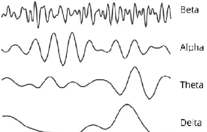
The human brain consists of billions of brain cells named by neurons. When communicating this neuron at the same time it generated a significant amount of electrical activity called brainwaves and measured in Hertz. The brainwaves are generally divided into five main frequencies: Beta waves, Alpha waves, Theta waves, Delta waves and Gamma waves. Not only brainwaves Different in frequency but also it was Different in amplitude.
Brainwaves are electrical waves of certain frequencies, which result from the work and activity of neurons in the brain, or "Neurons." Actually, brain waves are what represent our thoughts, feelings, feelings, and reactions.
Brainwaves are recorded based on a set of sensors placed on the head. The function of these sensors is to capture, record, process and display the electrical signals on the surface of the head. The process of capturing and recording signals can be done on the surface of the head, but it will be a difficult and complicated process due to the large interference that will be on the signals. The second way to record brainwaves is by placing the electrodes directly on the cerebral cortex according to the surgical procedure, and this method gives better and more accurate results. The process of recording and capturing brainwaves is known as EEG: Electroencephalography.
With regard to the details of the brainwaves themselves, they are divided into several bandwidths, each of which represents a particular pattern of mental activity. It is logical to note that high-frequency packets represent concentrated and intense mental activity (known as gamma waves) while low-frequency packets represent low mental activity, i.e., sleep situations (represented by the Delta).
Speech means that brain waves will vary according to the mental activity of the person and according to the feelings he feels. When we feel tired and relaxed, lower-frequency brain waves are the dominant and most prevalent, while high-frequency brain waves occur when we are in high concentration and intense activity.
It is worth mentioning here that this article aims to highlight the concept of brain waves and types in general and comprehensive, while the careful and practical approach to the subject of brain waves and applications requires more complex details, especially that brain waves represent many things according to the region where the brain.
The frequency of the brainwaves is measured in Hertz or CPS: Cycle per Second. The following is the classification of the brain waves according to the frequency bands to which they belong.
1- Delta Waves:
ISSN(Online): 2319-8753 ISSN (Print): 2347-6710
I
nternational
J
ournal of
I
nnovative
R
esearch in
S
cience,
E
ngineering and
T
echnology
(A High Impact Factor, Monthly, Peer Reviewed Journal)
Visit: www.ijirset.com
Vol. 7, Issue 3, March 2018
2- Theta Waves:
Most often, theta waves appear during sleep (not the deep sleep in which delta waves appear) and may sometimes appear in long and deep meditations. Theta waves are likened to our path and path to memories and information stored in the brain. Theta waves are responsible for drawing our senses from focusing on the surrounding medium, to focus on the signals generated within the brain. Theta waves are also responsible for dreams. Theta waves generate images, live scenes, information and knowledge that are not connected to the direct conscious perception we get from the senses of hearing, sight, smell, and others. [4]
3- Alpha Waves:
Alpha waves represent the quiet state of the brain, which means that the brain is conscious and aware of what is around it, but it is inactive or active and can be likened to the "Stand by" state of the computer, Where the computer does not perform any action requires the capabilities of the processor, but the computer is ready to receive any signal or alert. [5]
4- Beta Waves:
Beta waves appear dramatically when the brain is conscious and aware, and it also performs various functions and functions, all of which are related to the conscious perception of the senses. Beta waves represent the state of "activity" of the brain, and when they spread across the brain, it means that we do a variety of functions, such as thinking, problem-solving, looking and listening and receive various alerts. Beta waves are divided into three other bands:
Beta1: low-bandwidth waves, which cover the bandwidth of 12-15 Hz, represent the lowest state of alertness and cognitive-brain awareness. [6]
Beta2: A band that covers a frequency band of 15 - 22 Hz, which represents a growing concentration of mental brain activity.
Beta3: high waves: a band that covers a bandwidth of 22 - 39 Hz. These waves represent complex thoughts, learn new experiences, and stimulate brain states.
5- Gamma Waves:
Gamma waves are the fastest brain waves and highest in terms of frequency value. Gamma waves represent intense mental states, focused thinking, and also represent the response of several brain regions to contribute to a single focused thinking process. Previously, gamma waves were an additional brain activity that had no meaning; only research showed that they were actually the highest levels of brain activity. One of the puzzles about gamma waves is that it has a higher frequency than the frequency of signal transmission across the same neurons, and how the generation and emergence of gamma brain waves are still considered one of the important mysteries in the field of neuroscience. [7]
B. ELECTRICAL BRAIN SIGNALS:
The human brain consists of a group of cells known as neurons that communicate with each other in long neuronal axons. These neurons are in a working state every time we do something: move, think, feel, remember, etc. The neurons act as follows: When we want to do something, such as raising a glass of water to drink it, the neurons generate an electrical nerve signal, which moves through the axons to the nerve units, which are the muscles and bones, and the speed of transmission of these signals 250 mph 400 km per hour). These electrical signals are generated mainly via electrophoresis difference in neuronal membranes.
Although the electrical nerve pathway is insulated, it is not perfectly insulated, so some electrical nerve signals can leak out. What neuroscientists and neuroscientists do is capture and record these signals to determine the effectiveness and nervous activity of nerve cells, thus determining the state of the brain as a whole. The opposite can also be done, namely, determining the response of neurons to the brain when a neurotransmitter is moved from a sensitive organ such as the eye. When the eye sees a specific color, a neurotransmitter is sent to the brain to explain. The neurons in the brain are the ones who will interpret it, so they will also produce electrical signals during their work. So, we can know exactly how the brain works and how it understands the world around us.
ISSN(Online): 2319-8753 ISSN (Print): 2347-6710
I
nternational
J
ournal of
I
nnovative
R
esearch in
S
cience,
E
ngineering and
T
echnology
(A High Impact Factor, Monthly, Peer Reviewed Journal)
Visit: www.ijirset.com
Vol. 7, Issue 3, March 2018
C. ELECTROENCEPHALOGRAPHY (EEG):
German psychologist Hans Berger in 1924 first recorded electric activities in peoples by placing electrodes on the scalp and the galvanometer. In 1929, in spite of primitive instruments and strategies by that time, he was capable to define some low frequency vibrations called alpha waves. Following these parameters, the basic ideologies and processes for (EEG) planning have hardly transformed.
Electroencephalography (EEG) signals recorded from the surface of the scalp are the best signals uses to brain computer interface, because this method of signals the brain activity is easy to use and very inexpensive. The electrical brain activity is represented by electroencephalogram (EEG) signals. The best way to get EEG signals from surface of the scalp is use the electrode scheme as shown in (Figure1). [9]
Electroencephalography (EEG) is naturally a non-intrusive method (though, usually used as electrodes in special applications) to record the electric activities of the mind sideways the scalp. The brainwave chart actions fluctuation in the voltage generated by ionic currents in brain neurons. This technique is referred to in the clinical field as a recording of the automatic electric activities of the mind done a dated of period. It is measured by several electrodes located on the scalp. Diagnostic uses usually effort on the spectral contented of brainwave planning, Appear in signs of EEG. Brain planning is used extensively to analyze epilepsy, which grounds abnormal patterns in EEG reading. This mechanism is as well using to analyze slumber sicknesses, coma, cerebral anomalies, and mind decease. EEG was used as a primary diagnostic technique for tumors, hit and extra principal mind sicknesses. However, its use may decrease with the advent of high resolution anatomical imaging techniques, such as magnetic resonance imaging (MRI) and computed tomography (CT). Regardless of limited spatial precision, brain planning remains a valuable tool in the fields of research and diagnostics, particularly when a temporal resolution of a fraction of thousandths of a second is required, which is not provided by other methods like attractive character imagine and tomography. One of the brainwave-derived derivatives is called the excited effort (EP), which involves the equation of brainwave planning activity to be time-bound with exposure to a given stimulus (optical, tactile, or audit stimulus). Efforts associated with the ERP event refer to the modified brainwave planning replies that are time-bound for much multifaceted processes of stimuli; This method is using in the mental skills, mental psychology study.
Figure1: EEG electrode placement schemes.[10]
The human brain wave has four main frequencies are:
Delta: It is frequency ranges from 1 Hz to 4 Hz. Is known as high in amplitude and the slow wave.
Theta: It is frequency range between 4Hz and 8Hz. It is known as "slow" activity.
Alpha: It is frequency range between 8 Hz and 12 Hz. It higher in amplitude.
ISSN(Online): 2319-8753 ISSN (Print): 2347-6710
I
nternational
J
ournal of
I
nnovative
R
esearch in
S
cience,
E
ngineering and
T
echnology
(A High Impact Factor, Monthly, Peer Reviewed Journal)
Visit: www.ijirset.com
Vol. 7, Issue 3, March 2018
Figure 2: Brain wave samples to beta, alpha, theta, and delta.
D. FEATURES EXTRACTION:
In recent years, feature extraction technique has important role in signal processing, there are many feature extraction methods to extracting features from EEG signals such as Fourier Transform, Short-Time Fourier Transform and The Wavelet Transform. This step used to convert original brain wave to some values, important investigations in this step we can classified those values easily by using classification methods and then convert to commands by using machine learning as shown in Figure (3). We can extract features from EEG signals by using two various domains like Time and Frequency domain features (TDF) (FDF). [11]
Figure 3: The steps for EEG signals processing.
III. FEATYRE EXTRACTION METHODS
The unique brainwaves have a noise and we cannot use this wave for brain-computer interface easily. there are many steps for EEG signal processing. the most important steps use for brain-computer interface systems are feature extraction technique. this technique has the best role in extracting a feature from signals and converts to some values. There are many methods for extracting a feature from EEG signals and each one has advantages and disadvantages. The original EEG signal is that the time domain signals and also the signals power distribution square measure distributed. In order to extract features, the EEG signals is analyzed to allow a signals power description as a function of time or frequency. Based on previous studies, the characteristics of the extractor within the frequency domain are one of the most effective to spot mental tasks supported EEG signals analyses. [12]
ISSN(Online): 2319-8753 ISSN (Print): 2347-6710
I
nternational
J
ournal of
I
nnovative
R
esearch in
S
cience,
E
ngineering and
T
echnology
(A High Impact Factor, Monthly, Peer Reviewed Journal)
Visit: www.ijirset.com
Vol. 7, Issue 3, March 2018
eliminate the potential would like for info compression. Recently, a large form of strategies are accustomed extract options from graph signals. These embody time-frequency distribution (TFD), fast Fourier transform (FFT), eigenvector (EM), wavelet Transform (WT) Automatic reduction methodology (ARM), and so on. In general, graph analysis has been the topic of many studies, given its ability to supply associate objective methodology of recording brain stimulation that's wide utilized in pc interface analysis within the brain with application in diagnosing and rehabilitation engineering. Therefore, the needs of this paper can discuss some ancient strategies of extracting the graph feature, examination its performance to a particular task, and at last, recommending the foremost acceptable thanks to extract the performance-based advantage.
Electroencephalography (EEG) is one of the areas in the diagnosis of epilepsy. The EEG records can provide a valuable look and better understanding of the mechanisms that cause epileptic disorders. the Fourier transform (FFT) and wavelet conversion are used as spectral analysis tools for EEG signals. These methods can provide a time-varying frequency of EEG signals. Since frequency characteristics are important observable information from signals, FFT and wavelet conversion are among the best ways to analyze EEG signals. Comparisons are also made between these two methods. The result showed that wavelet conversion was better than FFT in the analysis of EEG signals.
A. Fast Fourier Transform (FFT) Method:
FFT is an algorithm can use for signal processing and signals feature extraction. this technique services some accurate incomes or tools or equations for analyzing EEG information. this method utilizes to change signals from time-domain to frequency-domain and vice versa. Features of the obtained EEG signal to be examined are calculated by power spectral density (PSD) estimation so as to by selection characterize the EEG models signal. But, four frequency bands cover the main characteristic waveforms of EEG spectrum. [13]
The PSD is studied by Fourier transforming the evaluated autocorrelation arrangement which is initiate by nonparametric approaches. the Welch's method is One of these methods. The data sequence is utilized to data windowing, the result modified periodograms. The information sequence xi(n) be represented as
( ) = ( + ), = , , , … , −
(1)
= , , , … , − ;
takes to be first point of start of sequence ℎ and then L takes for length of 2M perform information’s segments that are created, the outputs results give.
≈( )
( ) = ( ) ( ) ( )
In this function, named by window function, U bounces regularization feature of the power and is chosen like that.
= ( ) ( )
Where w(n) is the window function, the average of these modified periodograms gives Welch’s power spectral as follows:
ISSN(Online): 2319-8753 ISSN (Print): 2347-6710
I
nternational
J
ournal of
I
nnovative
R
esearch in
S
cience,
E
ngineering and
T
echnology
(A High Impact Factor, Monthly, Peer Reviewed Journal)
Visit: www.ijirset.com
Vol. 7, Issue 3, March 2018
B. Wavelet Transform (WT) Method:
P.Jahankhani and K.Revett have proposed Wavelet Transform (WT) to analyze various transient events in the field of biomedicine. They have described that WT is fit for non-fixed signals and has more benefit than spectral investigation. For timing, signal wave representations are an actual technique. The significant feature of WT is that they provide exact frequency data at low frequencies and exact period data on high frequencies. This belonging is significant in biomedical claims. Because most references in the biomedical area continuously cover high-frequency mechanisms with short period and low-frequency mechanisms with a long-time period. WT offers multiresolution examination of nonstationary signals. [14]
This method is a period frequency investigation; Wavelet analysis method defines an algorithm which involves the multistage window method. In this method the length of window has variable size this feature of wavelet is used in this technique. This method involves the use of large time interval where we need low frequency data and where we have to find high frequency data we need to set the length of window to be shorter so accordingly we can use this type of technique. Fourier transform cannot be useful to the transitory signals as plenty of signals in EEG contains non-stationary components so this technique cannot be applied for EEG so wavelet transform must be used in wavelet transform variety of probing function are used and depending upon the comparison with the threshold function window length can be control and we can easily extract features from the signals. This concept leads to the function for the continuous wavelet transform (CWT
( , ) = ( ) ∗, ( ) ( )
One major advantage of wavelet technique is its flexibility to perform general analysis that is, to analyze or can be apply to localize the window size and localization area of a larger signal is done. In the wavelet packet technique, signal is compressed and noise is removed by using the Fourier transform technique exactly same ideas can be developed to extract the feature of EEG signal the basic idea and technique will be the same as traditional method but the only difference is that we can easily analysis the complex problem in easy and flexible manner and here we have to deal with EEG signal as in wavelet analysis approximations method are used to spilt the details of signals as shown below and these details will be the required feature. The mined wavelet coefficients afford a compressed symbol that displays the energy distribution of the EEG signal in time and frequency. The subsequent numerical structures could be used to characterize the time–frequency distribution of the EEG signals such as mean of the complete standards of the coefficients, the power average of the wavelet coefficients in each sub-band and he subsequent numerical structures were used to characterize the time frequency distribution of EEG signals which are mean of the absolute of coefficients in each sub bands and Average power of wavelet coefficients in each sub band. These structures characterize the frequency distribution of signal. The coefficients Standard deviation in each sub bands and ratio of absolute mean values of adjacent sub bands represents the amount of changes in frequency distribution so these features were considered for the specific application and used for the classification of EEG signals.
1- Continuous Wavelet Transform (CWT) Method:
This can be expressed as
( , ) = ( ) ∗, ( ) ( )
( )standing for the unprocessed EEG, where standing for dilation, and representing translating feature. The
ᴪ , ( ) denotes the complex conjugate and can be calculated by
, ( ) =
| |
−
ISSN(Online): 2319-8753 ISSN (Print): 2347-6710
I
nternational
J
ournal of
I
nnovative
R
esearch in
S
cience,
E
ngineering and
T
echnology
(A High Impact Factor, Monthly, Peer Reviewed Journal)
Visit: www.ijirset.com
Vol. 7, Issue 3, March 2018
where ᴪ( ) means wavelet. Though, its main weaknesses is that evaluating parameters and translation parameters of CWT change continuously. Thus, the coefficients of the wavelet for all accessible gages after calculating will consume many of energy and crop many of amusing data.
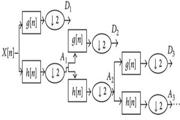
2- Discrete Wavelet Transform (DWT):
In demand to report the softness of the CWT, discrete wavelet transform (DWT) had stayed cleared on the base of multiscale feature image. All the gage below consideration characterizes an exclusive thickness of the EEG signal. The multiresolution decomposition of the raw EEG data ( ) is showed in Figure 4. Every stage covers two digital filters,
( ) and ℎ( ), also two down samplers by 2. The discrete mother wavelet ( ) is a high pass in nature, while its mirror image is ℎ( ) is a low-pass in nature.
Figure 4: Implementation of decomposition of DWT.
As in the in Figure 3 shown, every step output offers a details of the signal D and an estimation of the signal A, where
the latest changes an input for the following stage. The number of stages to which the wavelet decomposes is selected
dependent on the element of the EEG data with dominant frequency. [15]
The connection among WTs and filter h, that is, low pass, could be characterized as following function:
( ) ( ) + (− ) (− ) = ( )
Here, H(z) represents filter's h z-transform. The high-pass filter's complementary z-transform is expressing as
( ) = (− ) ( )
By accurately describing the signal segment characteristics within a specific frequency domain and localized domain
characteristics, there are many advantages that go beyond the high computing and memory requirements of traditional
convolution-based DWT implementation.
IV. CONCLUSION
ISSN(Online): 2319-8753 ISSN (Print): 2347-6710
I
nternational
J
ournal of
I
nnovative
R
esearch in
S
cience,
E
ngineering and
T
echnology
(A High Impact Factor, Monthly, Peer Reviewed Journal)
Visit: www.ijirset.com
Vol. 7, Issue 3, March 2018
devices, one of those way is (BCI). In this paper has two different methods discussed for electroencephalogram (EEG) signal processing, each one different with other, each one has some advantages and disadvantages such as connection speed, loss of important data, there is two type of signal domain like Frequency and time-frequency domain. Each one has a different method of analyzing EEG signals. Frequency domain ways don't offer high-quality performance for a few electroencephalogram signals whereas time-frequency strategies will not offer elaborate info regarding electroencephalogram information the maximum amount as frequency domain strategies. Thence in line with completely different mental task connected applications correct technique ought to be chosen for higher results. And each one of those methods has a different performance like Frequency Resolution and Spectral leakage.
REFERENCES
[1] L. Mayaud, M. Congedo, A. van Laghenhove, et al., “A comparison of recording modalities of P300 Event Related Potentials (ERP) for Brain-Computer Interface (BCI) paradigm,” Clinical Neurophysiology, vol. 43, no. 4, pp. 217–227, 2013.
[2] Nicolas-Alonso, L. F., & Gomez-Gil, J. (2012). Brain computer interfaces, a review. Sensors, 12(2), 1211-1279.
[3] Steriade, M. (2003). 1. Abstract 2. Neuronal substrates of brain disconnection during NREM sleep 3. Three types of brain rhythms during NREM sleep: the unified corticothalamic system 3.1. Spindles, a thalamic rhythm under neocortical influence 3.2. Delta waves: two different (thalamic and cortical) components 3.3. The neocortical slow oscillation groups thalamically generated NREM sleep rhythms. Frontiers in bioscience, 8, d878-899.
[4] Schacter, D. L. (1977). EEG theta waves and psychological phenomena: A review and analysis. Biological psychology, 5(1), 47-82. [5] Kirschfeld, K. (2005). The physical basis of alpha waves in the electroencephalogram and the origin of the “Berger effect”. Biological
Cybernetics, 92(3), 177-185.
[6] Dustman, R. E., Boswell, R. S., & Porter, P. B. (1962). Beta brain waves as an index of alertness. Science, 137(3529), 533-534. [7] Kirschfeld, K. (1992). Oscillations in the insect brain: do they correspond to the cortical gamma-waves of vertebrates?. Proceedings of
the National Academy of Sciences, 89(10), 4764-4768.
[8] Sutter, E. E. (1992). The brain response interface: communication through visually-induced electrical brain responses. Journal of Microcomputer Applications, 15(1), 31-45.
[9] Guger, C., Schlogl, A., Neuper, C., Walterspacher, D., Strein, T., & Pfurtscheller, G. (2001). Rapid prototyping of an EEG-based brain-computer interface (BCI). IEEE Transactions on Neural Systems and Rehabilitation Engineering, 9(1), 49-58.
[10] Light, G. A., Williams, L. E., Minow, F., Sprock, J., Rissling, A., Sharp, R., ... & Braff, D. L. (2010). Electroencephalography (EEG) and event related potentials (ERPs) with human participants. Current protocols in neuroscience, 6-25.
[11] Al-Fahoum, A. S., & Al-Fraihat, A. A. (2014). Methods of EEG signal features extraction using linear analysis in frequency and time-frequency domains. ISRN neuroscience, 2014.
[12] Lotte, F., Congedo, M., Lécuyer, A., Lamarche, F., & Arnaldi, B. (2007). A review of classification algorithms for EEG-based brain– computer interfaces. Journal of neural engineering, 4(2), R1.
[13] Polat, K., & Güneş, S. (2007). Classification of epileptiform EEG using a hybrid system based on decision tree classifier and fast Fourier transform. Applied Mathematics and Computation, 187(2), 1017-1026.
[14] Kıymık, M. K., Güler, I., Dizibüyük, A., & Akın, M. (2005). Comparison of STFT and wavelet transform methods in determining
epileptic seizure activity in EEG signals for real-time application. Computers in biology and medicine, 35(7), 603-616.