University of South Carolina
Scholar Commons
Theses and Dissertations
2016
Development of Protein-Polymer Core-Shell
Nanoparticles (PPCS-NPs) as Efficient Vehicles to
Deliver Therapeutic Agents Across Blood Brain
Barrier (BBB)
Napat Tandikul
University of South Carolina
Follow this and additional works at:https://scholarcommons.sc.edu/etd
Part of theChemistry Commons
This Open Access Thesis is brought to you by Scholar Commons. It has been accepted for inclusion in Theses and Dissertations by an authorized administrator of Scholar Commons. For more information, please [email protected].
Recommended Citation
Development of Protein-Polymer Core-Shell Nanoparticles (PPCS-NPs) as
efficient vehicles to deliver therapeutic agents across Blood Brain Barrier
(BBB)
by
Napat Tandikul
Bachelor of Science Chulalongkorn University, 2011
Submitted in Partial Fulfillment of the Requirements
For the Degree of Master of Science in
Chemistry
College of Arts and Sciences
University of South Carolina
2016
Accepted by:
Qian Wang, Director of Thesis
Thomas M. Makris, Reader
D
EDICATIONI would like to dedicate this thesis…
To my mom, my dad and my brother for all their forever love, care and support,
To Mae Noon, who is suffering from Her2 positive breast cancer that is now
penetrating her brain and Nong Einz, who passed away from Ependymoblastoma
A
CKNOWLEDGEMENTSFirst and foremost, I would like to thank Dr. Qian Wang for giving me a great
opportunity to join his research group and thank you for all his generous help during these
two and a half years at the University of South Carolina. I would also like to thank
Dr. Thomas Makris, Dr. Chuanbing Tang and Dr. Michael Wyatt for their suggestions for
my research plan and special thanks to Dr. Makris for always willing to help and for being
my thesis reader.
Thank you to all the members in Dr. Wang’s research group for sharing memories
together, especially, Dr. Gary Horvath for training me well since the first day I joined this
lab; Dr. Yuzhe Nie and Dr. Xiaolei Zhang for their help in MALDI-MS and peptide
analysis; and Dr. Jing Yan for providing me with a basic introduction to polymer and
self-assembly technique and for synthesizing PCL-py polymer.
I would like to also gratefully thank my former advisors, Dr.Weerapong
Prasongchean for his training and advice before and during my graduate study life,
Dr.Pakorn Winayanuwattikun, and Assoc. Prof. Dr. Vichien Rimphanitchayakit for their
support in the admission process to University of South Carolina. Thank you Dr. Nitsara
Karoonuthaisiri for being my role model, my inspiration, my forever favorite woman
scientist and my long-lost sister, thank you for all her suggestions, comments and support.
Thank you all professors at the University of South Carolina, especially Dr. Amy
Taylor-Perry for being my role model of a good “teacher”. I thank all my friends at the
University of South Carolina. I could not believe that I have friends from all around the
world. Thank you Malini, TT, John, Nikita, Evan and Safaa for their friendships and for
being so supportive and always listen to me when I have tough time. Special thanks to Gift,
Amie and Nikki for helping me settling down when I first moved to Columbia and for their
general advices.
Thank you my host family, Mr.Paul Rouppasong and Na Malivan for spoiling me
with good food and always providing me kindness and generosity. Of course, thank you
my long-lost sister, Nong Sala Dang, who brings laugh and happiness to my graduate life,
and take me to everywhere I want. I also thank Kevin, Blair, Briana, Eli and P’Bung for
spending good times and sharing good memories at Rouppasong’s house. I also thank Pa
Sri who taking good care of me, and sending me food and love even though we are far
apart. I thank P’Oa, my graduate student 101 teacher, who always shares her experiences
and gives me clear answers for all my questions about my poor graduate study life and all
her support.
Finally and most importantly, I would like to thank my parents, Mr. Piroj and Mrs.
Yanaporn Tandikul and my brother, for their faith in my dream, for rooting me with love,
support, patience and understanding throughout my life. I would not have made it today
without them. I would also like to thank my best friends, the gang, in Thailand, Patt, Ploy,
Nat, June, for their care and supports even if we are 9,144 miles apart. Our 10 years of
friendship will last forever. Special thanks to Nisachon, my best friend who always stands
Last but not least, thank you for all obstacles throughout my graduate life that make
me stronger, just like J.K.Rowling said “Rock bottom became the solid foundation on
A
BSTRACTBlood Brain Barrier (BBB) plays a main role as selective barrier which controls
and limits access of chemicals, molecules and therapeutic agents from blood to brain. The
BBB endothelial cells are connected by Tight Junctions (TJs) which close intracellular
spaces between the endothelial cells and block the free diffusion of substances, therefore
many potential drugs for treating human brain diseases cannot reach the brain in sufficient
concentration. Recently, many studies have thrown an interest in development of
nanoparticles for delivering drugs and imaging agents across BBB. Our research group has
developed protein-polymer core-shell nanoparticles (PPCS-NPs) which demonstrate great
potential for targeted delivery. In this work, Apolipoprotein E3 (ApoE3), which can be
specifically bound to LDLR receptor on BBB endothelial cells, was chosen as a targeted
motif. Nanoparticles conjugated with ApoE3 and fluorescently labelled ApoE3 (Fl-ApoE3)
were successfully synthesized. The synthesis of ApoE3/ Fl-ApoE3 NPs with encapsulation
of drugs and dyes is in progress. In vitro study of the uptake of ApoE3-NPs,
Fl-ApoE3-NPs with and without encapsulation of drugs and dyes will be further investigated by using
human umbilical vein endothelial cells (HUVECs) and brain microvascular endothelial cell
T
ABLE OFC
ONTENTSDEDICATION ... iii
ACKNOWLEDGEMENTS ... iv
ABSTRACT ... vii
LIST OF TABLES ...x
LIST OF FIGURES ... xi
CHAPTER 1:BACKGROUND AND SIGNIFICANCE...1
1.1 INTRODUCTION ...1
1.2 BLOOD BRAIN BARRIER (BBB) ...1
1.3NANOPARTICLE FOR DRUG DELIVERY ...5
1.4 NANOPARTICLES UPTAKE AND TRANSPORT ACROSS BBB ...7
1.5 STATEMENT OF WORK ...9
REFERENCES ...10
CHAPTER 2:PURIFIACATION OF APOLIPOPROTEIN E AND SYNTHESIS OF FLUORESCENTLY LABELLED APOE3(FL-APOE3) ...13
2.1APOLIPOPROTEIN E ...13
2.2PURIFICATION AND ANALYSIS OF APOLIPOPROTIEN E3,E4 ...16
2.3SYNTHESIS OF FLUORESCENTLY LABELLED APOE3(FL-APOE3) ...20
2.4 CONCLUSION ...22
2.5MATERIALS AND METHOD ...23
CHAPTER 3:SELF-ASSEMBLY AND CELL UPTAKE OF PROTEIN POLYMER CORE SHELL
NANOPARTICLES ...28
3.1INTRODUCTION ...28
3.2 SELF-ASSEMBLY OF APOE3-P4VPNANOPARTICLES (APOE3-P4VP-NPS) ...31
3.3 SELF-ASSEMBLY OF FLUORESCENTLY LABELLED APOE3-P4VP NANOPARTICLES (FL-APOE3-P4VP-NPS) ...33
3.4SELF-ASSEMBLY OF APOE3-P4VPNANOPARTICLES (APOE3-PCL-PY-NPS) ...35
3.5CELL VIABILITY ASSAY ...37
3.6NANOPARTICLES UPTAKE HUVECS AND HMSCS...39
3.7SELF-ASSEMBLY OF FLUORESCENTLY LABELED APOE3-P4VP ENCAPSULATED NILE RED NANOPARTICLES (FL-APOE3-P4VP/NR-NPS) ...44
3.8CONCLUSION ...45
3.9MATERIALS AND METHOD ...45
L
IST OFT
ABLESTable 2.1 Prevalence of the human ApoE isoforms and their key differences. Adapted with permission from Hatter, D.M. et al (2006) Trends Biochem Sci, 31, 445-454. ...15
Table 3.1 Sizes of ApoE3-P4VP-NPs measured by DLS (a-c) with different mass ratios of proteins to P4VP (MApoE3/MP4VP). ...33
Table 3.2 Sizes of Fl-ApoE3-P4VP-NPs measured by DLS (a-c) with different mass ratios of proteins to P4VP (MFl-ApoE3/MP4VP). ...34
Table 3.3 Sizes of ApoE3-PCL-py-NPs measured by DLS (a-c) with different mass ratios of proteins to PCL-py (MApoE3/MPCL-py). ...37
L
IST OFF
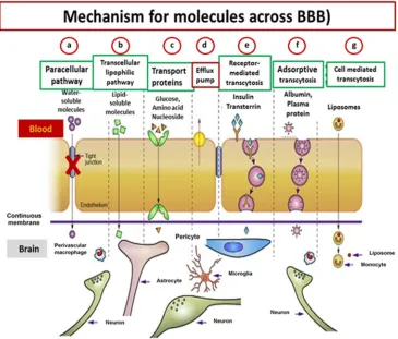
IGURESFigure 1.1 Blood Brain Barrier and cell association overview (top). The cell association at the BBB (bottom). The cerebral endothelial cells form tight junctions at the margins (bottom, red circle) which block the aqueous paracellular diffusional pathway. Pericytes which distribute along the endothelial cells ensheath the endothelium and contribute to the local basement membrane which forms basal lamina. Astrocytic endfeet of the astrocytes form a complex network and cell association around the capillaries which help in maintenance of the BBB properties. Microglia, which are the resident of immunocompetent cells, regulate BBB properties during embryogenesis and disease. Figure is adapted with permission from Abbott, N.J. et al. (2010) Structure and function of the blood-brain barrier. Neurobio Dis. 37(1), 13-25. ...2
Figure 1.2 Mechanism and different pathways for molecules across BBB. a) Paracellular pathway for water soluble molecules, the molecules were blocked by tight junction (TJs). b) Transcellular lipophilic pathway for lipid-soluble molecules. c) Carrier-mediated transport as occurs for glucose, amino acids, nucleosides, etc. d) Efflux pump, molecules can be pumped out by transporters on endothelial cell membrane. e) Receptor mediated transcytosis for peptide signaling and regulatory molecules, e.g. insulin, transferrin. f) Adsorptive transcytosis for positively charged cargo (serum proteins) transport. Figure is adapted with permission from Chen, Y. and Liu, L. (2012) Structure and function of the blood–brain barrier. Adv Drug Deliv Rev 64 (7), 640-665. ...4
Figure 1.3 Example of nanocarriers (adapted with permission from Dan, P. et al. (2007) Nature Nanotechnology 2, 751 – 760) ...6
Figure 2.1 ApoE3 has two structural domains, N-terminal domain (red) which contains receptor binding region and C-terminal domain (blue) which contains lipid binding region. Those two domains are linked by hinge region (gray). ApoE2, E3 and E4 isoforms are encoded by the ε2, ε3 and ε4 alleles on ApoE gene respectively. There are two polymorphic
positions, 112 and 158, that distinguish the three common isoforms. Figure is adapted with permission from Liu, C-C. et al (2013) Nat Rev Neurol, 9, 106-118. b) The model structure illustrating the full length human ApoE3 created by Chen, J. et al (2011) Proc. Natl. Acad. Sci, 108, p.14813. Solution NMR method was used in studying the structure of the ApoE3
Figure 2.2 a) SDS-PAGE analysis of the purified ApoE3. Protein were analyzed on a gradient (4-20%) gel and stained with Coomassie blue. Lane 1, ApoE3 with a concentration of 2.52 µg/mL; lane 2, ApoE3 with a concentration of 4.00 µg/mL; Lane 3, ApoE3 with a concentration of 6.00 µg/mL. The molecular weight of ApoE3 was a little higher than 34.0 kDa (Red arrow). b) Western blot analysis of purified ApoE3 against 1o antibody, Goat
anti human ApoE, and 2o antibody, Fab anti-goat IgG (Donkey antibody), shown ApoE
expression at molecular weight of about 34 kDa. Lane 1-5 was ApoE3 with a concentration of 2.0, 4.0, 6.0, 8.0, 10.0 ng/mL. c) MALDI-MS analysis of purified ApoE3 showed the molecular mass of unmodified ApoE3 as 35,085.56 m/z (red arrow). d) Peptide analysis
peak list of ApoE3 created by FlexAnalysis software. e) Peptide analysis by MASCOT
software, identical residues were highlighted in red with 62% recovery and MASCOT score of 75. ...18
Figure 2.3 ApoE4 determination by SDS-PAGE. Protein were analyzed on a gradient (4-20%) gel and stained with Coomassie blue. Lane 1, ApoE4 with a concentration of 2.5 µg/mL; Lane 2, ApoE4 with a concentration of 5.0 µg/mL; Lane 3, ApoE4 with a concentration of 9.90 µg/mL. The result showed a lot of impurities indicated that the purification process was not successfully done. ...20
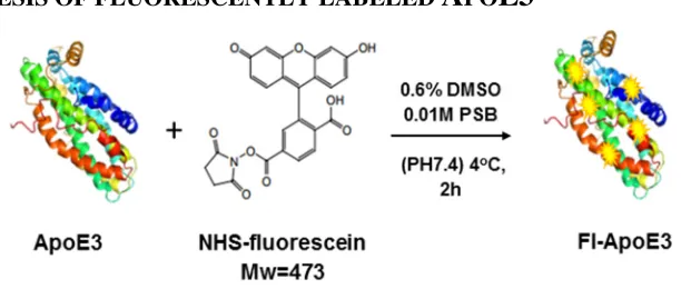
Figure 2.4 Schematic illustration of the synthesis of Fl-ApoE3 ...20
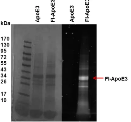
Figure 2.5 Fl-ApoE3 identification by SDS-PAGE visualized under EPI white (left) and UV-VIS (right). Fluorescent signal can only be seen in the lane loaded with Fl-ApoE3. The molecular mass of Fl-ApoE3 was approximately about 34 kDa. ...21
Figure 2.6 MALDI-MS analysis of Fl-ApoE3 showed the molecular mass of Fl-ApoE3 was 35,128.53 m/z (red arrow). ...22
Figure 3.1 Schematic illustration of the formation of Fl-ApoE3 nanoparticles (Fl-ApoE3 NPs) and the study of nanoparticles uptake in HUVECs cells. ...30
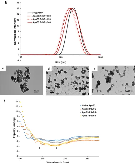
Figure 3.2 (a) Schematic illustration of the synthesis of ApoE3-P4VP-NPs. (b) DLS results of ApoE3-P4VP-NPs with different mass ratio (MApoE3/MP4VP): 0.60, 1.20 and 2.40. TEM
images of ApoE3-P4VP-NPs at different mass ratio, where (c) MApoE3/MP4VP = 0.60, (d)
MApoE3/MP4VP = 1.20, and (e) MApoE3/MP4VP = 2.40. (f) Circular dichroism of ApoE3 and
ApoE3-P4VP-NPs. ApoE3:P4VP-a: MApoE3/MP4VP = 0.60; ApoE3:P4VP-b: MApoE3/MP4VP
= 1.20; and ApoE3:P4VP-c: MApoE3/MP4VP = 2.40. Both native ApoE3 and
ApoE3-P4VP-NPs does not clearly show two minima at 208 (1) and 222 (2) nm. ...31
Figure 3.3 (a) Schematic illustration of the synthesis of Fl-ApoE3-P4VP-NPs. (b) DLS results of Fl-ApoE3-P4VP-NPs with different mass ratio (MFl-ApoE3/MP4VP: 0.60, 1.20 and
2.40). TEM images of Fl-ApoE3-P4VP-NPs at different mass ratio, where (c)
MFl-ApoE3/MP4VP = 0.60 (d) MFl-ApoE3/MP4VP = 1.20 (e) MFl-ApoE3/MP4VP = 2.40. ...33
Figure 3.4 (a) Schematic illustration of the synthesis of ApoE3-PCL-py-NPs. (b) DLS results of ApoE3-PCL-py-NPs with different mass ratio (MApoE3/MPCL-py) of 0.60, 1.20 and
ApoE3 and ApoE3-PCL-py-NPs, ApoE3:PCL-py-a is MApoE3/MPCL-py = 0.60;
ApoE3:PCL-py-b is MApoE3/MPCL-py = 1.20; ApoE3:PCL-py-c is MApoE3/MPCL-py = 2.40.
The CD spectra is not good enough to tell any conformational changes...35
Figure 3.5 Cell viability assay of (a) ApoE3-P4VP-NPs (b) ApoE3-PCL-py-NPs treated for 24 h. ...38
Figure 3.6 Fluorescent microscopic images of HUVECs (LDLR receptor+) cells incubated with Fl-ApoE3, Fl-ApoE3-P4VP-NPs, Fl-BSA-P4VP-NPs for 2, 24h and pre-treated with ApoE3 for 2 h follow by the incubation of Fl-ApoE3-P4VP-NPs for 2, 24 h. The blue nuclei of cells were stained with DAPI. The green fluorescence belonged to the Fl-ApoE3. The scale bars are 20 µm. ...40
Figure 3.7 High magnification fluorescent microscopic images of HUVECs (LDLR receptor+) cells incubated with Fl-ApoE3-P4VP-NPs, for 2, 24h. The blue nuclei of cells were stained with DAPI. The green fluorescence belonged to the Fl-ApoE3. The scale bars are 20 µm. ...41
Figure 3.8 Fluorescent microscopic images of HUVECs (LDLR receptor+) cells pre-incubated with ApoE3 for 2 h, then pre-incubated with Fl-ApoE3-P4VP-NPs for 2, 24h. The blue nuclei of cells were stained with DAPI. The green fluorescence belonged to the Fl-ApoE3. The scale bars are 20 µm. ...42
Figure 3.9 Fluorescent microscopic images of hMSCs (LDLR receptor-) cells incubated with Fl-ApoE3, Fl-ApoE3-P4VP-NPs, Fl-BSA-P4VP-NPs for 2, 24h and pre-treated with ApoE3 for 2 h follow by the incubation of Fl-ApoE3-P4VP-NPs for 2, 24 h. The blue nuclei of cells were stained with DAPI. The green fluorescence belonged to the Fl-ApoE3. The scale bars are 20 µm. ...43
CHAPTER
1
B
ACKGROUND ANDS
IGNIFICANCE1.1
I
NTRODUCTIONIn 2010, statistics from American Brain Tumor Association (ABTA) showed that
688,096 Americans were living with the diagnostic of a primary brain tumor. However,
Only 0.2% are living after diagnosis (America Brain Tumor Association, 2013). One in
nine older Americans, estimated 5.2 million Americans of all ages have Alzheimer’s
disease. Every year, approximately 150 billion US dollars have been paid for health care
and long term care (Alzheimer’s Association, 2014). Parkinson’s disease foundation
reports in 2014 that 1 million Americans are living with Parkinson’s disease, and 25 billion
US dollars have been paid for treatment in USA each year. One of the crucial problems of
human brain diseases treatment is the incapability in efficiently transporting drugs to the
brain because of the blood brain barrier.
1.2
B
LOODB
RAINB
ARRIER(BBB)
Blood brain barrier (BBB) is a selective barrier formed by the endothelial cells that
safeguard cerebral microvessels [1, 2]. BBB is the largest surface area for exchanging
substances between blood and brain, it is approximately 12-18 m2 in surface area [3]. Once
the BBB is crossed, diffusion distances to neurons and glial cell bodies for solutes and
cytotoxic agents by forming a very tight barrier called tight junctions (TJs) [3]. A diffusion
barrier, formed by TJs which present between cerebral endothelial cells, severely restrict
penetration of water soluble compounds and polar drugs into the brain [3]. The presence
of TJs divides plasma membrane of the vascular endothelial cells into two separate
domains, apical membrane which faces the blood, and basolateral membrane which faces
the brain tissue [4].
STRUCTURE OF BLOOD BRAIN BARRIER
The cell association at the BBB including endothelial cells, basal lamina,
astrocytes, pericytes and microglia [4, 5]. Astrocytic end-feet, cover more than 90% of the
endothelial cell surface, tightly ensheaths the vessel wall and takes part in the induction,
maintenance and robustness of the integrity of endothelial barrier [4, 5]. Astrocytes are
important in induction and maintenance of the barrier properties [3]. Pericytes are
contractile cells that embrace brain capillary and contributes to the development,
maintenance and regulation of BBB [5]. Microglia play a role in regulating BBB properties
during embryogenesis and disease (Figure 1.1).
Figure 1.1. Blood Brain Barrier and cell association overview (top). The cell association at the BBB (bottom). The cerebral endothelial cells form tight junctions at the margins (bottom, red circle) which block the aqueous paracellular diffusional pathway. Pericytes which distribute along the endothelial cells ensheath the endothelium and contribute to the local basement membrane which forms basal lamina. Astrocytic endfeet of the astrocytes form a complex network and cell association around the capillaries which help in maintenance of the BBB properties. Microglia, which are the resident of immunocompetent cells, regulate BBB properties during embryogenesis and disease. Figure is adapted with permission from Abbott, N.J. et al. (2010) Structure and function of the blood-brain barrier. Neurobio Dis. 37(1), 13-25.
Transport across the BBB
Normally, the TJs severely restrict penetration of water-soluble compounds,
including polar drugs by regulating paracellular flux [1, 3]. Water soluble or polar
compounds can penetrate only by paracellular transport which is limited to small
hydrophilic molecules [4, 5]. Lipid-soluble agents (smaller than 400 Da) can effectively
diffuse through the large surface area of the lipid membranes of the endothelial cells via
transcellular lipophilic pathway [6]. This process is driven by concentration gradient and
limited to small hydrophobic molecules [4]. Moreover, it is a main entry route to the brain
nucleosides, choline or other substances require the transporters as specific carriers [1, 4].
However, some transport proteins, such as P-glycoprotein (P-gp), localized on the apical
(luminal) side of the brain capillary endothelium, act as efflux transporters and restrict the
uptake of drugs into the brain [7]. Additionally, certain proteins, such as insulin and
transferrin, are taken up by specific receptor-mediated endocytosis, delivered through BBB
by transcytosis and exposed out of the cells by exocytosis [1, 5]. Adsorptive transcytosis
relies on transport of positively charged cargo in a non-specific manner [4] (Figure 1.2).
1.3
N
ANOPARTICLES FOR DRUG DELIVERYA critical problem in the treatment of brain tumor and neurodegenerative diseases
such as Alzheimer’s disease and Parkinson’s disease is the incapability of drugs to be
delivered across BBB in the brain [8]. To overcome this problem, many studies have shown
an interest in the development of nanoparticles as promising drug delivery agents that can
be transported across BBB and increase the uptake of appropriate drugs in the brain [8-12].
The development of nanocarrier-drug system as Trojan horse complex is one of a
promising drug targeting technology [4]. From this concept, natural or genetically
engineered proteins or small peptides conjugated with appropriate nanocarriers can
specifically transport a drug-payload which is directly coupled or encapsulated in the
nanocarriers [4]. The important advantages of therapeutic nanoparticles over free drugs are
the ability to: 1) prolong blood circulation; 2) control the bio-distribution and release of
drugs; 3) site-specific targeting; 4) stabilize labile molecules (e.g. protein, peptides, DNA)
on the particles’ surface from degradation; and 5) protect a drug from degradation [8, 13,
14]. Moreover, they can be modified to deliver a variety of drugs with improved delivery
efficiency and reduced side effects by targeted delivery [8].
DEFINITION OF NANOPARTICLES
The definition of nanoparticles in the Encyclopedia of Pharmaceutical Technology
and the Encyclopedia of Nanoscience and Nanotechnology is
“Nanoparticles for pharmaceutical purposes are solid colloidal particles ranging in size
from 1 to 1000 nm (1 µm) consisting of macromolecular materials in which the active
principle (drug or biologically active material) is dissolved, entrapped, or encapsulated,
On the other side, physicists and material scientists limit the size of nanoparticles
not to exceed 100 nm [15]. The definition from NNI (National Nanotechnology Initiative)
is “Nanoparticles are structure of sizes ranging from 1 to 100 nm in at least one
dimension”[14]. The size above 1000 nm shows no significant influence in cell uptake and
may lead to embolization in lung capillaries [15, 16].
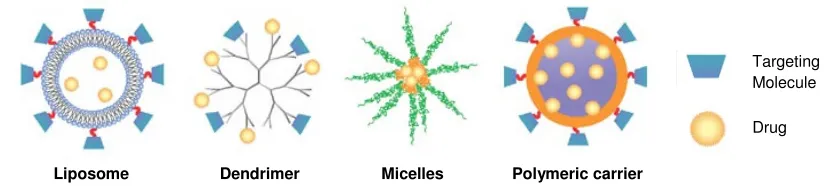
TYPES OF NANOPARTICLES
Nanocarriers in medical applications should be biocompatible (able to migrate
with a biological system without eliciting immune response or any negative effects), and
nontoxic (harmless to the given biological system) [14]. However, many types of
nanoparticles are either toxic or have undesirable effects to cells depending on their
hydrodynamic size, shape, amount, surface chemistry, route of administration, response of
the immune system, and resident time in blood stream [14]. There are several major types
of nanoparticle that have been widely used for development of nanomedicines, including
liposomes, dendrimers, polymeric micelles, polymeric carriers that made of biodegradable
polymers such as poly(butyl cyanoacrylate) (PBCA), poly(isohexyl cyanoacrelate)
(PIHCA), poly(lactic acid) (PLA), poly(lactide-co-glycolide) (PLGA), human serum
albumin (HSA), as well as chitosan [17-19], and inorganic carriers such as gold
nanoparticles, quantum dot etc.
Figure 1.3. Example of nanocarriers (adapted with permission from Dan, P. et al. (2007) Nature
Nanotechnology 2, 751 – 760).
Drug Targeting Molecule
1.4
N
ANOPARTICLES UPTAKE AND TRANSPORT ACROSSBBB
There are eight possible mechanisms of nanoparticles uptake and of bound drugs
into the brain: [16, 20, 21]
1) An increase in retention time of nanoparticles in blood that would lead to higher concentration gradient, and as a result, enhance the delivery to the brain. However, drugs can be subjected to and pumped out by the highly efficient efflux transporters such as P-gp. Thus, the concentration of drug in the brain does not achieve pharmacological effects.
2) Surfactant or coating agents such as polysorbate 80 (tween80) can be used to inhibit efflux system, especially P-glycoprotein (P-gp). However, Pgp cannot be completely blocked by low percentage of tween80.
3)-4) Permeabilization of blood brain barrier by toxic effects or surfactants.
5) Opening of the tight junctions. Then, the drug could permeate through the tight junctions in free form or together with the nanoparticles.
6)-7) Nanoparticles can be uptaken by endocytosis by endothelial cells followed by the release of the drugs within these cells and delivery to the brain or by transcytosis through the endothelial cells.
8) A combination of the above effects.
Many studies have reported that nanoparticles can be taken up by receptor-mediated
endocytosis, which takes place at the aptical or blood side and transported across the BBB
by transcytosis [4, 17, 22]. The particles can then be delivered towards the endothelial cells
by intracellular vesicular trafficking and exocytosed at the opposite surface [4]. However,
Surface modification of nanoparticles with specific targeted motifs is necessary for
facilitating the uptake of nanoparticles [17]. On top of that, surface properties of
nanoparticles, including properties of coating surfactant, core polymer, drugs and
stabilizers, play the most important role for their ability to deliver drugs to the brain [16,
25, 26]. Several studies have investigated the drug transport across BBB by covalent
attachment of targeting motifs such as apoA1, B, E, insulin, anti-insulin receptor
monoclonal antibody (29B4), transferrin, anti-transferrin antibody to nanoparticles [8,
27-30]. Due to the fact that these motifs can specifically interact with specific receptors, for
example, apoB and E with LDL receptor, apoA-I with the scavenger receptor class B typeI
(SR-BI) [31], the conjugated nanoparticles would mimic lipoprotein particles and enter and
across brain endothelial cells by endocytosis and transcytosis[16]. In particular,
apolipoprotein (ApoE) has gained more interest in many recent studies. The Apo E protein
specifically binds to a number of receptors on the endothelial cell membrane of BBB, such
as LDLR, LRP-1, very low density lipoprotein receptor (VLDLR), apolipoprotein
receptor-2 (Apo ER-2) and megalin/gp330, as well as receptors in other parts of central
nervous system [17]. Thus, the presence of ApoE on the nanoparticle surface can promote
the internalization of nanoparticles in the brain endothelial cells via the LDL receptors
expressed by these cells [17]. Moreover, ApoE, especially ApoE3 and ApoE4 isoforms,
also play the major role of amyloid-β (Aβ) aggregation and clearance which relate to
progression of Alzheimer’s disease (AD) [32, 33]. So, it is probable that ApoE conjugated
nanoparticles can be used to facilitate BBB uptake and target AD disease cells at the same
1.5
S
TATEMENT OF WORKIn this work, apolipoprotein E3 (ApoE3) was employed as a targeting motif in the
self-assembly process with two types of polymer, poly (4-vinylpyridine) (P4VP) and
poly(caprolactone-grafted-pyridine) (PCL-pyridine) in order to form protein-polymer core
shell nanoparticles (ApoE3-NPs). The ApoE3 was also modified with fluorescein dye and
self-assembled with the same types of polymer forming fluorescently labelled
protein-polymer core shell nanoparticles (Fl-ApoE3-NPs). The uptake of Fl-ApoE3-NPs in a blood
brain barrier cell model, human umbilical vein endothelial cells (HUVECs), was studied
R
EFERENCES1. Abbott, N.J., Ronnback, L., & Hansson, E. (2006). Astrocyte-endothelial interactions at the blood-brain barrier. Nature Reviews Neuroscience, 7(1). 41-53.
2. Nishitsuji, K., Hosono, T., Nakamura, T., Bu, G., & Michikawa, M. (2011). Apolipoprotein E regulates the integrity of tight junctions in an isoform-dependent manner in an in vitro blood-brain barrier model. The Journal of Biological Chemistry. 286(20). 17536-17542.
3. Abbott, N.J., Patabendige, A.A., Dolman, D.E., Yusof, S.R., & Begley, D.J. (2010). Structure and function of the blood brain barrier. Neurobiology of Disease, 37(1).
13-25.
4. Georgieva, J.V., Hoekstra, D., & Zuhorn, I.S. (2014). Smuggling Drugs into the Brain: An Overview of Ligands Targeting Transcytosis for Drug Delivery across the Blood-Brain Barrier. Pharmaceutics. 6(4). 557-583.
5. Deeken, J.F., & Löscher, W. (2007). The blood-brain barrier and cancer: transporters, treatment, and Trojan horses. Clinical Cancer Research. 13(6).
1663-1674.
6. Pardridge, W.M. (2005). The Blood-Brain Barrier: Bottleneck in Brain Drug Development. NeuroRx. 2(1). 3-14.
7. Löscher, W., & Potschka, H. (2005). Blood-Brain Barrier Active Efflux Transporters: ATP-Binding Cassette Gene Family. NeuroRx. 2(1). 86-98.
8. Shilo, M., Motiei, M., Hana, P., & Popovtzer, R. (2014).Transport of nanoparticles through the blood-brain barrier for imaging and therapeutic applications.
Nanoscale. 6(4). 2146-2152.
9. Denora, N., Trapani, A., Laquintana, V., Lopedota, A., & Trapani, G. (2009). Recent advances in medicinal chemistry and pharmaceutical technology--strategies for drug delivery to the brain. Current Topics in Medicinal Chemistry. 9(2).
182-196.
10. Gao, J.Q., Lv, Q., Li, L.M., Tang, X.J., Li, F.Z., Hu, Y.L., & Han, M. (2013). Glioma targeting and blood-brain barrier penetration by dual-targeting doxorubincin liposomes. Biomaterials. 34(22). 5628-5639.
11. Roney, C., Kulkarni, P., Arora, V., Antich, P., Bonte, F., Wu, A., Mallikarjuana, N.N., Manohar, S., Liang, H.F., Kulkarni, A.R., Sung, H.W., Sairam, M., & Aminabhavi, T.M. (2005). Targeted nanoparticles for drug delivery through the blood-brain barrier for Alzheimer's disease. Journal of Controlled Release. 108
12. Su, X., Wang, Z., Li, L., Zheng, M., Zheng, C., Gong, P., Zhao, P., Ma, Y., Tao, Q., & Cai, L. (2013). Lipid-polymer nanoparticles encapsulating doxorubicin and 2'-deoxy-5-azacytidine enhance the sensitivity of cancer cells to chemical therapeutics. Molecular Pharmaceutics. 10(5). 1901-1909.
13. Singh, R., & Lillard, J.W., Jr. (2009). Nanoparticle-based targeted drug delivery.
Experimental and Molecular Pathology. 86(3). 215-223.
14. Wilczewska, A.Z., Niemirowicz, K., Markiewicz, K.H., & Car, H. (2012). Nanoparticles as drug delivery systems. Pharmacological Reports. 64(5).
1020-1037.
15. Schafer, V., Von, B.H., Rübsamen-Waigmann, H., Steffan, A.M., Royer, C., & Kreuter, J. (1994). Phagocytosis and degradation of human serum albumin
microspheres and nanoparticles in human macrophages. Journal of
Microencapsulation : Micro and Nano Carriers. 11(3). 261-269.
16. Kreuter, J. (2014). Drug delivery to the central nervous system by polymeric nanoparticles: What do we know? Advanced Drug Delivery Reviews. 71. 2-14.
17. Wohlfart, S., Gelperina, S., & Kreuter, J. (2012). Transport of drugs across the blood-brain barrier by nanoparticles. Journal of Controlled Release. 161(2).
264-273.
18. Yang, H. (2010). Nanoparticle-mediated brain-specific drug delivery, imaging, and diagnosis. Pharmaceutical Research. 27(9). 1759-1771.
19. Wohlfart, S., Gelperina, S., & Kreuter, J. (2012). Transport of drugs across the blood brain barrier by nanoparticles. Journal of Controlled Release. 161(2).
264-273.
20. Kreuter, J. (2001). Nanoparticulate systems for brain delivery of drugs. Advanced Drug Delivery Reviews. 47(1). 65-81.
21. Kreuter, J. (2012) Nanoparticulate systems for brain delivery of drugs. Advanced Drug Delivery Reviews. 64. 213-222.
22. Ye, D., Raghnaill, M.N., Bramini, M., Mahon, E., Åberg, C., Salvati, A., & Dawson, K.A. (2013). Nanoparticle accumulation and transcytosis in brain endothelial cell layers. Nanoscale. 5(22). 11153-11165.
23. Bramini, M., Ye, D., Hallerbach, A., Raghnaill, M.N., Salvati, A., Aberg, C., & Dawson, K.A. (2014). Imaging approach to mechanistic study of nanoparticle interactions with the blood-brain barrier. ACS Nano. 8(5). 4304-4312.
25. Gelperina, S., Maksimenko, O., Khalansky, A., Vanchugova, L., Shipulo, E., Abbasova, K., Berdiev, R., Wohlfart, S., Chepurnova, N., & Kreuter, J. (2010). Drug delivery to the brain using surfactant-coated poly(lactide-co-glycolide) nanoparticles: Influence of the formulation parameters. European Journal of Pharmaceutics and Biopharmaceutics. 74(2). 157-163.
26. Wohlfart, S., Khalansky, A.S., Gelperina, S., Maksimenko, O., Bernreuther, C., Glatzel, M., & Kreuter, J. (2011). Efficient chemotherapy of rat glioblastoma using doxorubicin-loaded PLGA nanoparticles with different stabilizers. PLoS One. 6(5).
ID e19121.
27. Kratzer, I., Wernig, K., Panzenboeck, U., Bernhart, E., Reicher, H., Wronski, R., Windisch, M., Hammer, A., Malle, E., Zimmer, A., & Sattler, W. (2007). Apolipoprotein A-I coating of protamine-oligonucleotide nanoparticles increases particle uptake and transcytosis in an in vitro model of the blood-brain barrier.
Journal of Controlled Release. 117(3). 301-311.
28. Mulik, R.S., Mönkkönen, J., Juvonen, R.O., Mahadik, K.R., & Paradkar, A.R. (2010). ApoE3 mediated poly(butyl) cyanoacrylate nanoparticles containing curcumin: study of enhanced activity of curcumin against beta amyloid induced cytotoxicity using in vitro cell culture model. Molecular Pharmaceutics. 7(3).
815-825.
29. Mulik, R.S., Mönkkönen, J., Juvonen, R.O., Mahadik, K.R., & Paradkar, A.R. (2012). ApoE3 mediated polymeric nanoparticles containing curcumin: apoptosis induced in vitro anticancer activity against neuroblastoma cells. International Journal of Pharmaceutics. 437(1-2). 29-41.
30. Ulbrich, K., Knobloch, T., & Kreuter, J. (2011). Targeting the insulin receptor: nanoparticles for drug delivery across the blood-brain barrier (BBB). Journal of Drug Targeting. 19(2). 125-132.
31. Wagner, S., Zensi, A., Wien, S.L., Tschickardt, S.E., Maier, W., Vogel, T., Worek, F., Pietrzik, C.U., Kreuter, J., & Von B.H. (2012). Uptake mechanism of ApoE-modified nanoparticles on brain capillary endothelial cells as a blood-brain barrier model. PLoS One. 7(3). ID e32568.
32. Frieden, C., & Garai, K. (2012). Structural differences between apoE3 and apoE4 may be useful in developing therapeutic agents for Alzheimer's disease.
Proceedings of the National Academy of Sciences of the United States of America. 109(23). 8913-8918.
CHAPTER
2
P
URIFICATION OFA
POLIPOPROTEINE
ANDS
YNTHESIS OFF
LUORESCENTLYL
ABELLEDA
POE3
(F
L-A
POE3)
2.1
A
POLIPOPROTEINE
Human apolipoprotein E protein (ApoE) is a polymorphic glycoprotein of 299
amino acids with a molecular weight of 34 kDa [1-4]. It is produced by several cell types
but highly expressed in the liver and central nervous system (CNS), especially in astrocytes
and microglia [2, 5]. ApoE is a major apolipoprotein in brain and plays an important role
in the transportation of lipoprotein, cholesterol and other essential lipids to brain via ApoE
receptors which are members of low-density lipoprotein receptor (LDLR) family,
including LDLP and LRP1 [1, 5-7]. It also functions as a ligand in receptor mediated
endocytosis of lipoprotein particles in the CNS [2, 5].
ApoE proteins have 3 different isoforms which differ by only one or two amino
acids: ApoE2 (Cys112, Cys158), ApoE3 (Cys112, Arg158) and ApoE4 (Arg112, Arg158),
these differences vary ApoE structure and function (Figure 2.1a, c and Table 2.1) [1, 3-5].
All isoforms are coded from the same ApoE gene but expressed from different alleles (ε2,
ε3, ε4): ε2 is associated with a lower risk for Alzheimer’s disease (AD) [4, 5], ε3 is the
most common isoform found in human [2, 4], and ε4 is a strong risk factor of AD. In
addition, the risk of AD increases approximately three fold in people with one ε4 allele and
ApoA-I, ApoA-II, ApoA-IV, ApoD, ApoH and ApoJ, which can also be found in the brain
[2]. Among them, ApoE is very important to the drug development of neurodegenerative
diseases because there are evidences from human and animal studies which indicated that
the differences in ApoE isoform differentially affect Aβ aggregation and clearance in the
brain, a critical factor for AD therapy [1, 5]. The structure of ApoE (Figure 2.1a, c) shows
that there are two major functional domains with the 22-kDa N-terminal domain consisting
of four-helix bundle and containing the receptor-binding region on helix 4 (amino acid
residues 136-150) and the 10-kDa C-terminal domain consisting of two α-helices and
encompassing the lipid-binding region (amino acid residues 244-272) [3, 4, 8]. Two
domains are linked by a protease sensitive “hinge region” (amino acid residues 167-206)
[3] (Figure 2.1a). Three different isoforms of ApoE are distinguished by two polymorphic
positions at amino acid residue 112, 158 [1]. Structural difference of ApoE3 and ApoE4 is
a salt bridge between Arg-61 and Glu-255 that plays an important role in maintaining the
structure of ApoE4 [4, 8] (Figure 2.1c). More details about differences of ApoE isoforms
can be found in Table. 2.1.
. b)
Figure 2.1. a) ApoE3 has two structural domains, N-terminal domain (red) which contains receptor binding region and C-terminal domain (blue) which contains lipid binding region. Those two domains are linked by hinge region (gray). ApoE2, E3 and E4 isoforms are encoded by the ε2, ε3 and ε4 alleles on ApoE gene respectively. There are two polymorphic
positions, 112 and 158, that distinguish the three common isoforms. Figure is adapted with permission from Liu, C-C. et al (2013) Nat Rev Neurol, 9, 106-118. b) The model structure illustrating the full length human ApoE3 created by Chen, J. et al (2011) Proc. Natl. Acad. Sci, 108, p.14813. Solution NMR method was used in studying the structure of the ApoE3
and the picture was produced by PyMOL. c) Model of the structure of ApoE3 and ApoE4 and structural difference of ApoE3 and ApoE4. Key structural elements of ApoE are N-terminal domain which contains a four helix bundle (helix1, red; helix2, blue; helix3, green; helix4, yellow) and C-terminal domain (gray) which contains lipid binding elements. Main differences is a putative salt bridge between Arg-61 and Glu-255 presenting only in ApoE4 that stabilizes a closer contact between the N- and C- terminal domains. Figure is adapted with permission from Hatter, D.M. et al (2006) Trends Biochem Sci, 31, 445-454.
Table 2.1 Prevalence of the human ApoE isoforms and their key differences. Adapted with
permission from Hatter, D.M. et al (2006) Trends Biochem Sci, 31, 445-454.
According to the ability of ApoE to specifically bind LDLR receptor on BBB
endothelial cell membrane and the ability of ApoE to internalize within the cells by
receptor mediated endocytosis, we believe that ApoE is a good candidate in developing
drug carrier targeting BBB. Therefore, in this work, ApoE3, which is the most abundant
isoform in human body and ApoE4 which is a risk factor of AD were chosen as targeting
motif in the development of protein polymer core-shell nanoparticles (PPCS-NPs). We
propose that the capability of ApoE3 in targeting LDLR receptor together with
self-assembly protocol which was developed in our lab will generate well-defined PPCS-NPs
that can be used to specifically target BBB endothelial cell and further developed as drug
delivery vehicles across the BBB in the future. The development of PPCS-NPs will be
further explained in next chapter.
2.2
P
URIFICATION AND ANALYSIS OFA
POLIPOPROTEINE3
ANDE4
Human ApoE3 protein can be obtained from pre-engineered Human embryonic
kidney endothelial cell line (HEK 293T cells) which was provided from Dr. Dapin Fan,
School of Medicine, University of South Carolina. Briefly, the ApoE coding sequence (954
bp) was sub-cloned into the PWPI-GFP vectorusing single enzyme (PmeI) insertion and
direction screening generating lentiviral ApoE constructs. The lentiviral expression
plasmid for human ApoE3 were then transfected into HEK 293T cells using ProFection
mammalian transfection system to generate HEK 293T cells that can produce human
ApoE3 protein (HEK 293T-ApoE3). The HEK 293T-ApoE3 cells were cultured in DMEM
complete growth medium with standard conditions, 5% CO2 at 37 oC. ApoE3 protein was
CL-6B column and freeze-dried by lyophilization for long-term storage. Typical yield of
the purified ApoE3 was about 5 mg/150 mLculture medium. The ApoE3 was further
analyzed by SDS-PAGE using gradient (4-20%) gel and stained with Coomassie blue, then
confirmed by western blot and MALDI-MS. The molecular weight of ApoE3 is slightly
higher than 34 kDa as showed from SDS-PAGE analysis (Figure 2.2a). Two faint bands
below 34 kDa can also be seen on the gel. We assumed that they were degraded partial
fragments of ApoE3 after purification process, probably, N-terminal and C-terminal based
on approximate molecular weight. This assumption correlated to special structure of
ApoE3 protein which contained two terminal domains linked to each other by protease
sensitive hinge region [3, 9]. The purity of the purified ApoE3 was estimated to be ̴ 85%
based on the result from SDS-PAGE analysis. The identity of human ApoE3 was
confirmed by Western blot analysis of purified ApoE3 against primary antibody, Goat anti
human ApoE, and secondary antibody, Fab anti-goat IgG (Donkey antibody). The result
showed ApoE expression in purified ApoE3 solution at the molecular weight of about 34,
22 and 10 kDa (Figure 2.2b). Molecular weight of ApoE3 was further confirmed by
MALDI-MS anlysis. The molecular weight of unmodified full-length ApoE3 was
35,085.56 m/z (Figure 2.2c, red arrow) comparing to the theoretical molecular weight of
human ApoE protein which was 34,236 Da (Uniprot). However, the molecular weight of
ApoE3 from MS analysis may not be very accurate due to broad peaks and low signal
intensity. The MALDI-MS result correlated to the SDS-PAGE analysis, however the
difference in molecular weight of the purified ApoE3 may probably came from genetic
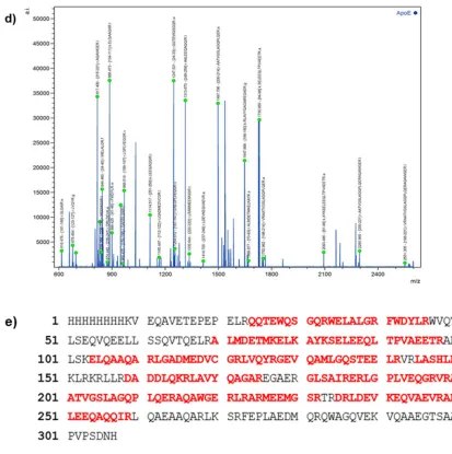
modification process producing HEK 293T-ApoE3 cells. For peptide analysis, ApoE3 was
peak list (Figure 2.2d) was generated by FlexAnalysis software and interpreted by
MASCOT software. The result showed that the purified ApoE3 soluiton was identical to
the full length ApoE3 with a 62% of protein sequence coverage (Figure 2.2e) and the
MASCOT score of 75 confirming the identity of the purified protein as ApoE3.
A p o E 3 , 1 + 2 2 .2 k D a p e p ti d e , 1 + 2 0 .9 k D a p e p ti d e , 1 + A p o E 3 , 2 +
Figure 2.2. a) SDS-PAGE analysis of the purified ApoE3. Protein were analyzed on a gradient (4-20%) gel and stained with Coomassie blue. Lane 1, ApoE3 with a concentration of 2.52 µg/mL; lane 2, ApoE3 with a concentration of 4.00 µg/mL; Lane 3, ApoE3 with a concentration of 6.00 µg/mL. The molecular weight of ApoE3 was a little higher than 34.0 kDa (Red arrow). b) Western blot analysis of purified ApoE3 against 1o antibody, Goat
anti human ApoE, and 2o antibody, Fab anti-goat IgG (Donkey antibody), shown ApoE
expression at molecular weight of about 34 kDa. Lane 1-5 was ApoE3 with a concentration of 2.0, 4.0, 6.0, 8.0, 10.0 ng/mL. c) MALDI-MS analysis of purified ApoE3 showed the molecular mass of unmodified ApoE3 as 35,085.56 m/z (red arrow). d) Peptide analysis
peak list of ApoE3 created by FlexAnalysis software. e) Peptide analysis by MASCOT
ApoE4 was purified from HEK293T-ApoE4 culture media followed the same
procedure as ApoE3. However, the purification of ApoE4 was not successful. The purified
eluent still had a lot of impurities as shown by the SDS-PAGE analysis. The impurities
may came from contamination during purification process by FPLC including binding
condition of protein to column, running and elution buffer condition or protein degradation
during purification process and storage technique. Another possible reason is problem with
protein expression in HEK293T-ApoE4 cells. MALDI-MS analysis was also inconclusive.
Figure 2.3. ApoE4 determination by SDS-PAGE. Protein were analyzed on a gradient
(4-20%) gel and stained with Coomassie blue. Lane 1, ApoE4 with a concentration of 2.5 µg/mL; Lane 2, ApoE4 with a concentration of 5.0 µg/mL; Lane 3, ApoE4 with a concentration of 9.90 µg/mL. The result showed a lot of impurities indicated that the purification process was not successfully done.
2.3
S
YNTHESIS OF FLUORESCENTLY LABELEDA
POE3
NHS-fluorescein conjugated ApoE3 (Fl-ApoE3) was synthesized and its molecular
weight was analyzed by SDS-PAGE using a gradient (4-20%) gel and staining with
Coomassie blue. The gel was visualized under the EPI white light comparing to under the
UV-VIS light in order to detect the fluorescent signal on the protein bands. Fluorescent
signal can be detected under UV-VIS only in Fl-ApoE3 not in unmodified ApoE3. The
result showed that molecular weight of Fl-ApoE3 was a little higher than 34 kDa (Figure
2.5). Fl-ApoE3 was further analyzed by MALDI-MS, the molecular weight of Fl-ApoE3
was 35,128.53 m/z (Figure 2.6, red arrow). The result showed that there were about 2
fluorescein molecules conjugated on an ApoE3 protein, comparing to the theoretical
molecular weight of ApoE3. If the fluorescein molecules were successfully conjugated to
ApoE3 at all 13 lysine residues, the theoretical molecular weight of Fl-ApoE3 would
increase to 40,390 Da. The broad peak as showed in the result and low signal intensity was
assumed to cause inaccurate molecular weight. The resultant proteins were used in the
co-assembly study with selected polymers as described in the consequent chapter.
Figure 2.5. Fl-ApoE3 identification by SDS-PAGE visualized under EPI white (left) and
Figure 2.6. MALDI-MS analysis of Fl-ApoE3 showed the molecular mass of Fl-ApoE3 was 35,128.53 m/z (red arrow).
2.4
C
ONCLUSIONApoE3 can be purified from HEK 293T-ApoE3 culture media by FPLC and
freeze-dried for long-term usage. ApoE3 was successfully produced in a large quantity
( ̴ 5.0 mg/150 mL culture media) and high purity. The molecular weight of purified ApoE3
which was identified by SDS-PAGE was approximately 34 kDa. The identity of purified
ApoE3 was further confirmed by western blot analysis, MALDI-MS, and peptide analysis.
The molecular mass of ApoE3 form MALDI-MS was 35,085.56 m/z which was consistant
to the theoretical mass. We did not successfully purified ApoE4, even though we have tried
to optimize the conditions of the purification process. Fl-ApoE3 was successfully
synthesized and analyzed by SDS-PAGE and MALDI-MS. The result from SDS-PAGE
showed molecular mass of Fl-ApoE3 was a little bit higher than 34 kDa and the result from
MALDI-MS showed the molecular mass of Fl-ApoE3 was 35,128.53 m/z. Both ApoE3
and Fl-ApoE3 would be further used in self-assembly process with selected polymers to
form protein-polymer core-shell nanoparticles for the BBB cellular uptake study.
F l-A p o E 3 , 1 + 2 5 .3 k D a p e p tid e , 1 +
22.2 kDa peptide, 1+ 12.8 kDa peptide, 1+
22.2 kDa peptide, 2+
2.5
M
ATERIALS ANDM
ETHODMATE RIALS
HEK 293T-ApoE3 and HEK 293T-ApoE4 cell line were obtained from Dr.Daping
Fan (University of South Carolina, School of Medicine), Dulbecco’s modified Eagle’s
medium (DMEM) and fetal bovine serum (FBS) was purchased from VWR. Trypsin/
EDTA solution and penicillin-streptomycin (P/S) was purchased from Hyclone.
Mini-Protein TGX Stain-free Procast Gel (4-20%), 10 well-comb, 50 µL/well was purchased
from Bio-Rad. Tris-Glycine-SDS, 10X Solution (Electrophoresis) was purchased from
Fisher Scientific. LabSafe gel blue was purchased from VWR. EZ-Run™ Prestained Rec
Protein Ladder was purchased from Fisher Scientific. Trypsin from bovine pancreas was
purchased from Sigma-Aldrich. NHS-fluorescein was purchased from Pierce. Snake skin
dialysis tubing 3.5K was purchased from Fisher Scientific. Nanosep 10K was purchased
from Pall. All the reagents were used as receive.
APOE3 HARVESTING
The HEK293T-ApoE3 cell line, which can produce Apolipoprotein E3, was
obtained from Dr.Daping Fan (University of South Carolina, School of Medicine). The
293T-ApoE3 Cells were maintained in two of 75 mm2 flasks in DMEM-high glucose
medium with 10% FBS and 1% P/S in a water-saturated atmosphere of 5% CO2 and 95%
air at 37 °C until it reaches 80% confluent. Then, the cells were split into twenty 100 mm2
dishes and cultured in complete growth medium with 10% FBS for two days. The cells
would be about 80% confluent, then changed the media to 1% FBS/DMEM/high glucose
centrifuge bottles, centrifuged at 5,000 rpm for 10 min in the SLA-1500 rotor. The media
were aliquoted to labeled 50 mL tubes and kept at -80 oC or use immediately for purification
by FPLC.
APOE4 HARVESTING
The HEK293T-ApoE4 cell line, which can produce Apolipoprotein E4, was
obtained from Dr. Daping Fan (University of South Carolina, School of Medicine). The
293T-ApoE4 Cells were maintained in the same condition and follow the ApoE3
harvesting protocol.
APOE3 PURIFICATION
ApoE3 can be purified from the media that were collected from HEK293T-ApoE3
cells by using a Heparin-Sepharose CL-6B column for purification, 200 mM NaCl in
10mM sodium phosphate buffer pH 7.4 as running buffer and 1M NaCl in 10mM sodium
phosphate buffer pH 7.4 as elution buffer. Then, the selected fractions were pooled and
dialyzed against 4 liters of 10 mM ammonium bicarbonate at 4 oC over night with two
buffer changes. The purified ApoE3 could be freeze-dried by lyophilization and kept in
APOE4 PURIFICATION
ApoE4 can be purified from the media that were collected from HEK293T-ApoE3
cells by using a Heparin-Sepharose CL-6B column for purification, 200, 300, 400, 600,
800 mM NaCl in 10mM sodium phosphate buffer pH 7.4 as running buffer and 1M NaCl
in 10mM sodium phosphate buffer pH 7.4 as elution buffer.
APOE3 ANALYSIS
The molecular weight of ApoE3 was determined by SDS-PAGE, using a gradient
(4-20%) gel, running at 200 V for 30 min and stained with Coomassie blue. The result can
be confirmed by MALDI-MS analysis which was done by Dr.Yuzhe Nie. For MS analysis,
ApoE3 protein solution in water was mixed in a ratio of 1:1 with 20 mg/mL α-cyano-4-
hydroxycinnamic acid matrix (CHCA matrix) in 70% acetonitrile (CAN) containing 0.1%
trifluoroacetic acid (TFA). The mixture was spotted on the MTP AnchorChip target TM
600/384TF (Bruker Daltonics), spectra were then acquired in the m/z range of
10,000-50,000 with Ultraflex TOF/TOF (Bruker Daltonics) MALDI time-of-flight mass
spectrometer. The spectrometer was operated in a linear positive ion mode with a laser
frequency of 20 Hz and 100% relative energy. External calibration was done based on the
average value of [M+H+] of BSA, m/z of 66,463. A total of 20,000 shots were used to
generate a spectrum from the spots. The export mass data was analyzed by mMass
software. Moving average smooth method was used to get better S/N with widow size 250
m/z. ApoE3 peptide analysis was done with help from Dr. Xiaolei Zhang. Brieftly, ApoE3
was extracted from SDS gel and incubated with trypsin at 37 oC for 4 h and further analyzed
antibody, Goat anti human ApoE, and secondary antibody, Fab anti-goat IgG (Donkey
antibody).
FLUORESCENT-CONJUGATED PROTEINS
A solution of NHS-fluorescein in DMSO (50 µL; 1 mg mL-1) was slowly added
(1 drop/10 s) into a solution of protein (2 mg mL-1 in PBS buffer pH 7.4) at 4 oC with gently
stirring. The solution mixture was incubated in dark at 4 oC for 2 h. Then, the excess
NHS-fluorescein was removed by using nanosep centrifugal system with Mw cutoff 10 kDa,
centrifuged at 5,000 × g for 5 mins, 2 times. The fluorescein conjugated protein was
R
EFERENCES1. Liu, C-C., Kanekiyo, T., Xu, H., & Bu, G. (2013). Apolipoprotein E and Alzheimer disease: risk, mechanisms and therapy. Nature Reviews Neurology. 9(2). 106-118.
2. Kim, J., Basak, J.M., & Holtzman, D.M. (2009). The role of apolipoprotein E in Alzheimer's disease. Neuron. 63(3). 287-303.
3. Frieden, C., & Garai, K. (2012). Structural differences between apoE3 and apoE4 may be useful in developing therapeutic agents for Alzheimer's disease.
Proceedings of the National Academy of Sciences of the United States of America.109(23). 8913-8918.
4. Chou, C.Y., Lin, Y.L., Huang, Y.C., Sheu, S.Y., Lin, T.H., Tsay, H.J., Chang, G.G., & Shiao M.S. (2005). Structural Variation in Human Apolipoprotein E3 and E4: Secondary Structure, Tertiary Structure, and Size Distribution. Biophysical Journal. 88(1). 455-466.
5. Holtzman, D.M., Herz, J., & Bu, G. (2012). Apolipoprotein E and apolipoprotein E receptors: normal biology and roles in Alzheimer disease. Cold Spring Harb Perspect Med. 2(3): ID a006312.
6. Michaelis, K., Hoffmann, M.M., Dreis, S., Herbert, E., Alyautdin, R.N., Michaelis, M., Kreuter, J., & Langer, K. (2006). Covalent linkage of apolipoprotein e to albumin nanoparticles strongly enhances drug transport into the brain. Journal of Pharmacology and Experimental Therapeutics. 317(3). 1246-1253.
7. Nishitsuji, K., Hosono, T., Nakamura, T., Bu, G., & Michikawa, M. (2011). Apolipoprotein E regulates the integrity of tight junctions in an isoform-dependent manner in an in vitro blood-brain barrier model. The Journal of Biological Chemistry. 286(20). 17536-17542.
8. Hatters, D.M., Peters-Libeu, C.A., & Weisgraber, K.H. (2006). Apolipoprotein E structure: insights into function. Trends in Biochemical Sciences. 31(8). 445-454.
CHAPTER
3
S
ELF-
ASSEMBLY AND CELL UPTAKE OF PROTEIN POLYMER CORE SHELL NANOPARTICLES3.1 INTRODUCTION
It is very challenging to develop drug delivery carriers to deliver payloads to brain
due to the restriction of the blood brain barrier (BBB) [1-3]. To overcome this problem,
many researchers are currently focusing on the development of safe and efficient methods
that promote the drug delivery across the BBB [3, 4]. One idea is to specifically target the
BBB endothelial cells and effectively transport the delivery vehicles across the BBB and
subsequently release drug at appropriate sites within the brain [3, 5].
Nanoparticles can help transporting drugs that normally cannot cross BBB [1, 6].
The first developed particles that were reported crossing BBB and led to important
pharmacological and therapeutic effects in the brain were the poly(butyl cyanoacrylate)
(PBCA) particles coated by polysorbate 80 (Tween80) [1, 2, 7-9]. The nanoparticles could
deliver drugs, such as loperamide, tubocurarine, doxorubicin, and peptides, such as
hexapeptide, endorphin, dalargin and dipeptide kyotorphin [1, 7, 10]. It was hypothesized
that the general toxic effect of polysorbate 80 might disrupt the tight junctions and result
in the translocation of nanoparticles across the BBB [3, 11]. However, Kreuter J et al. later
assumed that the surfactant coated nanoparticles led to the adsorption of apolipoprotein E
interaction with the lipoprotein receptors(LDLR) on the endothelial cells of the BBB [1,
3, 12-14]. The particles would then subsequently endocytose into the cells and transcytose
across the BBB [1, 15]. Although the Tween80-PBCA nanoparticles were proved to
increase the CNS bioavailability of rivastigmine and tacrine for treating AD, they also
showed several disadvantages, including limited drug loading capacity, low rate in in vivo
biodegradation, and the release of toxic formaldehyde residues [3, 16, 17].
Recently, Michaelis et al. developed ApoE coupled with human serum albumin
nanoparticles (ApoE-HSA-NPs) for transporting loperamide across the BBB [1]. These
biodegradable, surfactant-free nanoparticles can be prepared in defined size and carry
reactive groups, such as thiol, amino, and carboxylic groups, on their surfaces that can be
used for ligand binding and surface modifications [1]. The ApoE-HAS-NPs were able to
transport loperamide across the BBB while the unmodified HAS-NPs cannot do so [1].
This may lead to the conclusion that ApoE conjugated nanoparticles can facilitate drug
transport to the brain [1]. A similar study was confirmed by Zensi et al., where the
ApoE-HAS-NPs, size of approx. 250 nm, were shown to be uptaken by endothelial cells (in vivo)
within 15-30 min while pegylated nanoparticles, as a control, did not associate with the
cells [3, 18]. Intracellular trafficking was also studied in vitro in bEND3 cells, a BBB cell
model, and found that ApoE3-HAS-NPs were located in the intracellular compartment
after incubation for 2 h [3]. Other apolipoproteins, ApoA-I and ApoB-100 were also used
as targeting motifs for loperamide-HAS-NPs. The transportation of these nanoparticles
comparing to ApoE-HAS-NPs was studied in vivo scored by antinociceptive response 15
min after injection. The result showed that ApoE-HAS-NPs produced the highest
Our research group developed a one-step self-assembly protocol to prepare
protein-polymer core-shell nanoparticles (PPCS-NPs) with controlled structure and preserved
protein activity and conformation [21]. This self-assembly method is based on synergistic
interactions between proteins and water insoluble polymers tailored with pyridine units,
such as poly(4-vinylpyridine) (P4VP) and pyridine grafted poly(hydroxyethyl
methacrylate) (pHEMA) [21]. The process is driven by displacing protein between the
interface of water and polymers to minimize the interfacial energy, the same idea as
Pickering emulsions [21-23]. Proteins on the surface take part in stabilizing the
water-polymer interface, and the hydrogen bonding helps the stabilization of the final structures
[21, 22, 24, 25]. We recently developed a new pyridine-grafted polymer, i.e. poly
(caprolactone-grafted-pyridine) (PCL-py). Since ApoE3 can facilitate drug delivery across
BBB, here we use ApoE3 as a targeting motif, P4VP and PCL-py as polymer components,
to fabricate ApoE3 based PPCS-NPs and study the cellular uptake of the nanoparticles in
human umbilical vein endothelial cells (HUVECs), a BBB cell model (Figure 3.1).
Figure 3.1 Schematic illustration of the formation of Fl-ApoE3 nanoparticles (Fl-ApoE3
3.2
S
ELF-
ASSEMBLY OFA
POE3-P4VP
N
ANOPARTICLES(A
POE3-P4VP-NP
S)
Preparation of ApoE3-P4VP-NPs was easily conducted by slowly dropwise the
solution of P4VP in ethanol to an aqueous solution of ApoE3 proteins with stirring (Figure
3.2a). The size of the particles were controlled by varying mass ratio between protein and
polymer (MApoE3/MP4VP). We chose three MApoE3/MP4VP, i.e. 0.60, 1.20 and 2.40, in this
study. The size of the particles with different mass ratios was measured by dynamic light
scattering (DLS) technique (Figure 3.2b). The average size of ApoE3-P4VP-NPs was
ranging from 150-180 nm, polydispersity index (PDI) and zeta potential of the particles are
listed in Table. 3.1. TEM imaging was used to confirm the size and spontaneous assembly
of proteins and polymers into the PPCS-NPs. As shown in Figure 3.2c-e, the size of the
particles measured from TEM corresponded with results from DLS. The TEM result also
revealed that the particles seemed to have spherical shapes with well-defined particle
morphologies. The CD spectra did not clearly show two minima at 208 and 222 nm [26]
(Figure 3.2f), therefore it was unable to reveal the helical structure of ApoE3 or
conformational change of the ApoE3 after self-assembly process. One possible reason that
CD analysis is inconclusive is an inaccurate concentration of samples that was prepared by
serial dilution. Another possible reason is contamination of salt from PBS buffer that was
Figure 3.2. (a) Schematic illustration of the synthesis of ApoE3-P4VP-NPs. (b) DLS results of ApoE3-P4VP-NPs with different mass ratio (MApoE3/MP4VP): 0.60, 1.20 and 2.40.
TEM images of ApoE3-P4VP-NPs at different mass ratio, where (c) MApoE3/MP4VP = 0.60,
(d) MApoE3/MP4VP = 1.20, and (e) MApoE3/MP4VP = 2.40. (f) Circular dichroism of ApoE3
and ApoE3-P4VP-NPs. ApoE3:P4VP-a: MApoE3/MP4VP = 0.60; ApoE3:P4VP-b:
MApoE3/MP4VP = 1.20; and ApoE3:P4VP-c: MApoE3/MP4VP = 2.40. Both native ApoE3 and
ApoE3-P4VP-NPs does not clearly show two minima at 208 (1) and 222 (2) nm.
Table 3.1 Sizes of ApoE3-P4VP-NPs measured by DLS (a-c) with different mass ratios of proteins to P4VP (MApoE3/MP4VP).
System MApoE3/MP4VP Size (nm) PDI Zeta potential
Free P4VP - 204 ± 24 0.091 -9.34
ApoE3-P4VP-a 0.60 148 ± 14 0.116 -19.88
ApoE3-P4VP-b 1.20 166 ± 3 0.150 -20.87
ApoE3-P4VP-c 2.40 184 ± 5 0.133 -20.17
3.3
S
ELF-
ASSEMBLY OFF
LUORESCENTLY LABELEDA
POE3-P4VP
N
ANOPARTICLES(F
L-A
POE3-P4VP-NP
S)
Preparation of Fl-ApoE3-P4VP-NPs was followed the same method as the
preparation of the ApoE3-P4VP-NPs (Figure 3.3a). The size of the particles with mass
ratios of MFl-ApoE3/MP4VP: 0.60, 1.20 and 2.40, were measured by dynamic light scattering
(DLS) (Figure 3.3b). The average size was ranging from 135-165 nm as shown in Table.
3.2. TEM imaging results confirmed that the size of the particles was corresponded to the
DLS result. They also showed well-defined spherical structure that represented
spontaneous assembly of proteins and polymers into the PPCS-NPs as can be seen in Figure
Figure 3.3. (a) Schematic illustration of the synthesis of Fl-ApoE3-P4VP-NPs. (b) DLS
results of Fl-ApoE3-P4VP-NPs with different mass ratio (MFl-ApoE3/MP4VP: 0.60, 1.20 and
2.40). TEM images of Fl-ApoE3-P4VP-NPs at different mass ratio, where (c) MFl-ApoE3/MP4VP = 0.60 (d) MFl-ApoE3/MP4VP = 1.20 (e) MFl-ApoE3/MP4VP = 2.40.
Table 3.2 Sizes of Fl-ApoE3-P4VP-NPs measured by DLS (a-c) with different mass ratios
of proteins to P4VP (MFl-ApoE3/MP4VP).
System MFl-ApoE3/MP4VP Size (nm) PDI Zeta potential
Free P4VP - 316 ± 28 0.211 -11.38
Fl-ApoE3-P4VP-a 0.60 135 ± 7 0.111 -17.80
Fl-ApoE3-P4VP-b 1.20 147 ± 9 0.114 -20.62
3.4
S
ELF-
ASSEMBLY OFA
POE3-P4VP
N
ANOPARTICLES(A
POE3-PCL-
PY-NP
S)
Preparation of ApoE3-PCL-py-NPs was performed by self-assembly method, the
same manner as the preparation of the ApoE3-P4VP-NPs (Figure 3.4a). Three mass ratios
of MApoE3/MPCL-py; 0.60, 1.20 and 2.40 were chosen in this study. The size of synthesized
particles were measured by dynamic light scattering (DLS) technique (Figure 3.4b). The
average size was ranging from 108-150 nm, polydispersity index (PDI) and zeta potential
of the particles were shown in Table. 3.3. TEM imaging was used to confirm the size and
spontaneous assembly of proteins and polymers into the PPCS-NPs. TEM images
illustrated the ApoE3-PCl-py-NPs in spherical shape with small size distribution (Figure
3.4c-e). The conformational change of the ApoE3 protein on the surface of the particles
was studied by circular dichroism (CD). However, CD spectra as shown in Figure 3.2f was
not good enough to tell conformational change of ApoE3. We still believe that it is because
an inaccurate concentration of samples that was prepared by serial dilution and
contamination of salt from PBS buffer that was used in self-assembly process. Also,
degradation of protein or nanoparticles may be another possible reason.