Copyright2000 by the Genetics Society of America
Co-expression of the Mating-Type Genes Involved in Internuclear Recognition
Is Lethal in
Podospora anserina
Evelyne Coppin and Robert Debuchy
Institut de Ge´ne´tique et Microbiologie, UMR 8621 CNRS-Universite´ Paris Sud, F-91405 Orsay, France Manuscript received August 24, 1999
Accepted for publication March 1, 2000
ABSTRACT
In the heterothallic filamentous fungus Podospora anserina, four mating-type genes encoding transcrip-tional factors have been characterized: FPR1 in the mat⫹sequence and FMR1, SMR1, and SMR2 in the alternative mat⫺ sequence. Fertilization is controlled by FPR1 and FMR1. After fertilization, male and female nuclei, which have divided in the same cell, form mat⫹/mat⫺pairs during migration into the ascogenous hyphae. Previous data indicate that the formation of mat⫹/mat⫺pairs is controlled by FPR1, FMR1, and SMR2. SMR1 was postulated to be necessary for initial development of ascogenous hyphae. In this study, we investigated the transcriptional control of the mat genes by seeking mat transcripts during the vegetative and sexual phase and fusing their promoter to a reporter gene. The data indicate that FMR1 and FPR1 are expressed in both mycelia and perithecia, whereas SMR1 and SMR2 are transcribed in perithecia. Increased or induced vegetative expression of the four mat genes has no effect when the recombined gene is solely in the wild-type strain. However, the combination of resident FPR1 with deregu-lated SMR2 and overexpressed FMR1 in the same nucleus is lethal. This lethality is suppressed by the expression of SMR1, confirming that SMR1 operates downstream of the other mat genes.
T
HE mating-type locus of the filamentous ascomy- mat⫺nuclei (seeRajuandPerkins1994 and Thomp-cete Podospora anserina appears to be a master regu- son-CoffeandZickler1994). The success of the sexual latory locus, mainly controlling self-nonself recognition process relies on the proper association of mat⫹ and between cells at fertilization and between nuclei after mat⫺ nuclei in the ascogenous hyphae and requires fertilization. Four genes assumed to encode transcrip- that nuclei of each parent recognize each other as differ-tional factors were characterized (see Figure 1): FPR1 ent. This process will be referred to hereafter as in-in the mat⫹haplotype and FMR1, SMR1, and SMR2 in ternuclear recognition (IR). Mutations in FPR1, FMR1, the alternative mat⫺haplotype corresponding to com- or SMR2 were shown to lead to aberrant progeny with pletely different DNA sequences (DebuchyandCoppin non-Mendelian segregation and this phenotype was in-1992;Debuchy et al. 1993). At fertilization, FPR1 and terpreted as resulting from improper recognitionbe-FMR1 determine, respectively, mat⫹and mat⫺mating tween nuclei (Zickleret al. 1995). FPR1 was
character-specificity, mediating recognition between male ga- ized as the mat⫹gene involved in IR and FMR1/SMR2 metes and female organs (Coppinet al. 1993) probably as the mat⫺genes involved in IR (Zickleret al. 1995; through a pheromone/receptor system as in yeasts (re- Arnaiseet al. 1997). SMR1 is only required for postfertil-viewed inHerskowitz1988). After fertilization, all four ization development, but unlike FMR1, SMR2, and FPR1
mat genes control an initial stage of perithecial develop- it does not confer any mating-type identity to nuclei.
ment that requires recognition between mat⫹ and Crosses with transgenic strains indicate that SMR1 can mat⫺ nuclei (Zickler et al. 1995). In fact, mat⫹ and fulfill its function either in the mat⫺ parent or in the mat⫺ nuclei of female and male origin do not fuse mat⫹ parent or even in both parents (Arnaise et al. immediately after fertilization but proliferate in syncitial 1997). Consequently, although SMR1 lies at mat locus, conditions; afterwards, pairs of nuclei of opposite
mat-it does not behave as a mating-type gene sensus stricto. ing type migrate to specialized hyphae, the ascogenous
In crosses with SMR1 deletion mutants, perithecia are hyphae, which divide in an intricate manner: they form
blocked very early in their development and no progeny hook-shaped cells called croziers in which the dikaryotic
are recovered (Arnaiseet al. 1997). Indirect arguments (mat⫹/mat⫺) state is maintained. Nuclear fusion occurs
mainly based on epistatic relationships between muta-in the apical cell of the crozier and is followed by meiosis
tions in mat genes (S. Arnaise,personal communica-and formation of asci with a strict 1:1 ratio of mat⫹and
tion) suggest that SMR1 acts downstream of IR genes for initial development of the ascogenous hyphae after nuclear pairing. Its definite function is still unknown. Corresponding author: E. Coppin, Institut de Ge´ne´tique et
Microbiolo-IR is a brief event, occurring in the early stage of gie, Baˆtiment 400, Universite´ Paris Sud, F-91405 Orsay Cedex, France.
E-mail: [email protected] fruiting-body development. That implies evident
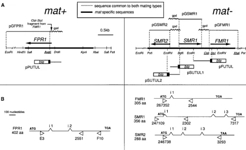
tion (Debuchy et al. 1993). A restriction map of the mat⫹ culties in observation and analysis. On the assumption
and mat⫺loci is presented in Figure 1A. that vegetative expression of mating-type genes may
Plasmids pPUTUL, pFUTUL, pSUTUL1, and pSUTUL2 mimic IR in the mycelium and aid in its analysis, we contain the ble gene (Drocourtet al. 1990) under the control investigated the control of expression of mating-type of the translation initiation and upstream sequence of FPR1,
FMR1, SMR1, and SMR2, respectively (Figure 1A). The ble gene,
genes in wild-type strains and the effects of deregulated
conferring resistance to phleomycin, was prepared using the IR genes (FMR1, SMR2, FPR1) and SMR1 during the
pUT703 plasmid (Calmelset al. 1991). All the plasmids were
vegetative phase. Expression studies showed that FMR1
based on pUL. Plasmid pPUTUL contains an in-frame fusion and FPR1 are active during both the vegetative and of the first 10 residues of FPR1, preceded by 1 kb of upstream sexual reproduction phase of P. anserina, while SMR1 untranslated region (UTR) with the ble gene at the NcoI site. The NcoI site has been introduced in FPR1 by amplification and SMR2 are not vegetatively transcribed. Deregulated
from KSRIRV (Debuchy and Coppin 1992) with a reverse SMR2 and SMR1 and vegetatively overexpressed FMR1
primer and 5⬘-CGCCATGGAGAAGGCTTCAAAATTGAA transgenes have been associated in various combina- GGC-3⬘ followed by digestion with NcoI. pFUTUL was con-tions by crossing. The association of FMR1 and SMR2 structed by ligation of the 1.02-kb EcoRI-StuI mat⫺fragment was found to lead to ascospore lethality in mat⫹genetic encoding the initial 13 residues of FMR1 with the ble gene. pSUTUL1 was constructed by ligation of the 0.59-kb EcoRI-context. Germination of ascospores was shown to be
BglII fragment encoding the first four residues of SMR1 with
restored by introducing a vegetatively expressed SMR1
the ble gene. The EcoRI-BglII fragment was prepared from transgene. In the frame of the functional model of the pULP-68. pSUTUL2 was constucted by ligation of a 0.74-kb mating types, we will discuss how these results can be BglII-EcoRV mat⫺fragment encoding the first 22 residues of SMR2 with the ble gene. The in-frame fusion between the interpreted and exploited for further investigations.
mat⫺ gene and the ble gene in pFUTUL, pSUTUL1, and pSUTUL2 has been reexamined by DNA sequencing. Plasmid pFLUT is a pUL, in which the ble coding sequence was cloned, MATERIALS AND METHODS ligated to the 60-bp ClaI-StuI mat⫺fragment. This fragment contains 21 bp upstream of the coding sequence of FMR1 and
P. anserina:genetic analysis, strains, transformation: The encodes the first 13 residues of FMR1 in frame with the ble ascus of P. anserina normally contains four ascopores that coding sequence.
develop around two nonsister nuclei after a single postmeiotic Plasmids pGFMR1, pGSMR1, and pGSMR2 (Figure 1A) are mitosis. However, 2 to 5% of asci contain five ascospores, two based on the pUL vector and contain the A. nidulans gpd of which are smaller and uninucleate, yielding homokaryotic promoter on a 2.3-kb EcoRI-NcoI fragment of pUT703 (Cal-mycelium. Tetrad analysis is routinely performed on five- melset al. 1991) fused to mat⫺genes. Plasmid pGFMR1 was spored asci. The⌬mat strain used in the study is derived from constructed by ligation of the gpd promoter with the 1.35-kb a mat⫹strain deleted for the mat locus (Coppinet al. 1993). ClaI-XbaI mat⫺fragment, resulting in a promoter fusion 21 The leu1-1 mat⫺ (SMR2::ura5) strain was obtained from a bp upstream of the translation start of FMR1. pGSMR1 has mat⫺strain in which the resident SMR2 gene was disrupted, been constructed by the ligation of the gpd promoter with the and the mat⫺(SMR1::ura5) strain carries a SMR1 disruption 1.35-kb NcoI-PstI fragment that contains the SMR1 gene at the at the resident mat⫺locus (Arnaiseet al. 1997). Transforma- start of the initiation codon. This fragment was obtained by tion was performed as previously described (Picard et al. amplification using the 48827 [5⬘-CCCCCCATGGACCACCGA 1991). When necessary, phleomycin (Cayla, France) or hygro- GATCTATCC-3⬘] and 48829 [5⬘-GGGGCTGCAGGATCATC mycin (Roche Diagnostics, Meylan, France) was added to pro- TCC-3⬘] primers on pULP. Plasmid pGSMR2 was constructed toplast regeneration medium at a concentration of 5g/ml using the ligation of the gpd promoter with a 1.04-kb NcoI-PstI and 100 g/ml, respectively. Segregation of antibiotic resis- fragment containing the SMR2 gene that starts at the initia-tance in the sexual crosses was scored on minimal medium tion codon. This fragment was obtained by amplification with containing either 20g/ml of phleomycin or 75g/ml hygro- the 46303 (5⬘-CCCCCCATGGATGTCTCCAACTCCAC-3⬘)
mycin. primer and reverse primer on pULP followed by enzymatic
informa-the 0.7-kb NcoI-HindIII fragment of pUT703 (Calmelset al. test for mat⫺progeny was possible, when necessary the pres-ence of the gpd::SMR1 transgene was established by PCR anal-1991) containing the ble gene in Bluescript KS digested with
EcoRI and HindIII. Plasmid pPaFMR1 was generated by clon- ysis.
DNA procedures:Genomic DNA of transformants was pre-ing the 2.3-kb EcoRI-XbaI fragment containpre-ing the entire FMR1
coding sequence and its 5⬘UTR (DebuchyandCoppin1992) pared as described previously (Coppin-Raynalet al. 1989). Minipreparations of DNA were done from cultures grown in pPable. Plasmid pPgFMR1 was generated by cloning in
pPable the 3.6-kb EcoRI-XbaI fragment encompassing the on a cellophane disk placed on agar minimal medium and recovered by scraping with a sterile spatula.
gpd::FMR1 fusion prepared from pGFMR1.
Determination of the phleomycin resistance of the trans- To confirm the genotype of transgenic progeny from sexual crosses, the presence of the mat transgenes was tested by PCR
formants carrying the 5ⴕmat::blegene fusion: The 5⬘mat::ble
fusions cloned in the pUL plasmid were introduced into the analysis. The position of pairs of primers is indicated in Figure 1B, and their sequence is as follows: FPR1: E3 [5⬘-GTCACTGG leu1-1 mat⫺ strain. Transformants (leu⫹) were then tested
on minimal medium containing 20g/ml phleomycin. They AACACACTCAAG-3⬘]; F10 [5⬘ -TTGACCGAAGATTTGGGC-3⬘]. FMR1: 267352 [5⬘ -GGCGGGAATCAACAGTATTTTGC-were considered resistant if growth was observed after 3 days
of incubation, since growth of the wild-type strain was totally 3⬘]; 2544 [5⬘-CATCCAAGGGCTTCCATGTA-3⬘]. SMR1: 247109 [5⬘-CGCGCATATAATGAATATCACGG-3⬘]; 7317 [5⬘ -abolished during this period. A total of 30 to 100 primary
(leu⫹) transformants obtained with each fusion were tested. CCCTCCAACTGAGATGCCAC-3⬘]; SMR2: 246738 [5⬘-GGAT GTCTCCAACTCCACTC-3⬘]; 3293 [5⬘-CGTTGAGATCCGCG The phleomycin phenotype was then more accurately
deter-mined, using two or three purified transformants issued from GTGGTC-3⬘] .
To analyze the structure of the integrated 5⬘mat::ble fusions, crosses of selected primary transformants with a leu1-1 mat⫹
strain. Since the transformants also contained leu1-1, the segre- PCR amplifications were performed using the ble2 primer [5⬘ -CACGAAGTGCACGCAGTT-3⬘], localized close to the stop gation of the integrated transforming vector was easily scored
as (leu⫹) phenotype, independent of expression of the codon in the ble gene, in association with a primer specific to the 5⬘UTR of the concerned mat gene: 573 [5⬘-CTAATAAGAA 5⬘mat::ble fusion. In these transformants, the integrity of the
fusion transgene was ascertained by PCR analysis. The minimal TAATGTAATG-3⬘], which is 540 nucleotides upstream of FMR1 start codon, 246738, close to the SMR2 start codon, the inhibitory concentration (MIC) of phleomycin was
deter-mined on mat⫹and mat⫺progeny carrying the construct. An reverse primer flanking the 5⬘-SMR1 sequence in pSUTUL1. The structure of the integrated gpd::mat fusions was analyzed identical MIC was obtained for strains of opposite mating type.
At 10g/ml phleomycin, growth of the wild-type strain was using the 39048 primer [5⬘-CCATCCTTCCCATCCCTTAT TCC-3⬘] localized in the gpd promoter 100 nucleotides up-inhibited forⵑ6 days; a residual growth was then observed.
The time lag before residual growth was increased when phleo- stream of the initiation codon in association with a primer localized at the 3⬘end of the relevant mat gene.
mycin was used at 20g/ml, and some implants showed zero
growth. Transformants with different levels of phleomycin Standard procedures for Southern blotting on Hybond N nylon filters (Amersham, France) were used. The probes were resistance were recovered, depending on the construct
intro-duced. MIC higher than 100g/ml phleomycin were not prepared using a random primer labeling kit (Roche Diagnos-tics).
tested.
Genetic analysis of the deregulatedmatgene associations: RNA extraction:To prepare RNA from mycelium, fungal cultures were made on a cellophane disk placed on agar mini-First, each mat⫹ SMR2 transformant containing a
constitu-tively transcribed ectopic SMR2 gene was crossed with the mal medium. After 2 days at 27⬚, the mycelium was recovered and transferred to a microcentrifuge tube. To prepare RNA mat⫺GFMR1-5 transformant carrying the pGFMR1 plasmid
(gpd::FMR1, leu1). At least 18 five-spored asci from each cross from perithecia, cultures of mat⫹and mat⫺strains were fertil-ized, respectively, with mat⫺and mat⫹microconidia on sepa-were submitted to genetic analysis and screened for sexual
phenotype (mating type and self-fertilization) and hygromycin rate petri dishes. Two hundred perithecia samples were col-lected 3 days after fertilization (production of mature resistance. Second, each SMR2 transformant was crossed with
transformants carrying the pPgFMR1 plasmid (gpd::FMR1, ble) ascospores begins on the fourth day). Perithecia were crushed with a conical grinder in 4mguanidium thiocyanate, 50 mm or the pPaFMR1 plasmid (FMR1, ble). At least 20 five-spored
asci from each cross were screened for sexual phenotype (mat- TRIS HCl pH 8, 10 mmEDTA pH 8, 2% N-lauroylsarcosine (sodium salt), and 1% -mercaptoethanol. The suspension ing type and self-fertilization) and hygromycin and
phleo-mycin resistance. In such crosses involving three genetic loci— was treated three times with phenol-chloroform (1:1) and nucleic acids were precipitated with 1 volume of isopropanol. the mating-type locus, the integration locus of the (SMR2,
hph) transgenes, and the integration locus of the (gpd::FMR1, After centrifugation the pellet was resuspended in water. LiCl was added to a final concentation of 2 m, the solution was leu1, or ble) transgenes (provided that these are not genetically
linked)—the eight genotypes of homokaryotic progeny listed centrifuged, and the pellet was resuspended in water; sodium acetate pH 5.2 was added to a final concentration of 0.3 m in Tables 3 and 4 are expected to be equivalent. When a
phenotypic class is lacking, tetrad analysis allows us to deter- and total RNA precipitated with 2 volumes of ethanol and recovered by centrifugation. The RNA pellet resuspended in mine whether it corresponds to ascospores that have not
ger-minated, genotypes of which can possibly be deduced from water was purified on a RNeasy Plant minikit (QIAGEN, Hil-den, Germany) according to the manufacturer’s indications. segregation of the genetic markers in the remaining viable
ascospores of the same ascus. Contaminating DNA was eliminated by Dnase digestion or centrifugation on a CsCl2 cushion. Complete degradation of
Construction ofmat⫹SMR2 gpd::FMR1 gpd::SMR1strain:
The mat⫹GSMR1-4 and mat⫹GSMR1-5 were crossed with the DNA was ascertained by PCR reaction seeking the internal transcribed sequences of RNA ribosomal genes, with one mat⫺SMR2-19 GFMR1-1 and mat⫺SMR2-19 GFMR1-2 strains.
In the four crosses, 15 five-spored asci were submitted to ge- primer [5⬘-CCGTTGGTGAACCAGCGGAGGGATC-3⬘] local-ized at the end of the 18S gene and the other [5⬘-TCCGCTTAT netic analysis. In three crosses an additional sample of 60 to
100 homokaryotic ascospores from five-spored asci were also TGATATGCTTAAG-3⬘] at the beginning of the 28S gene. For quantitative competitive RT-PCR, RNA were purified with analyzed. The segregation of the gpd::SMR1 in mat⫹progeny
was scored by ability to restore fertility in sexual cross with High Pure RNA kit (Roche Diagnostics).
The reverse transcriptase polymerase chain reaction
method (RT-PCR):Two micrograms of total RNA were used performed with RNA extracted from either mat⫹ or for RT-PCR with the Titan one Tube RT-PCR Kit (Roche mat⫺ mycelial cultures, respectively. No mature or pri-Diagnostics) according to the manufacturer’s specifications.
mary SMR1 and SMR2 transcripts were detected in RT-The following pairs of primers were used (DNA sequence of
PCR reactions from mat⫺ mycelial cultures, although some primers is cited in DNA procedures): FPR1: E3/2551
[5⬘-GATCTCAGAAGATCGACGAGG-3⬘]; FMR1: 267352/ several RNA preparations were tested with different 2544; SMR1 247109/7317; and SMR2 246738/3293. The pairs of primers. The cDNA of the four mat genes were gpd::SMR1 transcript was sought using the 2302 primer [5⬘- detected in RT-PCR reactions performed on RNA from GATTGACCTGGGGGTTGAGG-3⬘] localized downstream of
3-day perithecia (data not shown). The fragments were the first intron in association with the gpd specific primer
cloned and DNA sequencing demonstrated that they 39048. The localization of the primers is shown in Figure 1B.
QC-RT-PCR:Quantification of FMR1 mRNA was done by actually corresponded to cDNA with proper intron splic-competitive RT-PCR (reviewed inFreemanet al. 1999). The ing (Debuchy et al. 1993; R. Debuchy, unpublished competitive template for FMR1 was prepared according to the data).
double-cut method (McCullochet al. 1995) in which both
Transcriptional activity of the 5ⴕUTR of thematgenes
competitive and target molecules contain a unique restriction
during vegetative growth: FMR1 and FPR1 RNA detected
enzyme site. Any heteroduplexes will remain uncut and
sepa-rate from the competitor and target. The competitive mole- in whole mycelium extracts can be attributed to contam-cule was prepared by amplification of two overlapping frag- ination of the extracts by sexual organs (protoperithecia ments from FMR1 cDNA. One fragment resulted from the or microconidia) or to the expression of FMR1 and amplification with primer 267352 and IFMR1Hha [5⬘TTCTTC
FPR1 in vegetative hyphae. To test the expression of TTGGCGGGCTGACGCGGTGTGCCTTCCCG-3⬘] and the
FMR1 and FPR1 in vegetative hyphae, we constructed second fragment was obtained with primers FMRHha [5⬘-AGC
CCGCCAAGAAGAAGGTCAACGGTTTCATGCGC-3⬘] and fusions between FPR1 and FMR1 and the ble reporter 2544. Overlap extension of these two fragments, followed by gene (materials and methods). Fusions of SMR1 and PCR with 267352 and 2544, allows us to prepare the competi- SMR2 with ble were also tested to confirm the negative tive molecule that differs from FMR1 cDNA by the loss of a
results obtained in RT-PCR assays. Expression in
vegeta-HhaI site at 300 bp from primer 2544 and a new site vegeta-HhaI at
tive hyphae could be easily measured by determining 250 bp from primer 2544. The competitive DNA was cloned
into pGEM-T (Promega, Madison, WI) to produce the resistance level to phleomycin conferred by the fu-pGMFMR1dH. Amplifications were performed with primers sions (materials and methods). The constructs were 267352 and 2544, the PCR products were digested with HhaI, made in the pUL transformation plasmid containing and bands at 300 bp (target) and 250 bp (competitor) were
the leu1 gene as selective marker. The 5⬘UTR and the compared for fluorescence on a 2% agarose gel. Target and
origin of the coding region of FPR1 (mat⫹), FMR1, competitor DNA molecules have been checked for identical
amplification kinetic. Competitor RNA was prepared from SMR1, and SMR2 (mat⫺) were fused in-frame with the pGMFMR1dH by transcription with T7 RNA polymerase after entire coding sequence of the ble gene, leading to linearization with SalI. The RNA was purified with the High pPUTUL, pFUTUL, pSUTUL1, and pSUTUL2 plas-Pure RNA kit (Roche Diagnostics) and its integrity was
mids, respectively (Figure 1A and Table 1). The in-frame checked by gel electophoresis. To quantitate FMR1 mRNA,
fusions were reexamined by DNA sequencing. 10-fold serial dilutions ranging from 1 femtomole to 0.001
attomole of competitive RNA and 200 ng of total RNA were Two series of controls were performed. First, we deter-added to each tube. After RT-PCR (Titan One Tube, Roche mined if the 5⬘UTR sequences were or were not compe-Diagnostics) for 30 cycles, the PCR products were analyzed as tent to promote transcription of their native gene. indicated above.
Therefore, each mat gene with the upstream sequence used for the fusion with ble was introduced by transfor-mation in the suitable recipient and was shown to be RESULTS
functional in assays similar to those described for the
Search for themattranscripts in vegetative and sexual gpd::mat fusions (see below). Second, as plasmids mainly
Figure1.—Functional structure of the mat⫹and mat⫺mating types. (A) Restriction map of the mat locus (on chromosome I, the centromere-proximal end is left of this map). The underlined sites for restriction enzymes are not unique. Arrows indicate position and orientation of coding sequences. The mat-specific sequences fused to the ble reporter gene and to the gpd promoter and the name of the relevant plasmids are shown below and above the map, respectively. (B) Detailed structure of the genes with position of their introns (i 1, i 2, i 3). Arrowheads below the map indicate the position of the oligonucleotides used in RT-PCR experiments and/or RT-PCR analysis of the integrated transgene in transformants.
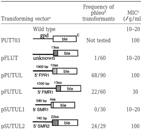
formants were recovered at a higher frequency with to phleomycin as the wild-type strain. PCR assays (seematerials and methods) and Southern analysis pPUTUL (75%) and pSUTUL2 (83%) than with pFUTUL
(37%). Primary transformants were expected to mani- of DNA from three purified strains transformed with pSUTUL1 were carried out and confirmed that the fest a heterogeneity of basic resistance to phleomycin.
First, they contained variable proportions of trans- 5⬘SMR1::ble transgenes were not rearranged (data not shown).
formed and untransformed nuclei because protoplasts
are often plurinucleate. Second, transforming DNA in- Vegetative repression ofSMR2transcription requires an upstreamciselement:The two above-cited methods tegrates at random chromosomal locations that may
have different cis-effects on the transcription activity of gave conflicting data with respect to SMR2, since SMR2 mRNA were not detected in mycelium by RT-PCR analy-the ble gene. Third, inactivation of an active transgene
during integration of the transforming DNA is a fre- sis whereas the 5⬘SMR2::ble fusion was found to be ex-pressed vegetatively. These data suggest that the vegeta-quent event. The potential heterogeneity was examined
by testing 30–100 primary (leu⫹) transformants ob- tive transcription of the 5⬘SMR2::ble fusion results from the loss of a regulatory cis element involved in transcrip-tained with each fusion for their resistance to
phleo-mycin, and the MIC was determined for two or three tional repression or in transcript instability during the vegetative phase. RT-PCR assays were performed on purified transformants (Table 1). Purified
trans-formants carrying the FPR1::ble fusion (pPUTUL) and RNA prepared from mycelia of mat⫹and mat⫺ trans-formants bearing different transgenic constructs
con-SMR2::ble fusion (pSUTUL2) could still grow at 100g/
ml phleomycin while transformants carrying the taining SMR2. SMR2 cDNA was not detected in mat⫹ transformants bearing the SMR2 coding sequence and
FMR1::ble fusion were inhibited at 30g/ml. These data
suggest that the expression level of the FPR1::ble and 4.7 kb upstream of the SMR2 translation initiation cov-ering the entire mat⫺region at an ectopic position (see SMR2::ble fusions is higher than the expression level
TABLE 1 notype. The phenotypic assays described in Table 2 in-dicate whether the gpd::mat fusion is functional but do
Resistance level to phleomycin displayed by the
not give information on its expression level.
Transfor-transformants carrying the 5ⴕmat::blegene fusion
mants with the expected sexual phenotype were recov-ered with each of the four constructs. The data were as Frequency of
phleoR MICb
follows: Transforming vectora transformants (g/ml)
1. Transformants carrying gpd::FPR1 displayed full Wild type 10–20 mat⫹activity (fertilization of a mat⫺partner giving
rise to fertile perithecia producing asci).
PUT703 Not tested ⬎100
2. Transformants carrying gpd::FMR1 displayed partial mat⫺activity (fertilization of a mat⫹partner giving
pFLUT 1/60 10–20 rise to poorly fertile perithecia). The postfertilization
function was examined by introducing simultane-ously into the⌬mat strain pGFMR1 and pSUT12
con-pPUTUL 68/90 ⬎100
taining the two other mat⫺genes (SMR1 and SMR2) with the ble selective marker. The cotransformants
pFUTUL 22/60 ⵑ30
displayed a wild-type mat⫺activity (fertilization of a mat⫹partner giving rise to fertile perithecia
produc-pSUTUL1 0/30 10–20 ing asci).
3. The cotransformants containing both the gpd::SMR1 fusion and a mat⫹ transgenic information were
pSUTUL2 24/29 ⬎100
crossed with the mat⫺ (SMR1::ura5) mutant,
dis-aFor each construct the size of the 5⬘UTR (in base pairs)
rupted within SMR1 at the resident mat⫺locus. The and the size of the coding sequence (in amino acids) belong- cross yielded abundant progeny, whereas a cross of ing to the mat gene are indicated. Localization of those
se-the mat⫺(SMR1::ura5) mutant with the mat⫹ wild-quences in the physical map is shown in Figure 1A. In pFLUT
type strain was sterile. Internuclear complementa-and pFUTUL the fusion gene contains the sequence coding
the first 13 amino acids of FMR1. tion, previously demonstrated with the native SMR1
bThe MIC of phleomycin was determined on two or three
gene (Arnaise et al. 1997), was thus also observed purified transformants harboring each fusion (seematerials for the gpd::SMR1 fusion.
and methods). When a high frequency of transformants
resis-4. The pGSMR2 plasmid was introduced into a leu1-1 tant to phleomycin was obtained, only resistant transformants
mat⫺(SMR2::ura5) recipient since only intranuclear were purified.
cThe ble gene is driven by the A. nidulans gpd promoter. complementation was observed for the native SMR2
gene (Arnaiseet al. 1997). The mat⫺(SMR2::ura5) strain provided only uniparental progeny in crosses with a mat⫹tester (Arnaiseet al. 1997), whereas the SMR2 coding sequence and 1.4 kb upstream of the
transformants containing the gpd::SMR2 fusion also SMR2 first codon. These data suggest that an element
gave biparental progeny, thus indicating efficient required for the repression of SMR2 transcription is
complementation of the mutant phenotype. present in the region between 1.4 kb and 4.7 kb
up-stream of the SMR2 translation start. The data demonstrate that the four mat genes are
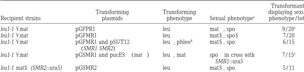
Deregulatedmatgenes complementmatmutants and active when driven by the foreign gpd promoter. More-do not alter vegetative phenotype:To promote expres- over, during these tests, no gpd::mat fusion was found sion or overexpression of mat genes during vegetative to produce an effect on viability, growth, or morphology growth, we constructed gene fusions between the glyc- of the recipient strain.
ero-phosphate-3-dehydrogenase (gpd) promoter of A. The functional assays allowed us to determine
nidulans and the coding region of the four mat genes, whether the gpd::mat fusions were active during sexual
including the initiation codon. Constructs were cloned reproduction but did not allow us to determine whether in the pUL plasmid carrying the leu1 selective marker, they were expressed vegetatively. In particular, replace-giving rise to pGFPR1, pGFMR1, pGSMR1, and ment of the native promoter of SMR1 by the gpd pro-pGSMR2 plasmids (Figure 1A). Each plasmid was intro- moter was expected to induce its vegetative expression. duced into a strain suitable for examining the functions To check the presence of gpd::SMR1 mRNA, RT-PCR of the gpd::mat fusion, generally the ⌬mat mutant de- assays were performed on RNA prepared from mycelia leted for mat information (Coppin et al. 1993). When of one transformant bearing a functional gpd::SMR1 necessary additive wild-type mat genes were introduced fusion. A product of the expected size was detected simultaneously to the gpd::mat fusion. Between 10 and using a primer localized close to the stop codon in 20 (leu⫹) primary transformants carrying each plasmid association with a gpd specific primer, indicating that
phe-TABLE 2
Functional tests performed to determine expression of thematgenes under control of theA. nidulans gpdpromoter
Transformants
Transforming Transforming displaying sexual
Recipient strains plasmids phenotype Sexual phenotypea phenotype/total
leu1-1⌬mat pGFPR1 leu⫹ mat⫹, spo⫹ 9/20b
leu1-1⌬mat pGFMR1 leu⫹ mat⫺, spo⫺ 7/20
leu1-1⌬mat pGFMR1 and pSUT12 leu⫹, phleoR mat⫺, spo⫹ 6/15
(SMR1 SMR2)
leu1-1⌬mat pGSMR1 and pucES⫹(mat⫹) leu⫹, mat⫹ spo⫹in cross with 7/15b
SMR1::ura5
leu1-1 mat⫺(SMR2::ura5) pGSMR2 leu⫹ mat⫺, spo⫹ 5/11
aExpression of the gpd::mat genes can be tested by determining the fertilization ability (mat⫹or mat⫺) on a tester of opposite
mating type and/or the production of a progeny (spo⫹, abundant progeny with Mendelian segregation of the parental genetic markers; spo⫺, no progeny.
bIntegrity of the transgenic fusion gene was checked by PCR analysis in one or two transformants.
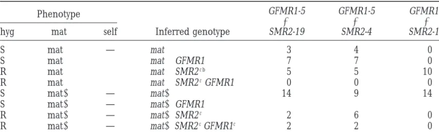
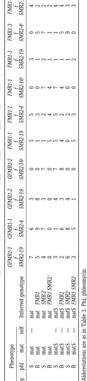
The combination of deregulatedmatgenes produces SMR2-16, and SMR2-19 : in the nomenclature SMR2-x, x specifies the integration locus of the SMR2 transgene.
a lethal phenotype: With the help of the transgenic
strains obtained by transformation with the different mat The same nomenclature is used to design each mat gene introduced by integrative transformation. Trans-constructs, we performed numerous crosses to obtain
different combinations of the deregulated mat genes. formants carrying the pCBSMR2 construct must express SMR2 in hyphae because the gene does not contain the In the course of the genetic analysis of some crosses,
we observed a high frequency of ascospores unable to negative cis element that represses its transcription in mycelium. This prediction is confirmed by the detection germinate. Tetrad analysis allowed us to determine their
genotype: the lethal ascospore carried the mat⫹resident of a fragment with the expected size for cDNA in RT-PCR assays performed on RNA extracts from the mat⫹ mating type (FPR1) and both gpd::FMR1 and SMR2
transgenes, that is to say an artificial association of the SMR2-19 strain (data not shown). SMR2c will
subse-quently designate this constitutive SMR2 transgene. three IR genes. To further investigate this phenomenon,
new transformants more suitable for genetic analysis In the same way, (phleoR) mat⫹ primary trans-formants obtained with the pPgFMR1 or the pPaFMR1 were constructed. For that purpose, the gpd::FMR1
fu-sion was cloned in a plasmid carrying the ble gene as plasmids were used to generate homokaryotic mat⫹and mat⫺ progeny carrying the gpd::FMR1 fusion (GFMR1-selective marker (pPgFMR1). A plasmid containing the
native FMR1 gene with its own 5⬘UTR (pPaFMR1) was 1, GFMR1-2) or the FMR1 transgene (FMR1-1, FMR1-3, FMR1-7). Introduction of these plasmids into a mat⫹ also constructed to examine the role of the promoter.
A SMR2 gene with a 1.4-kb 5⬘ UTR was cloned in a recipient induced self-fertilization and ability to fertilize a mat⫹tester provided the FMR1 transgene was active. plasmid carrying the hph gene, conferring resistance to
hygromycin (pCBSMR2). The segregation of FMR1 and Only primary transformants exhibiting this phenotype were selected. QC-RT-PCR detection of FMR1 tran-SMR2 transgenes could thus be easily followed through
resistance to phleomycin (phleoR) and hygromycin (hy- scripts in total RNA extracted from GFMR1-1, FMR1-1, and wild-type mat⫺strains indicated that transcription groR), respectively.
Transformants (hygroR) were recovered upon trans- of FMR1 is at least 10 times higher in GFMR1-1 strain than in FMR1-1 and wild-type strains (materials and formation of the mat⫹ recipient with the pCBSMR2
plasmid. Introduction of the SMR2 transgene into a methods). The GFMR1-5 transformant was previously obtained with the pGFMR1 plasmid carrying the mat⫹strain was previously found to induce the
enlarge-ment of female organs that do not develop if they are gpd::FMR1 fusion associated with the leu1 gene. Crosses were performed between mat⫹ strains car-not fertilized (S. Arnaise, personal communication).
This phenotype was used to determine whether the (hy- rying a SMR2c transgene and mat⫺ strains carrying a
gpd::FMR1 fusion and submitted to genetic analysis. groR) transformants carried a functional SMR2. Such
transformants were genetically purified by crossing with Data are presented in Table 3 and in Table 4 (first four columns). The most important finding is that no viable a mat⫺ wild-type strain, and homokaryotic mat⫹ and
mat⫺progeny harboring the SMR2 and hph transgenes mat⫹SMR2cGFMR1 homokaryotic progeny was
recov-ered, while the seven other possible genotypes were were recovered. Three homokaryotic transformants
were generated from three independent primary trans- obtained at equivalent frequency (except in crosses with
SMR2-16 because the transgene is linked to mat⫹).
Tet-formants in which the plasmid had integrated at
TABLE 3
Homokaryotic progeny obtained in crosses ofmat⫹strains carrying the (SMR2,hph) transgenes (SMR2-19,SMR2-4,SMR2-16) with themat⫺strain carrying the (gpd::FMR1,leu1) transgene (GFMR1-5)
Phenotype GFMR1-5 GFMR1-5 GFMR1-5
⫻ ⫻ ⫻
hyg mat self Inferred genotype SMR2-19 SMR2-4 SMR2-16a
S mat⫹ — mat⫹ 3 4 0
S mat⫹ ⫹ mat⫹GFMR1 7 7 0
R mat⫹ ⫹ mat⫹SMR2c b 5 5 10
R mat⫹ ⫹ mat⫹SMR2cGFMR1 0 0 0
S mat⫺ — mat⫺ 14 9 14
S mat⫺ — mat⫺GFMR1
R mat⫺ — mat⫺SMR2c 2 6 0
R mat⫺ — mat⫺SMR2cGFMR1c 2 2 0
Abbreviations are as follows: hyg, hygromycin; R and S, resistant and sensitive; mat, resident mating type; self, formation of perithecia or microperithecia on the homokaryotic mycelium.
a(SMR2-16, hph) are genetically linked to mat⫹.
bStrains unable to fertilize a mat⫹strain, a function expected if GFMR1-5 were present. cUnpigmented, female sterile mycelia.
the absent genotype was attributable to immature asco- fer an ascospore lethal phenotype in a mat⫹strain, they nonetheless impaired the growth rate and morphology spores that had not germinated. This genetic association
of mat genes was thus responsible for an autonomous of the mycelium, which was devoid of aerial hyphae. In a mat⫺strain, they conferred the phenotypic alteration ascospore lethal phenotype, that is, a phenotype
con-trolled by the nuclei within the ascospore itself. Abun- (unpigmented female sterile mycelium) already ob-served in mat⫺SMR2cGFMR1 strains (Table 5). By
con-dant asci with morphologically normal ascospores were
produced in all crosses, indicating that proper sexual trast, SMR2c
FMR1-1 associations did not impair the phenotype, whatever might be the mating-type resident development occurred before ascospore delimitation.
Heterokaryotic ascospores interpreted as containing haplotype (Table 5). One may assume that the vegeta-tive effects are less drastic because the FMR1 transgenes both mat⫹ SMR2c GFMR1 and mat⫺ wild-type nuclei
were unable to germinate, which indicated that the le- are less expressed than are the gpd::FMR1 fusions as indicated by QC-RT-PCR experiments. Moreover, the thality was dominant. Contrary to the mat⫹ SMR2c
GFMR1 ascospores, the mat⫺SMR2cGFMR1 ascospores integration site of the ectopic FMR1 copy may influence
its expression level, which could be lower in FMR1-1 were viable. Nevertheless, on growth medium they gave
rise to an unpigmented and flat mycelium that grew as than in FMR1-3 and FMR1-7 transformants. The same rationale can be applied to SMR2 to explain the pheno-well as the wild-type strain but failed to form aerial
hyphae and rarely differentiated female organs (at least typic differences between mat⫹ FMR1-3 SMR2-4 and
mat⫹ FMR1-3 SMR2-19 strains (Table 5).
200 times less than the wild type). Consequently, when
used as female parent in a cross, only 10 to 100 fruiting In conclusion, the association of a FPR1 (mat⫹) resi-dent gene with a vegetatively expressed SMR2 transgene bodies were produced on a petri dish in contrast to the
thousands produced by a wild-type cross. The six other and a gpd::FMR1 transgene appears to be lethal. Re-placement of the gpd::FMR1 by the FMR1 transgene genotypes listed in Tables 3 and 4 did not confer any
par-ticular phenotype. Finally, a mat⫺ SMR2-19 GFMR1-2 results in viable strains that nevertheless exhibited im-paired growth rate and mycelium morphology. Vegeta-strain issued from this analysis was crossed with a mat⫹
strain. As previously, mat⫹SMR2-19 GFMR1-2 progeny tive expression of SMR2 was confirmed by detection of a fragment with the expected size for cDNA in RT-PCR were not obtained. The data indicated that the lethality
phenomenon occurs whatever the initial association of assays performed on RNA extracts from the mat⫺ SMR2-19 GFMR1-2 strains (data not shown).
the SMR2 and GFMR1 transgenes in the parental strains
(one transgene in each parent as in Table 3 and Table To examine the role of the resident FPR1 gene, the SMR2 and gpd::FMR1 transgenes were introduced into 4 or both in the same parent).
Crosses were also performed between mat⫹ SMR2c a ⌬mat strain by crossing the mat⫺ SMR2-19 GFMR1-1
strain with a⌬mat strain carrying the 6.3-kb EcoRI-Sal I strains and mat⫺ FMR1 strains (Table 4, last six
col-umns). By contrast to crosses with mat⫺GFMR1 strains, fragment from the mat⫹locus (see Figure 1A). Progeny with the⌬mat SMR2-19 GFMR1-1 genotype were recov-mature mat⫹SMR2cFMR1 ascospores giving rise to
con-ascospores definitely confirms that the deregulated mat⫺genes must be associated with the resident FPR1 (mat⫹) gene to confer the ascospore lethal phenotype. Observations made in the course of our study show that P. anserina transgenic strains exhibit some instabil-ity. In particular, sectors with increased growth rate ap-peared frequently from the poorly growing mat⫹
SMR2-19 FMR1-3 or mat⫹ SMR2-19 FMR1-7 mycelium. This
phenomenon was investigated by crossing several sec-tors with the mat⫺wild-type strain. Although resistance to hygromycin (SMR2, hph) and phleomycin (FMR1, ble) segregated normally in the offspring, the sexual phenotype associated with either SMR2 or FMR1 transgene was lost. PCR reactions were performed on DNA extracted from sectors and from some of their progeny with a pair of primers specific to the inactive transgene: no band was detected, whereas the specific band was present in assays with DNA from the original mycelium (data not shown). The growth improvement in the sectors is thus caused by the loss of one of the transgenes without concomitant loss of the associated resistance marker. Consequently, to avoid any misinter-pretation of a phenotype, the presence of a transgene was never deduced solely from the phleR or hygroR phenotype, but also using a functional test when possi-ble and/or a PCR assay when necessary. To date, exci-sion of an integrated plasmid or transgene in P. anserina transformants was considered a rare event, perhaps mis-takenly. The situation we report is particularly well adapted to reveal such an event since the transgenes are “toxic.” First, there is a selection pressure in favor of nuclei that have lost one transgene. Second, the loss of a transgene is associated with an increased growth rate and aerial hyphae production and thus with a di-rectly observable phenotypic change.
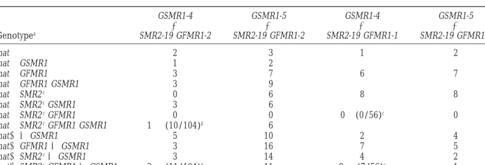
Integration of a gpd::SMR1 fusion restores viability to themat⫹SMR2-19 GFMR1-2strain: FPR1, FMR1, and
SMR2 control a sexual development-specific function, internuclear recognition. As explained in the Introduc-tion, the fourth mat gene, SMR1 (mat⫺), is assumed to act downstream of FPR1, FMR1, and SMR2. We therefore tested the effect of the gpd::SMR1 fusion on the pheno-type resulting from the expression of FPR1, FMR1, and SMR2 in the same nucleus. The gpd::SMR1 transcrip-tional fusion was used to force expression of SMR1 in vegetative mycelium since transcription of SMR1 was observed only in perithecia (see above). The mat⫺
SMR2-19 GFMR1-1 and mat⫺SMR2-19 GFMR1-2 strains
were crossed with mat⫹strains harboring the gpd::SMR1 fusion (GSMR1-4 and GSMR1-5). These were obtained in two steps. First, the mat⫹leu1-1 strain was transformed with the pGSMR1 plasmid and (leu⫹) primary trans-formants were recovered. Second, the transtrans-formants were crossed with the mat⫺ (SMR1::ura5) strain. The sterility of this strain was complemented by the gpd::SMR1 transgene carried by the mating partner,
TA BL E 4 Homokaryotic progeny obtained in crosses of strains carrying the ( SMR2 , hph ) transgenes ( SMR2-19 , SMR2-4 , SMR2-16 ) w ith strains carrying the ( gpd::FMR1 , ble )o r( FMR1 , ble ) transgenes ( GFMR1-1 , GFMR1-2 and FMR1-1 , FMR1-3 , FMR1-7 ) Phenotype GFMR1-1 G FMR1-1 G FMR1-2 G FMR1-2 FMR1-1 FMR1-1 FMR1-1 F MR1-3 F MR1-3 F MR1-7 ⫻ ⫻ ⫻ ⫻ ⫻ ⫻ ⫻ ⫻⫻⫻ hyg phl mat self Inferred genotype SMR2-19 SMR2-4 S MR2-19 SMR2-16 a SMR2-19 SMR2-4 SMR2-16 a SMR2-19 SMR2-4 b SMR2-19 SS m a t ⫹ — mat ⫹ 7 6 3 0 5 5 0 304 SR m a t ⫹⫹ mat ⫹ FMR1 5 9 8 0 1 3 0 152 RS m a t ⫹⫹ mat ⫹ SMR 2 c 4 7 3 7 1 2 7 272 RR m a t ⫹⫹ mat ⫹ FMR1 SMR2 c 0 0 0 0 5 4 4 112 SS m a t ⫺ — mat ⫺ 3 1 4 7 5 5 7 114 SR m a t ⫺ — mat ⫺ FMR1 2 6 3 8 4 2 4 154 RS m a t ⫺ — mat ⫺ SMR2 c 6 8 4 0 0 2 0 193 RR m a t ⫺ — mat ⫺ FMR1 SMR2 c 3 5 1 0 5 3 0 203 Abbreviations are as in Table 3 . Phl, phleomycin. a(SMR2-16 , hph ) are genetically linked to mat ⫹ . b(SMR2-4 , hph ) are genetically linked to (FMR1-3 , ble ).
TABLE 5
Phenotypes ofmat⫹FMR1 SMR2andmat⫺FMR1 SMR2strains
Phenotype
mat⫹context mat⫺context
Ascospore Mycelial Female
Genotype germination Growth Growth pigmentation fertilitya
WT ⫹ ⫹ ⫹ ⫹ ⫹
GFMR1 SMR2c — — ⫹ — —
FMR1-1 SMR2-19 ⫹ ⫹ ⫹ ⫹ ⫹
FMR1-1 SMR2-4 ⫹ ⫹ ⫹ ⫹ ⫹
FMR1-1 SMR2-16 ⫹ ⫹ Not determined
FMR1-3 SMR2-19 ⫹ Poor ⫹ — —
FMR1-3 SMR2-4 ⫹ ⫹ Not determined
FMR1-7 SMR2-19 ⫹ Poor ⫹ — —
a⫹, protoperithecia are formed; —, no (or few) protoperithecia are formed.
Data of the crosses mat⫺SMR2-19 GFMR1-1/GFMR1-2⫻ at a 6.25% frequency of the homokaryotic ascospores (16 genotypes at equivalent frequency were expected
mat⫹GSMR1-4/GSMR1-5 are presented in Table 6. The
segregation of the gpd::SMR1 in mat⫹ progeny was in a cross involving four unlinked genetic markers). Progeny with this phenotype were not obtained in scored by ability to restore fertility in sexual cross with
the mat⫺(SMR1::ura5) mutant. Since no simple func- crosses with mat⫹SMR2-19 GFMR1-1 (two final columns of Table 6). In contrast, in crosses of mat⫹ SMR2-19 tional test for gpd::SMR1 in mat⫺progeny was possible,
the presence of the gpd::SMR1 transgene was established GFMR1-2 with mat⫹GSMR1-4 and mat⫹GSMR1-5 (first two columns of Table 6), 8.4% and 6.7%, respectively, by PCR analysis when necessary. If the gpd::SMR1 fusion
gene acts as a suppressor of ascospore lethality, (mat⫹ of viable homokaryotic ascospores generating mycelium with the expected phenotype were found. The presence hygroR phleoR) mycelium corresponding to the mat⫹
SMR2-19 GFMR1 GSMR1 genotype should be recovered of the gpd::SMR1, SMR2, and GFMR1 transgenes
de-TABLE 6
Homokaryotic progeny obtained in crosses of strains carrying the (SMR2,hph) and (gpd::FMR1,ble) transgenes (SMR2-19 GFMR1-1andSMR2-19 GFMR1-2) with strains carrying the (gpd::SMR1,leu1) transgenes
(GSMR1-4andGSMR1-5)
GSMR1-4 GSMR1-5 GSMR1-4 GSMR1-5
⫻ ⫻ ⫻ ⫻
Genotypea SMR2-19 GFMR1-2 SMR2-19 GFMR1-2 SMR2-19 GFMR1-1 SMR2-19 GFMR1-1
mat⫹ 2 3 1 2
mat⫹GSMR1 1 2
mat⫹GFMR1 3 7 6 7
mat⫹GFMR1 GSMR1 3 9
mat⫹SMR2c 0 6 8 8
mat⫹SMR2cGSMR1 3 6
mat⫹SMR2cGFMR1 0 0 0⫹(0/56)c 0
mat⫹SMR2cGFMR1 GSMR1 1⫹(10/104)b 6
mat⫺ ⫾GSMR1 5 10 2 4
mat⫺GFMR1⫾GSMR1 3 16 7 5
mat⫺SMR2c⫾GSMR1 3 14 4 2
mat⫺SMR2cGFMR1⫾GSMR1 3⫹(11/104)b 11 0⫹(7/56)c 1
Abbreviations are as in Table 5.
aGenotypes were deduced from the phenotypic tests presented in Table 4; segregation of gpd::SMR1 in the mat⫹progeny was
determined by crossing with mat⫺(SMR1::ura5) only when (mat⫹phleR hygR) progeny were obtained (two first columns). In the two last columns each number is for two genotypes⫾GSMR1.
bAn additive cross was performed and 104 homokaryotic progeny analyzed. Ten (mat⫹hygR phlR) progeny were obtained
and were found to carry the gpd::SMR1 transgene.
duced from functional tests was also confirmed by geno- vegetative cells or only in reproductive structures. The authors proposed that translation of mt A-2 and mt A-3 mic DNA PCR analysis in some progeny. Although the
mat⫹SMR2-19 GFMR1-2 GSMR1 ascospores germinated messengers may be developmentally regulated through
small open reading frames that are present downstream well, the colonies displayed a slight morphological
alter-ation on the germinalter-ation medium as compared to wild of the 5⬘end in these messengers.
Since transcriptional regulation is the primary regula-type (smaller thallus with a less regular margin).
More-over, after transfer on minimal medium, mycelial tory control of expression of the P. anserina mat genes, we deregulated them by replacing their natural pro-growth is very slow and irregular. The (mat⫺ hygroR
phleoR) progeny were submitted to PCR analysis to test moter with the A. nidulans gpd promoter. Each of the four gpd::mat fusions was found to be functional since the presence of the gpd::SMR1 transgene. In fact these
progeny corresponded to two genotypes, mat⫺ SMR2- it can complement a null or a mutated allele. No effect was noticed when one fusion was introduced in a mat⫹
19 GFMR1-2 and mat⫺SMR2-19 GFMR1-2 GSMR1, which
gave the same altered phenotype: unpigmented female or mat⫺ wild-type strain. Crosses were performed be-tween transgenic strains to associate the three IR genes sterile mycelia with no aerial hyphae. These genetic
data indicated that SMR1 did not suppress the mutant in the same nucleus. We were unable to construct mat⫹ (FPR1) strains containing a constitutively transcribed phenotype conferred by the mat⫺ SMR2-19 GFMR1-2
association. SMR2c gene and a gpd::FMR1 fusion (Table 5).
Asco-spores with this genotype were nevertheless recovered Finally, to confirm the suppression of lethality by
SMR1, a mat⫺SMR2-19 GFMR1-2 GSMR1-5 progeny was but they were unable to germinate, which indicated that
this genetic association was lethal. By contrast, homokar-crossed with a mat⫹ GSMR1-5 strain. The gpd::SMR1
transgene was present in both parents, and only viable yotic mat⫹ (FPR1) strains containing a constitutively transcribed SMR2c transgene and a FMR1 transgene
ascospores were recovered. Among the 80 homokaryotic
ascospores analyzed, 11% were (mat⫹hygroR phleoR) driven by its own promoter were obtained (Table 5). They exhibited a flat mycelium with reduced growth. and 9% were (mat⫺ hygroR phleoR), in agreement
with the 12.5% expected for each class. The gpd::FMR1 fusion was found to be at least 10 times more transcribed than FMR1 driven by its own pro-moter, suggesting that the overexpression of FMR1 in DISCUSSION
a SMR2cmat⫹strain is lethal, while a lower expression
of FMR1 has a less drastic effect. Coexpression of the We have studied the means by which mating-type
genes are regulated during the life cycle of P. anserina three IR genes thus results in a partial or complete inhibition of growth. We propose that this phenomenon to choose a strategy to deregulate them. We have further
examined the physiological consequences of forcing mimics the events that occur during sexual reproduc-tion. A cross between a mat⫹ strain and the mat⫺ their expression in vegetative hyphae.
RT-PCR analyses and fusions of mating-type genes (SMR1::ura5) mutant, which contains functional FMR1 and SMR2 genes, displays a phenotype in agreement with reporter genes revealed that during the vegetative
phase FMR1 and FPR1 are expressed whereas SMR1 with this interpretation. This cross is sterile, although fertilization has occurred. Cytological observations indi-and SMR2 are transcriptionally silent. The vegetative
expression of FMR1 and FPR1 genes is in agreement cate that development of perithecia is blocked before formation of ascogenous hyphae (Arnaiseet al. 1997). with their role in fertilization (Debuchy andCoppin
1992) and with the observation that hyphae can occa- Since IR genes are functional, it is likely that IR occurs normally in this cross and that development arrests sionally substitute to microconidia for fertilization.
Ma-ture transcripts of the four mating-type genes have been shortly after the recognition stage. We postulate that this developmental arrest is a programmed event similar detected in the fertilized female organs, suggesting that
some unknown factors control the transcription of to the growth arrest observed in the mycelium as a result of the expression of IR genes. In the fruiting body, this SMR1 and SMR2 in the perithecium. A negative
cis-acting element has been localized in a region 1.4 kb arrest may be required for the synchronization of mat⫹ and mat⫺ nuclei before entry into the ascogenous hy-to 4.7 kb upstream of SMR2. Preliminary experiments
suggest that the SMR2 silencer is present in the FMR1 phae, where they undergo simultaneous mitoses ( Simo-netandZickler1972). Construction of a mat⫹(FPR1) sequence, 2.4 kb upstream of SMR2 translation start.
A comparison of the transcription pattern of Neuro- strain containing a SMR2c and a gpd::FMR1 transgene
was found to be possible by simultaneously introducing
spora crassa (Ferreiraet al. 1996) and P. anserina
mating-type genes indicates that these genes are regulated at the gpd::SMR1 fusion. The suppression of vegetative growth inhibition resulting from vegetative coexpres-different steps of their expression in the the two fungi.
In N. crassa, mt A-1 (similar to FMR1), mt A-2 (similar sion of FPR1, FMR1, and SMR2 by a gpd::SMR1 fusion confirms that SMR1 operates downstream of IR genes. to SMR1), and mt A-3 (similar to SMR2) are transcribed
SMR1 is required after IR for the initial development genetic screen for selecting suppressors and identifying possible target genes of the transcription factors en-of biparental ascogenous hyphae. In the fruiting bodies,
SMR1 function would thus be required to remove the coded by the mating-type genes. Moreover, in nonlethal
associations leading to a reduced mycelial growth rate, developmental inhibition triggered by IR.
The hypothesis that recently proposed that IR is medi- we have available biological material for the identifica-tion of specific mRNA by differential hybridizaidentifica-tion with ated by a pheromone response pathway (Debuchy
1999) may help us to understand what happens at the RNA from a wild-type strain.
molecular level when IR genes are expressed. The possi- We are grateful to H. D. Osiewacz for providing the pRP81-1 plas-ble involvement of a pheromone cascade in IR suggests mid. We thank S. Arnaise, V. Berteaux-Lecellier, M. Chablat, and M.
Picard for critical reading of the manuscript. that the accompanying growth arrest may be similar to
the G1 cell-cycle arrest observed in Saccharomyces cerevis-iae in response to the activation of the pheromone
re-sponse pathway (reviewed inCrosset al. 1988). In yeast, LITERATURE CITED
constitutive activation of this pathway caused by disrup- Arnaise, S., R. DebuchyandM. Picard,1997 What is a bona fide mating-type gene? Internuclear complementation of mat mutants tion of the structural gene for the G␣ protein results
in Podospora anserina. Mol. Gen. Genet. 256: 169–178. in a haploid-specific lethal phenotype due to cell-cycle
Boyer, H. W.,andD. Roulland-Dussoix,1969 A complementation arrest (Miyajimaet al. 1987). Similarly, in P. anserina, analysis of the restriction and modification of DNA in Escherichia
coli. J. Mol. Biol. 41: 459–472. heterochronic vegetative coexpression of the three IR
Calmels, T., M. Parriche, H. DurandandG. Tiraby,1991 High genes in the same nucleus could generate a constitutive
efficiency transformation of Tolypocladium geodes conidiospores activation of the pheromone signal transduction path- to phleomycine resistance. Curr. Genet. 20: 309–314.
Carroll, A. M., J. A. SweigardandB. Valent, 1994 Improved way and thus provoke nuclear arrest in G1.
vectors for selecting resistance to hygromycin. Fungal Genet. The association of the SMR2c and the gpd::FMR1
Newsl. 41: 22.
transgenes, lethal in the mat⫹ strain, is viable in the Coppin, E., S. Arnaise, V. ContamineandM. Picard,1993 Deletion of the mating-type sequences in Podospora anserina abolishes mat-mat⫺ strains but leads to phenotypic alterations. The
ing without affecting vegetative functions and sexual differentia-mycelium of all mat⫺ SMR2c GFMR1 and some mat⫺
tion. Mol. Gen. Genet. 241: 409–414.
SMR2c FMR1 strains is unpigmented, devoid of aerial
Coppin-Raynal, E., M. PicardandS. Arnaise,1989 Transforma-tion by integraTransforma-tion in Podospora anserina III. Replacement of a hyphae, and almost totally female sterile (Table 5).
How-chromosome segment by a two-step process. Mol. Gen. Genet. ever, these strains grow as well as the wild-type strain in
219:270–276.
contrast to the mat⫹SMR2c
FMR1 strains. The finding Cross, F., L. H. Hartwell, C. JacksonandJ. B. Konopka,1988
Conjugation in Saccharomyces cerevisiae. Annu. Rev. Cell Biol. 4: that the⌬mat SMR2-19 GFMR1-1 strain has a wild-type
429–457. phenotype in contrast to the mat⫺ SMR2-19 GFMR1-1
Debuchy, R.,1999 Internuclear recognition: a possible connection strain that displayed a mutant phenotype demonstrates between euascomycetes and Homobasidiomycetes. Fungal Genet.
Biol. 27: 218–223. that the resident mat⫺information is important in
de-Debuchy, R.,andE. Coppin,1992 The mating types of Podospora termining the morphological alterations. It has been
anserina: functional analysis and sequence of the fertilization do-established that differentiation of sexual reproductive mains. Mol. Gen. Genet. 233: 113–121.
Debuchy, R., S. ArnaiseandG. Lecellier,1993 The mat⫺allele structures is not controlled by the mating-type genes,
of Podospora anserina contains three regulatory genes required since it occurs in the⌬mat mutants (Coppinet al. 1993).
for the development of fertilized female organs. Mol. Gen. Genet. Consequently, female sterility caused by forced vegeta- 241:667–673.
Drocourt, D., T. Calmels, J. P. Reynes, M. BaronandG. Tiraby, tive expression of SMR2 and FMR1 in the mat⫺context
1990 Cassettes of the Streptoalloteichus hindustanus ble gene for
(and the other phenotypic alterations) may be an
indi-transformation of lower and higher eukaryotes to phleomycin rect effect resulting from deregulation of the expression resistance. Nucleic Acids Res. 18: 4009.
Ferreira, A. V. B., S. SaupeandN. L. Glass,1996 Transcriptional of the mat⫺genes. This deregulation might alter fungal
analysis of the mt A idiomorph of Neurospora crassa identifies two physiology and, more particularly, generate a bypass of
genes in addition to mt A-1. Mol. Gen. Genet. 250: 767–774. the female differentiation pathway. In accord with this Freeman, W. M., S. J. WalkerandK. E. Vrana,1999 Quantitative
RT-PCR: pitfalls and potential. Biotechniques 26: 112–125. hypothesis, the pleiotropic phenotype of the mat⫺
Herskowitz, I.,1988 Life cycle of the budding yeast Saccharomyces
SMR2c
GFMR1 strains is not suppressed by the gpd::SMR1
cerevisiae. Microbiol. Rev. 52: 536–553.
fusion, contrary to the lethal phenotype resulting from McCulloch, R. K., C. S. Choong andD. M. Hurley,1995 An evaluation of competitor type and size for use in the determina-association of the three IR genes.
tion of mRNA by competitive PCR. PCR Methods Appl. 4: 219– All available lines of evidence suggest that vegetative
226.
expression of mating-type genes results in the activation Miyajima, I., M. Nakafuku, N. Nakayama, C. Brenner, A. Miyajima et al., 1987 GPA1, a haploid-specific essential gene, encodes a of their target genes and mimics the transitory events
yeast homolog of mammalian G protein which may be involved occurring within the fruiting body. Ectopic expression
in mating factor signal transduction. Cell 50: 1011–1019. of the mating-type genes is a valuable tool for the com- Picard, M., R. DebuchyandE. Coppin,1991 Cloning the mating
types of the heterothallic fungus Podospora anserina: develop-prehension of fruiting-body development. The
charac-mental features of haploid transformants carrying both mating terization of vegetative growth inhibition triggered by
types. Genetics 128: 539–547.
glyceraldehyde-3-phosphate dehydrogenase gene of Aspergillus ni- Turcq, B.,1989 Clonage direct de genes par comple´mentation chez dulans. Gene 69: 49–57. le champignon Podospora anserina : application a` l’e´tude de ge`nes Raju, N. B.,andD. D. Perkins,1994 Diverse programs of ascus d’incompatibilite´. Ph.D. Thesis, University of Bordeaux II,
development in pseudohomothallic species of Neurospora, Gelasi- France.
nospora and Podospora. Dev. Genet. 15: 104–118. Yanisch-Perron, C., J. VieiraandJ. Messing,1985 Improved M13 Ridder, R.,andH. D. Osiewacz,1992 Sequence analysis of the gene phage cloning vectors and host strains: nucleotide sequences of
coding for glyceraldehyde-3-phosphate dehydrogenase (gpd) of the M13mp18 and pUC19 vectors. Gene 33: 103–109.
Podospora anserina: use of homologous regulatory sequences to Zickler, D., S. Arnaise, E. Coppin, R. Debuchy andM. Picard,
improve transformation efficiency. Curr. Genet. 21: 207–213. 1995 Altered mating-type identity in the fungus Podospora
anse-Simonet, J.-M.,andD. Zickler,1972 Mutations affecting meiosis rina leads to selfish nuclei, uniparental progeny, and haploid
in Podospora anserina. Chromosoma 37: 327–351. meiosis. Genetics 140: 493–503.
Thompson-Coffe, C.,andD. Zickler,1994 How the cytoskeleton
recognizes and sorts nuclei of opposite mating type during the Communicating editor:P. J. Pukkila