JOURNAL OF VIROLOGY, Nov. 1994,p.6871-6882 0022-538X/94/$04.00+0
CopyrightC 1994, American Society for Microbiology
Induction of
Herpes
Simplex
Virus Type 1 Immediate-Early Gene
Expression by
a
Cellular Activity Expressed in
Vero
and
NB41A3 Cells after Growth Arrest-Release
WALTERM. RALPH,JR.,"12MARK S. CABATINGAN,1 ANDPRISCILLA A. SCHAFFERl12* Laboratory of TumorVirus Genetics, Dana-Farber Cancer Institute,1 and Department of Microbiology
andMolecularGenetics, Harvard Medical School,2Boston, Massachusetts02115 Received 25January 1994/Accepted 27July1994
Infectedcell protein 0 (ICPO), amajor immediate-early regulatory protein of herpes simplex virustype 1 (HSV-1), activates expression ofall classesofHSVgenesaswellas avarietyofheterologous viral and cellular
genes.Previous studies have shown thatacellular activity expressedmaximallyin Vero cells 8 h after release
from growtharrestintheG0/G1 phaseofthe cellcyclecanenhance plaqueformationandgeneexpression of a mutant virus (7134) lacking both copies ofthe gene encoding ICPO (W. Cai and P. Schaffer, J. Virol.
65:4078-4090, 1991). This observation suggests that thecellularactivity cansubstitute for ICPO toactivate viralgeneexpression.Tofurther characterizethis cellularactivity,Vero andNB41A3 (mouse neuroblastoma) cells were transfected at various times after release from growth arrest with promoter-chloramphenicol acetyltransferase (CAT)constructscontainingpromotersrepresentingthe majorkineticclassesofHSVgenes,
andCATactivitywasmeasuredfrom2to24 h postrelease. The resultsofthesetestsdemonstrate that CAT expressionfrom
immediate-early
promoter-CATplasmidswasenhanced10- and 3-fold when Vero and NB41A3 cells weretransfectedat 6 and 2 h postrelease, respectively. In contrast, only lowlevels ofimmediate-early promoter-driven CAT activity were apparent when cells were transfected at later times postrelease. No significantstimulation of CATactivitywas observed from promoter-CAT plasmids containing representativeearly or late HSV promoters or a heterologous viral (simian virus 40 early) promoter. Differences in the
efficiencyofuptake ofplasmid DNAby cells atvarious times postrelease did not account forthe observed
differences in CAT expression. Unlike Vero cells, inwhich cell division resumed after release from growth arrest, division of NB41A3cellsdid notresume.Rather,these cells displayed morphologicalfeatures suggestive ofadifferentiatedphenotype. Collectively,these findingsdemonstratethatacellularactivityexpressedin Vero
and NB41A3 cells after release from growth arrest can activate HSV gene expression by enhancing immediate-earlygeneexpression.
The hallmarkofherpesvirus infection is theestablishment of latency associated with periodic reactivation (reviewed in references 36and72). Inacute infectionwithherpes simplex virustype1(HSV-1),thisphenomenon is clinicallyapparentas
acute gingivostomatitis or pharyngitis followed by periodic
recurrences at the initial site of inoculation, resulting in recurrentdisease (72).Duringacuteinfection,the virusenters dendritic terminiand travels viaretrogradeaxonal transportto neuronal cell bodies in ganglia.Viral DNAexists in a
quies-cent, probably episomalstate inneuronalnuclei, where gene
expression is restricted to the latency-associated transcripts (LATs) (25,59-61). Atsomelater time(weekstoyears),viral
gene expression is derepressed in response to a variety of stress-associated stimuli, resulting in the resumption of viral
gene expression, synthesis of new virus, anterograde axonal
transportof virustothe initialsite ofinfection, andrecurrent infection ofepithelialcellsat thissite (15).
The molecular mechanism by which expression of the HSV-1 genome is repressed and subsequently reactivated in
neurons is unknown. Although cellular factors are likely
re-sponsiblefor thesuppressionofproductiveinfectionleadingto latencyand theresumptionofproductiveinfection leadingto reactivation, viral gene expression is thought to be mediated
*Corresponding author. Mailing address: Division of Molecular Genetics,Dana-Farber CancerInstitute,B406,44BinneySt., Boston,
MA 02115. Phone: (617) 375-8240. Fax: (617) 375-8255. Electronic
mail address:[email protected].
through the activities of viralimmediate-early regulatory
pro-teins in response to these cellular factors (41, 62). Although not expressed during latent infection, immediate-early genes
are the firstto be expressed during productive infection, and immediate-early proteins are required for the timely and coordinate expression of early and late viralgenes (37, 38).
Consequently,the activities ofimmediate-early proteins during productive infection must be repressed duringthe establish-mentoflatencyandreinitiatedduringreactivation.
The roles of immediate-early proteins in regulating viral
gene expression during productive infection have been fairly wellcharacterized; however,the molecular mechanisms under-lying these roleshavenot.Of the fiveimmediate-early proteins (infected cell protein 0 [ICPO], ICP4, ICP22, ICP27, and ICP47) only two, ICP4 and ICP27, are essential for virus replication (22, 47). ICP4 activates transcription ofearlyand lategenesandrepressesitsowntranscriptionaswellasthatof other immediate-early genes (19, 20, 30, 53). ICP27 is also involved in regulating expression of early and late genes,
possibly byalteringthe stabilities ofmRNAs whosesynthesisis activated by ICP4 and ICPO, by modifyingthe rates of
tran-scription of late messages, or by a combination of both activities (39, 49, 58, 73). Very little is known about the functions ofimmediate-early proteins ICP22andICP47. Avail-able information suggests, however, thatICP22is alsoaviral regulatory protein (3, 69). Although not essential for viral growth in cellculture,ICPO istheonlyimmediate-earlyprotein able to activate expression from immediate-early promoters,
6871
Vol. 68, No. 11
on November 9, 2019 by guest
http://jvi.asm.org/
and thus ICP0 is the
only
immediate-early protein able to activateexpression
of all classes of HSV-1 genes(11,
26, 27,30).
Despite
its broad and potenttransactivating activity,
the mechanismby
whichICPO activates viral geneexpressionisnot known. Because of its strongactivating activity, especially
forimmediate-early
genes,ICPO is anexcellentcandidateproteintoinitiate thecascade of viral gene
expression
atthe onsetof reactivation. If this is the case, one could postulate that thetransactivating activity
of ICPO isrepressed by
cellular factorsduring
the establishment and maintenance of latency andderepressed by
cellular factors atthe onsetof reactivation. As notedabove,
arole forcellular factors inmediating
the switch fromproductive
infection tolatency
andviceversa has beenpostulated. Indeed,
cellularproteins
involved in the activation and repression ofimmediate-early gene expressionhave been described. First, the virion-associated protein,
VP16,
and the cellularoctamer-binding
factor, Oct-1, activateimmediate-early
genesthrough sequence-specific
interactionswith viral promoters
(48, 52, 57).
VP16 alone exhibits notransactivating activity. Second, repression
of transcriptionfromtheICP4promoterwasshowntobemediatedbyafactor in neuroblastoma cells that interacts with the same sequence that binds the
VP16/Oct-1 complex
in immediate-early pro-moters(40).
Acentralquestion
thatremainstobe answeredis,What cellular factors are involved in the initial event that
signals
reactivation of HSV geneexpression?
Recently, Caiand Schaffer have shown that a cellular activity expressed
maximally
6 hafter release of Vero cells fromgrowtharrestin theGo/Gl
stageofthe cellcycle
can substitute operationallyfor ICPO to enhance viral gene expression
(12).
This cellularactivity
is ofspecial
interest withregard tothe mechanism ofreactivation,
since it isultimately
abletoenhanceexpressionof all classes of viral genes inICPO-deletionmutant-infected cells. Theactivity
isexpressed only
after release of cells fromgrowth arrestinGO/GI
of the cellcycle.
Neuronslatentlyinfected with HSV in vivoaregrowth
arrested inGo
andareinducedto enter G1following
stress-associated stimulation(14,
32). Events at the molecular level associated with the transition ofneurons fromG,JG1
tolater stages ofthecellcycle
maywellplayarole in reactivationof HSV fromlatency.
As afirst step in
characterizing
the cellularactivity initiallydescribed
by
Cai and Schaffer(12),
weexamined whether theactivity expressed by growth-arrested-released
Vero cells is able to stimulateexpression
from individual classes(immedi-ate-early, early,
andlate)
of HSV-1 promoters. We also examined whether a similar activity is expressed in cells of neuralorigin following
growth arrest-release. Our results indicate thatanactivity
abletostimulateimmediate-early,but notearly
orlate,
promoters is expressed in both Vero and NB41A3 cells afterrelease from growth arrest. Notably, thestimulatory activity
wasexpressedin NB41A3 cellsdespitethe factthat these cells ceasedtodividebut remained fully viableafterreleasefromgrowtharrest.
MATERIALSAND METHODS
Cells.African greenmonkey kidneycells(Vero;ATCC CCL
81)
werepropagated
in Dulbecco's modified Eagle's mediumsupplemented
with 10% fetal bovine serum. NB41A3 cells(ATCC
CCL147)
were propagated in Ham's F-10 mediumsupplemented
with 15% horse serum and 2.5% fetal bovineserum. All cells were grown and maintained as monolayer
cultures at
37°C
in5%CO2.
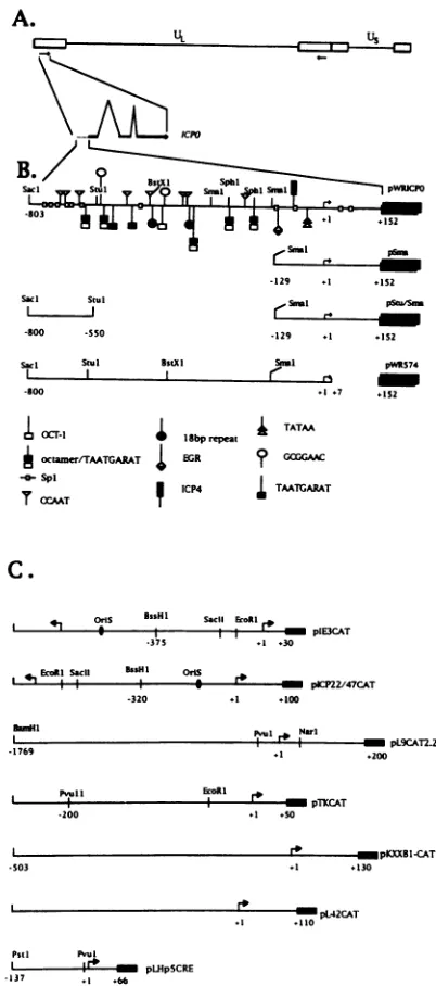
Construction of plasmids. Plasmid
pWRICPO
expresseschloramphenicol
acetyltransferase (CAT)under control of thewild-type
promoter/regulatory
sequences of theICPO
gene(45) (nucleotides125062to124105bythe
numbering
systemof Perry et al. [55, 56]) (Fig. 1B). pWRICPOwasconstructedby
removing the 1,597-bp SacI-BamHI fragment ofpW3
(which
contains the entire ICP0 promoter,leader, first exon, and part of the first intron) and cloningit into theSacI-BamHI site of
pUC19 (New England Biolabs, Beverly,
Mass.)
to createpW3SB (63). TheuniqueNcoI site inpW3SB, whichincludes the initiation codon of the ICPO open reading frame, was cleaved with Ncol, blunt ended following incubation with the Klenow fragment of DNA polymerase I, ligated to HindIll
linkers, and recircularized to create plasmid pW3SH (55). A 955-bp Sacl-HindIIl fragment was isolated from pW3SH and cloned into the SacI-HindIll site of the CATexpressionvector pGemCAT (5), placing the ICPO promoter/regulatory se-quences (-803 to +152 relative to the start of transcription
[45, 56]) immediately 5' to the openreadingframe of theCAT gene (Fig. 1B). Three tandem copies of the simian virus 40 (SV40) polyadenylation signalincluded inan810-bpfragment
were inserted immediately 5' of the ICPO promoter to mini-mize the influence of readthrough transcriptsoriginating from cryptic promoters within vectorsequences(46).The sequence content and orientation of ICPO promoter/regulatory se-quences inthis plasmid were confirmed bydideoxynucleotide
sequencing(Promega, Madison, Wis.).
The ICPO-luciferase reporter geneplasmid, pMCICPO,was constructedby isolating a 955-bp SacI-HindIll fragment from pWRICPO, which contains the ICPO promoter/regulatory se-quences, and cloning it into the Sacl-HindIll site of the luciferase expression vector Basic (Promega). pGL2-Control, anSV40 early promoter-luciferase reporterplasmid,
wasobtained from Promega.
Deletion mutations were introduced into the 5' region of ICPO sequences as follows. pWRICPO was doubly digested with StuI and SmaI or with Sacl and SmaI, ligated to XbaI linkers or Sacl linkers, and recircularized to create plasmids pStu/Sma and pSma, respectively (Fig. 1B). The deletion in pStu/Sma is 431 bp in length (-559 to - 128), and that in pSma is 675 bp (-803 to -128). pWR574 was constructed by synthesizing a 574-bp StuI-HindIII fragment by PCR (Perkin-ElmerCetus, Norwalk, Conn.), usingpWRICPO as a template and 20-nucleotide primers (5'-TGCAAGAGGCCTTGTTC
CGC-3' and 5'-TTAAGCTTGCGACCAGACTGTCGGGC
CA-3') designed to specify the boundaries of the fragment as -559 and +7 of the wild-type sequence (55). The amplified fragment was cloned into theStuI-HindIII site of
pWRICPO,
replacing the wild-type sequence and resulting in a plasmid lacking 145 bp ofthe 152-bp 5' untranslated leader sequence ofICPO. The identity of all mutations was confirmed by di-deoxynucleotide sequencing (Promega).
Theconstruction and characterization of HSV-1 promoter-CAT plasmidscontaining the promoter of the early gene UL9 (pL9CAT2.2), the delayed-early gene encoding glycoprotein B (pKXXBI-CAT), the gene specifying the LATs(pLHpSCRE), the immediate-early gene encoding ICP4 (pIE3-CAT), the early gene encoding thymidine kinase (pTK-CAT), and the true late gene, L42 (pL42-CAT), of HSV-1 have been de-scribed previously(5, 11, 21, 42). CAT plasmidspLSHp5 (17), pICP22/47-CAT, and pHCMV-CAT (79) contain the SV40 early promoter, the promoter/regulatory sequences of the immediate-early genes encoding ICP22/47, and the human cytomegalovirus (HCMV) transcriptional regulatory region of lE1, respectively.
Effect ofisoleucine deprivation on cell growth. The growth of Vero and NB41A3 cells in medium containing and lacking isoleucine was assessed by seeding 3 x 10⁵ cells in 35-mm-diameter petridishes in appropriate growth medium for 24 h,
on November 9, 2019 by guest
http://jvi.asm.org/
ACTIVATION OF HSV-1 IE GENE EXPRESSION 6873
A.
US
15 an * K
-129 .1 *152
SacI StuPS.SIau
L JI r r
-800 -S50 -129 .1 *IS2
Saci Stul
-800
A XT-I
O octamerTATMTGAT
-0-Spi
TccAT
B3tXI Saul pWR574
r rt1. .
.1 .7 -152
lSbprepeat
BGR
ICP4
I TATAA
? GOGGAC
I TAATGARAT
C.
OrlS BssHI Sacil EcoSl
-3S pIE3CAT
-375 .1 .30
EcoRlSacil 35sH1
-320
OrlS _
IpK3P22/47CAT
.1 .100
faIl-H1p,, arl
I !a,I& I pL9CAT2.2
-1769 .1 200
Pvu1TIEIoRI
I ---I_ pTKCAT
-200 .1 So0
re, _---~ pKXXBI-CAT
-503 .1 *130
1 .110
Pstl PwlI
t _ pLHpSCRE
-137 .1 66
FIG. 1. Wild-type andmutantpromoter-CATplasmids usedinthis study. (A) Diagram of the HSV-1 genome showing the locations of bothcopies of the ICPOtranscriptionalunitwithinthelongrepeatsof HSVDNA.Beneath thediagram of theHSV-1 genome isanexpanded diagramof theICPO transcriptional unit showing thepromoterregion (dotted line), exons (thick horizontal lines), and two introns (thin diagonallines). (B) Expanded diagram of the wild-typeICPO promot-er/regulatory region from -803 to +152 in pWRICPO. Relevant restriction sitesareshown abovetheline. Theblack boxontheright represents the CAT gene, including its 5' untranslated leader and coding sequences. The locations of deletions in ICPO regulatory sequences in plasmids pSma, pStu/Sma, and pWR574 are shown beneath the diagram ofpWRICP0. Below the plasmids aresymbols representing previously identified and potential regulatory elements within the ICPO promoter region. 18bp repeat; an 18-bp repeat sequencenotpreviouslydescribed;EGR,cis-element target for cellu-larimmediate-early proteinsEgr-1(mouse)and EGR2(human) (54); ICP4,binding site for HSV-1regulatory protein ICP4; GCGGAAC, GA-rich element(4, 10, 75,80). (C)Diagramofpromoter/regulatory sequences in immediate-early (ICP4, pIE3CAT; ICP22/47, pICP22/ 47CAT), early (TK, pTKCAT; UL9, pCAT22), delayed-early (gB, pKXXB1-CAT),andlate(L42,pL42CAT)reporter genes.Alsoshown
transferring the cells to medium lacking isoleucine for 42 h, andthen returning the cells to medium containing isoleucine. Cells were counted in triplicate before, during, and after cultivation in mediumlacking isoleucine.
CAT assays in growth-arrested-released Vero and NB41A3 cells. (i) Growth arrest-release. Vero and NB41A3 cells were growth arrested by isoleucine deprivation essentially as de-scribed by Cai and Schaffer(12), Hamlin andPardee (34), and Ley and Tobey (43). Briefly, cells were seeded in 100-mm-diameter petri dishes at densities of 2.5 x 10⁶ Vero cells or 4.0 X 10⁶ NB41A3 cells in their respective growth media and incubated at 37°C for 24 h. Growth medium was then removed and replaced with medium lacking isoleucine and supple-mentedwith10% dialyzed fetal bovine serum(forVerocells)
or 15% dialyzed horse serumand 2.5% dialyzedfetal bovine serum (for NB41A3 cells). Isoleucine-free Dulbecco's modi-fiedEagle's medium(productno.57-861-501) and isoleucine-free Ham's F-10 (product no. 57-965-501) were prepared as instructed by the manufacturer (JRH Biosciences, Lenexa,
Kans.). Following incubation for 42 h in medium lacking isoleucine, cells were returned to isoleucine-containing me-dium to reverse theblock andwere transfectedat the
desig-natedtimes. Vero cells incubated in medium lacking isoleucine have been shown to be growth arrested at apoint within the
GJG1 stageof the cellcycle (12).
(ii) Transfection. In an effort to maximize theefficiencyof transfection while minimizing the time needed to achieve detectable levels of CAT activity, we used Lipofectin
(Be-thesda Research Laboratories, Gaithersburg, Md.) to
intro-duce plasmid DNA into cells (28). Twenty micrograms of plasmid DNA in 2.5 ml of theappropriategrowthmediumwas mixed with 2.5 ml ofgrowth medium containing 75 mg of Lipofectin and incubatedat25°Cfor 15 min. At thedesignated
timesafterreturnofculturestoisoleucine-containing medium,
growth mediumwas removed frommonolayers andreplaced
with 5 mloftheDNA-Lipofectin mixture.DNAand cellswere incubated at 37°C in 5% C02, and transfected cells were harvested andprocessedfor CATactivityorluciferaseactivity
6 haftertransfection.
(iii) CAT assays. Extracts of transfected cellswereprepared byscrapingmonolayersin100-mm-diameterpetridishesinto 6 ml of CAT buffer (0.25M Tris-HCl [pH 7.4]), concentrating
thecellpelletby centrifugationat4,000xg,andresuspending
thepelletin 100
RIl
ofCAT buffer. After celllysisinasonicator(Heat Systems-Ultrasonics, Inc.,Plainview,N.Y.)for1minat
60%efficiency,thesolubleproteinfractionwasseparatedfrom cellular debrisbycentrifugationat 16,000 xgfor 15min, and the protein concentration in the soluble extract was deter-minedbythe method of Bradford(8).CAT assayswerecarried outby the addition of 200 mg ofproteinextract to30 ml of 20 mMacetyl coenzyme A
(Boehringer
Mannheim,Indianapolis,
Ind.) and 100 nCi of[4C]chloramphenicol (Dupont,
NEN ResearchProducts, Boston,Mass.). Sampleswereincubatedat37°C for 90 min and
analyzed
bythin-layer
chromatography
(Eastman Kodak, Rochester,N.Y.) afterethylacetate extrac-tion. The results of CAT assaysarepresented
as the percent conversion of substrate to acetylatedforms ofchlorampheni-col. All assays were repeated at least three times. Because absolute values varied among tests,
meaningful
averagescould not be calculated. Relative values werehighly
reproducible,
however.
isadiagramofthe promoterregulatoryregionofthe genespecifying
the LATs in reporterplasmid pLHp5CRE. VOL. 68, 1994
on November 9, 2019 by guest
http://jvi.asm.org/
[image:3.612.75.276.76.530.2](iv) Luciferase assays. Extracts of transfected cells were
prepared by scraping monolayers in 100-mm-diameter petri dishes into 1 ml of extraction buffer (100 mM KPO4 [pH 7.8], 1 mM dithiothreitol), concentrating the cell pellet by centrif-ugation at 4,000 x g, andresuspendingthe pelletin 100 ,ul of
extractionbuffer. After cell lysis inasonicator (Heat Systems-Ultrasonics) for 1 min at 60% efficiency, the soluble protein fraction wasseparated from cellular debrisby centrifugationat
16,000 x g for 15 min, and the protein concentration in the
soluble extract was determined by the method of Bradford (8). Luciferase assays were performed by adding 50 ,u1 of cell extract to 100 ,u1 of luciferase assay reagent (Promega) and measuring the reaction in anLKB-Wallac 1251 luminometer (BioOrbit, Turku, Finland).
Slot blot hybridization. To measure transfection efficiency, cells in either 100- or 60-mm-diameter petri dishes were
washed in ice-cold phosphate-buffered saline (PBS; 0.13 M NaCl, 2.6 mMKCI, 10mM Na2HPO4, 1.3mM KH2PO4 [pH 7.4]), scrapedinto ice-cold PBS,transferred toa15-mlconical
tube (CorningGlass Works, Corning,N.Y.), andcentrifugedat
2,000x g. After the supernatant fluid was removed, the cell
pelletwas washed three times in PBS,and the final pelletwas
resuspended in three times the packed cell volume of ice-cold hypotonicbuffer (10 mM NaCl, 10 mM Tris [pH 7.4], 3 mM MgCl2). After incubation on ice for 10 min, the swollen cells were transferred to an ice-cold Dounce homogenizer (Wheaton) and homogenized with 10 strokes of a type B pestle, releasingnucleiinto the medium.The mixturewasthen
transferred to an Eppendorf microcentrifuge tube and centri-fuged at 16,000 x g for 10 min at 4°C, pelleting nuclei and cellular debris.Following removalof thesupernatantfluid,the pellet was resuspended in 200 ml of TE buffer (10 mM Tris HCl, 1 mM EDTA [pH 8.0]) and 400 mlofNonidet P-40 lysis buffer(10 mM NaCl, 10 mM Tris [pH 7.4], 3mM MgCl2, 0.5% Nonidet P-40) and allowed to incubate at 25°C for 1 min to
disrupt nuclear membranes. Ten microliters of RNase A (Sigma, St. Louis, Mo.) was then added, and the reaction mixture was incubated for an additional 5 min and then subjected to extraction with an equal volume of a 25:24:1
mixture of phenol, chloroform, and isoamyl alcohol (Sigma). Fourhundredfifty microliters of the aqueousphasecontaining both cellular and plasmid DNA was transferred to a fresh
EppendorfMicrofuge tubeandtreatedwith 0.1 volume of 3 M NaOHfor 5 min at 68°Cto denature DNA. After neutraliza-tionby the addition of 1.0 volume of sodiumacetate (pH5.0), DNA was transferred to a nylon membrane in a slot blot
apparatus (Minifold II; Schleicher & Schuell, Inc., Keene, N.H.) as instructed by the manufacturer. Known amounts of purified plasmid DNA were also transferred to serve as
standards. The blot was probed with purified plasmid DNA radiolabeled by nick translation (65), and the amount of
hybridizing radiolabeled DNA in each slot was measured by Phosphorlmager (model 400E; Molecular Dynamics,
Sunny-vale, Calif.) scanning. The amount of plasmid DNA in the
nucleiof transfectedcellswascalculatedbycomparison witha
standard curve generated from the signals of known
concen-trations ofplasmid DNAused asstandards. RESULTS
Activation of an immediate-early promoter in
growth-ar-rested-released Vero cells. Results of previous studies in this
laboratory have demonstrated that the plating efficiency of a
mutant virus(7134) lacking both copies of theICPOgene was
enhanced approximately fourfold when plated on Vero cell
monolayers8 h after release from growth arrestrelative to 24
c
a.2
0C
0 U
ZU.U-
15.0-10.0
5.0-0.c.I ,
0 24 6 8 101214 1618202224262830
Time of transfection (hours) after addition of isoleucine-containing medium FIG. 2. CAT expression in Vero cells transfected with pWRICPOat various times after release from growth arrest induced by isoleucine deprivation. Vero cells (2.5 x 10O cells per 100-mm-diameter petri dish) were growth arrested by incubation in isoleucine-free medium for 42 h (12). At 42 h, isoleucine-free cell culture medium was replaced by isoleucine-containing medium, and cells were transfected with 20
,ug
ofpWRICPO at various times thereafter. Six hours after transfec-tion, cells were harvested and processed for CAT activity. Percent conversion of unacetylated to acetylated substrate was used as the measure of promoter activity over the 6-h interval between the timeof transfection and the time of harvest. This experiment was repeated four times.h after release(12). This difference in plating efficiency was not observed with the wild-type virus or with the mutant virus plated onICPO-expressing cells. Thetime ofmaximum expres-sion of this plaque-enhancing activity (8 h postrelease) corre-lated with progression from
GO/GI
to the S phase of the cell cycle,asdeterminedbyflowcytometryof cells synchronizedby isoleucine deprivation. These observations suggested the pos-sibility that afactor (or factors), expressed maximally in Vero cells 8 h after release from growth arrest inGo/Gl,
can substitute for thedefect in the mutantbyenhancing viralgene expression, and hence plaque formation, in the absence of functional ICPO. In an effort to better understand the basis of this enhancing cellular activity, we attempted to determine whether a factor(s) in Vero cells could enhance gene expres-sion from each of the major classes of viral promoters (imme-diate-early, early, and late) 8 h after growth arrest-release.To determine whether the cellular activity is able toenhance HSV immediate-early gene expression, we constructed a chi-mericICPOpromoter-CAT plasmid, pWRICPO(Fig.1B), and tested its inducibility following transfection of Vero cellsat2-h intervals after release from growth arrest (Fig. 2). Under the conditions ofgrowth arrest-release used in these tests, greater than 80% of cells would be expected to be in synchrony throughout the 24-h test period (12). Each time pointshown in Fig. 2 represents cumulative CAT activity over the 6-hinterval between the time of transfection withpWRICPOandthetime of harvest of cells for CAT assays. This relatively shortinterval allowed us to monitor CAT expression roughly as afunctionof the time after release of transfected cells fromgrowth arrest. In this assay, maximum expression of CAT from pWRICPO occurredwhen Vero cell monolayers were transfected 6 hafter release from growth arrest. These findings are consistentwith results obtained in previous studies in which the maximum enhancing effect on the platingefficiencyof 7134 inVerocells
on November 9, 2019 by guest
http://jvi.asm.org/
[image:4.612.344.527.76.248.2]ACTIVATION OF HSV-1 IE GENE EXPRESSION 6875
occurred whenmonolayerswereinfected 8 h after release from
growtharrest (12).
Expression of the cellular enhancing activity was not ob-servedinexponentially growing cells that hadnotbeengrowth arrested, nor was the activity detected again when growth-arrested-released Vero cellswere tested after completion of
onesynchronous growth cycle (24 h) (data notshown).Thus,
asinthecaseof theplaque-enhancing activity describedby Cai
and Schaffer (12), the cellular activityappearstobe expressed specifically following release from growth arrest near the
GJG1 borderandnotinactivelycycling cells.
Transfection efficiency of growth-arrested-released Vero cells. To test the possibility that the differences in CAT expressionobservedwereduetodifferential uptake of plasmid DNAby Vero cells transfectedatdifferent times after release from growtharrest,andnot todifferences in the ability of cells to enhance gene expression at these times,we measured the
levelofplasmidDNA innuclei of cellstransfectedat6 and 24 hpostreleaseby slot blot hybridization (Table 1). Although the 6-hsamplescontainedanaverageof1.7,ug of DNA (8.6% of
input DNA), the24-h samples containedan averageof 4.9 ,ug
of DNA (24% of input DNA). This greater than twofold increase in theamountof DNAwasanticipated, given thatat
24h afterrelease from growth arrest the cell populationhad completedoneround ofcell division andtwice as many cells
were available to takeup DNA (12) (Table 1). This
observa-tionindicates that the efficiency of uptake of plasmid DNAwas
slightly greater in cells transfected at 24 h than in cells transfected at 6 h after release from growtharrest.However, Vero cells transfected at 6 h postrelease exhibited approxi-mately16-fold-greater CAT activitypermicrogram of nuclear
plasmidDNA thancellstransfectedat24 hpostrelease (Table 1).Thus, the increase inCATactivityat6 h inFig.2wasdue
not togreaterefficiencyintheuptakeoftransfectedpWRICPO DNAbuttothe enhancedabilityof this DNAtobeexpressed at6 h.
Responseof other HSV-1 andheterologousviralpromoters tothe cellularactivity. Toassess theresponsiveness of other HSV-1 promoter-CATconstructs tothe cellularactivity,CAT plasmidscontainingpromotersrepresentingeach of themajor kinetic classes of HSV genes were tested in Vero cells after growth arrest-release (Fig. 3). In these studies, cells were
transfectedonly duringthe 6-hperiods postreleasethat
corre-spondedto maximum (6to 12h) and minimum (24to 30h) expression of the cellular activity in assays with the ICPO promoter(Fig.2).The results of thesetestsshow thatonlythe reporter plasmids containing the immediate-early ICPO and ICP4promotersrespondedtothe cellularactivityinVero cells (Fig. 3A). ICPO promoter-driven CAT activity was sixfold
greaterat6 h thanat24h after release fromgrowtharrest,and ICP4promoter-driven activitywasfourfoldgreater.No
signif-icant differences in CATexpression in cells transfected at 6 and 24 h postreleasewereobserved for plasmids driven byan
early (UL9) or late (L42) promoter (Fig. 3A). Studies of plasmids in which CAT expression was driven by non-HSV
viral promoters revealed that SV40 early promoter-driven CAT activitiesweresimilarat6 and 24 h postrelease andthat HCMV immediate-early promoter-CAT activity was 2.3-fold
greater at 6 thanat 24h (Fig. 3B). Collectively, these results indicate that a cellular activity induced in Vero cells
trans-fected 6 h after release from growth arrest is selective in its abilitytoenhanceexpression fromatleasttwoimmediate-early promotersofHSV and (toalesser extent) HCMV.
Growth properties ofa neural cell line following release
fromgrowth arrest inducedby isoleucine deprivation. In an
efforttodetermine whether cells of neural origin also specify
an enhancing activity for HSV-1 immediate-early promoters,
we performed studiessimilarto those described above, using NB41A3 cells. Originally established from a murine neural
neoplastic tumor (C1300), this clonal cell line (41A3) shares
manyproperties of sympathetic neuronal cells, including the development of neurofilaments and neurotubulesaswellasthe
synthesis and storage of catecholamines (9, 68). Because of difficulties associated withthegrowtharrest-release ofprimary
neurons, we chose to use NB41A3 cells because they have
proven useful for studies of biochemical and metabolic
pro-cessesassociatedwithneurons (9, 51, 70, 76).
Priortoconducting CATassaysingrowth-arrested-released NB41A3 cells, we examined the effect of the growth arrest-release procedure on NB41A3 cell growth andviability. The
growth propertiesof these cellswereexaminedby comparing
the numberofviablecells atregular intervalsbefore, during, and after incubation in medium lacking isoleucine with the number of viable cellsgrowncontinuouslyinmedium contain-ing isoleucine (Fig. 4A). The results of similar tests of Vero cells are provided for comparison (Fig. 4B). Figure 4 shows that neither NB41A3 nor Vero cells increased in number
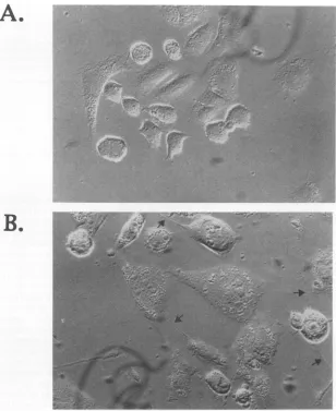
during the 42-h incubation in medium lacking isoleucine. However,unlike Verocells,whichresumed cell division within 36 h after return to isoleucine-containing medium, NB41A3 cells remainedgrowtharrested forthe remainder of the test. Flowcytometric analysisofNB41A3 cellsfollowingisoleucine deprivationconfirmed the abortedprogression throughthecell cycleafter return to isoleucine-containing medium (data not shown).Because these cells failedtoprogress throughthe cell cycle,itwasdifficult to assess the stage of the cycleatwhich cellswere arrested. Microscopically, these cells exhibited
al-teredmorphology (Fig. SB)relativetocells that hadnotbeen subjectedtogrowtharrest(Fig. 5A). Thus,the cellsappeared enlarged and flattened and exhibited limited neurite out-growth. Although they failed to resume cell division, 100% of the cells shown in Fig. SB excluded trypan blue forup to
TABLE 1. Transfectionefficiencyofgrowth-arrested-releasedVerocells
Inputplasmid DNAd(,ug)
Time(h) oftransfection C no. Total protein Protein used N at time of: % ofinput plasmid
after release from
Cell+
no. T* rotei inCAT Conversion' DNAin nucleiatgrowtharrest (10'avg±S mg,avg± , assay(mg) (avg± SD) Transfection Harvest timeofharvest
(avg±SD)
6 2.4±0.1 0.58±0.03 0.2 14.1±3.8 20 1.72 0.32 8.6
12 4.8±0.2 1.32±0.03 0.2 2.6± 2.0 20 4.87±0.26 24.3
aValues are calculated averages of determinations in three 100-mm-diameter petri dishes each seeded with 2.5 x 10⁶ cells and treatedtoachieve growth
arrest-releaseasoutlined in Materials and Methods.
bPerculture,asdeterminedbythe method of Bradford(7).
See Materials andMethods.
d SeeMaterialsandMethods.
VOL. 68,1994
on November 9, 2019 by guest
http://jvi.asm.org/
A.
9.8
10.0-8.0
-0
6.0-0
3.0.0
2.0-
16 24 6 24 6 24 6 24 Mock
Promoter: ICPO ICP4 UL9 L42
B.
- 30.0-29.0
-28.0
27.0 -26.0
-25.0
-12.0
C 11.0
0
;i 10.0
> 9.0
0
L 8.0 7.0 6.0
5.0
4.0-3.0
-2.0
1.0
0.0
A.
_. 12.0
uo
r-
10.0-0
8.0-u 0 CL 6.0
U 4.0
6 z
a , I I ...I I
0 24 48 72 96 120
14 192 26. .2
144 168 192 216 240
B.
27.0 1.9
" 07 ~~~~1.1
6 24 6 24
Promoter: ICPO SV40
U
11.5
'no0
4-0
U'
S
U-0
z
30.0-
25.0-
20.0-
15.0-
10.0-
5.0-0 24 48 72 96 120 144
/'
'03 02
1
_06AC7
6 24 6 24
HCMV MOCK
Time of transfection (hours) after addition of isoleucine-containing medium
FIG. 3. CAT expression in growth-arrested-released Vero cells transfectedwithreporterplasmids containingpromotersrepresenting themajor HSVgenes ornon-HSV viralpromoters.Plasmids contain-ing the promoter/regulatory regions of HSV immediate-early (ICPO andICP4), early (UL9),and late (L42)genes(A)andSV40late and HCMVimmediate-earlygenes(B)weretransfectedat6 and 24 hafter
addition of isoleucine-containing medium. Experiments were
per-formedessentiallyasdescribed in thelegendtoFig.2exceptthatcells
weretransfectedonlyatthetimes of maximum(6 h) and minimum (24 h) ICPO promoter-CAT activity. Experimentswere repeated atleast
twotimes.
300 h after return to isoleucine-containing medium, demon-strating thattheywereviable. Weconcludefrom these obser-vations thatisoleucinedeprivation irreversibly growth-arrested NB41A3cells butnotVero cells. Whether the cessation of cell division isaresult ofterminal differentiation is unclear.
Activation of theICPOpromoterin NB41A3 cells following incubation in medium lacking isoleucine. Although NB41A3
168 192 216 240
Addition of Addition of 1- medium 1+ medium
Hour post seeding
FIG. 4. Effectsof isoleucinedeprivationonthegrowthof NB41A3 and Vero cells. NB41A3 (A) orVero (B) cells (3 x 10⁵ cellsper 60-mm-diameterpetri dish)weregrowth arrested asdescribedinthe legendtoFig.2. Afterseeding,cellswereharvestedandcountedatthe indicated times before, during, and after incubation at 37°C in isoleucine-freemedium.Eachpointrepresentstheaveragenumber of cellsin each ofthree dishes.Squares,cellsincubatedinisoleucine-free medium; diamonds,cellsincubatedcontinuouslyin isoleucine-contain-ingmedium.
cells ceased todividefollowing incubation in mediumlacking
isoleucine, we were interested in determining what effect growtharrestwould haveonexpression of the CAT gene from
pWRICPOtransfectedatvarious times afterreturn tomedium
containing isoleucine. The time courseexperiment presented
inFig. 6wasconducted ina manneridenticaltothatdescribed above for Vero cells (Fig.2). The results of this test demon-strate thatasforVerocells returned toisoleucine-containing medium,asinglepeakof CATactivitywasevident. However, NB41A3 cells reproducibly exhibited peak CAT expression
m -_ r
on November 9, 2019 by guest
http://jvi.asm.org/
[image:6.612.93.262.66.534.2] [image:6.612.306.551.74.522.2]ACTIVATION OF HSV-1 IE GENE EXPRESSION 6877
A ;
S.
-~
...
~~~~~~~~~~~~~~~~~~~~~~~~..
....c....-.S.B
92
o-S...
... [image:7.612.153.461.76.453.2]...
. .
FIG. 5.
Morphological
changesin NB41A3cellsfollowing
incubation in isoleucine-free medium.Cellswerephotographed
before(A)andafter(B)42hof incubation in isoleucine-free medium.
Magnification,
X63. Arrowsindicate neuriteoutgrowth.when transfected at 2 h after return to
isoleucine-containing
medium(i.e.,when CATactivitywasmeasured from 2to8
h).
Thus, peakICPOpromoter-activating activitywas detectedat anearlier time afterreturn toisoleucine-containing
medium in NB41A3 than in Verocells.Responses of other HSV promotersto thecellular activity
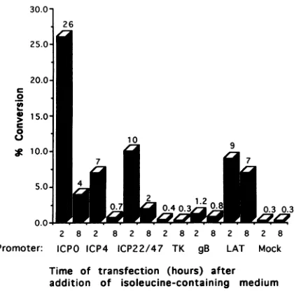
expressed by NB4LA3cells. ReporterCAT
plasmids
contain-ing HSV promoters
representing
themajor
kinetic classes of HSV genes were screened for theability
torespond
to the cellularactivityinNB41A3cells(Fig.
7).
Thetwo6-h intervals chosen fortesting representedtheperiods
of maximal(2
to 8h)and minimal (8to 14
h)
expressionof the ICPOpromoter-activating
activity
(Fig.
6).
The results of these tests demon-strated that thepromoter-CATplasmids
containing
theimme-diate-early promoters,
ICPO, ICP4,
andICP22/47,
responded
to the cellularactivitymore
strongly
when transfectedat 2 h than at 8 h after return toisoleucine-containing
medium,
although the absolute levels of CAT
activity
varied from promoter to promoter. The results of slot blotanalysis
ofpWRICPO
DNA recovered from the nuclei of NB41A3 cells transfected at 2-h intervals afterreturn toisoleucine-contain-ingmedium revealedno
significant
differences in the accumu-lation of plasmid DNA (data notshown).
Promoter-CATplasmids pTK-CAT (TK, early) andpKXXBI-CAT
(gB,
de-layed-early) as well as
pLHp5CRE (LATs)
wereminimally
responsive to the cellular
activity expressed
from 2 to 8 hpostrelease in NB41A3cells.
Notably,
thebasalactivity
of the LAT promoter in NB41A3 cellswassignificantly higher
than the basal activities of thegB and TK promoters(Fig.
7).
Weconclude from these studies that thecellular
activity
in NB41A3 cellsas in Vero cells isspecific
forimmediate-early
promoters. Moreover, the cellular
activity
enhanced CATexpression from the ICPO and ICP4 promoters twice as
efficiently in NB41A3 cells asin Verocells.
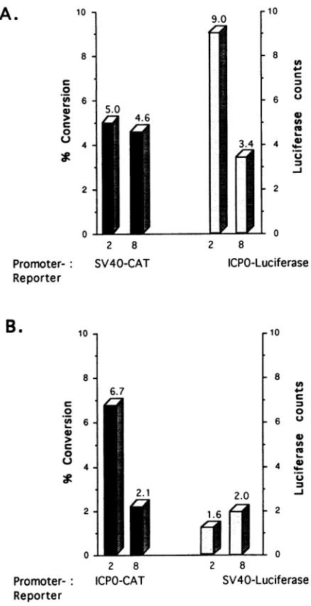
In anefforttoconfirm the
specificity
ofthe cellularactivity
forimmediate-early promoters,weexaminedthe responsesof
the ICPOandSV40
early
promotersto the cellularactivity
in cells cotransfected with plasmidscontaining
each promoter. TheSV40earlypromoterhad been showntobenonresponsive
to the cellular
activity
inprevious
tests.Thus,
NB41A3 cells were cotransfected at either 2 and at 8 h after release fromgrowth arrest with ICPO-CAT
(pWRICPO)
andSV40-lucif-erase
(pGL2-Control)
reporterplasmids,
and extracts weretested for both CAT and luciferase activities. To control for differences in thesensitivities of thetwoassays,cellswerealso cotransfected with ICPO-luciferase
(pMCICPO)
andSV40-VOL.68, 1994
on November 9, 2019 by guest
http://jvi.asm.org/
c
0
0
u
16.0-14.0l
12.0-
10.0- 8.0-6.0 4.0
2.0'- ... ...
0 2 4 6 8 1012141618202224262830 Time of transfection (hour) after addition of isoleucine-containing medium
FIG. 6. CAT expression in NB41A3 cells transfected with
pWRICPOatvarious after addition of isoleucine-containing medium
tocellsgrownin isoleucine-free mediumfor 42 h. NB41A3 cells (4.0 x
106 cells per 100-mm-diameter petri dish) were growth arrested as
described in the legend to Fig. 2. Atvarious times after addition of
isoleucine-containing medium, cells were transfected with 20,ug of
pWRICPO. Six hours after transfection, cells were harvested and
processed for CAT activity. Percent conversion was used as the
measureofpromoteractivity during the6-h interval between thetime
of transfection and the timeof harvest. This experimentwasrepeated
three times.
CAT (pCAT-Control) reporter plasmids, and extracts were tested for both CAT and luciferase activities. In these
experi-ments, the ICPO promoter, whether linked to the luciferase (Fig. 8A)orCAT(Fig. 8B)reportergene, directed expression
30.0]
25.0-
20.0-c 0
rn
, 15
0
u
* 10,
10
5.0.
0.0-2 8 2 8 2 8 2 8 2 8 Promoter: ICPO ICP4 ICP22/47 TK gB
0.3 0.3 2 8 2 8
LAT Mock
Time of transfection (hours) after
addition of isoleucine-containing medium
FIG. 7. CATexpression inNB41A3isoleucine-deprivedcells
trans-fected 2 or 8 h after addition of isoleucine-containing medium.
Experimentswereperformed essentially asdescribed in thelegend to
Fig. 2exceptthatcellsweretransfected onlyat2or8h afteraddition
ofisoleucine-containing medium. CAT expressionfrom HSV
[image:8.612.87.269.75.251.2]promot-er-CAT plasmids representing immediate-early (ICPO, ICP4, and ICP22/47),early(TK), anddelayed-early (gB)genesand LATs(LAT) is shown. Experimentswererepeatedtwice.
TABLE 2. Induction of CAT expression from wild-type and mutant ICPO promoter test plasmids in growth-arrested NB41A3 cells
after addition of isoleucine-containingmedium
% Conversion after transfectionat
2 and 8 h afterrelease from Fold Plasmida growth arrestb (avg ± SD) inductionc
2h 8h
pWRICPO 6.9±0.1 0.8 ±0.01 8.5
pStu/Sma 1.0 ±0.1 0.8 ±0.3 1.3
pSma 0.8 ± 0.01 0.60±0.01 1.3
pWR574 5.3 ±0.4 0.7±0.03 7.5
Plasmids are depicted in Fig. 1.
bSeeMaterials and Methods.
Level of CATactivityat2hrelative to the level of CATactivityat 8h after
release fromgrowth arrest.
more efficiently when cells were transfected at 2 hthan when they were transfected at 8 h after release from growth arrest. In contrast, the levels of expression of the SV40 early promoter within the same cells were similar at the two time points. In contrast to experiments mentioned above, in which approxi-mately sixfold induction was routinelyobserved, induction of the ICPO promoter was only approximately threefold in these cotransfection experiments. This reduction in expression is likely due to the fact that only 10,ug of each reporter plasmid was used in cotransfection experiments, compared with 20 ,ug in the aforementioned single-plasmid transfection experi-ments. Taken together, these results support the conclusion that immediate-early promoters arespecificallyresponsiveto a cellular activity induced in neural cells (NB41A3) after addi-tion of isoleucine-containing medium to cells irreversibly growth arrested by isoleucine deprivation.
The region of the ICPO immediate-early promoter respon-sive to the cellular activity. To identify elements within the promoter/regulatory region ofpWRICPO that respond to the enhancing activity expressed in NB41A3 cells, several mutant
ICPOpromoter-CAT plasmids wereconstructed. Each plasmid contains a unique deletionmutationengineered in either the 5' upstream or 5' untranslated region of the ICPO gene with respect to the transcriptionalstart site at + 1 (45, 55) (Fig.1B). The uninduced levels of expression of all of themutants were comparable to that of the wild-type plasmid (pWRICPO), as determined by standard CATassays in exponentially growing (i.e., non-growth-arrested) NB41A3 cells(data not shown). To assess the responsiveness of each mutant promoter to the cellular activity,NB41A3 cells were growth arrested in medium lacking isoleucine and returned to isoleucine-containing me-dium essentially as described above. Growth-arrested-released
cells were then transfected at thetimes ofmaximum (2h) and minimum (8 h) activation of theICPO promoter by the cellular activity (Fig. 6).
In this series ofexperiments, NB41A3 cells transfected with pSma and pStu/Sma at 2 hexhibited nearly the same levels of CAT activity as cells transfected at 8 h after release from growth arrest. In contrast, the levels of CAT activity in cells transfected withpWRICPOandpWR574 at 2 h were 8.5- and 7.5-foldgreater than cells transfected at 8 h after release from growth arrest (Table 2). The observation that nearly equal levels of CAT expression were induced from pWRICPO and pWR574 when cells were transfected at 2 h after return to isoleucine-containing medium indicates that the cis-acting elements thatrespond to the cellular activity are located in the promoter and not the 5' untranslated region of theICPOgene. Moreover, the failure to detect CAT activity in cells
on November 9, 2019 by guest
http://jvi.asm.org/
[image:8.612.318.561.106.194.2] [image:8.612.79.289.444.652.2]ACTIVATION OF HSV-1IE GENE EXPRESSION 6879
fected with pSma or pStu/Sma at 2 h at levels above those obtained 8 h after return to isoleucine-containing medium indicates that cis-acting elements distinct from those responsi-ble forbasal activity are responsive to the cellular activity and thatthese responsive elements lie between nucleotides -550 and -129upstream of theICPOtranscriptional start site.
DISCUSSION
Anunderstanding of the molecular mechanisms that under-lie the establishment and reactivation of HSV-1 latency will requireabetterunderstanding of the interplay between cellu-lar and viral factors that mediate the repression and activation of viral genes. Because immediate-early viral genes are re-quired to orchestrate the expression of early and late genes, cellular factors that affect the expression of this class of
immediate-earlygenes are of special significance to studies of latency. In this study, we have demonstrated that a cell
cycle-regulated activity expressed in cells of neural and non-neural origin is able to enhance expression from immediate-early, but not early or late, HSV-1 promoters.
Theidentificationof a cell cycle-regulated factor(s) able to
complementanICPO null mutant (7134) for plaque formation on Vero cell monolayers was first described by Cai and Schaffer (12). These investigators demonstrated that impair-ment of the plating efficiency and synthesis of viral proteins
characteristic of the ICPO null mutant, 7134, on 24-h-old monolayers can be overcome by plating virus on synchronized Vero cellmonolayers 8 h after cells have entered the cell cycle at some point in GJ/G1. In contrast to 7134, the plating
efficiency ofthe wild-type virus was independent of cell cycle status. A reasonable explanation for this observation is that upon entry into the
post-G,JGl
stage of the cell cycle, Vero cells express factors that facilitate replication of HSV-1 ge-nomes unable to express ICPO. Because the wild-type virus expresses ICPO, it is able to replicate efficiently in cells independent of the expression of such factors and, hence, independent of the stage of the cell cycle. Because we wish to isolate, clone, and characterize the cellular gene(s) whoseprotein products are able to substitute for ICPO, we have attempted to identify the class(es) of viral genes, and the subset of sequences within the regulatory regions of these genes, thatrespond to the cellular activity.
Time ofpeakexpression of the cellular activity in Vero cells and NB41A3 cells. Theobservation that expression of anICPO
promoter-CATtestplasmid was maximal when Vero cells were
transfected 6 h after release from growth arrest induced by isoleucine deprivation is in excellent agreement with the results of Cai and Schaffer (12), in which the peak in 7134 platingefficiency occurred in Vero cell monolayers at 8 h after release fromgrowth arrest. The 2-h difference in the times of peak promoter activity and peak plating efficiency is most likely due to differences in the times required for the two assays. Platingefficiency was measured during the 1-h window needed for virus adsorption (12), whereas CAT assays measured cumulative promoter activity in a 6-h time period. The 6-h
periodof maximum CAT activity in transient assays with Vero cells(6 to 12 h)includes the 1-h window of maximum plating efficiency (8 to 9 h).
Because theexistence of a cellular activity able to enhance HSV-1 gene expression may be significant with regard to the mechanism ofreactivation of HSV from latency, we attempted to determine whether cells of neural origin are capable of expressing a similar activity. For this purpose, we extended our experiments to include tests in the mouse neuroblastoma cell line, NB41A3. This clonal cell line and its parent have been
used to study several metabolic processes associated with cells of neural origin (i.e., nerve growth factor secretion, catechol-amine metabolism, choline metabolism, and neurotoxicity) as well as to model certain interactions of HSV with neurons (2, 9, 18, 24, 51, 70, 78). Like Vero cells, NB41A3 cells deprived of isoleucine entered a state of growth arrest. However, upon return to isoleucine-containing medium, NB41A3 cells re-mained growth arrested for at least 6 days, whereas Vero cells resumed growth within 24 h. This observation demonstrates that isoleucine deprivation can induce physiological alterations inNB41A3 cells leading to irreversible growth arrest. Whether the suspended growth of NB41A3 cells reflects a state of terminal differentiation is not known. The morphological changes observed following return to isoleucine-containing medium (i.e., flattening of cells and limited neurite outgrowth) are, however, suggestive of a differentiated phenotype, in that they are similar to changes observed in neuroblastoma cells after treatment with sodium butyrate, an agent reported to induce differentiation of neuroblastoma cells, inhibit cell divi-sion, and increase the activity of enzymes involved in neuro-transmitter metabolism (2, 24, 67, 68).
Like growth-arrested Vero cells, growth-arrested NB41A3 cells express an enhancing activity for the immediate-early promoters after addition of isoleucine. The pattern of
ICPO-driven CAT expression over a 24-h period was similar to that observed in Vero cells, except that maximum promoter en-hancing activity occurred in the period 2 to 8 h after return to isoleucine-containing medium. This period of peak CAT ex-pression occurs 4 h earlier than it does in Vero cells, which may reflect differences in the overall kinetics of expression of the cellular activity in the two cell types. Alternatively, NB41A3 cells may simply be more efficient in the uptake and expression ofplasmid DNA than Vero cells. The observed shift of the peak of CAT enhancing activity observed in Vero cells (high, 6 h;low, 10 h) to a time period 4 h earlier in NB41A3 cells (high, 2h;low, 6 h) suggests that the overall kinetics of expression of the respective cellular activities, and not just DNA uptake, occur at an earlier time in NB41A3 cells than in Vero cells. Although the stimulatory activities in the two cell types exhibit the same immediate-early promoter specificity, it is not clear whether the activity expressed in NB41A3 cells represents the neural counterpart of the same activity observed in Vero cells. That the activity in NB41A3 cells is expressed at significant levelsdespite the inability of these cells to resume cell division after reintroduction of isoleucine to the growth medium is notable because it demonstrates that resumption of cell divi-sion is not a requirement for expression of the activity.
Thecellular activity is specific for the promoters of imme-diate-early genes. Tests of the promoter specificity of the cellular activity in Vero and NB41A3 cells indicate that of the classes of HSV-1 promoters tested, only those of the immedi-ate-early class responded to the cellular enhancing activity in the time window used to study the ICPO promoter-CAT plasmid,
pWRICPO.
In addition, we observed that expression from an immediate-early promoter (IE1) of HCMV, a lym-photropic betaherpesvirus, was also enhanced, although only twofold in Vero cells. In contrast, the SV40 early promoter expressed alone or coexpressed with the ICPOpromoter was not enhanced (Fig. 3 and 8).Itshould be noted that plasmid pLHp5CRE, used in these studies to assess the responsiveness of the promoter of the LATs to the cellular activity in NB41A3 cells, contains only 203 bp of its 5'regulatory sequence (+66 to -137), with respect to the start site of the primary LAT [23]). Several reports have demonstrated the requirement of cis-acting sequences up-stream of this basal promoter for both neural cell-specific
VOL.68, 1994
on November 9, 2019 by guest
http://jvi.asm.org/
A 10
-8
-C
0
uFn
6-5-- _
C
2 8
Promoter-: SV40-CAT
Reporter
B.
10C
0
W) 6
C
0
c-4
2
0
Promoter-: Reporter
6.7
2 8
ICPO-CAT
[image:10.612.64.283.70.496.2]Time of cotransfection addition of isoleucine FIG. 8. CAT expression and luciferas isoleucine-deprived cells. Experimentswei
described in the legendto Fig. 2exceptth only at 2 and 8 h with either 10,ug of
pCAT-Control(A)or 10,ugof pWRICPO (B). CAT expression (solid bars) and luci
bars)areshown.
activity and induced activity in neura
lightofthese findings, conclusions drn
ofinducibilityof theLATpromoterto
7)wouldbe premature.
The results presented in thisrepor
cellular activity expressed in cells of
origin ispromoter specific, (ii) this ac
specificenhancerofherpesvirusimme
sionduringproductiveinfectionandr(
and(iii)theactivitiesobserved inVeri representcell-type-specifichomologs cis-acting response elements in ti
10 immediate-early genes. We have shown that deletion of
se-quences-550to-129of the ICPO
promoter/regulatory
region
render the ICPOpromoter
unresponsive to the cellularactivity8 inNB41A3cells
(Table
2).
Contained within thisregion
of the ICPO promoter are cis-acting response elements orbinding
sites for severalpreviouslyidentifiedcellular activatorsaswell
-6
aspotential
cis elements for otherpossible regulators
of gene* expression (Fig. 1).Deletion of thisregion
effectively
removes all but one of theoverlapping octamer/TAATGARAT motifs 34 _ and all of the single octamer and TAATGARAT consensus motifs(Fig.
1B), potentially impairing the ability ofplasmidri.:3 pSma or pStu/Sma torespond toactivation by the
Oct-1NVP16
X complex (10, 31, 45, 52). In the absence of VP16, however,
Oct-1 does not appear to activate
transcription
ofmostpoly-merase II-responsive promoters that contain TATAA
ele-LP_
___ments_(66,
74).
Thus,
it isunlikely
thatOct-1
alone isrespon-0 sible for the immediate-early promoter-enhancing activity 2 8 observed in NB41A3 cells, in which no VP16 is present. The ICPO-Luciferase possibility remains, however, that (i) a cellular factor expressed in NB41A3 cells can enhance gene expression incombination with Oct-1 or (ii) octamer-binding proteins other than
Oct-1
can function as transcriptional activators in the absence of
VP16.Indeed, He et al. have described alarge family of POU 10 domain regulatory proteins in mammalian brain tissue that are expressed transiently during development of the nervous sys-tem, manyof which havebindingaffinityfor the
Oct-1
binding8 motif (35).
Potential role of the cellular activity in the life cycle of HSV-1. The demonstration of a cellular activity able to activate
0
6 u
immediate-early
promotersingrowth-arrested-released
neural * cells suggests the possibility that this activity functions in the reactivation of HSV fromlatency
in neuronsfollowing
stimu-4 lation by stress-related factors. Isoleucine deprivation causesNB41A3 cells to enter a state of growth arrest that may have 2.0 3 features similar to the growth-arrested state of differentiated 2 neurons. Addition of isoleucine to
growth-arrested
NB41A3
i.6 ]
~
cells may trigger an increase in metabolic activity similar to thatwhich occurs in stimulated neurons (14). Changes in themicroenvironment associated with neuronal stimulation in-2 8 0
clude
increases in second messenger molecules, such as cyclicSV40-Luciferase
AMP (cAMP),cGMP, andprotein kinase C,among others(1,13, 44, 76). Treatment of neuronal cells in culture with a variety of these second messenger molecules as well as the stimulation i (hours) after of neuron-specific receptors have been shown to elicit transient
-containing
medium
expression of certain transcription factors (e.g., c-Fos, c-Jun, se expression in NB41A3 andOct-1)
able to regulate gene expression in such diverse reperformed essentially as processes as cellular differentiation andmitogenesis
(16,
32, iatcellswere cotransfected 33,50,51).As in thecaseofgrowth-arrested-released
NB41A3
pMCICPO and 10 ,ug of cells,latentlyinfectedneuronsmayalsoexpressanactivitythat and 10 ,ug of pGL-Control enhances immediate-early gene expression. Expression of im-iferase expression (stippled mediate-early genes is a prerequisite for reactivation of HSV from latency since it constitutes a resumption of the program of gene expression characteristic of productive infection. Evi-dence to support the notion that latent viral gene expression is
d1
cells (6, 7, 29, 81). In regulated by cellular factors is available from in vitro models of awn concerning the lack HSV latency in which reactivation has been shown to occur in thecellular activity (Fig. response to the activation of second messenger pathways (64, 71). Moreover, Wheatley et al. have used immortalized sensory tdemonstrate that (i) a neurons to show that agents that raise intracellular levels of f neural and nonneural cAMP also increase expression of the ICPO gene as well as the -tivity mayfunction as a permissivity of these cells for HSV infection (77), and Leib et diate-early gene expres- al. have demonstrated that the reactivation of HSV-1 fromeactivationfromlatency, dissociated latently infected murine trigeminal ganglia is
ac-oandNB41A3 cells may celerated by cAMP analogs or adenylate cyclase activators of the sameactivity. (42). Although the cellular pathway leading to activation of ie promoters of HSV-1 immediate-early genes in NB41A3 cells is unknown, having
on November 9, 2019 by guest
http://jvi.asm.org/
ACTIVATION OF HSV-1IE GENE EXPRESSION 6881
identifiedaregionof the ICPOpromoter that respondstothe
cellular activity,weshouldnowbe abletofinemapindividual
response elements, isolate the effector proteins that bind to
these elements, andclone the genes that encode the effector
proteins.
ACKNOWLEDGMENTS
WethankChristine Dabrowski, MaryAnnHardwicke, andRobert
Jordan for helpful discussions and Monica Shea for manuscript
preparation.
These studies were supported by Public Health Service research grantR37 CA20260from the National Cancer Institute andprogram projectgrantP01AI24010fromtheNational Institute of Allergyand
Infectious Diseases.W.M.R.wassupported in partbytraininggrant
T32 GM11676 as part of the Harvard Medical School M.D./Ph.D
program.
REFERENCES
1. Abraham,I.,K. E. Sampson,E. A. Powers, J. K Mayo,V. A.Ruff,
and K. L. Leach. 1991. Increased PKA and PKC activities
accompanyneuronal differentiation ofNT2/D1cells.J. Neurosci. Res.28:29-39.
2. Ash,R. J.1986.Butyrate-inducedreversal of herpes simplexvirus
restriction in neuroblastomacells. Virology155:584-592.
3. Astor,T., and P. A.Schaffer.Personal communication.
4. Bailey, A. C., and R. Thompson. 1992. Asequence-specific
DNA-binding protein recognising a GA-rich element that cooperates
with Oct-1 at the herpes simplex virus type 1 IE3 promoter. Intervirology34:74-85.
5. Baradaran, K.1993. Personal communication.
6. Batchelor,A.H., and P. O'Hare. 1990.Regulation and
cell-type-specific activity of a promoter locatedupstream of the
latency-associated transcript of herpes simplex virus type 1. J. Virol.
64:3269-3279.
7. Batchelor,A. H.,and P. O'Hare. 1992. Localization of cis-acting
sequencerequirementsin thepromoterofthe latency-associated transcript of herpes simplexvirustype 1 required for cell-type-specific activity. J. Virol. 66:3573-3582.
8. Bradford, M. M. 1976. A rapid and sensitive method for the
quantitation of microgram quantities of protein utilizing the principle of protein-dyebinding. Anal. Biochem. 72:248-254. 9. Breakfield,X.O.,E.A. Neale,J. H.Neale,andD.M.Jacobowitz.
1975.Localized catecholaminestorageassociated with granulesin murineneuroblastoma cells.Brain Res. 92:237-256.
10. Bzik,D.J., andC. M. Preston. 1986.Analysisof DNAsequences
whichregulate the transcriptionof herpes simplex virus immediate earlygene3: DNAsequences required forenhancer-like activity andresponsetotrans-activationbyavirionpolypeptide.Nucleic Acids Res. 14:929-943.
11. Cai,W., and P. A.Schaffer.1989. Herpessimplex virustype1 ICPO
plays a critical role in the de novo synthesis of infectious virus
followingtransfectionofviralDNA. J. Virol.63:4579-4589. 12. Cai, W., andP.A.Schaffer. 1991.Acellular functioncanenhance
geneexpression and plating efficiency ofamutantdefective inthe
genefor ICPO,a transactivatingprotein of herpes simplexvirus type 1.J. Virol.65:4078-4090.
13. Ciesielski-Treska,J., andG. Ulrich. 1980. Monoamineregulation of adenosine 3',5'-monophosphate in homogeneous neuronal
cul-turesfromchick brainhemispheres.Neurochem. Res. 5:1097-1106. 14. Cole, A. J., D. W.Saffen,J. M. Baraban, and P.F. Worley.1989. Rapid increase ofanimmediate earlygene messenger RNA in
hippocampal neurons by synaptic NMDA receptor activation. Nature(London)340:474-476.
15. Cook,M., andJ.Stevens. 1973. Pathogenesis ofherpes neuritis and ganglions in mice: evidence for intra-axonal transport of infection. Infect. Immun.7:272-288.
16. Curran, T., and J. I. Morgan. 1986. Barium modulates c-fos expression andpost-translationalmodification. Proc.Natl. Acad. Sci.USA83:8521-8524.
17. Dabrowski, C.,and J.Alwine.Personal communication.
18. de La Pefia, N. C., E. Bal, L. Puricelli,A. Diaz,andE.S. deLustig.
1978. Interferon in the replication of herpes simplex virus in
normal and pathological nerve cells.IARC Sci. Publ. 24:1055-1066.
19. DeLuca, N., and P. A.Schaffer.1987. Activities of herpes simplex virus-1 ICP4 genes specifying nonsense peptides. Nucleic Acids Res. 15:4491-4511.
20. DeLuca, N., and P. A. Schaffer. 1988. Physical and functional domains of the herpes simplex virus transcriptional regulatory protein ICP4. J. Virol.62:732-743.
21. DeLuca, N.A.,and P.A.Schaffer. 1985. Activation of immediate-early, early, and late promoters by temperature-sensitive and wild-type forms of herpessimplex virus type 1 protein ICP4. Mol. Cell. Biol.5:1997-2008.
22. Dixon, R.,and P. A. Schaffer. 1980. Fine structure mapping and functional analysis oftemperature-sensitive mutants in the gene encoding the herpes simplex virustype 1 immediate early protein VP175.J. Virol.36:189-203.
23. Dobson, A. T., F. Sederati, G. Devi-Rao, W. M. Flanagan, M. J. Farrell, J. G. Stevens,E. K. Wagner, and L. T. Feldman. 1989. Identification of the latency-associated transcript promoter by expression ofrabbit beta-globin mRNA in mouse sensory nerve ganglia latentlyinfected with arecombinant herpes simplex virus. J.Virol.63:3844-3851.
24. Dreyer, L. L.,R.J. Sydiskis, and N. Bashirelahi. 1989. Effect of dexamethasone on herpes simplex virus replication in mouse neuroblastoma cells(NB41A3): receptor characteristics. J. Clin. Lab. Anal.3:236-243.
25. Efstathiou, S., A. C. Minson, H. J. Field, J. R. Anderson, and P. Wildy. 1986. Detection of herpes simplex virus-specific DNA sequences in latently infected mice and in humans. J. Virol. 57:446-455.
26. Everett, R. 1984. Transactivation of transcription by a herpes simplex virus product: requirement for two HSV-1 immediate earlypolypeptidesformaximalactivity. EMBO J. 3:3135-3141. 27. Everett,R.D. 1986. The products ofherpes simplex virus type 1
(HSV-1) immediate early genes 1, 2, and 3 can activate HSV-1 gene expressionintrans. J. Gen.Virol. 67:2507-2513.
28. Felgner, P. L., T.R.Gadek, M. Holm,R.Roman, H. W. Chan, M. Wenz, J. P. Northrop, G. M. Ringold, and M. Danielsen. 1987. Lipofection: a highly efficient, lipid-mediated DNA-transfection procedure.Proc. Natl. Acad. Sci. USA84:7413-7417.
29. Frazier, D., and P. A.Schaffer. 1993. Personal communication. 30. Gelman,I. H., and S.Silverstein. 1985.Identification of
immedi-ate-early genes from herpes simplexvirus that transactivate the virusthymidinekinase gene. Proc. Natl. Acad. Sci. USA 82:5265-5269.
31. Gerster, T., andR.G.Roeder. 1988. A herpes virustrans-activating proteininteractswithtranscriptionfactorOTF-1 and other cellu-larproteins.Proc. Natl. Acad. Sci. USA85:6347-6351.
32. Greenberg,M.E.,L. A.Greene,and E. B. Zif. 1985. Nerve growth factor andepidermalgrowth factor inducerapidtransient changes in proto-oncogene transcription in PC12 cells. J. Biol. Chem. 26:14101-14110.
33. Greenberg, M. E., E. B. Zif, and L. A. Greene. 1986. Stimulation ofneuronal acetylcholine receptorsinducesrapid gene transcrip-tion.Science234:80-83.
34. Hamlin, J. L., and A. B. Pardee. 1976. S phase synchrony in monolayerCHOcultures. Exp. Cell Res. 100:265-275.
35. He, X., M. N. Treacy, D. M. Simmons, H. A. Ingraham, L. W. Swanson, and M. G.Rosenfeld. 1989.Expressionof alargefamily ofPOU-domain regulatory genes in mammalian brain develop-ment. Nature (London) 340:35-41.
36. Hill, T. J. 1985. Herpes simplex virus latency, p. 175-190. In B. Roizman (ed.), Theherpesviruses,vol. 3.PlenumPress, NewYork. 37. Honess,R.W., and B.Roizman. 1974. Regulation of herpesvirus macromolecular synthesis. I.Cascaderegulationofthe synthesisof three groups of viralproteins.J.Virol.14:8-19.
38. Honess, R. W., and B.Roizman.1975. Regulation of herpesvirus macromolecular synthesis: sequential transition ofpolypeptide synthesis requiresfunctionalviralpolypeptides. Proc. Natl.Acad. Sci. USA72:1276-1280.
39. Jean, S., and D. Knipe. Personalcommunication.
40. Kemp, L. M., L. C. Dent, and D. S. Latchman. 1990. Octamer motifmediatestranscriptional repressionofHSVimmediate-early
VOL. 68,1994