ISSN: 2319-8753
I
nternational
J
ournal of
I
nnovative
R
esearch in
S
cience,
E
ngineering and
T
echnology
(An ISO 3297: 2007 Certified Organization)
Vol.2, Issue 11, November 2013
Copyright to IJIRSET www.ijirset.com 6412
Effect of Ginger Extract Consumption On Renal Function
during Ethanol Withdrawal Induced-Stress
Swaroopa Maralla
1Division Of Molecular Biology and Exercise Physiology, Department Of Zoology, Sri Venkateswara University, Tirupati –
517 502 , Andhra Pradesh, India1
ABSTRACT: From times unknown, ginger has become a subject of interest because of its beneficial effects on human health. The purpose of the present study was to investigate the effects of daily oral administration of ginger extract for 6 weeks on kidney functions in withdrawal rats to evaluate the ameliorating effects in alcohol induced-withdrawal rats. Rats (130-150gm) were divided into 4 groups; normal control rats, alcoholic control rats, ethanol withdrawal rats and ethanol withdrawal rats pretreated with ginger. Ginger extract was administered orally for 6 weeks to pre-treated rats, and they were compared with the normal and alcoholic groups, respectively. The treatment with ginger extract had significant effect on Plasma Electrolyte Profiles. Low plasma sodium level and increased plasma potassium levels were observed in ginger pre-treated ethanol withdrawal group. The plasma creatinine, urea and uric acid levels were significantly reduced in this group compared to alcoholic control rats and ethanol withdrawal rats. It is concluded that the consumption of ginger produced a significant anti- nephrotoxic effect in ethanol withdrawal rats. In addition, ginger is showing properties of anti-hypertensive drugs and is capable of improving impaired kidney function in ethanol withdrawal rats.
Keywords: Ginger, Kidney Function, Alcohol, Plasma, Lipids, Withdrawal, Rats.
I.INTRODUCTION
Long-term alcohol misuse is associated with water and salt retention, causing an expanded extracellular volume [1]. Impaired renal function, secondary to the long-term effects of alcohol misuse, also results in a metabolic acidosis, as well as other electrolyte disturbances such as hypomagnesaemia, hypophosphataemia and hypocalcaemia. Severe alcohol misuse predisposes to acute renal failure [1]. Rarely, bladder dysfunction occurs with alcohol misuse, possibly secondary to an alcohol-induced neuropathic bladder [2]. Urinary retention and abdominal distension can result. Excessive alcohol consumption can have profound negative effects on the kidneys and their function in maintaining the body’s fluid, electrolyte, and acid-base balance, leaving alcoholic people vulnerable to a host of kidney-related health problems that include decreased ability to excrete body wastes, inability to maintain body fluid and electrolyte balance and decreased synthesis of essential hormones [3].
ISSN: 2319-8753
I
nternational
J
ournal of
I
nnovative
R
esearch in
S
cience,
E
ngineering and
T
echnology
(An ISO 3297: 2007 Certified Organization)
Vol.2, Issue 11, November 2013
Copyright to IJIRSET www.ijirset.com 6413
Plant derived products have been used for medicinal purposes for centuries. At present, it is estimated that about 80% of the world population relies on botanical preparations as medicines to meet their health needs [11]. Herbs and spices are generally considered safe and proved to be effective against many diseases ([12], [13]). Ginger has a long history of use in traditional medicine in the treatment of over a wide range of ailments including renal disorders. Ginger was proved more potent renoprotective agent in both acute and chronic renal failure (CRF) and the mechanisms underlying the effects of renal failure by ischemia–reperfusion ([14], [15]). The study of Ajith et al., 2007 [16] reaffirmed the protective effect of ginger against cisplatin-induced oxidative stress and acute renal failure in kidneys of mice. In addition, this study observed the effect of pre-treatment with ginger on the levels of serum creatinine and urea, and concluded that the administration of ethanol extract of ginger before and after cisplatin injection significantly lowered the elevated levels of serum creatinine and urea.
In light of these observations an attempt was made in this study to evaluate the nephroprotective ability of ginger extract during acute withdrawal from chronic alcohol ingestion in male wistar rats.
II.MATERIALS AND METHODS
Ginger extract: The fresh rhizomes of Zingiber officinale were obtained from local market and identified by the herbarium staff of the Botany Department, SV University, India. Whole rhizome of ginger was thoroughly washed, sliced , grated and grind to fine paste . A weighed quantity (30gm) of the paste was subjected to continuous extraction in Soxhlet apparatus using double distilled water. The extract was evaporated under reduced pressure using rotary evaporator and then lyophilized until all the solvent has been removed to give an extract sample and stored at 4c for further studies.
Experimental animals: The study involved young (3–4 months old; 200 - 220g ) male albino rats of Wistar strain purchased from Sri Venkateswara Traders Pvt. Limited, Bangalore, maintained in the animal house of the department in polypropylene cages. The animals were allowed to habituate to the animal facilities for at least for two weeks adaptation period upon arrival and were maintained under standard conditions of humidity (50% relative humidity), room temperature (25 - 28ºC) and 12 h light/ dark cycle (6:00 A.M. to 6:00 P.M.). A standard rodent diet (M/s Hindustan Lever Ltd., Mumbai), and water were provided ad libitum. All experimental procedures were approved by the CPCSEA on Animal Care.
Experimental protocol: The experimental animals were divided into 4 groups; each group contained six animals: Control group G1 (normal without treatment), alcoholic group G2 (injected with 2 gm/ kg body wt ( p.o.) ethanol), alcoholic rats treated with ginger extract (200 mg/kg body weight) for 6 weeks G3(firstly, the rats were injected with ethanol along with ginger extract), alcoholic rats subjected to withdrawal G4(first, the rats were given Etoh for 6 weeks and then subjected to withdrawal from alcohol for 3 days after the last dose), withdrawal rats treated with ginger extract(200 mg/kg body weight) G5(first, the rats were given ginger for 6 weeks along with Etoh and then subjected to withdrawal from alcohol for 3 days after the last dose). Ginger extract was given orally (200 mg/kg body weight) to the rats through a gastric tube daily for 6 weeks.
Collection of blood samples: At the end of the 6 weeks of post-treatment, blood samples were collected by sacrificing the animals and the blood was collected in clean EDTA tubes, then plasma was separated by centrifugation and stored at-20°C for biochemical analysis. While the groups of rats pretreated with ginger were decapitated after 3 days of abstinence from chronic alcohol treatment and blood samples were collected as mentioned above.
ISSN: 2319-8753
I
nternational
J
ournal of
I
nnovative
R
esearch in
S
cience,
E
ngineering and
T
echnology
(An ISO 3297: 2007 Certified Organization)
Vol.2, Issue 11, November 2013
Copyright to IJIRSET www.ijirset.com 6414
Biochemical analysis: Creatinine and urea were determined by enzymatic method according to the method (Patton and Crouch, 1977) [17]. The flame photometry method of Vogel, 1960 [18] was used for the determination of sodium ion (Na+) and potassium ion (K+) concentration in serum. Inorganic phosphate (Pi) levels in serum was estimated according to Fiske and Subbarow, 1925 [19] method while serum Calcium and Magnesium were estimated using AAS according to the methods adapted from Thin and Thomson, 1967 [20].
Statistical analysis: Data were statistically analyzed by one-way analysis of variance followed by Duncan's test (SPSS). Finally, significant difference (L.S.D) was used to test the difference among treatments. Results were considered statistically significant when (P < 0.05).
III.RESULTS
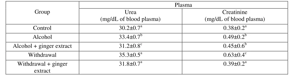
Kidney Functions: As shown in Table 1, the alcohol produced significant increase in the levels of plasma creatinine, urea and uric acid when compared with normal group, while, administration of ginger extract to the alcoholic rats significantly reduced the levels of plasma creatinine, urea and uric acid when compared with the alcoholic group, but no significant changes were observed when compared with the normal rats. This indicates that, coadministration with ginger extract normalized the plasma creatinine, urea and uric acid. On the other hand, the pre-treatment of Etoh followed by induction of withdrawal decreased significantly the levels of plasma creatinine and uric acids were decreased significantly when compared with the alcoholic group. Furthermore, they are still significantly higher than normal rats. In contrast, non-significant increase was observed in plasma urea when compared with alcoholic group, but it was still non-significantly higher than normal rats as shown in Table 1.
Table 1: Effect of ginger extract on Renal markers during chronic ethanol consumption and ethanol withdrawal
Group
Plasma Urea
(mg/dL of blood plasma)
Creatinine (mg/dL of blood plasma)
Control 30.2±0.7a 0.38±0.2a
Alcohol 33.4±0.7b 0.49±0.2b
Alcohol + ginger extract 31.2±0.8c 0.45±0.6b
Withdrawal 35.3±0.5a 0.63±0.4c
Withdrawal + ginger extract
31.8±0.7a 0.39±0.2a
Data are expressed as mean ± SE (n=6). The statistical test conducted was ANOVA followed by multiple two-tail t-test and data with different superscript (a,b,c) in a specific vertical column differed from each other significantly (p < 0.05).
ISSN: 2319-8753
I
nternational
J
ournal of
I
nnovative
R
esearch in
S
cience,
E
ngineering and
T
echnology
(An ISO 3297: 2007 Certified Organization)
Vol.2, Issue 11, November 2013
Copyright to IJIRSET www.ijirset.com 6415
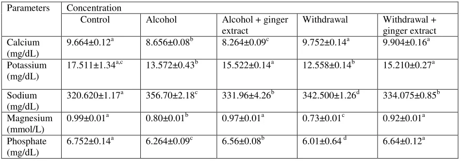
Table 2: Effect of ginger extract on serum electrolyte abnormalities during chronic ethanol consumption and ethanol withdrawal
Parameters Concentration
Control Alcohol Alcohol + ginger
extract
Withdrawal Withdrawal +
ginger extract Calcium
(mg/dL)
9.664±0.12a 8.656±0.08b 8.264±0.09c 9.752±0.14a 9.904±0.16a
Potassium (mg/dL)
17.511±1.34a,c 13.572±0.43b 15.522±0.14a 12.558±0.14b 15.210±0.27a
Sodium (mg/dL)
320.620±1.17a 356.70±2.18c 331.96±4.26b 342.500±1.26d 334.075±0.85b
Magnesium (mmol/L)
0.99±0.01a 0.80±0.01b 0.97±0.01a 0.73±0.01c 0.92±0.01a
Phosphate (mg/dL)
6.752±0.14a 6.264±0.09c 6.56±0.08b 6.01±0.64 d 6.64±0.12a
Values are Mean ± S.E.M., n=6, per group.
a,b,c Values in the same row with different superscripts are significantly different at P<0.05.
IV.DISCUSSION
The present study demonstrated that withdrawal induces the elevation of the plasma urea and creatinine in alcoholic rats, which are considered a significant marker of renal dysfunction [21]. In the present study the effect of ginger on the kidney functions was assessed by the determination the levels of plasma creatinine, urea and uric acid, and the study revealed that pre administration of ginger extract to the alcoholic rats reduced and normalized the levels of plasma creatinine, urea and uric acid. On the other hand, the pre-treatment with ginger before the induction of diabetes inhibited the higher increase of plasma creatinine and uric acid resulted from the induced-stress by EW but they did not normalized. Moreover, the study of Ajith et al [16] demonstrated that ethanol extract of ginger rendered significant protection against induced nephrotoxicity, which was evident from the lowered serum urea, and creatinine levels in the mice that were pre-treated with ginger extract, and this study concluded that ginger extract significantly protected the elevation of serum creatinine and urea levels. Furthermore, the treatment of ginger extract could significantly prevent the depletion of antioxidant concentration and antioxidant enzymes activities in the kidneys. In addition, studies reported that the presence of polyphenols and flavonoids in ginger extract might be responsible for the antioxidant nephroprotective activities and the reduction of serum urea and creatinine levels [16].
Withdrawal may also have an important impact on fluid and electrolyte status. Almost all patients in acute withdrawal are dehydrated as a result of diaphoresis, hyperthermia, vomiting, and tachypnea. Hypokalemia is common due to renal and extra-renal losses, alterations in aldosterone levels, and changes in potassium distribution across the cell membrane. Hypomagnesemia occurs frequently with DTs and may predispose to withdrawal seizures [8]. Hypophosphatemia may occur due to malnutrition, may be symptomatic, and if severe, may contribute to cardiac failure and rhabdomyolysis.
ISSN: 2319-8753
I
nternational
J
ournal of
I
nnovative
R
esearch in
S
cience,
E
ngineering and
T
echnology
(An ISO 3297: 2007 Certified Organization)
Vol.2, Issue 11, November 2013
Copyright to IJIRSET www.ijirset.com 6416
hypokalemia may result in different cardiac rhythm disorders, cardiomyopathia and paralyticileus ([22], [23], [24]); hypomagnesemia causes cardiac arrhythmias, mental and emotional changes [24]. So, electrolyte evaluation and appropriate correction must be done during management of chronic alcoholic patients in intensive care units, especially those with alcohol withdrawal.
Potassium: Normally the kidneys are a major route of potassium ion excretion and serve as an important site of potassium regulation. Alcohol consumption historically is found to reduce the amount of potassium excreted by the kidneys [25],
although the body’s hydration state may help determine whether potassium excretion will increase or decrease in response to alcohol. Levels of potassium, like those of sodium, also can affect the way the kidneys handle fluid elimination or retention. In addition, potassium depletion has been proposed to exacerbate hyponatremia through any of several mechanisms [26].
Sodium :Alcohol does appear to directly influence the kidney’s handling of sodium and other electrolytes, potentially
resulting in hypernatremia. In a study by Rubini and colleagues, 1955 [25], subjects who consistently drank about 4 ounces(oz) of 100-proof bourbon whiskey experienced decreased sodium, potassium, and chloride excretion (i.e., increased retention of solutes). Although some exceptions exist, several historical studies have reported similar modest reductions in sodium and potassium excretion following alcohol use ([27], [28], [29], [30], [31], [32], [33]).
The serum sodium level is determined by the balance of fluid in relation to that of sodium: Not enough fluid in the body results in a sodium concentration that is too high (i.e., hypernatremia), whereas excessive amounts of fluid produce a sodium concentration that is too low (i.e., hyponatremia). Hyponatremia does not constitute merely a biochemical abnormality but most likely has clinical consequences as well (e.g., impaired mental activity, neurological symptoms, and in extreme instances, seizures).
Phosphate: Another potential cause of hypophosphatemia in alcoholic patients is hyperventilation, which can occur during alcohol withdrawal. Prolonged rapid, shallow breathing results inexcessive loss of carbon dioxide and decreased blood acidity (i.e., alkalosis), which in turn activates an enzyme that enhances glucose breakdown. In glucose breakdown, phosphate becomes incorporated into various metabolic compounds, ultimately lowering blood levels of phosphate. As the rate of glucose breakdown increases, profound hypophosphatemia potentially can result. Alcoholic patients also may develop low blood levels of phosphate by excreting too much of this ion into their urine. Alcohol can induce abnormally high phosphate levels (i.e., hyperphosphatemia) as well as abnormally low levels. In fact, hyperphosphatemia often precedes hypophosphatemia. Alcohol consumption apparently leads to excessive phosphate levels by altering muscle cell integrity and causing the muscle cells to release phosphate. This transfer of phosphate out of muscle cells and into the
bloodstream results in an increased amount of phosphate passing through the kidneys’ filtering system.
There are a number of published reports showing that serum potassium concentration falls during alcohol withdrawal, especially if complicated by delirium tremens ([34], [35]). Some studies even show evidence that potassium could be useful as an indicator to predict delirium tremens ([7], [36]).
ISSN: 2319-8753
I
nternational
J
ournal of
I
nnovative
R
esearch in
S
cience,
E
ngineering and
T
echnology
(An ISO 3297: 2007 Certified Organization)
Vol.2, Issue 11, November 2013
Copyright to IJIRSET www.ijirset.com 6417
Calcium: Early studies showed that alcohol consumption markedly increases calcium loss in urine. In severely ill alcoholic patients, low blood levels of calcium occur about as often as low blood levels of phosphate and can cause convulsions or potentially life-threatening muscle spasms when respiratory muscles are involved. Alcoholic patients with liver disease often have abnormally low levels of a calcium-binding protein, albumin, and also may have impaired vitamin D metabolism; either of these two factors could result in reduced blood levels of calcium (i.e.,hypocalcemia). Muscle breakdown and magnesium deficiency are other potential causes of hypocalcemia in alcoholic patients. A direct effect of alcohol in reducing calcium levels is suggested by at least one experimental study: Dogs became hypocalcemic after administration of alcohol above a critical threshold amount of approximately 1 g/kg [37].
Conclusion: From the data obtained, it is concluded that post-treatment and pre-treatment with ginger extract produced a significant anti-nephrotoxiic effect. Furthermore, ginger is capable of improving hyperlipidemia and the impaired kidney functions in ethanol induced- withdrawal rats maintaining proper kidney functioning as well as maintaining electrolyte balance preventing retention of solutes.
Despite the significance of alcohol’s effects on the kidney, however, relatively few recent studies have been conducted to
characterize and elucidate their pathophysiology. More studies are required in animals and humans on the kinetics of ginger and its constituents relating the effects of ginger consumption over a long period of time during nephrotoxicity in cases of alcohol abstinence. It is hoped that future investigations will focus on this important subject area.
Acknowledgement: The corresponding author sincerely thank Dr.K.Sathyavelu Reddy, Professor, Sri Venkateswara University, Tirupati and Dr.W.Rajendra, Professor, Sri Venkateswara University, Tirupati for the kind support and timely help, for allotting valuable time for healthy discussions and consultation regarding the problem under investigation and also for providing laboratory aid.
Conflict of Interest: None Declared.
REFERENCES
[1]. S.Vamvakas, M. Teschner, U. Bahner and A. Heidland , “Alcohol abuse: potential role in electrolyte disturbances and kidney diseases”, Clinical Nephrology, Vol. 49, pp 205–213, 1998.
[2]. I. Jun-Ichi, T. Takahide and O.Tetsuro, “Acute abdominal distension secondary to urinary retention in a patient after alcohol withdrawal”, Alcohol and Alcoholism, Vol. 40, pp 86–87, 2005.
[3]. M. Emily, “Toxicity. In: Encyclopedia of Earth” Eds. Cutler J. Cleveland, Washington D.C. 2007.
[4]. I. Al-Sanouri, M. Dikin, A.O. Soubani , “Critical Care Aspects of Alcohol Abuse”, South Med J, Vol. 98, No.3, pp 372-381, 2005. [5]. S.B. Chabria, “Inpatient management of alcohol withdrawal: a practical approach”, Signa Vitae, Vol. 3, No.1, 24 - 29, 2008.
[6]. M.Elisaf, E.Liberopoulos, E.Bairaktari, K.Siamopoulos, “Hypokalaemia in alcoholic patients”, Drug Alcohol Rev, Vol. 21, No.73, 2002. [7]. J. Wadstein and G. Skude, “Does hypokalaemia precede delirium tremens?,” Lancet, Vol. 2, No.549, 1978.
[8]. M.Victor, “The role of hypomagnesemia and respiratory alkalosis in the genesis of alcohol-withdrawal symptoms,” Ann N Y Acad Sci, Vol. 215, No. 235, 1973.
[9]. L. Larsson, K. Rebel, B. Sorbo, “Severe hypophosphatemia--a hospital survey”, Acta Med Scand, Vol. 214, No.3, pp 221, 1983.
[10]. A.L.King, D.A. Sica, G.Miller, S.Pierpaoli, “Severe hypophosphatemia in a general hospital population”, South Med J, Vol. 80, No.7, pp 831-835, 1987.
[11]. J.N.M. Shri, “Ginger: It’s Role in Xenobiotic Metabolism”, ICMR Bulletin; Vol.33, No.6, pp 57-63, 2003.
[12]. J.F. Deng, “Clinical Toxicity of Herbal Medicine in Taiwan”, 7th International Conference on Health Problems Related to the Chinese. 1994 [13]. M. O’Hara, D. Kiefer, K. Farrel, K.Kemper. “A review of 12 commonly used medicinal herbs”, Arch Fam Med, Vol. 7, No.6, pp 523- 536,1998 [14]. M.F. Mahmoud, A.A. Diaai and F. Ahmed,“Evaluation of the Efficacy of Ginger, Arabic Gum, and Boswellia in Acute and Chronic Renal
Failure”,Renal Failure, Vol. 34, No.1, pp 73-82, 2012.
[15]. E. Uz, O.F. Karatas, E. Mete, R. Bayrak, O.Bayrak, A.F.Atmaca, O. Atis, M.E. Yildirim, A.Akcay,“The effect of dietary ginger (Zingiber officinale Rosc) on renal ischemia/reperfusion injury in rat kidneys”, Ren Fail, Vol.31, No.4, pp 251-60, 2009.
ISSN: 2319-8753
I
nternational
J
ournal of
I
nnovative
R
esearch in
S
cience,
E
ngineering and
T
echnology
(An ISO 3297: 2007 Certified Organization)
Vol.2, Issue 11, November 2013
Copyright to IJIRSET www.ijirset.com 6418
[17]. C.J.Patton and S.R. Crouch, “Spectrophotometeric and Kinetics investigation of the Berthelot reaction for determination of ammonia”, Anal. Chem, Vol.49, pp 464-469, 1977.
[18]. A.I. Vogel, “A Textbook of Quantitative Inorganic Analysis”. Longman Group Ltd. London,3rd ed, pp 882-885, 1960. [19]. C. H. Fiske & Y. Subbarow, “The colorimetric determination of phosphorous”, J Biol Chem, Vol. 66, pp 375-400, 1925.
[20]. C.G. Thin and P.A. Thomson, “Estimation of calcium and magnesium in serum and urine by atomic absorption spectrophotometry”,J Clin Pathol, Vol. 20, No.3, pp 280–282, 1967.
[21]. T.P. Almdal and H.Vilstrup, “Strict insulin treatment normalizes the organic nitrogen contents and the capacity of urea-N synthesis in experimental diabetes in rats”, Diabetologica Vol.31, pp 114-118, 1988.
[22]. D.J.Adamson, R.B.Laing, D.Nathwani, “Alcoholism, hyponatraemia and central neurological damage”, Scott Med J, Vol.37, pp 1006-12, l992. [23]. M.R. Burin, C.C.Cook, “Alcohol withdrawal and hypokalaemia: a case report”. Alcohol Alcohol, Vol.35, No.2, pp 188-9, 2000.
[24]. G. Kaysen and R.H. Noth, “The effects of Alcohol on blood pressure and electrolytes”, The Med Clin North Amer, Vol. 68, pp 221-47, 1984. [25]. M.E. Rubini, C.R. Kluman and E. Lamdin, “Studies on alcohol diuresis. I. The effect of ethyl alcohol ingestion on water, electrolyte, and acid-base
metabolism”, J. Clin. Invest, Vol. 34, pp 439-447, 1955.
[26]. M.Epstein, “Alcohol and the kidney. In: Lieber, C.S.,ed.Medical and Nutritional Complications of Alcoholism: Mechanisms and Management”, New York : Plenum Medical Book Company, pp. 495–513,1992.
[27].M.B. Strauss, J.D. Rosenbaum and W.P. Nelson, “The effect of alcohol on the renal excretion of water and electrolyte”,J Clin Invest, Vol.29, No. 8, pp 1053–1058, 1950.
[28]. W.Q. Sargent, J.R.Simpson, J.D.Beard, “Twenty-Four-Hour Fluid Intake and Renal Handling of Electrolytes after Various Doses of Ethanol”, Alcohol Clin Exp Res, Vol.4, No.1, pp 74-83, 1980.
[29]. W.R.Miles, “The comparative concentrations of alcohol in human blood and urine at intervals after ingestion”, J. Pharm. & Exper. Therap, Vol. 20, No. 4, pp 265-319, 1922.
[30]. M. Bruger, “The effects of alcohol on the normal and pathologic kidney: a review”, Quart. J. Studies on Alcohol,Vol.1, No. 85, 1940. [31]. W.M. Nicholson and H.M. Taylor, “The effect of alcohol on the water and electrolyte balance in man”, J. Clin. Invest, Vol. 17, No. 279, 1938. [32]. J.D. Beard and D.H. Knott, “The Effect of Alcohol on Fluid and Electrolyte Metabolism”, The Biology of Alcoholism, Vol.1, pp 353-376,1971. [33]. M. Tang and J.L. Falk, “Chronic alcohol dependence and water-electrolyte status”, Alcohol, Vol.3, No.1, pp 33–37, 1986.
[34]. G. Carl and E. Holzbach, “Reversible hypokalemia and hypomagnesemia during alcohol withdrawal syndrome”, Nervenarzt, Vol. 65, No.3, pp 206–211, 1994.
[35]. F.J.Laso, J.M.Gonzalez-Buitrago, C.Martin-Ruiz, E.Vicens and J.C.Moyano, “Inter-relationship between serum potassium and plasma
catecholamines and 3′,5′ cyclic monophosphate in alcohol withdrawal”, Drug and Alcohol Dependence Vol. 26, pp 183–188, 1990.
[36]. T. Wetterling, R.D. Kanitz, C.Veltrup and M. Driessen, “Clinical predictors of alcohol withdrawal delirium”, Alcoholism: Clinical and Experimental Research, Vol.18, pp 1100–1102, 1994.