The development of quantitative live cell imaging techniques and
their applications in the study of inter-cellular communication
and sarcoma cell motility
James Edward Monypenny
University College London
And
Cancer Research UK
Principal supervisor: Dr Daniel Zicha
Second supervisor: Dr Anne Ridley
A thesis submitted in partial fulfilment of the requirements of the
degree of Doctor of Philosophy,
University of London.
ProQuest Number: U 643255
All rights reserved
INFO RM A TIO N TO ALL U SER S
The quality of this reproduction is dependent upon the quality of the copy submitted.
In the unlikely event that the author did not send a complete manuscript
and there are missing pages, these will be noted. Also, if material had to be removed, a note will indicate the deletion.
uest.
ProQuest U643255
Published by ProQuest LLC(2016). Copyright of the Dissertation is held by the Author.
All rights reserved.
This work is protected against unauthorized copying under Title 17, United States Code. Microform Edition © ProQuest LLC.
ProQuest LLC
789 East Eisenhower Parkway P.O. Box 1346
ABSTRACT
The aim of the project was to develop and apply quantitative light microscopy and image
analysis techniques to the study o f two essential aspects o f cell behaviour: gap
junction-mediated intercellular communication and cell motility and chemotaxis.
Gap junction-m ediated intercellular communication is essential for the regulated diffusion of
small molecules and ions between adjacent cells. Mutations in connexin proteins, the main
components of gap junctions, result in a variety o f diseases that include skin disorders,
neuropathy, and deafness. In this study a combination o f microinjection, multi-channel time-
lapse microscopy, and image processing was used to investigate in vitro the effects of
different disease associated connexin 31 mutations on intercellular communication and cell
survival. This study revealed that a deafiiess/peripheral neuropathy mutation and a mutation
previously reported as a polymorphic variant o f connexin 31 were associated with impaired
intercellular communication, while mutations linked to skin disorders were associated with
cell death.
Cell motility and chemotaxis are believed to play an important role in the metastatic spread of
cancer. In order to investigate the role of cytoskeletal signalling molecules in the motility and
chemotaxis o f metastatic sarcoma cells, the Dunn chemotaxis chamber was applied in
combination with microinjection and time-lapse microscopy. The study focused on the Rho
GTPases Cdc42, and TclO and their effectors PAKl and N-WASP, proteins previously
identified as regulators o f polarity in other cell systems. This study revealed that PAKl was
N-WASP were all necessary for efficient motility they were not found to be important for
chemotaxis. Comparative studies in fibroblasts confirmed the importance o f Cdc42 in
motility but not directionality. In additional studies the novel fluorescence localisation after
photobleaching technique revealed increased actin dynamics in metastatic sarcoma cells
compared to non-metastatic cells. Finally, the subcellular interaction o f Cdc42 with PAKl
and N-WASP was explored using fluorescence lifetime imaging microscopy in order to
reveal the dynamics o f Cdc42/effector interactions in single cells. Cdc42 and N-WASP
associated constitutively in the majority o f cells studied, and this interaction was largely
confined to a perinuclear region. In contrast, Cdc42 and PAKl association was inducible,
AKNOWLEDGEMENTS
Foremost I would like to thank Daniel Zicha for giving me the opportunity to work within his
laboratory and for his constant guidance, supervision and support. I am also indebted to all
the members of the Department of Light Microscopy for their support, and above all
tolerance over the past four years: special thanks go to Colin Gray, Ian Dobbie, Debbie
Aubyn, Peter Jordan, Alastair Nicol, Yan Gu, and Tamara Cavanna. I would especially like
to thank Tony Ng for his interest in my work and for many useful discussions. Above all I
would like to thank those people who, either through financial donation or investment of
time, have provided the excellent facilities at Cancer Research UK, within which I have had
TABLE OF CONTENTS
1 PREFACE 10
2 GAP JUNCTION-M EDIATED IN TERCELLULAR COMM UNICATION 11
2.1 INTRODUCTION 11
2.1.1 The structure of gap junctions 11 2.1.2 Connexins in physiology and pathology 15
2.1.3 Cx31 mutation and disease 19
2.1.4 Study aims 24
2.2 METHODS 25
2.2.1 Cell Culture 25
2.2.2 Microinjection 26
2.2.3 Time-lapse microscopy 27
2.2.4 EGFP fasion constructs 29
2.2.5 The conjugation of fluorescent dyes to antibodies 29 2.2.6 In vitro dye transfer assay for the study of intercellular communication 30 2.2.7 Measurement of dye transfer rate 32 2.2.8 Evaluation of cell death in NEBl kératinocytes 35 2.2.9 Dynamic assessment of cell death in NIH 3T3 fibroblasts 36 2.2.10 Assay for assessing the intercellular transmission of a cell death signal 40
2.3 RESULTS 42
2.3.1 Expression of (wt)Cx31-EGFP results in the formation of functional intercellular channels 42 2.3.2 The R32W and 66DelD Cx31 mutations inhibit channel function 47 2.3.3 Quantitative analysis of the effects of Cx31 mutation on the rate of dye transfer 51 2.3.4 The R42P and C86S Cx31 mutations induce death in NEB 1 kératinocytes but only
C86S permits channel function 58
2.3.5 Skin disease-associated Cx31 mutations are lethal in NIH 3T3 fibroblasts 69
2.4 DISCUSSION 74
2.4.1 The R32W polymorphism and 66DelD deafness/peripheral neuropathy mutation disrupt
Cx31 -mediated intercellular communication 75 2.4.2 Cell death is a characteristic of skin disease-associated Cx31 mutations 78
3 THE CHEMOTAXIS AND MOTILITY OF SARCOMA CELLS
AND FIBROBLASTS
84
3.1 INTRODUCTION 84
3.1.1 Chemotaxis in physiology and pathology 84
3.1.2 Metastatic disease 86
3.1.3 Mechanisms of chemotaxis 94
3.1.4 Regulators of cell polarity 95
3.1.5 Cdc42 96
3.1.6 TclO 100
3.1.7 The WASP-family proteins 101
3.1.8 PAK serine/threonine kinases 106 3.1.9 Sarcoma cell lines derived from inbred rats as a model for metastasis 111 3.1.10 Methods of analysing chemotactic behaviour 113 3.1.11 Imaging protein dynamics in living cells 118 3.1.12 Fluorescence localisation after photobleaching (FLAP) 119 3.1.13 Fluorescence resonance energy transfer (FRET) 121 3.1.14 Microscopy-based techniques for FRET detection 124 3.1.15 Fluorescence lifetime imaging microscopy (FLIM) 128
3.1.16 Time domain FLIM 129
3.1.17 Frequency domain FLIM 135
3.1.18 The application of FLIM in cell biology 139
3.1.19 Study Aims 142
3.2 METHODS 144
3.2.1 Large-scale preparation of plasmid DNA stocks 144
3.2.2 Gel electrophoresis 145
3.2.3 Polymerase Chain Reaction (PCR) 146 3.2.4 Restriction digestion and ligation of cDNA 147
3.2.5 Gel extraction 147
3.2.6 DNA sequencing 148
3.2.7 Generation of EGFP/ECFP/EYFP fusion constructs 149
3.2.8 Cell Culture 153
3.2.9 Preparation of coverslips 153
3.2.10 Application of the Dunn chemotaxis chamber 154 3.2.11 Application of the random walk chamber 160 3.2.12 Analysis of cell motility 163
3.2.13 Intensity analysis 166
3.2.14 FLAP 170
3.2.15 Preparation of fixed cell specimens for FLIM/FRET analysis 172 3.2.16 Preparation of live cells for FLIM/FRET experiments 173
3.2.17 Time domain FLIM 174
3.2.18 Frequency domain FLIM 181
3.3 RESULTS 186
3.3.9 Swiss and NIH 3T3 fibroblasts exhibit chemotaxis in PDGF-BB gradients 228 3.3.10 Swiss 3T3 fibroblasts exhibit Cdc42 independent chemotaxis 229 3.3.11 Cdc42 is required for the efficient motility of NIH 3T3 fibroblasts 234 3.3.12 FLAP reveals distinct patterns of actin dynamics in sarcoma cells 237 3.3.13 Studying Cdc42-related signalling pathways in single cells 243 3.3.14 N-WASP associates constitutively with Cdc42 in NIH 3T3 fibroblasts 244 3.3.15 N-WASP/Cdc42 association is dependent on the nucleotide state of Cdc42 246 3.3.16 N-WASP associates with Arp2/3 in the presence of Cdc42 247 3.3.17 EGF stimulates Cdc42 activation and association with PAKl 255 3.3.18 Cdc42/PAK1 association is dependent on the nucleotide state of Cdc42 256
3.4 DISCUSSION 260
3.4.1 Cdc42, TclO and N-WASP regulate sarcoma cell motility but not directionality 262 3.4.2 PAKl regulates chemotaxis and motility in sarcoma cells 269 3.4.3 Analysis of fluorescence intensity in studying the effects of varying levels of protein
expression on cell motility 277
3.4.4 Imaging actin dynamics in metastatic and non-metastatic cells 279 3.4.5 Imaging Cdc42/N-WASP/Arp2/3 interactions in single cells 280 3.4.6 Imaging Cdc42 activation in single living cells 283
3.5 CONCLUSION 285
4 CONCLUDING REMARKS 287
5 APPENDICES 300
5.1 MATHEMATICA ROUTINES FOR THE QUANTITATION OF DYE TRANSFER 300
5.2 MATHEMATICA ROUTINES FOR THE EXTRACTION OF CELL INTENSITIES 303
5.3 LIST OF MANUFACTURERS 308
LIST OF FIGURES
Figure 1. The structure of gap junctions 13 Figure 2. The germline mutations of Cx31 and their associated disorders 22 Figure 3. Summary of the in vitro dye transfer assay for the assessment connexin function 33 Figure 4. Summary of the in vitro assay for the assessment of NIH 3T3 cell death 33 Figure 5. Three-channel fluorescence imaging 38 Figure 6. Expression of (wt)Cx31-EGFP enhances dye transfer in NEB 1 kératinocytes 45 Figure 7. Expression of (wt)Cx31-EGFP enhances dye transfer in NIH 3T3 fibroblasts 46 Figure 8. Intercellular dye transfer in NEB 1 kératinocytes 49 Figure 9. Intercellular dye transfer in NEH 3T3 fibroblasts 50 Figure 10, Derivation of the dye transfer rate for a (wt)Cx31-EGFP film sequence 53 Figure 11. Derivation of the dye transfer rate for a (R3 2W)Cx31-EGFP film sequence 54 Figure 12. Derivation of the dye transfer rate for a (66DelD)Cx31-EGFP film sequence 55 Figure 13. Effects of Cx31 mutation on dye transfer rates in NEB 1 and NIH 3T3 cells 56 Figure 14. (R42P)Cx31-EGFP expression induces death in NEBl cells 62 Figure 15. Confirmation of cell death in NEBl cells 63 Figure 16. Effects of EKV-associated mutation on channel function 64 Figure 17. Dye transfer and death in (C86S)Cx31-EGFP expressing NEBl cells 65 Figure 18. Summary of the effects of EKV associated Cx31 mutations on cell survival 66 Figure 19. Cell death is confined to the (C86S)Cx31-EGFP expressing population 68 Figure 20. (R42P)Cx31-EGFP expression rapidly induces deatii in NIH 3T3 cells 71 Figure 21. (C86S)Cx31-EGFP expression rapidly induces death in NIH 3T3 cells 72 Figure 22. (wt)Cx31-EGFP expression has no effect on NIH 3T3 survival 73
Figure 23. The metastatic cascade 88
Figure 24. The Rho GTPases 98
Figure 25. Activation of N-WASP 105
Figure 26. Activation of PAK 1 108
Figure 27. The Dunn chemotaxis Chamber 117 Figure 28. Fluorescence resonance energy transfer (FRET) 123 Figure 29. Analysis of FRET by acceptor photobleaching 127 Figure 30. Analysis of FRET by donor photobleaching 127 Figure 31. The characteristics of fluorescence emission 130 Figure 32. Time domain FLIM based on time-gated image acquisition 131 Figure 33. Time domain FLIM based on single photon counting 134
Figure 34. Frequency domain FLIM 136
Figure 54, Cell trajectories demonstrating the persistence of sarcoma cell motility 217 Figure 55. Box and whisker charts summarising the persistence of sarcoma cell motility 218 Figure 56. The expression profile of EGFP fusion proteins 221 Figure 57. Extraction of cell intensities from an EGFP-PAK1(83 - 149) film sequence 222 Figure 58. Extraction of cell intensities firom an EGFP-N-WASP(wt) film sequence 223 Figure 59. Effects of increasing EGFP expression on cell speed 224 Figure 60. Cell speed and the intensity of expression of dominant negative proteins 226 Figure 61. Cell speed and the intensity of expression of wild type proteins 227 Figure 62. The chemotaxis of Swiss and NIH 3T3 fibroblasts 231 Figure 63. The effects of PDGF-BB on fibroblast motility 232 Figure 64. Film sequences of chemotaxing Swiss 3T3 fibroblasts 232 Figure 65. Swiss 3T3 fibroblasts exhibit Cdc42 independent chemotaxis 233 Figure 66. Cell trajectories of NIH 3T3s expressing wild type or dominant negative cdc42 236 Figure 67. Box and whisker charts summarising the speed of NIH 3T3 motility 236 Figure 68. Actin dynamics in the leading edge of a T15 sarcoma cell 240 Figure 69. High temporal resolution imaging of actin dynamics in a T15 sarcoma cell 241 Figure 70. Actin dynamics in a K2 sarcoma cell 242 Figure 71. N-WASP/Cdc42 association in situ 249 Figure 72. The localisation of N-WASP/Cdc42 association in situ 249 Figure 73. Differing patterns of N-WASP/ Cdc42 localisation in situ 250 Figure 74. N-WASP/Cdc42 association measured by Frequency Domain FLIM 251 Figure 75. Lifetime scatter plots for N-WASP in the presence of Cdc42(wt) acceptor 251 Figure 76. N-WASP/Cdc42(T17N) association measured by Frequency Domain FLIM 252 Figure 77. Lifetime scatter plots for N-WASP in the presence of Cdc42(T17N) acceptor 252 Figure 78. N-WASP/Arp2/3 association requires Cdc42 253 Figure 79. Localisation of N-WASP/Arp2/3 association in situ 254 Figure 80. Imaging Cdc42 activation in Hving cells 258 Figure 81. The nucleotide state of Cdc42 is critical for PAKl association 258
LIST OF TABLES
1 PREFACE
In order to maintain a logical structure this thesis is presented in two parts: the first is a study
o f connexin mutation and gap junction-mediated intercellular communication, while the
second is a study o f sarcoma cell chemotaxis and motility. While both parts essentially
represents independent bodies o f work, the similarities in the experimental approach adopted
in each provide a common theme. A key feature o f this work has been the development and
application o f novel microscopy and image analysis techniques to the study o f specific
biological problems. The use o f fluorescence imaging has been essential for the study of cell
behaviour in both parts. The development of techniques that combine multi-channel
time-lapse microscopy with post-acquisition image processing and analysis has enabled
observations o f cell behaviour to be interpreted objectively, in a quantifiable and statistically
2 GAP JUNCTION-MEDIATED INTERCELLULAR
COMMUNICATION
2.1 INTRODUCTION
The regulated exchange o f ions and small metabolites between adjacent cells is essential for
the maintenance o f tissue homeostasis and the control o f synchronised cellular responses
(De Maio et al., 2002). This process is mediated by gap junctions, intercellular channels that
directly couple the cytoplasm o f neighbouring cells (Evans and Martin, 2002). Gap junctions
couple cells in most, if not all, metazoan organisms (Simon and Goodenough, 1998). In
mammals gap junctions are found within a variety o f tissues where they adopt diverse
functional roles including the coordination o f biomechanical processes, the synchronisation
o f neuronal activity and cellular metabolism, the control o f differentiation, and tumour
suppression. Abnormalities in gap junction communication have been found to underlie a
wide range o f human disorders.
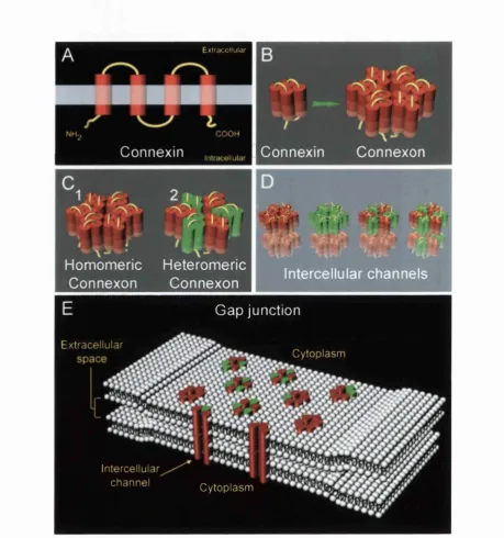
2.1.1 The structure o f gap junctions
Gap junction channels are multimeric structures comprised o f protein subunits known as
connexins. Each connexin is an integral membrane protein consisting o f four transmembrane
domains, two extracellular loops, a cytoplasmic loop, and cytoplasmic amino and carboxyl
termini (Oviedo-Orta and Evans, 2002). During transit to the plasma membrane individual
connexins associate to form hexameric hemichannels known as connexons. The six subunits
of each connexon are arranged in a radial manner such that they form an aqueous pore at the
that reside within the plasma membrane o f apposing cells. Clusters o f tens to thousands of
intercellular channels, often arranged in tight, regular arrays, form the gap junctions,
ultrastructures that directly link the cytoplasm o f adjacent cells (Figure 1, page 13).
Connexins are encoded by a large gene family comprising at least twenty family members
(De Maio et al., 2002). Individual connexin proteins are classified according to their
molecular weight (e.g. the nomenclature for the 31 kDa connexin is Cx31) and groups of
connexins assigned to subfamilies according to their homology (a, p, y, and unclassified).
Hemichannels can be composed o f connexins o f either the same or different types enabling
the formation o f either homomeric or heteromeric connexons respectively. Furthermore,
selective interactions between homomeric and heteromeric connexons on the surface of
apposing cells enable the creation o f both homotypic and heterotypic intercellular channels.
The extracellular loops o f the connexin subunits govern the interactions o f apposing
connexon hemichannels while their intracellular loops and carboxyl termini, which
represents the regions of greatest sequence divergence between connexin family members,
provide sites for channel regulation which include phosphorylation, and pH and voltage
sensing. The molecular permeabilities, conductance properties, and modes or regulation of
individual gap junction channels can vary and are determined by the properties of the
individual connexins from which they are derived (Beyer et al., 2001). Given the capacity for
different connexin types to associate with one another and given that many o f these proteins
are co-expressed in a wide range o f tissues, the potential exists for the generation of
Extracellular
Connexin
I
Connexin Connexonf
11
"i''T1 P'Ls&iS^
r m Homomeric Heteromeric
Connexon Connexon Intercellular channels
Gap junction
Extracellular
Cytoplasm
Intercellular
channel Cytoplasm
Figure 1. The structure o f gap junctions
Each connexin is a four trans-membrane spanning protein with intracellular N- and C-termini (A). Six connexins combine within the endoplasmic reticulum to form a hexameric connexon hemi-channel (B). Connexins o f the same type interact to form homomeric connexons (Cl) while the interaction o f different fam ily members produce heteromeric connexons (C2). Connexons from neighbouring cells couple to produce intercellular membrane channels directly linking the cytoplasm o f adjacent cells (D). The interaction o f homomeric and heteromeric connexons enables the formation o f both homotypic and heterotypic channels (D). Selective interactions between different connexons allow the formation o f a large variety o f channels with varying conductance properties. Large clusters
The exchange o f ions and small molecules between adjacent cells through gap junction
channels occurs due to passive diffusion. Owing to the relatively small pore size of these
channels passage is restricted to molecules o f less than approximately 1.0 - 1.2 kDa in size.
Consequently gap junctions are primarily involved in the transfer o f inorganic ions and low
molecular weight metabolites such as calcium ions, potassium ions, glucose, and cyclic
nucleotides (De Maio et al., 2002). However, pore size is not the sole determinant o f channel
permeability. Indeed many gap junction channels display highly selective conductances,
permitting the passage o f only those ions or molecules o f a particular charge. For instance
Cx32 is selectively permeable to anionic molecules while Cx26 permits the passage o f
molecules independent of their charge (Elfgang et al., 1995). Furthermore the specific
conductance properties conferred by a single connexin type can be modified when
incorporated into channels with other connexin species. Therefore, a heterotypic channel can
acquire electrophysiological properties that are markedly different from those o f homotypic
channels formed from single connexin types. Indeed, some connexins (e.g. Cx29) are unable
to form functional homotypic channels and appear to have evolved specifically to modify the
electrophysiological properties o f other connexin types through heterotypic associations
(Altevogt et al., 2002). The ensemble o f different connexins expressed within a particular
organ or tissue is therefore likely to reflect the specific function bestowed upon the gap
2.1.2 Connexins in physiology and pathology
Connexin proteins are associated with many hereditary and acquired human disorders that
range from mild hearing loss and cataractogenesis (Donaldson et al., 2001; Kelsell et al.,
2001) to cardiovascular disease and cancer (Carystinos et al., 2001; Severs et al., 2001).
Furthermore, different germline mutations in the same connexin gene can result in
profoundly different clinical phenotypes (Kelsell et al., 2001). Table 1, page 16 provides a
summary, but by no means an exhaustive list, o f many o f the connexins identified to date,
their respective tissue distributions, ascribed functions, and known disorders associated with
their dysfunction. Some o f these examples are also discussed further below. It is interesting
to note that while some connexins are expressed extensively in many tissues others are more
restricted in their distribution; for instance Cx43 is the most abundantly expressed connexin
and is found in most tissues including the heart, smooth muscle, brain, bone, and kidney,
while Cx50 appears to be expressed exclusively within the lens o f the eye.
Gap junctions are crucial for the coordination o f many biomechanical processes. Within the
walls o f the cardiac chambers gap junctions permit the interconnection o f vast networks of
myocytes, enabling the synchronised transmission o f calcium waves required for coordinated
heart contraction (Severs et al., 2001). They are also found in abundance in smooth muscle
tissue where they have been implicated in the control and maintenance o f vascular tone (Hill
et al., 2001) and the peristaltic movements of the gastrointestinal tract (Suzuki, 2000). Also,
an increase in the formation o f gap junctions between cells o f the myometrium is observed
during mid- to late-term pregnancy and is believed to occur in preparation for the
T he c o n n e x in s
Connexin Gene Subfamily Tissue distribution Proposed functions Disease association
Cx43 GJA1 a
A widely expressed connexin found in most gap junction containing tissues including heart [1], sm ooth muscle [11, CNS*[2], bone [3], ovary [4], adrenal gland [5], term pregnant myometrium (6j.
Control of heart contraction [11, vascular tone [11, myométrial activation [6).
Cardiovascular disease [1): Ventricle arhythmia, coronary arteriosclerosis, possibly hypertension.
Cx46 GJA3 a lens of eye [7]. Regulation of lens microcirculation [7). C ataractogenesis [8).
Cx37 GJA4 a
CNS [2], vascular endothelium [9], ovary [10).
Control of vascular tone [11, developm ent of ovarian follicles [101.
Arteriosclerosis (9), possibly female infertility [10).
Cx40 GJA5 a
CNS [21. heart [1), vascular endothelium [1), vascular sm ooth muscle [1), kidney [5).
Control of heart
contraction [1) and vascular tone[1).
Cardiovascular disease [1): atrial fibrillation, possibly hypertension.
Cx45 GJA7 a
CNS [21, heart [1L restricted expresion in vascular sm ooth muscle [1).
Control of heart contraction [11.
Of unknown significance.
CxSO GJA8 a lens of eye [7). microcirculation [7).Regulation of lens Cataractogenesis [11).
Cx32 GJB1 P
CNS[21,PNS**[121, Liver [131. Control of liver metabolism [131, peripheral nerve function [121.
X-linked Charcot Marie Tooth disease [12).
Cx26 GJB2 P
CNS [21, Epidermis [141, Inner ear [12).
Epidrmal growth and differentiation [14), auditory transduction [12).
Nonsyndromic and syndromic hearing loss [12), Vohwinkel syndrome [14).
Cx31 GJB3 P
PNS[15], Epidermis [141, Inner ear [12).
Epidrmal growth and differentiation [14), auditory and peripheral nerve transduction [12,15).
Erythrokeratoderma variablis [141, Nonsyndromic and syndromic hearing loss [12), peripheral neuropathy [15).
CX30.3 GJB4 P
Epidermis [141. Epidrmal grow th and differentiation [14).
Erythrokeratoderma variablis [14).
Cx30 GJB6 P
Epidermis [16), Inner ear [12). Epidrmal growth and differentiation [14), auditory transduction [12).
Hydrotic Ectodermal Dysplasia [161, Nonsyndromic and syndromic hearing loss [12).
Cx36 CX36 U/C***
CNS [2L retunal tissue: rod photoreceptors, cone pipolar cells, All amicrlne celts [17,18).
Vision [191, Neuronal developm ent [17].
Retinitis pigm entosa (proposed indirect pathological effect) [19).
*CNS, Central nervous system (1](Severs et al., 2001),[2](Rouach et al., 2002),[3](Rodriguez.,E),[4](S)mon et al., 1998),[5](Serre-Beinieret al., 2002), •*PNS, Peripheral nervous system [6](Carbillon et al., 2001),[7](Donaldson et al., 2001),(8](Pal et al., 2000),[9](Boerma et al., 1999),[101(Simon et al., — U/C, Unclassified 1997),[11](Pal et al., 1999),[121(Rabionet et al., 2002),[13](Nelles et al., 1996),[14)(Kelsell et al., 2001),[15](Lopez-Bigas
et al., 2001 ),(16](Common et al., 2002),[17](Sohl et al., 1998),[18]{Deans et al., 2002),|19)(Ripps., 2002)
Table 1. The physiology and pathology o f connexins
is expressed extensively in cardiac and smooth muscle and is a major component o f gap
junctions in these tissues. It is also becoming increasingly evident that Cx43 is o f significant
importance in the progressive development of cardiovascular disease: abnormalities in Cx43
expression and inappropriate patterns in Cx43-dependent gap junctionial coupling in
myocardial tissue have been associated with lethal ventricular arrhythmias, while conversely
the early stages o f coronary arteriosclerosis have been linked with an increase in Cx43
expression within the smooth muscle o f vascular tissue (Severs et al., 2001).
Gap junction communication has also been implicated in neuronal signalling and dysfimction
(Rouach et al., 2002). Within the central nervous system gap junctions provide an alternative
to chemical synaptic transmission, enabling the direct electronic coupling o f adjacent
neurones. These electrical synapses are thought to synchronise neuronal activity.
Neurological disorders such as epileptic seizure, which are characterised by excessive and
highly synchronised neuronal firing, are in part believed to be the consequence o f
abnormalities in gap junction signalling (Perez Velazquez and Carlen, 2000). Gap junctions
also appear to be o f importance in the peripheral nervous system where they interconnect the
many layers of the myelinating Schwann cells (Baker, 2002) and mutations in connexins
expressed in these tissues have been associated with peripheral nerve dysfunction and
neuropathy (Rabionet et al., 2002). Within the mammalian eye the propagation o f rod signals
across the retina requires the coupling o f adjacent cells via gap junction channels (Deans et
al., 2002). In the congenital disorder retinitis pigmentosa, a disease characterised by severe
visual impairment due to mutations in the rhodopsin gene, the transfer o f apoptotic signal
(Ripps, 2002). Although not functionally impaired themselves, the gap junctions contribute
to the pathogenesis o f this disease by permitting the spread o f cytotoxic signals from one cell
type to another. Within the inner ear gap junction channels enable the recycling o f potassium
ions within the cochlea, a requirement for the normal function o f the mammalian hearing
system. Cx26 is expressed in all inner ear cells presenting gap junctions, and as much as
30 - 50 % o f all hereditary hearing loss is a result o f congenital mutations in this protein
(Avraham, 2001).
Gap junctions are also necessary for coordinating the metabolic activities o f a number of
organ tissues. For instance glucose metabolism within the liver is synchronised via the
extensive coupling o f hepatocytes via gap junction channels (Nelles et al., 1996). Similarly,
the coordinated production and release o f secretary factors from endocrine and exocrine
glands such as the pituitary, adrenal gland, and pancreas is believed to be mediated by gap
junction communication (Serre-Beinier et al., 2002). The specialised anatomical design o f the
mammalian lens relies heavily on the properties of gap junctions; intercellular channels
permit the formation o f a highly specialised internal microcirculation between the fibre cells
of the lens, enabling tissue transparency by removing the need for a true vasculature
(Donaldson et al., 2001). Disruption of this microcirculation results in tissue necrosis and the
development o f cataracts (Pal et al., 1999; Pal et al., 2000). Intercellular communication
mediated by gap junctions is also critical for the normal development and differentiation of
the epidermis, and mutations in at least four different connexins expressed in epidermal
related tissues have been associated with a range of hyperproliferative skin disorders (Kelsell
The importance o f gap junctions in cancer biology has been known for many years. A loss of
gap junctionial communication is invariably associated with carcinogenisis and strongly
correlates with the metastatic potential o f a number o f different cell lines (Carystinos et al.,
2001; Nicolson et al., 1988; Saunders et al., 2001). Furthermore, re-induction o f gap
junctionial communication has been shown to inhibit the growth o f transformed cells
(Goldberg et al., 2000). A variety o f known chemical tumour promoters (e.g. phorbol esters)
have been shown to disrupt gap junction signalling while, conversely, various anticancer
agents (e.g. retinoids) seem to promote gap junction-mediated cell communication (Trosko
and Chang, 2001).
2 ,1 3 Cx31 mutation and disease
The majority o f hereditary disorders linked to connexin abnormalities have been associated
with mutations in P-connexin genes (GJB). The p-connexins, which include Cx32, Cx26,
Cx31, Cx30.3, and Cx30, are highly expressed in tissue o f ectodermal origin including the
epidermis, neuronal tissue, and the epithelium of the inner ear. Germline mutations in
p-connexins underlie a range o f clinically diverse human disorders including deafiiess, skin
disease, and peripheral neuropathy (Kelsell et al., 2001; Rabionet et al., 2002). These
disorders can occur either in isolation or in combination with one another and present with
varying degrees o f severity depending upon the specific connexin and mutation in question.
Mutations in Cx26 and Cx30, which are co-expressed in various tissues of the inner ear and
the epidermis are associated with both dominant and recessive hearing loss and a range of
syndrome (Cx26, MIM 124500) and Hidrotic Ectodermal Dysplasia (Cx30, MIM 129500).
Mutations in the epidermally expressed Cx30.3 result in the skin disease erythrokeratoderma
variables (EKV) (MIM 133200), while mutations in Cx32 which is expressed to a high
degree in peripheral nervous tissue result in the X-linked form o f the neurodegenerative
disorder Charcot-Marie-Tooth disease (CMTX)(MIM 302800) (reviewed, Kelsell et al.,
2001).
An interesting phenomenon associated with many p-connexin-associated disorders is the
widely differing clinical phenotypes that can arise from various mutations within the same
connexin protein. On o f the most striking example o f this is provided by Cx31. Cx31 is
expressed in the epidermis (Di et al., 2001), the myelinating cells o f peripheral nerves, and
the spiral ligament and limbus o f the inner ear (Lopez-Bigas et al., 2001a), and mutation in
this protein can result in disorders associated with all these tissues. As with many p-
connexins Cx31 has been implicated in the normal growth and development o f the epidermis
(Di et al., 2001; Kelsell et al., 2001). Various dominant germline mutations in this protein
have been associated with EKV, a skin disorder characterised by localised hyperkeratrosis
and generalised erythema (Rook et al., 1998). The disease typically presents as a diffuse
thickening of the palmoplantar epidermis due to the growth o f hyperkeratotic plaques, but
patients can also suffer from independent and transient erythematous patches that affect the
whole body and which only persist for short periods. Both hyperkeratotic plaques and
erythematous patches can be triggered by a number of environmental factors ranging from
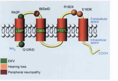
trauma to the skin, and exposure to ultraviolet irradiation to emotional stress. To date six
(Gottfried et al., 2002; Kelsell et al., 2001). Interestingly, these mutations do not seem to be
confined to specific fimctional domains but instead are distributed throughout the molecule.
For instance single amino acid changes in the amino terminus (G12D/R), the first
extracellular loop (R42P), or the second transmembrane domain (C86S) o f Cx31 have all
been associated with EKV.
That mutations located in many o f the different fimctional domains o f Cx31 cause EKV
would suggest that any impairment o f protein function per se should result in this skin disorder. However, two dominant mutations identified in the second extracellular loop of
Cx31 (R180X and E183K) have both been associated with non-syndromic (i.e. of single
clinical phenotype) hearing loss with no additional abnormalities associated with the
epidermis (Xia et al., 1998). Cx31 is expressed both within the inner ear and the auditory
nerve and therefore it is not surprising that disruptions in Cx31-related gap junction
signalling can result in hearing dysfunction. However, what is surprising is that different
mutations within the same molecule can result in independent and unrelated clinical
phenotypes. A further complication to the pathology of Cx31 has been provided by the recent
identification o f an additional and rare dominant mutation within the first extracellular loop
of Cx31 (66DelD), which has been associated with syndromic hearing loss (i.e. o f multiple
clinical phenotype) (Lopez-Bigas et al., 2001a). This mutation is also associated with
reduced conduction velocities in motor and sensory nerves, resulting in diminished muscle
reflexes and severe peripheral neuropathy. Cx31 is co-expressed with Cx32 in the
myelinating Schwann cells o f peripheral nerves. Mutations in Cx32 result in the
66DelD R42P
G12R/D
EKV
Hearing loss
Peripheral neuropathy
R180X
E183K
Extracellular sp a ce
Intracellular sp a ce
\ / c O O H
Figure 2. The germline mutations o f Cx31 and their associated disorders
identification o f the 66DelD mutation in Cx31 confirms the importance of both these
connexins in peripheral nerve function.
Cx31 mutation can therefore result in multiple and varying clinical phenotypes. The factors
that determine the differential effects that particular mutations confer on various tissues are
likely to be complex and many possible explmations exist. For instance the co-expression of
different connexin types within a particular tissue may allow for a degree of redundancy,
enabling one connexin type to adopt the role o f another. In tissues where redundancy is not
permitted due to the relatively restricted expression o f other connexin types, the severity o f a
particular mutation may be greater. Howevei, it has been reported that at least ten connexin
types are expressed in the human epidermis (Di et al., 2001), while only four have been
detected in the inner ear (Rabionet et al., 2002), and yet certain Cx31 mutations can have
deleterious effects on the former tissue and not the latter. Redundancy alone is therefore
unlikely to provide a sufficient explanation for the variation in clinical phenotypes observed.
The severity of a particular mutation may also be governed by the capacity for Cx31 to form
heterotypic associations with other connexins within a particularly tissue. For instance a
specific Cx31 mutation may confer a more detrimental effect on homotypic channel fimction
than on heterotypic channel fimction as the presence o f other connexin types within the same
channel may in part compensate for the deleterious effects o f the mutation. Therefore in
tissues where the protein is largely incorporated into heterotypic channels the effects of the
mutation may be more subdued than in those tissues where homotypic channels are the
predominant type. It is interesting to note that no apparent correlation exists between the
specific skin disease and deafness mutations are spread across the length o f the molecule and
appear within various functional domains (Figure 2, page 19). It is therefore o f particular
interest to determine whether Cx31 mutations associated with specific disorders elicit
differing effects at the cellular level.
2,L4 Study aims
Although much is known about the genotype-phenotype differences between distinct Cx31
mutations at the clinical level, little is known about these differences at the cellular level.
This study represents an attempt to bridge this gap. A number o f functional assays combining
microinjection, wide-field multi-channel digital time-lapse microscopy, and image
processing were used to investigate in vitro the effects o f specific disease associated Cx31 mutations on both intercellular channel function and cell morphology in cultured
keratinocyte and fibroblast cell lines. The EKV-associated mutations, R42P & C86S, the
deafness and peripheral neuropathy mutation 66DelD, and a Cx31 polymorphic variant,
R32W, o f unknown disease significance provided the main focus o f this study. Here new
insights are provided into how distinct Cx31 mutations elicit different effects on epidermal
2.2 METHODS
2,2,1 Cell Culture
NEH 3T3 mouse fibroblasts were grown in Dulbecco’s Modified Essential Medium (DMEM)
supplemented with 10% foetal calf serum (FCS) (Sigma), 1.6% L-glutamine, 5 mM
glucose, and 100 U/pg/ml penicillin-streptomycin, and phenol red indicator. Adherent cell
cultures were passaged every 2 to 3 days to maintain continuous cell growth. Cells were
maintained in culture up until their 10^’’ passage and individual cultures never permitted to
grow to confluency. Cultures were kept in a humidified incubator (Heraeus Instruments) set
to 37°C with 10 % ambient CO2. The periodic passaging o f cells involved a brief wash with
phosphate-buffered saline (PBS) followed by treatment with a 0.02 % porcine trypsin
(Beckton Dickinson)/5 mM EDTA solution to induce cell detachment. Culture medium was
subsequently added to the cell suspension to inhibit further trypsin/EDTA activity and the
cells then used to re-seed new cell cultures o f varying densities. Sterile tissue culture plastics
were used throughout (Falcon).
NEB-1 cells, originally derived from HPV16 immortalized human kératinocytes (Morley et
al., 1995), were kindly grown and maintained by Wei-Li Di, Centre for Cutaneous Research,
Queen Mary Hospital, London. Cells were cultured in 3:1 DMEM/F12 medium
supplemented with 10 % FCS, 0.4 pg/ml hydrocortisone, 5 pg/ml insulin, 10 ng/ml
epidermal growth factor (EOF), 10 x 10*'® M choleratoxin, 5 pg/ml transferrin, 2 x 10*'* M
2,2,2 Microinjection
Nuclear microinjection provided the main tool for introducing expression constructs into
cells while cytoplasmic microinjection was used to introduce fluorescent dye into the cells
during dye transfer experiments. Microinjection needles were generated from glass
capillaries (GC120TF-10, Fisher Scientific) using an electronic microfilament puller (P-97,
Sutter Instrument Co.). Solutions prepared for microinjection containing either cDNA
constructs, fluorescent antibodies, or fluorescent dyes were spun at 14000 rpm for 20 min in
a desktop microcentrifuge (Eppendorf) pre-cooled to 4 °C in order to sediment particles that
could otherwise have blocked the microinjection needle. Samples were kept on ice
throughout the microinjection process and microinjection needles were loaded with solution
taken from only the very top o f the spun sample.
Nuclear Microinjection: All nuclear microinjection was carried out using a transjector and micromanipulator (Eppendorf) mounted on a Zeiss Axiovert 135 TV inverted microscope
(Carl Zeiss) equipped with a tungsten lamp for bright field illumination, a long-working
distance condenser, and long-working distance objectives. The microscope body rested on an
optical table isolator (Melles Griot) to prevent ambient vibrations from disturbing the
microinjection needle. A heated stage enabled cell cultures to be maintained at 37 ®C during
the microinjection process. A conventional CCD camera (Sony) was fitted to the front
camera port o f the microscope to enable microinjection to be visualized on an analogue
monitor. All microinjection was carried out using a x20, NA 0.3, long-working distance,
Cytoplasmic microinjection: During dye transfer experiments cytoplasmic microinjection was used to introduce Alexa 568 dye into cells expressing enhanced green fluorescent protein
(EGFP) fusions o f different Cx31 variants. A modified microscope configuration was
therefore required that permitted fluorescence imaging. The microscope system used for
cytoplasmic microinjection was the same as that used for time-lapse microscopy.
2,2,3 Time-lapse microscopy
Digital time-lapse microscopy was used to record cell behaviour during dye transfer and cell
death experiments. All time-lapse experiments were conducted on the same Zeiss Axiovert
TV 135 (Carl Zeiss) inverted microscope. The microscope was optimised for multi-channel
digital time-lapse microscopy and to accommodate the long-term imaging o f mammalian
cells in culture. The addition o f a transjector and micromanipulator unit (Eppendorf) also
enabled cells to be microinjected and subsequently filmed on the same system. This was
particularly crucial for dye transfer experiments where the time between microinjection and
imaging had to be kept to a minimum.
The microscope was fitted with tungsten and mercury lamps to enable both bright field and
fluorescence imaging, and was equipped with separate bright field and fluorescence shutters
(Uniblitz), a shutter controller (Sutter Instruments), and a highly sensitive CCD camera (Orca
ER, Hamamatsu). All peripheral devices were under the control o f image acquisition
software (Acquisition Manager, Kinetic Imaging). The CCD camera was mounted directly
under the sample on the lower camera port o f the microscope in order to minimise the
maximising the fluorescence signal. Blocks containing excitation and emission filters and
compatible dichromatic mirrors enabled a range o f fluorophore species to be imaged. In
addition, motorised excitation and emission filter wheels containing a selection o f different
fluorescence filters enabled the rapid sequential imaging o f multiple fluoropbores during a
single time-lapse experiment when used in conjunction with appropriate multiple dichromatic
mirrors. Filters and dichromatic mirrors were purchased from Omega Optical, Inc. and
permitted the imaging o f EGFP (FX100-2 Alpha Vivid: Ex 475 AF40, DRLP 505, Em 535
AF45), Alexa 568/Cy3/Cy3.5 (FX108-2 Alpha Vivid: Ex 525 AF45, DRLP 560, Em 695
AF55), and Cy5 (FXllO-2 Alpha Vivid: Ex 630 AF50, DRLP 650, Em 535 AF45)
fluorescence.
The microscope was housed in a purpose built Perspex environment chamber accurately
maintained at 37 °C using an in-house built controlled heater. The main body o f the
microscope rested on an optical table isolator (Melles Griot) in order to minimize the effects
o f ambient vibrations on both microinjection and image acquisition. A choice of short-
working and long-working distance condensers enabled experiments to be conducted either
with or without the additional use o f microinjection. Cytoplasmic microinjection was
conducted using a x20, NA 0.5, short working distance, phase contrast objective. A
conventional CCD camera (Sony) was mounted on the front camera port o f the microscope
The time-lapse interval, total duration of the recording, and the specific filter configurations
depended upon the experimental assay in question and are therefore described in their
relevant sections.
2.2.4 EGFP fusion constructs
Expression constructs encoding EGFP fusions o f wild type Cx31 and the mutant variants
R42P, C86S, R32W, and 66DelD were constructed and kindly provided by Wei-Li Di. All
Cx31 variants were encoded in the pEGFP-Nl expression vector (ClonTech) in-frame and
upstream o f EGFP. The native Cx31 stop codon was altered (TGA to GGA) to enable read-
through and consequently transcription o f the downstream fluorophore. Cx31 variants were
therefore expressed as N-terminal fusions to the EGFP. The wild-type and R42P mutant form
o f Cx31 were originally generated via PCR from human genomic DNA, while the R32W,
C86S, and 66DelD mutations were generated independently via site directed mutagenesis
using the wild-type Cx31-EGFP template (Di et al., 2002).
2.2.5 The conjugation o f fluorescent dyes to antibodies
Fluorescent dyes were conjugated to antibodies for use as microinjection markers during
various experiments. Conjugation reactions were performed as follows: lyophilised, reagent-
grade sheep IgG (Sigma) was reconstituted to a concentration of 2 mg/ml in 100 mM
Na2C0 3, pH 9.3. 1 ml o f IgG solution was then applied to a single pre-packed vial o f either
Cy3.5 or Cy5 fluorescent dye (Amersham Biosciences, cat: PA23500 & PA25000). The
reaction was then incubated at room temperature for 30 min with one brief additional mixing
step half way through. Conjugation reactions were always protected from light. Dye-
conjugated antibodies were then separated from free dye using the QuickSep dialysis system
(Tripple Red). Briefly, the 1 ml reaction volume was placed into a dialysis cassette
containing a 16,000 MW cut-off membrane and subsequently immersed in 400 ml of
phosphate buffered saline, pH 7.5 (PBS). Dialysis was preformed over 48 hours at 4 ®C with
periodic buffer changes. A magnetic stirrer ensured thorough mixing throughout. Dialysed
Cy3.5-IgG and CyS-IgG stocks were stored at -20 °C until required.
2,2,6 In vitro dye transfer assay fo r the study o f intercellular communication
Preparation o f experimental cultures: Freshly trypsinised NEBl kératinocytes and NIH 3T3 fibroblasts were seeded in 35 mm diameter glass-bottomed micro well dishes (MatTek)
containing 3 ml o f the appropriate tissue culture medium for the cell type in question. Cells
were allowed three days to grow to confluency before experimentation.
For all microinjection and imaging cells were transferred to fresh phenol red-free tissue
culture medium supplemented with 20 mM HEPES, pH 7.5 (experimental culture medium).
The presence o f phenol red in culture medium was not desirable during fluorescence imaging
due to the autofluorescent properties of this compound. HEPES was added to culture medium
to compensate for the low ambient levels o f CO2 during both microinjection and imaging,
therefore removing the need for more complicated CO2 delivery systems.
plasmid DNA encoding one o f the EGFP-Cx31 variants. Expression constructs were all
microinjected at a concentration o f 0.05 pg/pl. Following microinjection cells were
transferred to fresh tissue culture medium and returned to the incubator. Cells were allowed
4-5 hours to express the microinjected constructs.
Cytoplasmic microinjection and time-lapse microscopy: The cytoplasmic microinjection of fluorescent dye into cells was conducted on the same Zeiss Axiovert 135 TV inverted
microscope (Carl Zeiss) that was subsequently used for the imaging o f dye-transfer through
cells. For a detailed description o f the microscope specifications see section 2.2.3, page 19.
The culture dish was positioned on the stage of the microscope in a glass-topped, steel
humidity chamber (constructed in-house) that was designed to prevent evaporation during the
course o f time-lapse experiments. The lid of the chamber was removed during
microinjection. For all dye transfer experiments patches o f fluorescent cells were identified
under the microscope and single cells close to the patch perimeter were microinjected in the
cytoplasm with 2 mM Alexa 568 dye (Molecular Probes). Upon the successful cytoplasmic
microinjection of a single fluorescent cell the microinjection needle was quickly removed
from the microscope and the lid o f the steel humidity chamber replaced, completely
enclosing the culture dish. The occurrence of dye transfer between adjacent cells was then
determined using multi-channel digital time-lapse microscopy. The acquisition software was
used to acquire images every 20 seconds in three separate channels (phase contrast, EGFP
fluorescence, and Alexa 568 fluorescence) using the 20 x NA 0.5 objective, the CCD camera
and the multiple dichromatic mirror in conjunction with motorised excitation and emission
rounds). Although the exposure times used for imaging EGFP fluorescence tended to vary
between dye transfer experiments, the exposure time used for imaging Alexa 568
fluorescence was kept constant as fluorescence intensity o f the dye did not vary considerably.
The time elapsed from the injection o f dye to commencement o f image acquisition was
approximately a minute. The procedure for studying intercellular dye transfer is summarised
in Figure 3, page 19.
2,2,7 Measurement o f dye transfer rate
The time-lapse acquisition of Alexa 568 fluorescence images over the course o f dye transfer
experiments provided accurate information concerning the temporal dynamics of dye spread
through cell populations expressing different Cx31 variants. Image processing software was
specially developed in order to numerically quantify this process, enabling the statistical
comparison o f the effects of different Cx31 mutations on the rate o f dye transfer. By
comparing the various rates at which dye transferred through cell populations expressing the
wild type Cx31 protein with those found for cell populations expressing particular Cx31
variants, significances could be derived that reflected the relative degree to which different
Cx31 mutations impeded or enhanced intercellular communication.
Preparation o f film sequences fo r image analysis: Prior to image analysis film sequences were cropped (IMD analysis. Kinetic Imaging) to incorporate a region that contained the
EGFP expressing cells and dispersing dye. Interactive tracking (Motion Analysis, Kinetic
Imaging) was then used to mark, using the mouse pointer, the position o f the cell originally
Figure 3. Summary o f the in vitro dye transfer assay for the assessment connexin function
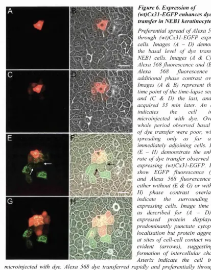
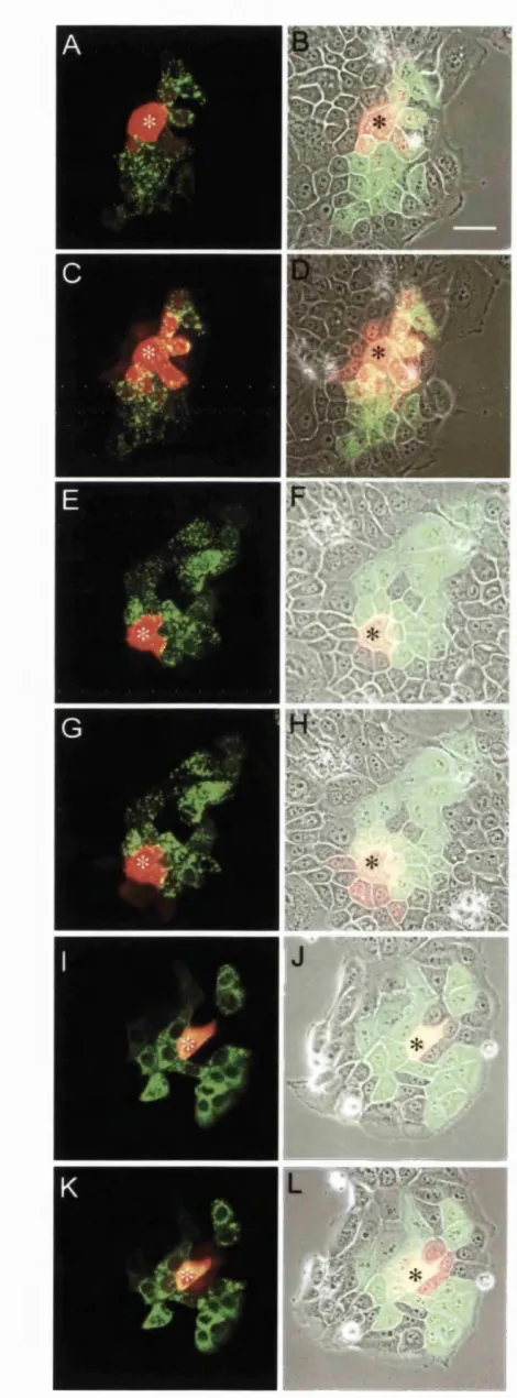
(A) Groups o f adjacent cells within a monolayer culture were microinjected in the nucleus with 0.05 jug/pi plasmid DNA encoding one o f the CxSl-EGFP fusion constructs under investigation. (B) Cultures were left fo r 4 — 5 hours to allow the expression o f microinjected constructs. This resulted in the formation o f tight patches o f fluorescent cells. (C) A single expressing cell close to the patch perimeter was then selected and microinjected in the cytoplasm with 2 mM Alexa 568 dye. Multi-channel time-lapse microscopy was then used to monitor the spread o f Alexa dye through the surrounding cell population: Images were acquired at 20 s time intervals in three separate channels (phase contrast, EGFP, and Cy3.5 fluorescence). Each fd m sequences consisted o f 100 acquisition rounds (33 min duration).
Figure 4. Summary o f the in vitro assay fo r the assessment o f NIH 3T3 cell death
and was defined as the centroid of the microinjected cell within the first frame o f the film
sequence. Cell movement within the monolayer was minimal over the course o f time-lapse
experiments and so it was considered unnecessary to score the cell position for each fi'ame
within the film sequence.
Image analysis: A single numerical value representing the normalised mean distance o f dye transfer {NMD) was generated for each Alexa 568 fluorescence image within a film sequence and collectively these values then used to calculate the mean rate o f dye transfer for a
particular experiment. Alexa 568 fluorescence images were extracted from film sequences,
binned to a pixel size of 2.6 x 2.6 pm ( 4 x 4 pixel binning) in order to remove high fi*equency
noise, and thresholded in order to remove background. Single NMD values were then calculated for each image as:
h ^ x y t
where / is the intensity at pixel (x, y) at time t and d is the distance o f that pixel fi'om the site of dye microinjection (defined by the pixel co-ordinate o f the centroid o f the cell initially
microinjected with Alexa 568 dye). In multiplying pixel intensity by distance {Ixyt x dxyt),
increasing bias was given to those intensity values that lay furthest fi’om the site o f initial dye
microinjection. By increasing the significance o f distant pixel values, greater discrimination
could be made between those treatments that enhanced the rate o f dye transfer and those that
impeded this process. All pixel intensity-distance values were summed, !>{Ixyt x dxyl), to produce a single value that represents the degree o f dye displacement for that image time
point. This value was then divided by the total image intensity in order to mean and
all Alexa 568 images within the first 10 min o f a film sequence were then used to derive a
single value that represented the mean rate o f dye transfer for that particular experiment.
Linear regression analysis (least squares fit) was applied to the NMD data and the slope of the resulting regression function taken to represent the mean rate o f dye transfer.
Significances in the differences in the mean rate o f dye transfer between groups o f sequences
were evaluated by the two-sided t-test. All image-analysis software was written in the
Mathematica programming language (Wolfram Research). The software code for the
derivation o f NMD values can be found in Appendices 5.1, page 19.
2.2.8 Evaluation o f cell death in N EBl kératinocytes
Nuclear microinjection: Confluent cell cultures o f NEB-1 kératinocytes were prepared as described in section 2.2.6, page 19. Cells were transferred to experimental culture medium
and confluent patches o f cells within the central microwell o f each dishes then selected for
microinjection. Cells were microinjected in the nucleus with 0.05 pg/ml o f plasmid DNA
encoding either (wt)Cx31-EGFP, (R42P)Cx31-EGFP, or (C86S)Cx31 -EGFP. Following
microinjection cultures were transferred to fresh tissue culture medium and returned to the
incubator where they were allowed 5 hours to express the microinjected constructs.
Assessment o f cell death: Cells were treated with 0.5 pg/ml o f the membrane-impermeable fluorescent dye propidium iodide (Sigma). After a 10 min incubation at 37 °C, expressing
•cells were located under the microscope and imaged in three separate channels (phase
contrast, EGFP fluorescence, and propidium iodide fluorescence). Propidium iodide
the same Zeiss Axiovert 135 TV inverted microscope described in section 2.2.3, page 19
using the 20 x objective, NA 0.5, the CCD camera and the multiple dichromatic mirror in
conjinction with motorised excitation and emission filter wheels. Once acquired propidium
iodide and EGFP fluorescence images were overlaid and interactive tracking subsequently
used to label cells as either dead or viable. Expressing cells were assigned to one o f three
possible categories: either (1) dead cells which exhibited bright propidium iodide nuclear
staining and nuclear shrinkage, (2) cells in the process o f dying which exhibited nuclear
shrinkage but which were not yet positive for propidium iodide staining, and (3) viable cells
that displayed a normal morphology and which were also negative for propidium iodide
staining. Only cells that clearly exhibited nuclear staining with propidium iodide were scored
as dead while only cells that exhibited an unaltered morphology as well as propidium iodide-
negative staining were scored as viable. Therefore, cells that fell into the second category
were regarded as indeterminate and ignored. Data were presented as percentages of the total
number o f cells scored for each treatment group, and tested for significant differences by
analysis o f variance (ANOVA).
2,2,9 Dynamic assessment o f cell death in NIH 3 T3 fibroblasts
Nuclear microinjection: Confluent cultures o f NIH 3T3 fibroblast were prepared as described in section 2.2.6, page 19. Cells were transferred to experimental culture medium and
confluent patches o f cells within the central microwell o f each dish selected for
microinjection. Patches of adjacent cells were microinjected in the nucleus with a mixture of
1 pg/p.1 Cy3.5-conjugated sheep IgG (Cy3.5-IgG) and 0.05 pg/pl o f plasmid DNA encoding
fresh tissue culture medium and returned to the incubator where they were allowed 20 min to
recover from the process o f microinjection.
Time-lapse microscopy: The occurrence o f cell death following the expression of microinjected constructs was determined using multi-channel digital time-lapse microscopy.
All experiments were conducted using the same Zeiss Axiovert 135 TV inverted microscope
used for dye transfer experiments (see section 2.2.3, page 19). Prior to time-lapse recording
cells were transferred to fresh experimental medium supplemented with 0.5 pg/ml propidium
iodide. The culture dish was placed on the stage of the microscope in a steel humidity
chamber to protect the sample from evaporation during the course o f the experiment. The co
microinjection of Cy3.5-IgG provided a fluorescent nuclear marker that enabled
microinjected cells to be re-located under the microscope prior to the expression o f EGFP
fusion constructs. A single patch o f microinjected cells was selected for observation using
time-lapse recording. Images were acquired every 60 seconds in four separate channels
(phase contrast, EGFP fluorescence, Cy3.5 fluorescence, and propidium iodide fluorescence)
using the 20 x objective NA 0.5, the CCD camera and the multiple dichromatic mirror in
conjunction with motorised excitation and emission filter wheels. Cell behaviour was
recorded for 100 min (100 acquisition rounds).
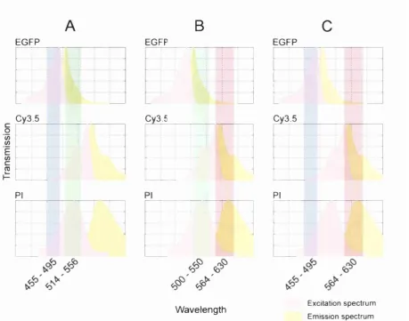
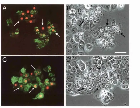
Three-channel fluorescence imaging: A specially developed technique enabled the sequential imaging o f EGFP, Cy3.5, and propidium iodide fluorescence despite the partial overlap that
A
B
EGFP EGFP 0 1 E (0 Cy3.5 Cy3.5 PI EGFPc
Cy3.5 PI •AW avelength Excitation spectrum
Em ission spectrum
Figure 5. Three-channel fluorescence imaging
The sequential acquisition o f EGFP, Cy3.5, and propidium iodide fluorescence was achieved by combining different excitation and emission filter combinations. The use o f motorised excitation and emission filter wheels and a multiple dichromatic mirror enabled the different filter configurations to be selected automatically during the filming process. (A) EGFP fluorescence was imaged using a conventional configuration enabling fluorophore excitation between 455 - 495 nm and detection o f fluorescence emission between 514 - 556 nm. This configuration did not allow significant excitation o f Cy3.5 but did enable a degree o f propidium iodide excitation. However, the emission filter only permitted the collection o f EGFP fluorescence emission. (B) A conventional TRITC filter configuration was used to image Cy3.5 fluorescence providing excitation between 500 — 550 nm and detection o f fluorescence emission between 564 - 630 nm. This enabled the effective excitation and fluorescence detection o f Cy3.5 and propidium iodide but not EGFP. (C) The selective imaging o f propidium iodide fluorescence was enabled by using the EGFP excitation filter (455 - 495 nm) in combination with the TRITC emission filter (564 - 630 nm). This enabled excitation o f both EGFP and propidium iodide but not Cy3.5, while enabling the detection o f propidium iodide and Cy3.5 fluorescence emission but not EGFP. Therefore, this fd ter
afforded by the use o f motorised excitation and emission filter wheels and is described in
detail in Figure 5, page 19. Briefly, while EGFP fluorescence and Cy3.5 fluorescence could
be monitored independently using separated EGFP and TRITC filter configurations,
propidium iodide fluorescence was detected using a miss-matched filter combination that is
similar in principle to that used in sensitised emission FRET studies. By exploiting the
relatively broad excitation spectrum o f propidium iodide EGFP excitation light could be used
to excite the fluorophore while its fluorescence emission could be detected using the
TRITC/Cy3.5 emission filter specification. This allowed effective discrimination between all
three fluorophores, while using filter sets essentially designed to discriminate between only
two. The acquisition o f EGFP fluorescence enabled the onset o f protein expression to be
monitored while the occurrence of cell death was determined by the subsequent uptake and
accumulation o f propidium within cell nuclei. Fluorescence images were pseudocoloured so
that EGFP fluorescence appeared green, Cy3.5 fluorescence appeared red, and propidium
iodide fluorescence appeared blue. Dead cells were therefore detected by a gradual change in
the colour o f nuclei from red (Cy3.5) to purple (Cy3.5 + propidium iodide). If expressed
proteins did not result in death then cell nuclei would remain red throughout the entire
recording period. The ambient concentration of propidium iodide within the surrounding
culture medium was sufficiently low that it was undetectable. The assay developed for the
study o f NEH cell death is summarised in Figure 4, page 19.
Assessment o f cell death: Acquisition Manager software was used to overlay the three separate fluorescence channels o f film sequences applying the pseudocolour combinations
described above. The final frame of the resulting merged film sequences was then extracted