Article
1
Synthesis and Structure-Activity Relationships of
2
Resorcinol Derivatives as Highly Potent Tyrosinase
3
Inhibitors
4
You Zhou 1, Qihang Li2, Ronggeng Fu 3, Hongyu Yang 2, Jun Mo 2, Yao Chen 4 and Qingyou Xia 5,*
5
1 College of Biotechnology, Southwest University, Chongqing 400715, China; [email protected]
6
2 School of Pharmacy, China Pharmaceutical University, Nanjing 210009, China; [email protected](L.Q.);
7
[email protected] (H.Y.); [email protected] (J.M.)
8
3 School of Pharmacy, Hunan University of Chinese Medicine, Changsha 410208, China;
9
10
4 School of Pharmacy, Nanjing University of Chinese Medicine, Nanjing 210023, China; [email protected]
11
5 State Key Laboratory of Silkworm Genome Biology, Southwest University, Chongqing 400715, China;
12
13
* Correspondence: [email protected]; Tel.: +86-139-9636-8108
14
15
Abstract: Compounds with tyrosinase inhibitory efficacy could be effective as depigmenting
16
agents. Although a large number of natural and synthetic tyrosinase inhibitors have been reported,
17
few of them are used as skin-whitening agents due to poor activity and safety concerns.
18
3-(2,4-Dihydroxyphenyl)propionic acid (DPPA), a naturally occurring compound isolated from
19
Ficus carica, was previously discovered as a moderate tyrosinase inhibitor. In this study, the
20
structure-activity relationship study of DPPA was conducted. Compound 3g, with the
21
2,4-resorcinol subunit and terminal hydrophobic di-butylamino group, was identified with low
22
nanomolar enzymatic IC50 value. Additionally, compound 3g could effectively reduce melanin
23
levels in B16-F10 melanoma cells treated with α-melanocyte-stimulating hormone (α-MSH)
24
without affecting cell viability and proliferation. All these results indicated that compound 3g
25
could be considered as a promising candidate for the treatment of diseases associated with
26
hyperpigmentation.
27
Keywords: hyperpigmentation; tyrosinase inhibitors; 3-(2,4-dihydroxyphenyl)propionic acid;
28
structure-activity relationship study; B16-F10 cellular melanogenesis inhibition
29
1. Introduction
30
Tyrosinase [EC 1.14.18.1] is a multifunctional copper-containing enzyme widely distributed in
31
nature and is a key enzyme in the biosynthesis of melanin pigments. It is well-known that
32
tyrosinase can catalyze the first two steps in the conversion of tyrosine to melanin: hydroxylation of
33
tyrosine to L-DOPA and oxidation of L-DOPA to dopaquinone [1]. Previous investigations have
34
shown that this enzyme was responsible not only for the browning of some fruits and vegetables,
35
decreasing the commercial value of the produce, but also for the formation of some dermatological
36
problems, such as melisma, postinflammatory melanoderma, age spots and freckles[2]. Moreover,
37
tyrosinase is important for the insect moulting process[3] and adhesion in marine organisms[4].
38
Furthermore, melanin pigments are found in the mammalian brain, and tyrosinase activity is linked
39
to neurodegeneration associated with Parkinson’s[5], Alzheimer’s, and Huntington’s diseases. Thus,
40
seeking potent tyrosinase inhibitors is a major concern in agricultural, pharmaceutical and cosmetic
41
fields. The well-known tyrosinase inhibitors include hydroquinone, arbutin and kojic acid.
42
[6]Unfortunately, most reported compounds are generally limited in practical application with
43
regard to their poor potency and high cytotoxicity[7]. Thus, it is necessary to search for and develop
44
novel tyrosinase inhibitors with excellent potency and a low toxicity to resolve issues related to the
45
tyrosinase activity.
46
So far, crystal structures from different species such as plant (e.g., Juglans regia), mushroom
47
(e.g., Agaricus bisporus), and bacteria (e.g., Bacillus megaterium) were reported. While preliminary
48
crystallographic studies on human tyrosinase have been reported very recently, no structures are
49
available to date at atomic resolution. In general, the structure of tyrosinase can be classified into
50
three domains, namely, the central, the N-terminal, and the C-terminal domains[8]. Among
51
tyrosinases from different species, the central domain is the most conserved domain, which
52
comprises two close copper centers (CuA and CuB) coordinated by six histidine residues[9]. To
53
obtain new potent tyrosinase inhibitors (> To study/investigate the mode of inhibition (or mode of
54
action) of tyrosinase inhibitors in an atomic level, we used mushroom tyrosinase in our study since
55
its X-ray crystal structure and in vitro tyrosinase assay were available). In addition, mouse B16-F10
56
melanoma cell line, a tyrosinase-expressing cell line, was employed to investigate the tyrosinase
57
activity across species in the presence of our compound by performing cytotoxicity and melanin
58
biosynthesis assays. As we know, mushroom tyrosinase and murine tyrosinase share 58% identity
59
within 6 Å of the two copper centers.
60
DPPA, is a naturally occurring tyrosinase inhibitor isolated from Ficus carica, with reported IC50
61
of 3.2 μM[10]. Previous efforts have proved that chemical modification of carboxyl group of DPPA
62
could augment its tyrosinase inhibitory activity[11]. Compared to other natural and synthetic
63
substances, it shows superiority in designing tyrosinase inhibitors for following reasons: (1) the
64
resorcinol scaffold can induce tyrosinase inactivation by the Quintox inactivation mechanism in
65
which a portion of the resorcinol substrate is subjected to monooxygenase action leading to
66
irreversible elimination of Cu0 from the active site; (2) the short synthetic route of DPPA makes it
67
suitable for further development. DPPA was, therefore, selected by our laboratory as an ideal
68
starting point for the development of novel and highly potent tyrosinase inhibitors. Here, we
69
reported the structure-activity relationship of DPPA focusing on the carboxyl group, 4-substituted
70
resorcinol moiety, and the ethylidene linker. Anti-tyrosinase efficacy of all targeted compounds
71
were tested and compound 3g was identified with low nanomolar enzymatic IC50 values.
72
Furthermore, compound 3g effectively reduced melanin levels in B16-F10 melanoma cell lines
73
treated with α-MSH without affecting cell viability and proliferation.
74
75
2. Results and Discussions
76
The synthesis of compounds 3a-3l was accomplished in 2 steps (Scheme 1). The reaction
77
between resorcinol (1) and acrylic acid generated compound 2, which was involved with
78
esterification followed by ring closure [12]. Compound 2 was then reacted with various amines in
79
DMF to give 3a-3l in high yield. The preparation of target compounds 5a-5f and 7 are described in
80
Scheme 2. Dibutylamine was reacted with various substituted phenylpropionic acids and
81
3-(4-chlorophenoxy)propionic acid in CH2Cl2 to give 5a-5f and 7 in good yield. All target
82
compounds were purified by column chromatography and their structures were characterized by
83
1H NMR, 13C NMR and HRMS.
84
86
Scheme 1. Reagents and conditions: (i) CH2=CHCO2H, Amberlyst 15, toluene, reflux; (ii) amine,
87
DMF, 100 °C.
88
89
Scheme 2. Reagents and conditions: (i) CDI, CH2Cl2, r.t, 12h.
90
Anti-tyrosinase efficacy of all synthesized compounds was tested on both enzyme activities
91
stages: L-tyrosine as substrate to L-DOPA and the oxidation of the later to o-quinone. Kojic acid, a
92
R1
R3
O
OH
R1
R3
O
N
4a-4f 5a-5f
i
R2 R2
a: R1= OCH3, R2= H, R3= OCH3
Cl
O
6 7
i
OH
O
Cl
O N
O
b: R1= OH, R2= H, R3= H
c: R1= H, R2= H, R3= OH
d: R1= H, R2= OH, R3= OH
e: R1= Cl, R2= H, R3= H
well-known tyrosinase inhibitor was selected as a positive control (Table 1). Compounds 3a-3h,
93
with the 2,4-resorcinol skeleton and different terminal amino groups indicated higher tyrosinase
94
inhibitory activity with the first stage than that of the second stage. Compound 3g, containing a
95
hydrophobic di-n-butylamino substituent, exhibited the best activity. As shown in Table 1,
96
compound 3g inhibited the first stage with low nanomolar concentration (IC50 = 12.2 nM) and
97
presented potent inhibition of the second stage (IC50 = 200.7 nM). Similarly, compounds (3a, 3f and
98
3h) with other terminal hydrophobic groups potently inhibited the first stage with IC50 values < 1
99
μM. These results, to some extent, were consistent with those found in previous study that addition
100
of a hydrophobic moiety that contains a minor bulky group to the 2,4-resorcinol skeleton was
101
beneficial to increase the tyrosinase inhibition potency [11]. Intriguingly, we found that the
102
existence of terminal hydrophilic group (3b, 3d and 3e) also played an important role in biological
103
potency, which may interact with the hydrophilic pocket around the binuclear copper ions leading
104
to improved enzyme-inhibitor binding affinity. It is noteworthy that the introduction of morpholino
105
group (3c) to DPPA apparently decrease its in vitro enzyme activity, suggesting that introduction of
106
the morpholino group has a detrimental effect on the inhibition potency.
107
In contrast to 3a-3h, compounds 3i-3l containing different terminal phenylamino groups
108
possessed higher tyrosinase inhibitory activity with the second stage than that of the first stage,
109
indicating a different mode of action involved (Table 1). Compound 3i, which contains a p-methyl
110
substituted aromatic benzene ring, inhibited the second stage with an low nanomolar concentration
111
(IC50 = 84.8 nM) and showed potent inhibition of the first stage (IC50 = 3501.0 nM). In comparison
112
with 3i, compounds containing unsubstituted benzene ring or other substituted benzene rings
113
demonstrated weaker enzyme inhibitory potency with the second stage.
114
When the 2, 4-resorcinol subunit was replaced with other substituted benzene rings, the
115
resulting compounds, 5a-5f, displayed very low inhibitory potency with both of the enzyme
116
activities stages (Table 1). It suggested that the 2,4-resorcinol subunit is the key pharmacophore
117
required for potency of the novel series of DPPA amide derivatives. In comparison with 3g, the
118
introduction of an oxygen atom in the ethylidene linker produced the analogue (compound 7)
119
without any inhibitory potency (Table 1).
120
121
122
123
124
125
126
127
128
129
130
131
Table 1. Inhibitory effects on mushroom tyrosinase activity of target compounds.
133
Compounds L-Tyrosine (IC50, nM) L-DOPA (IC50, nM)
3a
3b
3c
3d
3e
3f
3g
3h
3i
3j
3k
3l
5a
5b
5c
5d
5e
5f
7
DPPA
Kojic acid
239.5
436.0
34410.0
80.3
251.4
41.6
12.2
101.3
3501.0
2364.0
1142.0
6085.0
>50000
>50000
>50000
>50000
>50000
>50000
>50000
9031.0
9031.0
3260.0
3361.0
63460.0
1277.0
1117.0
1112.0
200.7
1603.0
84.8
303.4
154.8
307.3
>50000
>50000
>50000
>50000
>50000
>50000
>50000
8279.0
8279.0
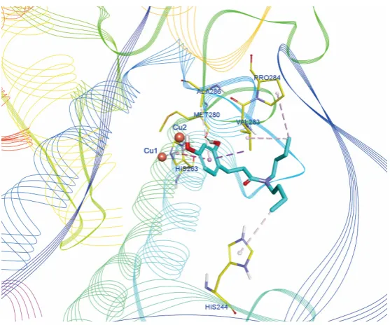
Figure 1. Binding mode prediction of compound 3g with mushroom tyrosinase (PDB ID: 2Y9X).
134
are depicted in yellow thin stick mode. The copper ions are represented by orange spheres. The
136
hydrophobic contact as well as the π-π stacking are depicted in purple dot line, while H-bonds are
137
in green dot line. Only polar hydrogen atoms are shown.
138
Next, the binding mode of the most active compound 3g to mushroom tyrosinase was
139
predicted by using molecular docking method (Figure. 1). The binding mode revealed that the C2
140
phenolic hydroxyl group of compound 3g formed a hydrogen bond with Met280, and the C4
141
phenolic hydroxyl group could chelate one of the binuclear copper ions. Furthermore, hydrophobic
142
interactions as well as π-π stacking were also found between the benzene ring of compound 3g and
143
the side chains of His263, Val283 and Ala286. Additionally, the di-n-butylamino group at the end of
144
3g also could form hydrophobic interactions with His244, Val283 and Pro284. These results clearly
145
emphasized the importance of the 2,4-resorcinol moiety and hydrophobic side chain in potent
146
tyrosinase inhibitors.
147
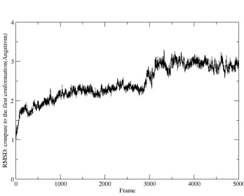
The stability and energy profile of the docking poses of compound 3g was investigated
148
through MD simulations and the calculations of binding free energies. In Figure 2, the RMSD values
149
of simulation converged after about 3000 frames (60 ns), indicating that the system is stable and
150
equilibrated. The root-mean-square deviations (RMSD) values of complex compared first
151
conformation of MD swing at 2~3 Å indicated that the complex of 3g and tyrosinase become stable
152
after the first short stage of variation and the docking pose is reliable.
153
154
Figure 2. The RMSD values of complex 3g-tyrosinase changes with the frames of MD comparing to
155
the first conformation of complex 3g-tyrosinase.
156
The MMPBSA method was used to calculated the binding free energies and gain information
157
on the different components of interactions energy that contribute to complex 3g-tyrosinase.
158
Detailed results are shown in Figure 3. Figure 3(a) indicates that electrostatic energy (EEL)and van
159
Non-polar solvation energy (ESURF) are also did favorable contribution to complex 3g-tyrosinase
161
binding (-3.3965 Kcal/mol). Meanwhile, Polar solvation energy(EGB) does not favor for 3g binding.
162
The binding free energy of compound 3g and tyrosinase is -18.8305 Kcal/mol indicates that the
163
binding of 3g and tyrosinase is favored in terms of energy.
164
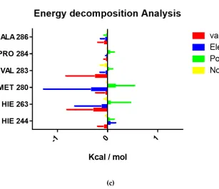
For identification of key residues contributing to binding affinity, we also carried out the
165
energy decomposition analysis. As shown in Figure 3(b) and (c), the interaction energy between the
166
residue and the ligand is negative would be considered to be hot residues, the hot residue are
167
HIE263,MET280,VAL283 and ALA286. These hot residues and energy decomposition analysis
168
provides a theoretical basis for further lead optimization and discussions.
169
170
(a)
171
172
174
(c)
175
Figure 3. MD Results of compound 3g and tyrosinase. (a): Binding energy difference of complex
176
3g-tyrosinase, (delta total: total binding free energy; DELTA G solv: total solvation free energy;
177
DELTA G gas: Total gas phase free energy; ESURF: Non-polar solvation energy; EGB: Polar
178
solvation energy; EEL: Electrostatic energy; VDWAALS: van der Waals energy). (b): Residue
179
contributions of potential hot residues (potential hot residues are decorated in different colors
180
meanwhile not-potential hot residues are decorated in black). (c): Energy decomposition of
181
potential hot residues. For all energies, unit is Kcal/mol.
182
183
184
Figure 4. Effect of compound 3g on cell viability. Cells were treated with various concentrations of
185
compound 3g (10-100 μM), and examined by using CCK-8 assay. Values represents the mean ± SE
186
188
Figure 5. Ability of compound 3g to inhibit melanogenesis after treatment with 100 nM α-MSH in
189
B16-F10 melanoma cells. Melanin levels were measured at 405 nm. Data are expressed as
190
percentages of the control, and the values are expressed as the mean ± SE of three determinations.
191
#p < 0.05 vs. the untreated control group; *p < 0.05 vs. the group treated with 100 nM α-MSH alone.
192
193
We then explored whether compound 3g that showed good tyrosinase inhibition activity was
194
cytotoxic to B16-F10 melanoma cells. The CCK-8 assay was used to estimate the cytotoxicity of 3g
195
and the results indicated that compound 3g was not cytotoxic up to 100 μM (Figure 4). Next, we
196
quantified the melanin content in B16-F10 melanoma cells treated with 3g to assess its inhibitory
197
effects on melanogenesis. As shown in Figure 5, compound 3g could effectively decreased melanin
198
production stimulated by the α-MSH in a dose-related manner. The percentage of cellular melanin
199
content following treatment with compound 3g was 125.07% at 1 μM, 103.75% at 5 μM, and 59.08%
200
at 10 μM, compared to 100 nM α-MSH alone (256.72%) and the control group without using α-MSH
201
(100%). Based on cytotoxic results, the inhibition of melanogenesis by compound 3g did not appear
202
to be due to either cytotoxic effects or cell growth inhibition. The suppression of melanogenesis by
203
compound 3g could be attributed to its inhibition of murine-derived tyrosinase[13].
204
3. Materials and Methods
205
3.1. General
206
Mushroom tyrosinase (T3824), L-tyrosine (91515), L-DOPA (D9628) were purchased from
207
Sigma-Aldrich (Shanghai, China). α-MSH (ab120189) was purchased from Abcam (Shanghai,
208
China). Synthetic melanin (M8631) and other chemical reagents were purchased from Aldrich
209
(Shanghai, China). Murine B16-F10 melanoma cells were obtained from Shanghai Institutes for
210
Biological Sciences, Chinese Academy of Sciences.
211
1H and 13C NMR spectra were measured on a Bruker Advance AC-300 spectrometer. MS
212
spectra were recorded on a Mariner Mass Spectrum (ESI) or a LC/MSD TOF HR-MS Spectrum.
213
Melting points were determined on Mel-TEMP II melting point apparatus. Shimadzu 160
214
spectrophotometer was used in the biological assays.
215
3.2. Chemical Synthesis
217
3.2.1 General Procedure for the Preparation of Compounds (3a - 3l):
218
To a solution of 1 (3 g, 30 mmol) in toluene (70 mL) was added acrylic acid (2 g, 30 mmol) and
219
Amberlyst 15 (5 g), and the mixture was stirred and refluxed for 2 h. The mixture was then filtered to
220
remove Amberlyst 15, and the filtrate was removed under vacuum and subjected to silica-gel
221
column chromatography (PE : EA = 4 : 1) to give the 2 as white powder. To a solution of 2 (0.4 g, 2.44
222
mmol) in DMF (2 mL) was added the specific amine (3 eq). The mixture was stirred for 12 h at 100
223
°C. Upon completion (TLC), H2O and EA were added and the layers separated. The organic layer
224
was washed with brine, dried over anhydrous Na2SO4, concentrated under reduced pressure and
225
then subjected to silica-gel column chromatography (CH2Cl2 : MeOH = 30 : 1) to give the target
226
product.
227
228
7-Hydroxychroman-2-one (2)
229
White powder; Yield 45%; Mp = 162-163 °C (Previous literature reported mp = 162-163 °C).
230
231
3-(2,4-Dihydroxyphenyl)-1-(pyrrolidin-1-yl)propan-1-one (3a)
232
White powder; Yield 85%; Mp = 137-139 °C; 1H NMR (300 MHz, CDCl3,δ ppm): 8.21 (s, 1H),
233
7.98 (s, 1H), 6.84 (d, J = 8.2 Hz, 1H), 6.43 (d, J = 2.5 Hz, 1H), 6.35 (dd, J = 8.2, 2.5 Hz, 1H), 3.47 (s, 2H),
234
3.41 (t, J = 6.7 Hz, 4H), 3.29 (d, J = 6.8 Hz, 2H), 2.83 - 2.79 (m, 2H), 2.59 - 2.55 (m, 2H); 13C NMR (151
235
MHz, DMSO, δ ppm): 171.5 (C), 156.9 (C), 156.2 (C), 130.7 (CH), 118.4 (C), 106.4 (CH), 102.9 (CH),
236
41.7 (CH2), 33.5 (CH2), 25.9 (CH2), 14.6 (CH2), 13.5 (CH2); HRMS (ESI) m/z calcd for C13H17NO3
237
[M + H]+ 236.1281; found 236.1276.
238
239
3-(2,4-Dihydroxyphenyl)-1-(piperidin-1-yl)propan-1-one (3b)
240
Yellow powder; Yield 80%; Mp = 102-105 °C; 1H NMR (300 MHz, DMSO-d6, δ ppm): 9.24 (br s,
241
1H, -OH), 8.97 (br s, 1H, -OH), 6.81 (d, J = 8.2 Hz, 1H), 6.24 (d, J = 1.8 Hz, 1H), 6.13 (dd, J = 8.2 Hz,
242
1.8Hz, 1H), 3.40 (m, 2H), 3.28 (m, 2H), 2.57 (d, J = 6.1 Hz, 2H), 2.45 (d, J = 6.1 Hz, 2H), 1.57 (m, 2H),
243
1.35-1.44 (m, 4H); 13C NMR (151 MHz, DMSO, δ ppm): 171.0 (C), 156.9 (C), 156.2 (C), 130.7 (CH),
244
118.5 (C), 106.4 (CH), 103.0 (CH), 46.3 (CH2), 45.7 (CH2), 35.3 (CH2), 26.0 (CH2), 25.1 (CH2), 24.4
245
(CH2); HRMS (ESI) m/z calcd for C14H19NO3 [M + H]+ 250.1438; found 250.1447.
246
247
3-(2,4-Dihydroxyphenyl)-1-morpholinopropan-1-one (3c)
248
Yellow powder; Yield 87%; Mp = 155-157 °C; 1H NMR (300 MHz, DMSO-d6, δ ppm): 9.15 (br s,
249
1H, -OH), 9.89 (br s, 1H, -OH), 6.73 (d, J = 8.2 Hz, 1H), 6.16 (d, J = 1.8 Hz, 1H), 6.03 (dd, J = 8.2 Hz,
250
1.8Hz, 1H), 3.41 (m, 4H), 3.36 (m, 4H), 2.51 (d, J = 6.1 Hz, 2H), 2.38 (m, 2H); 13C NMR (151 MHz,
251
DMSO, δ ppm): 171.3 (C), 157.0 (C), 156.2 (C), 130.7 (CH), 118.1 (C), 106.4 (CH), 102.8 (CH), 66.5
252
(CH2), 66.5 (CH2), 45.8 (CH2), 41.8 (CH2), 33.4 (CH2), 25.8 (CH2); HRMS (ESI): m/z calcd for
253
C13H17NO4 [M + H]+: 252.1230, found: 252.1231.
254
255
N-(tert-butyl)-3-(2,4-dihydroxyphenyl)propanamide (3d)
256
White powder; Yield 86%; Mp = 142-144 °C; 1H NMR (300 MHz, DMSO-d6, δ ppm):δ 9.11 (s,
257
1H), 8.92 (s, 1H), 7.32 (s, 1H), 6.79 (d, J = 8.2 Hz, 1H), 6.25 (d, J = 2.4 Hz, 1H), 6.11 (dd, J = 8.1, 2.4 Hz,
258
1H), 2.58 (m, J = 8.9, 6.6 Hz, 2H), 2.21 (m, J = 8.9, 6.7 Hz, 2H), 1.23 (s, 9H); 13C NMR (151 MHz,
259
DMSO, δ ppm): 172.1 (C), 156.7 (C), 156.1 (C), 130.3 (CH), 118.5 (C), 106.3 (CH), 102.8 (CH), 50.2 (C),
260
36.2 (CH2), 29.0 (CH3), 25.5 (CH2); HRMS (ESI): m/z calcd for C13H19NO3 [M + H]+: 238.1438,
261
found: 238.1431.
262
263
3-(2,4-Dihydroxyphenyl)-N,N-diethylpropanamide (3e)
264
White powder; Yield 90%; Mp = 132-133 °C; 1H NMR (300 MHz, DMSO-d6, δ ppm): 9.21 (br s,
265
1H, -OH), 8.95 (br s, 1H, -OH), 6.79 (d, J = 8.2 Hz, 1H), 6.23 (d, J = 1.8 Hz, 1H), 6.09 (dd, J = 8.2 Hz,
266
1.8Hz, 1H), 3.24 (m, 2H), 3.22 (m, 2H), 2.59 (d, J = 6.1 Hz, 2H), 2.41 (d, J = 6.1 Hz, 2H), 1.03 (t, J = 6.1
267
130.3 (CH), 118.5 (C), 106.3 (CH), 102.8 (CH), 50.2 (CH2), 37.0 (CH2), 29.0 (CH2), 25.5 (CH3); HRMS
269
(ESI): m/z calcd for C13H19NO3 [M + H]+: 238.1438, found: 238.1432.
270
271
3-(2,4-Dihydroxyphenyl)-N,N-dipropylpropanamide (3f)
272
White powder; Yield 85%; Mp = 129-130 °C; 1H NMR (300 MHz, CDCl3, δ ppm): 6.85 (d, J = 8.2
273
Hz, 1H), 6.46 (s, 1H), 6.38 (d, J = 8.2 Hz, 1H), 3.24 (m, 2H), 3.13 (m, 2H), 2.84 (d, J = 6.1 Hz, 2H), 2.64 (d,
274
J = 6.1 Hz, 2H), 1.46-1.53(m, 4H), 0.79-0.88 (m, 6H, -CH3*2); 13C NMR (151 MHz, DMSO, δ ppm):
275
172.7 (C), 157.0 (C), 156.3 (C), 130.6 (CH), 118.3 (C), 106.4 (CH), 102.9 (CH), 54.9 (CH2), 52.8 (CH2),
276
33.9 (CH2), 27.8 (CH2), 26.5 (CH2), 26.4 (CH2), 20.5 (CH3), 20.2 (CH3); HRMS (ESI): m/z calcd for
277
C15H23NO3 [M + H]+: 266.1751, found: 266.1761.
278
279
N,N-dibutyl-3-(2,4-dihydroxyphenyl)propanamide (3g)
280
Light yellow oil; Yield 82%; 1H NMR (300 MHz, CDCl3, δ ppm): 6.86 (d, J = 8.2 Hz, 1H), 6.45 (s,
281
1H), 6.37 (d, J = 8.2 Hz, 1H), 3.28 (m, 2H), 3.16 (m, 2H), 2.85 (d, J = 6.1 Hz, 2H), 2.64 (d, J = 6.1 Hz, 2H),
282
1.40-1.48 (m, 4H), 1.20-1.30 (m, 4H), 0.86-0.93 (m, 6H, -CH3*2); 13C NMR (151 MHz, DMSO, δ ppm):
283
171.9 (C), 157.0 (C), 156.3 (C), 130.7 (CH), 118.3 (C), 106.4 (CH), 102.9 (CH), 47.3 (CH2), 45.2 (CH2),
284
33.6 (CH2), 31.2 (CH2), 30.0 (CH2), 26.2 (CH2), 20.1 (CH2), 19.9 (CH2), 14.2 (CH3), 14.1 (CH3); HRMS
285
(ESI): m/z calcd for C17H27NO3 [M + H]+: 294.2064, found: 294.2070.
286
287
3-(2,4-Dihydroxyphenyl)-N,N-diisobutylpropanamide (3h)
288
Yellow oil; Yield 82%; 1H NMR (300 MHz, CDCl3, δ ppm): 9.55 (s, 1H), 6.88 (d, J = 8.2 Hz, 1H),
289
6.44 (d, J = 2.5 Hz, 1H), 6.36 (dd, J = 8.2, 2.5 Hz, 1H), 6.21 (s, 1H), 3.20 (d, J = 7.6 Hz, 2H), 3.06 (d, J = 7.6
290
Hz, 2H), 2.86 (m, 2H), 2.71 (m, 2H), 2.00 - 1.87 (m, 2H), 0.84 (dd, J = 13.8, 6.7 Hz, 12H); 13C NMR (75
291
MHz, DMSO, δ ppm): 172.8 (C), 157.0 (C), 156.3 (C), 130.6 (CH), 118.3 (C), 106.4 (CH), 103.3 (CH),
292
55.0 (CH2), 52.8 (CH2), 33.9 (CH2), 27.8 (CH2), 26.5 (CH2), 26.3 (CH2), 20.4 (CH3), 20.19 (CH3);
293
HRMS (ESI): m/z calcd for C17H27NO3 [M + H]+: 294.2064, found: 294.2073.
294
295
3-(2,4-Dihydroxyphenyl)-N-(p-tolyl)propanamide (3i)
296
White powder; Yield 85%; Mp = 130-132 °C; 1H NMR (300 MHz, DMSO, δ ppm): 9.75 (s, 1H),
297
9.17 (s, 1H), 8.97 (s, 1H), 7.46 (d, J = 7.7 Hz, 2H), 7.08 (d, J = 8.0 Hz, 2H), 6.83 (d, J = 8.2 Hz, 1H), 6.27 (s,
298
1H), 6.12 (d, J = 8.2 Hz, 1H), 2.69 (m, J = 7.4 Hz, 2H), 2.47 (m, J = 7.3 Hz, 2H), 2.24 (s, 3H); 13C NMR
299
(151 MHz, CDCl3, δ ppm): 174.1 (C), 157.9 (C), 156.5 (C), 156.4 (C), 155.5 (C), 131.3 (CH), 130.8 (CH),
300
129.5 (CH), 120.70 (CH), 107.6 (CH), 107.4 (CH), 104.8 (CH), 35.5 (CH2), 23.9 (CH2), 20.8 (CH3);
301
HRMS (ESI): m/z calcd for C16H17NO3 [M + H]+: 272.1281, found: 272.1288.
302
303
3-(2,4-Dihydroxyphenyl)-N-(4-isopropylphenyl) propanamide (3j)
304
White powder; Yield 86%; Mp 142-145 °C; 1H NMR (300 MHz, DMSO, δ ppm): 9.76 (s, 1H), 9.17
305
(s, 1H), 8.97 (s, 1H), 7.48 (d, J = 8.1 Hz, 2H), 7.14 (d, J = 8.1 Hz, 2H), 6.83 (d, J = 8.1 Hz, 1H), 6.27 (s, 1H),
306
6.12 (d, J = 7.9 Hz, 1H), 2.81 (m, J = 4.9 Hz, 1H), 2.71 (m, J = 7.7 Hz, 2H), 2.47 (m, J = 7.5 Hz, 2H), 1.17
307
(d, J = 6.7 Hz, 6H); 13C NMR (151 MHz, DMSO, δ ppm): 171.2 (C), 156.9 (C), 156.2 (C), 143.4 (C), 137.5
308
(C), 130.4 (CH), 126.7 (CH), 119.6 (C), 118.1 (CH), 106.3 (CH), 102.8 (CH), 37.2 (CH2), 33.3 (CH), 25.5
309
(CH2), 24.4 (CH3); HRMS (ESI): m/z calcd for C18H21NO3 [M + H]+: 300.1594, found: 300.1598.
310
311
3-(2,4-Dihydroxyphenyl)-N-(4-methoxyphenyl) propanamide (3k)
312
White powder; Yield 90%; Mp 149-152 °C; 1H NMR (300 MHz, DMSO, δ ppm): 9.70 (s, 1H), 9.17
313
(s, 1H), 8.98 (s, 1H), 7.49 (d, J = 8.7 Hz, 2H), 6.85 (t, J = 7.7 Hz, 3H), 6.27 (s, 1H), 6.12 (d, J = 8.2 Hz, 1H),
314
3.71 (s, 3H), 2.69 (m, J = 7.8 Hz, 2H), 2.47 (m, 2H); 13C NMR (151 MHz, DMSO, δ ppm): 171.0 (C),
315
156.9 (C), 156.2 (C), 155.4 (C), 132.9 (C), 130.4 (CH), 121.0 (CH), 118.2 (CH), 114.2 (CH), 106.3 (CH),
316
102.8 (CH), 55.5 (CH3), 37.2 (CH2), 25.6 (CH2); HRMS (ESI): m/z calcd for C16H17NO4 [M + H]+:
317
288.1230, found: 288.1236.
318
3-(2,4-Dihydroxyphenyl)-N-phenylpropanamide (3l)
321
White powder; Yield 92%; Mp 150-152 °C; 1H NMR (300 MHz, DMSO, δ ppm): 9.86 (s, 1H), 9.25
322
(s, 1H), 9.05 (s, 1H), 7.57 (d, J = 8.1 Hz, 2H), 7.28 (t, J = 7.6 Hz, 2H), 7.01 (t, J = 7.4 Hz, 1H), 6.84 (d, J =
323
7.9 Hz, 1H), 6.26 (s, 1H), 6.12 (d, J = 8.0 Hz, 1H), 2.70 (d, J = 7.7 Hz, 2H), 2.50 (s, 2H); 13C NMR (151
324
MHz, DMSO, δ ppm): 171.5 (C), 156.9 (C), 156.2 (C), 139.8 (C), 130.4 (CH), 129.1 (CH), 123.3 (CH),
325
119.5 (CH), 118.1 (CH), 106.3 (CH), 102.8 (CH), 37.3 (CH2), 25.5 (CH2); HRMS (ESI): m/z calcd for
326
C15H15NO3 [M + H]+: 258.1125, found: 258.1128.
327
328
3.2.2 General Procedure for the Preparation of Compounds (5a - 5f, 7):
329
A solution of phenylpropionic acid (0.2g) in CH2Cl2 (10 mL) was stirred at room temperature
330
for 60 min. Then CDI (1.2 eq) was added and stirred at room temperature for another 12 h. Upon
331
completion (TLC), H2O were added and the layers separated. The organic layer was washed with
332
brine, dried over anhydrous Na2SO4, concentrated under reduced pressure and then subjected to
333
silica-gel column chromatography (CH2Cl2 : MeOH = 30 : 1) to give the product as light yellow oil.
334
335
N,N-dibutyl-3-(2,4-dimethoxyphenyl)propanamide (5a)
336
Light yellow oil; Yield 91%; 1H NMR (300 MHz, CDCl3, δ ppm): 7.09 (d, J = 8.1 Hz, 1H), 6.48 -
337
6.38 (m, 2H), 3.81 (d, J = 2.8 Hz, 6H,-OCH3*2), 3.37 - 3.25 (m, 2H), 3.24 - 3.10 (m, 2H), 2.94 - 2.80 (m,
338
2H), 2.62 - 2.46 (m, 2H), 1.58 - 1.42 (m, 4H), 1.29 (m, J = 5.8 Hz, 5H), 0.93 (m, J = 7.2 Hz, 6H,-CH3*2);
339
13C NMR (151 MHz, CDCl3, δ ppm): 172.4 (C), 159.4 (C), 158.3 (C), 130.5 (CH), 122.0 (C), 103.7 (CH),
340
98.4 (CH), 55.3 (CH3), 55.1 (CH3), 47.6 (CH2), 45.6 (CH2), 33.6 (CH2), 31.2 (CH2), 29.9 (CH2), 26.5
341
(CH2), 20.29, 20.11, 13.9 (CH3), 13.8 (CH3); HRMS (ESI): m/z calcd for C19H31NO3 [M + H]+:
342
322.2377, found: 322.2385.
343
344
N,N-dibutyl-3-(2-hydroxyphenyl)propanamide (5b)
345
Light yellow oil; Yield 84%; 1H NMR (300 MHz, CDCl3,,δ ppm): 9.78 (s, 1H), 7.18 - 7.04 (m, 2H),
346
6.93 (d, J = 8.0 Hz, 1H), 6.84 (t, J = 7.3 Hz, 1H), 3.40 - 3.26 (m, 2H), 3.25 - 3.13 (m, 2H), 3.02 - 2.86 (m,
347
2H), 2.80 - 2.67 (m, 2H), 1.57 - 1.42 (m, 4H), 1.36 - 1.20 (m, 4H), 0.93 (m, J = 13.0, 7.0 Hz, 6H, -CH3*2);
348
13C NMR (151 MHz, CDCl3, δ ppm): 173.4 (C), 155.5 (C), 130.6 (CH), 128.3 (C), 127.9 (CH), 119.9
349
(CH), 117.9 (CH), 47.7 (CH2), 46.4 (CH2), 34.9 (CH2), 30.6 (CH2), 29.6 (CH2), 24.9 (CH2), 20.2 (CH2),
350
20.1 (CH2), 13.8 (CH3), 13.8 (CH3); HRMS (ESI): m/z calcd for C18H29NO2 [M + H]+: 292.2271,
351
found: 292.2266.
352
353
N,N-dibutyl-3-(4-hydroxyphenyl)propanamide (5c)
354
Light yellow oil; Yield 92%; 1H NMR (300 MHz, CDCl3, δ ppm): 9.78 (s, 1H), 7.14 (d, J = 7.8 Hz,
355
1H), 7.07 (d, J = 7.5 Hz, 1H), 6.93 (d, J = 8.0 Hz, 1H), 6.84 (t, J = 7.3 Hz, 1H), 3.37 - 3.27 (m, 2H), 3.23 -
356
3.15 (m, 2H), 3.02 - 2.93 (m, 2H), 2.78 - 2.69 (m, 2H), 1.56 - 1.42 (m, 4H), 1.36 - 1.19 (m, 4H), 0.93 (m, J =
357
13.0, 7.0 Hz, 6H, -CH3*2); 13C NMR (151 MHz, CDCl3, δ ppm): 172.5 (C), 154.9 (C), 129.4 (C), 129.1
358
(CH), 121.8 (CH), 115.4 (CH), 47.9 (CH2), 46.0 (CH2), 35.4 (CH2), 31.1 (CH2), 30.1 (CH2), 29.8 (CH2),
359
20.2 (CH2), 20.0 (CH2), 20.0 (CH2), 13.8 (CH3), 13.8 (CH3); HRMS (ESI): m/z calcd for C18H29NO2
360
[M + H]+: 292.2271, found: 292.2265.
361
362
N,N-dibutyl-3-(3,4-dihydroxyphenyl)propanamide (5d)
363
Light yellow oil; Yield 86%; 1H NMR (300 MHz, CDCl3, δ ppm): 6.94 (d, J = 7.9 Hz, 1H), 6.87 (d,
364
J = 6.1 Hz, 1H), 6.73 (d, J = 6.7 Hz, 1H), 3.40 (m, J = 13.2, 5.8 Hz, 2H), 3.38 - 3.30 (m, 2H), 2.89 (m, J = 7.5
365
Hz, 2H), 2.63 (m, J = 14.5, 7.4 Hz, 2H), 1.75 - 1.54 (m, 4H), 1.48 - 1.25 (m, 4H), 0.97 (d, J = 7.4 Hz,
366
6H,-CH3*2); 13C NMR (151 MHz, CDCl3, δ ppm): 178.0 (C), 155.3 (C), 147.9 (C), 139.0 (C), 138.5
367
(CH), 122.1 (CH), 118.7 (CH), 47.9 (CH2), 47.6 (CH2), 35.7 (CH2), 35.6 (CH2), 30.7 (CH2), 30.1 (CH2),
368
30.0 (CH2), 20.0 (CH2), 20.0 (CH2), 13.8 (CH3); HRMS (ESI): m/z calcd for C17H27NO3 [M + H]+:
369
294.2064, found: 294.2069.
370
N,N-dibutyl-3-(2-chlorophenyl)propanamide (5e)
373
Light yellow oil; Yield 89%; 1H NMR (300 MHz, DMSO, δ ppm): 7.41 (d, J = 7.6 Hz, 1H), 7.35 (d,
374
J = 6.7 Hz, 1H), 7.30 - 7.19 (m, 2H), 3.26 - 3.13 (m, 4H), 2.92 (m, J = 7.7 Hz, 2H), 2.56 (m, J = 7.7 Hz, 2H),
375
1.40 (m, 4H), 1.21 (m, J = 7.1 Hz, 4H), 0.86 (m, J = 7.1 Hz, 6H, -CH3*2); 13C NMR (151 MHz, CDCl3, δ
376
ppm): 178.1 (C), 137.8 (C), 133.9 (C), 130.4 (CH), 129.6 (CH), 127.9 (CH), 126.9 (CH), 47.9 (CH2), 46.0
377
(CH2), 32.9 (CH2), 31.1 (CH2), 30.0 (CH2), 28.7 (CH2), 20.2 (CH2), 20.0 (CH2), 13.9 (CH3), 13.8 (CH3);
378
HRMS (ESI): m/z calcd for C17H26ClNO [M + H]+: 296.1776, found: 296.1773.
379
380
N,N-dibutyl-3-(4-chlorophenyl)propanamide (5f)
381
Light yellow oil; Yield 83%; 1H NMR (300 MHz, CDCl3, δ ppm): 7.25 (d, J = 8.2 Hz, 2H), 7.16 (d,
382
J = 8.2 Hz, 2H), 3.36 - 3.25 (m, 2H), 3.19 - 3.08 (m, 2H), 2.96 (t, J = 7.7 Hz, 2H), 2.57 (t, J = 7.7 Hz, 2H),
383
1.55 - 1.40 (m, 4H), 1.36 - 1.21 (m, 4H), 0.92 (m, J = 8.1, 6.3 Hz, 6H, -CH3*2); 13C NMR (151 MHz,
384
CDCl3, δ ppm): 171.2 (C), 140.1 (C), 131.7 (C), 129.8 (CH), 128.5 (CH), 47.7 (CH2), 45.8 (CH2), 34.8
385
(CH2), 31.1 (CH2), 30.9 (CH2), 29.9 (CH2), 20.2 (CH2), 20.1 (CH2), 13.9 (CH3), 13.8 (CH3); HRMS
386
(ESI): m/z calcd for C17H26ClNO [M + H]+: 296.1776, found: 296.1767.
387
388
N,N-dibutyl-3-(4-chlorophenoxy)propanamide (7)
389
Light yellow oil; Yield 91%; 1H NMR (300 MHz, CDCl3, δ ppm): 7.23 (d, J = 8.8 Hz, 2H), 6.85 (d,
390
J = 8.6 Hz, 2H), 4.31 (t, J = 6.7 Hz, 2H), 3.50 - 3.17 (m, 4H), 2.82 (t, J = 6.7 Hz, 2H), 1.73 - 1.46 (m, 4H),
391
1.34 (dt, J = 15.5, 7.7 Hz, 4H), 1.07 - 0.81 (m, 6H, -CH3*2); 13C NMR (151 MHz, CDCl3, δ ppm): 169.4
392
(C), 154.6 (C), 129.4 (C), 125.1 (CH), 116.7 (CH), 52.7 (CH2), 50.2 (CH2), 30.7 (CH2), 29.7 (CH2), 29.6
393
(CH2), 20.1 (CH2), 19.8 (CH2), 13.8 (CH3), 13.4 (CH3); HRMS (ESI): m/z calcd for C17H26ClNO2 [M
394
+ H]+: 311.1652, found: 311.1662.
395
396
3.3. In Vitro Tyrosinase Inhibition Assay
397
The inhibition of target compounds on the monophenolase and diphenolase activity of
398
mushroom tyrosinase were carried out according to the method reported by Wei Yi et al [14] with
399
some modifications,using L-tyrosine or L-DOPA as substrate, respectively. All the inhibitors were
400
dissolved in methanol and phosphate buffer (pH = 6.8) was utilized to dilute the methanol stock
401
solution of test compounds. The test samples were first pre-incubated with L-tyrosine or L-DOPA
402
solution (1.25 mM), in 0.05 M phosphate buffer solution (pH 6.8), for 10 min at 25 °C. Then, 5 μL of
403
mushroom tyrosinase solution (333 U/mL) was added to the mixture. The assay mixture containing
404
L-tyrosine was incubated at 25 °C for 30 min, and the assay mixture containing L-DOPA was
405
incubated at 25 °C for 5 min. The amount of DOPAchrome produced in the reaction mixture was
406
determined at 475 nm in the microplate reader. The percentage of the inhibition of mushroom
407
tyrosinase activity was calculated as following: inhibition (%) = [(A - B) - (C - D)]/(A – B) x 100, where
408
A is the absorbance of assay buffer and enzyme, B is the absorbance of assay buffer, C is the
409
absorbance of assay buffer, test sample and enzyme, D is the absorbance of assay buffer and test
410
sample. All experiments were performed in triplicate. Calculation of the IC50 values was performed
411
with Graph Pad Prism 5.0. As the positive control, the IC50 of kojic acid was also determined.
412
413
3.4. In Vitro Cytotoxicity Assay
414
Cell viability was measured by the WST-8
415
[2-(2-methoxy-4-nitrophenyl)-3-(4-nitrophenyl)-5-(2,4-disulfophenyl)-2H-tetrazolium, monosodium
416
salt] assay using CCK-8. Briefly, 100 μL of B16-F10 melanoma cell suspension was dispensed in a
417
96-well plate (5,000 cells/well). After 24 h incubation of cells in a humidified incubator, various
418
concentrations of test compounds were added into the culture media in the plate (25 μL/well). The
419
plate was incubated at 37 °C in a humidified incubator with 5% (v/v) CO2 for 48 h. After removing
420
original culture medium, fresh complete medium containing CCK-8 solution (10 %) was added to
421
each well of the plate, and the reaction mixture was further incubated for 1h at 37 °C. The absorbance
422
at 450 nm was measured using a microplate reader with a reference filter of 650 nm. The percentage
423
As)/(Ac - Ab)] x 100, where As is the absorbance from the cells incubated with CCK-8 and test
425
compound, Ac is the absorbance from the cells incubated with CCK-8 and DMSO, and Ab is the
426
background absorbance from medium containing CCK-8 and DMSO.
427
428
3.5. Melanin Biosynthesis Assay
429
The melanin assay was performed as previously reported[15]. B16-F10 cells were transferred to
430
a 24-well plate at a density of 5 × 10⁴ cells / well and incubated in the presence or absence of 100 nM
431
α-MSH for 24 h. Cells were then treated with various concentrations of test compounds (0 - 10 μM)
432
for a further 24 h. After removing culture medium, 100 μL of 1 N NaOH was added to each well. The
433
samples were mixed immediately and incubated at 60 °C for 1 h to solubilize melanin. The amount
434
of melanin in reaction mixture was determined according to spectrophotometric analysis at an
435
absorbance of 405 nm. Absorbance at 405 nm was compared with a standard curve of synthetic
436
melanin.
437
438
3.6. Molecular Docking
439
The structure of compound 3g was prepared in DS 3.0 and then minimized by 2000 steps of
440
steepest decent method followed by 2000 steps of conjugate gradient method. The 3D structure of
441
mushroom tyrosinase was downloaded from protein data bank (PDB ID: 2Y9X) and prepared by
442
using the “Prepare protein” module in DS 3.0, with parameters set as default. The molecular docking
443
simulation was performed by DS 3.0. The binding site is defined by residues around the native
444
binder tropolone in radius 7.5 Å. Other parameters were kept as default. The intermolecular
445
interactions were shown in DS 3.0.
446
447
3.7.Molecular dynamics (MD) simulations
448
MD simulations were performed using the PMEMD module in AMBER 16[16] accelerated by
449
GPU system consist of the NVIDIA CUDA processor. The initial structures of 3g as the ligand
450
docked with the receptor from PDB ID:2Y9X, the details have been described in the Molecular
451
Docking section. The protein was assigned with the AMBER ff99SB force field[17], while the
452
ANTECHAMBER module and the general AMBER force field (GAFF)[18] was applied to the ligand.
453
All hydrogen atoms of the proteins and ligands were added using the reduce module. The systems
454
were solvated in a TIP3P water box in a 9 Å hexahedron. Sodium ions were added with the purpose
455
of neutralize the systems. To remove possible steric stresses, the systems were minimized for 1,0000
456
steps with the steepest descent method, followed by application of conjugate gradients for another
457
1,0000 steps. All two systems were linearly heated from 0 to 300 K using a Langevin thermostat and
458
weak restraints of 10 kcal/mol on the protein backbone atoms over 1ns. Then the whole system was
459
equilibrated in PMEMD with CPU code for 0.1ns to make the system density be converged. Finally,
460
dynamics simulations of 100ns NPT ensemble at 1 atm and 300K. After MD simulation, 5000 frames
461
were extracted from the 100 ns of trajectory for CPPTRAJ[19] analysis. The MMPBSA[20] method in
462
the AMBER 16 was used to calculate the binding free energies and energy decomposition.
463
4. Conclusion
464
In conclusion, we obtained a highly potent tyrosinase inhibitor (3g) combining series synthesis
465
,in vitro enzyme inhibition assay and cellular activity assays. Structure-activity relationship study
466
indicated that the 2,4-resorcinol moiety and terminal, minor bulky hydrophobic group were key
467
pharmacophores required for potency of the novel 4-resorcinol derivatives, and modification of the
468
ethylidene linker produced the analogue without any inhibitory potency. Moreover, we
469
demonstrated that 3g could efficiently inhibit the melanin production in B16-F10 cells without
470
affecting cell viability and proliferation. These results strongly indicated that compound 3g might be
471
used as an new candidate for therapeutic agents in the treatment of diseases associated with
472
hyperpigmentation.
473
Acknowledgments:
475
We gratefully thank the support from the Fundamental Research Funds for the Central Universities
476
(XDJK2014C066,SWU113080).
477
Author Contributions:
478
X.Q. and Z.Y. conceived and designed the experiments; Z.Y., L.Q. and Y.H. performed the experiments; L.Q,
479
F.R., Y.H. and C.Y. analyzed the data; M.J. and C.Y. contributed reagents/materials/analysis tools; Z.Y. and
480
L.Q. wrote the paper.
481
Conflicts of Interest:
482
The authors declare no conflict of interest. The founding sponsors had no role in the design of the study; in the
483
collection, analyses, or interpretation of data; in the writing of the manuscript, and in the decision to publish the
484
results.
485
References
486
1. Bard, M.; Woods, R. A.; Barton, D. H.; Corrie, J. E.; Widdowson, D. A., Sterol mutants of Saccharomyces
487
cerevisiae: chromatographic analyses. Lipids 1977, 12 (8), 645-54.
488
2. (a) Khan, K. M.; Maharvi, G. M.; Khan, M. T.; Jabbar Shaikh, A.; Perveen, S.; Begum, S.; Choudhary, M. I.,
489
Tetraketones: a new class of tyrosinase inhibitors. Bioorganic & medicinal chemistry 2006, 14 (2), 344-51; (b)
490
Nerya, O.; Musa, R.; Khatib, S.; Tamir, S.; Vaya, J., Chalcones as potent tyrosinase inhibitors: the effect of
491
hydroxyl positions and numbers. Phytochemistry 2004, 65 (10), 1389-1395.
492
3. Sugumaran, M., Molecular mechanisms for mammalian melanogenesis. Comparison with insect cuticular
493
sclerotization. FEBS letters 1991, 295 (1-3), 233-9.
494
4. Okombi, S.; Rival, D.; Bonnet, S.; Mariotte, A. M.; Perrier, E.; Boumendjel, A., Discovery of
495
benzylidenebenzofuran-3(2H)-one (aurones) as inhibitors of tyrosinase derived from human melanocytes.
496
Journal of medicinal chemistry 2006, 49 (1), 329-33.
497
5. (a) Xu, Y.; Stokes, A. H.; Freeman, W. M.; Kumer, S. C.; Vogt, B. A.; Vrana, K. E., Tyrosine mRNA is
498
expressed in human substantia nigra. Molecular Brain Research 1997, 45 (1), 159-162; (b) Asanuma, M.;
499
Miyazaki, I.; Ogawa, N., Dopamine- or L-DOPA-induced neurotoxicity: the role of dopamine quinone
500
formation and tyrosinase in a model of Parkinson's disease. Neurotox Res 2003, 5 (3), 165-76.
501
6. Fisk, W. A.; Agbai, O.; Levtov, H. A.; Sivamani, R. K., The use of botanically derived agents for
502
hyperpigmentation: a systematic review. Journal of the American Academy of Dermatology 2014, 70 (2),
503
352-365.
504
7. Lee, S. Y.; Baek, N.; Nam, T. G., Natural, semisynthetic and synthetic tyrosinase inhibitors. Journal of
505
enzyme inhibition and medicinal chemistry 2016, 31 (1), 1-13.
506
8. van Gelder, C. W.; Flurkey, W. H.; Wichers, H. J., Sequence and structural features of plant and fungal
507
tyrosinases. Phytochemistry 1997, 45 (7), 1309-23.
508
9. Pillaiyar, T.; Namasivayam, V.; Manickam, M.; Jung, S. H., Inhibitors of Melanogenesis: An Updated
509
Review. Journal of medicinal chemistry 2018, 61 (17), 7395-7418.
510
10. Mcevily, A. J., Inhibition of Polyphenol Oxidase by Phenolic Compounds. Acs Symposium 1992, 318-325.
511
11. Khatib, S.; Nerya, O.; Musa, R.; Tamir, S.; Peter, T.; Vaya, J., Enhanced substituted resorcinol
512
hydrophobicity augments tyrosinase inhibition potency. Journal of medicinal chemistry 2007, 50 (11), 2676-81.
513
12. Gonzalez-Gomez, J. C.; Santana, L.; Uriarte, E., Regioselective Synthesis of
514
Dihydrofuro[3,2-g]coumarin-6-one. 2003.
515
13. (a) Han, Y. K.; Park, Y. J.; Ha, Y. M.; Park, D.; Lee, J. Y.; Lee, N.; Yoon, J. H.; Moon, H. R.; Chung, H. Y.,
516
acid (MHY384). Biochimica et biophysica acta 2012, 1820 (4), 542-9; (b) Ha, Y. M.; Yun, J. P.; Ji, Y. L.; Park, D.;
518
Choi, Y. J.; Lee, E. K.; Ji, M. K.; Kim, J. A.; Ji, Y. P.; Lee, H. J., Design, synthesis and biological evaluation of
519
2-(substituted phenyl)thiazolidine-4-carboxylic acid derivatives as novel tyrosinase inhibitors. Biochimie 2012,
520
94 (2), 533-540.
521
14. You, A.; Zhou, J.; Song, S.; Zhu, G.; Song, H.; Yi, W., Structure-based modification of
522
3-/4-aminoacetophenones giving a profound change of activity on tyrosinase: from potent activators to highly
523
efficient inhibitors. European journal of medicinal chemistry 2015, 93, 255-62.
524
15. Bae, S. J.; Ha, Y. M.; Park, Y. J.; Park, J. Y.; Song, Y. M.; Ha, T. K.; Chun, P.; Moon, H. R.; Chung, H. Y.,
525
Design, synthesis, and evaluation of (E)-N-substituted benzylidene-aniline derivatives as tyrosinase inhibitors.
526
European journal of medicinal chemistry 2012, 57, 383-390.
527
16. Case, D. A.; Betz, R. M.; Cerutti, D. S.; Cheatham, T. E.; III; Darden, T. A.; Duke, R. E.; Giese, T. J.; Gohlke,
528
H.; Goetz, A. W., AMBER 2016, University of California, San Francisco. 2016.
529
17. Hornak, V.; Abel, R.; Okur, A.; Strockbine, B.; Roitberg, A.; Simmerling, C., Comparison of multiple amber
530
force fields and development of improved protein backbone parameters. Proteins 2006, 65 (3), 712-725.
531
18. Wei, Z.; Hou, T.; Xuebin Qiao, A.; Xu, X., Parameters for the Generalized Born Model Consistent with
532
RESP Atomic Partial Charge Assignment Protocol. J Phys Chem B 2003, 107 (34), 9071-9078.
533
19. Roe, D. R.; Cheatham, T. E., 3rd, PTRAJ and CPPTRAJ: Software for Processing and Analysis of Molecular
534
Dynamics Trajectory Data. J Chem Theory Comput 2013, 9 (7), 3084-95.
535
20. Iii, B. R. M.; Jr, M. G.; Swails, J. M.; Homeyer, N.; Gohlke, H.; Roitberg, A. E., MMPBSA.py: An Efficient
536
Program for End-State Free Energy Calculations. Journal of Chemical Theory & Computation 2012, 8 (9), 3314.