_____________________________________________________________________________________________________
www.sciencedomain.org
Postural Changes during First Pregnancy
S. Kouhkan
1, A. Rahimi
1*, M. Ghasemi
1, S. S. Naimi
1and A. Akbarzadeh Baghban
21Physiotherapy Department, Shahid Beheshti University of Medical Sciences, Tehran, Iran.
2Basic Sciences Department,Biostatistics Section, Shahid Beheshti University of Medical Sciences,
Tehran, Iran.
Authors’ contributions
This work was carried out in collaboration between all authors. Author AR designed and supervised the whole study, wrote the protocol, revised the manuscript. Author SK wrote the protocol, carried out the work in hospital, managed the experimental process and wrote the first draft of the manuscript. Authors MG and SSN helped in designing the work and revising the literature searches. All authors read and approved the final manuscript.
Article Information
DOI: 10.9734/BJMMR/2015/16730 Editor(s): (1) Dongbao Chen,Division of Maternal-Fetal Medicine, Perinatal Research Laboratories, Department of Obstetrics and Gynecology, University of California Irvine, Irvine, CA 92697, USA. (2) Chan Shen,Department of Biostatistics, MD Anderson Cancer Center, University of Texas, USA. Reviewers: (1) T. J. Ellapen, University of Kwa Zulu Natal, South Africa. (2) Abdelmonem Hegazy, Zagazig University, Egypt. Complete Peer review History:http://www.sciencedomain.org/review-history.php?iid=949&id=12&aid=8555
Received 12th February 2015 Accepted 7th March 2015 Published 23rd March 2015
ABSTRACT
Aims: The precise changes in postures of each part of the spine of primigravid women are still ambiguous. This study aimed to find out the spinal curvature and pelvic tilt changes during the first pregnancy.
Study Design: Observational longitudinal study (Cohort study).
Place and Duration of Study: The study was conducted at a hospital clinic in Iran, between April 2014 and December 2014.
Methodology: Thirty primigravid women at the first, second and third trimesters of pregnancy were recruited in this cohort study and their lumbar and thoracic curves changes and pelvic inclination angles were studied and compared with 18 age-BMI matched non-pregnant women. The curves were measured by use of a flexible ruler and the pelvic inclination angle was measured by a pelvic inclinometer device. Before starting the main study, the reliability and repeatability of the measuring tools were confirmed in this study (ICC>0.87). A one-way ANOVA and independent
test were used for statistical analysis.
Results: Marked increased curvature and inclination angles were revealed as pregnancy advanced (P<0.05). The lumbar lordosis significantly increased when the first trimester was compared with the second and third ones. The thoracic curvature showed significantly increased curvature between the first and third and between the second and third trimesters. None of the lumbar or thoracic curve showed significantly difference between the control group and pregnant women during the first trimester of pregnancy. Although ten weeks pregnancy increased lumbar lordosis and thoracic kyphosis (4.2%, 8%, respectively), but it was not significant when compared with no pregnant women. The pelvic inclination angle showed to be more sensitive than spinal curvatures as it showed significant increase within all trimesters and when compared to the control group. Interestingly, it increased nearly 2.5 times more at the end of pregnancy (236%).
Conclusion: The present study showed a significant increase in most variables that became higher as the months of pregnancy increased. The results might help clinicians for prescribing suitable exercises or spinal orthoses during pregnancy. More research is recommended in this area in women intending to get pregnant.
Keywords: Primigravid women; spinal curvatures; pelvic tilt; flexible ruler; pelvic inclinometer.
1. INTRODUCTION
Pregnancy as a physiologic phenomenon characterizes via simultaneous enlargement of both fetus and mother [1]. The fetal enlargement results in changes in mother’s center of mass and hence changes the weight distribution pattern of mother [2,3]. This means that the continuous postural changes occurring during pregnancy is counter balanced with compensatory changes in spinal column [2]. The pelvic tilt and lumbar and thoracic curvature changes are the most common modifications in pregnancy, which are still controversial in literature [2,4,5]. While some research reported increased lumbar, thoracic and pelvic tilt angles [1], others stated a decreased lumbar angle [6,7] during pregnancy. Some researchers have also reported no significant changes in pelvic tilt [2,5], thoracic [5,7-8] and lumbar curvatures [9] during pregnancy. Low back pain due to postural changes, is the second common neurological disorder seen in 50-90% of pregnant women in the USA [1,10-12]. Ee et al. [13] reported an increased rate of post-partum back pain in pregnant women who had back pain during pregnancy. Wang et al. [11] emphasized a relationship between back pain and insomnia and disturbance in their quality of life. This might be due to the significant role of the pelvis and spinal curvatures in both static and dynamic postures in human to reduce its energy expenditure [4,14-15]. Very little studies have closely monitored any postural changes in pregnant women [5]. Each postural change may cause compensatory alterations in the body, particularly in lower extremities, to keep the line of gravity as close as its normal condition [16]. In
addition, postural changes may increase the risk of fall in pregnant women [1]. An interesting study in the USA shows the rate of falling in pregnant women is as high as elderly people with age 65 years old [15]. Vullo et al. [17] found the hip and then knee joints as the most vulnerable joints experiencing pain in pregnancy. Another study reported that 88% of pregnant women complained of neurological symptoms in lower limbs during pregnancy and 42% from general pain after the delivery [18]. Obviously returning to the pre-pregnancy status is not possible in a short time following delivery [19] and any postural changes may last for months 8, which are detrimental for human musculo-skeletal system [4]. Bullock-Saxton reported increased spinal curvatures for at least two months following the delivery [19]. Since physiotherapists prescribe remedial exercises for pregnant women with pain at their thoracic and lumbar areas to help to return their faulty postures to their normal condition fast, it is necessary to find out the trends of spinal curvatures during pregnancy to choose the best intervention for their postural corrections [20]. The current study aimed to examine the lumbar and thoracic curvatures and pelvic tilt changes during trimesters of pregnancy in primigravid women.
2. MATERIALS AND METHODS
2.1 Pilot Study
used in this study were reliable enough to continue the test with more subjects.
2.2 Main Study
2.2.1 Study population
The current study was carried out when the Shahid Beheshti Medical University’s Ethical Committee approved the test. Based upon the inclusion and exclusion criteria, thirty pregnant women with average of age 25.4±0.7 years old, height 160.8±0.9 Cm and BMI 23.8±0.7 kg/m2, at their 10th weeks of pregnancy were recruited. As control group, eighteen nulliparous women were selected and matched with them in terms of age, sex, BMI and the level of physical activities. The pregnant group was assessed at 10th, 21st and 32nd weeks (2.5, 5 and 8 months) of pregnancy as representative of the first, second and third trimesters of pregnancy. The control group was only tested at weeks 10th and 32nd (at the beginning and end of the study). Based on the inclusion criteria, the age of subjects should be between 18 and 35 years old, being primigravida (having no pregnancy or abortion before), no history of lower limb fractures, diabetes or other neuropathies, no alignment or musculo-skeletal deformities on their spine and lower extremities with no history of surgery for spine or lower extremities. The exclusion criteria were having a high risk pregnancy (based on her gynecologist’s advice), acute trauma to their spine and lower extremities and pregnancy with twins or more babies.
2.2.2 Procedures
Firstly, both the subjects and their husbands completed the consent forms and were informed about the details of the study. They were also informed that they can depart the study whenever with any reasons and this will not affect on their treatment status in the hospital. The essential demographic information was gathered via interview with subjects and filling the related forms and questionnaires. In addition, the researcher measured their visual analog scale (VAS), spinal curvatures and pelvic inclination angles. The main test was carried out in two steps: Firstly, a flexible ruler (Kearing Model) was used to measure the subjects’ thoracic and lumbar curvatures (Fig. 1). The validity and reliability of the ruler has already been reported by Hart et al. (ICC=0.97), Walker et al. (ICC=0.90), and Nourbakhsh et al. (ICC=0.88) for lumbar lordosis; and by Teixeira &
Carvalho (ICC=0.90 & 0.97) for thoracic curvature [21-24]. In this test, while the subject stood free and comfortable with her knees extended and arms adducted to the body, the examiner marked the spinous proceses of C7, T12 and S2 vertebrae (Youdas Method) [21,25,26]. To find out the related bony prominence, the Hoppenfeld & Magee’s method was used [27,28]. Based on this method, the 12th ribs were touched by the examiner’s thumbs on both sides of the subject’s back and were connected to each other by a virtual line. The point on the spine that this line crosses was marked as the spinous process of T12. Then, both posterior superior iliac spine (PSIS) points were found on the sacrum and were connected to each other by a virtual line too. This line passed the spinous process of S2. The curves between C7 and T12 were assumed as the thoracic curvature and between T12 and S2 were assumed as the lumbar curvature. When the points were found, the flexible ruler was placed on the subjects’ spinal column and pressed to form curves on it. Then, the ruler was removed and positioned on an A3 paper to sketch the curves on the paper. The following standard formula was used to calculate the thoracic and lumbar curvature angles:
Where, θ is the angle of the curve to be found; L the length between the beginning and the end of the curve; and H as the vertical length between L line and the apex of the curve [21,26].
At the second step, a pelvic inclinometer was used to measure the anterior/posterior inclination of the pelvis of the subjects (Fig. 2). An inclinometer has a 23 Cm length base and two 15 Cm arms in each side that freely move 360 degrees in horizontal plane. The ends of the arms are placed on the bony landmarks. A goniometer is placed on the base of the inclinometer with a plumb line hanging from it. When the arms are placed on right points of the body, the angle between this line and the point of 90 degree on the goniometer shows the pelvic inclination angle. The intra-tester and inter-tester reliability of this tool has already been reported as very excellent (98% and 93%, respectively) [29].
as wide as their shoulders and their hands were crossed on their chests. Then, one arm of the tool was placed on the subjects’ anterior superior iliac spine (ASIS) and the second one on their PSIS of the same side. The angle shown on the
goniometer was reported as the
anterior/posterior pelvic inclination angle [29,30].
2.3 Statistical Analysis
The SPSS version 16 was used to analyze the data and the graphs were plotted by Excel 2010. α was set at 0.05 as the significance level and the paired t-test, repeated measures ANOVA and Independent t-test were used for statistical analysis.
3. RESULTS
3.1 Pilto Study
Before starting the main study, the repeatability of the flexible ruler used in this study was tested
and found as 0.87 and the pelvic inclinometer as 0.91. To find out if the data are normally distributed, a Kulmogrov-Smrinov statistical analysis was used (P>.05). Since the data were normally distributed, the parametric statistics were used in this study.
3.2 Descriptive Analysis
Based upon our inclusion and exclusion criteria, fifty women were recruited in this study as the experimental group. However, twenty subjects were excluded (one for abortion, three for being high-risk pregnancy and 16 rejected the project for personal reasons) and the study ended with thirty subjects. These subjects were compared with eighteen age-BMI matched subjects as the control group. Table 1 shows the demographic characteristics of the subjects.
Fig. 1. The flexible ruler used in this study to measure the spinal curves
The control group was also tested twice at the beginnings and end of the project (weeks 10th and 32nd). Using a paired t-test analysis showed that all measured data in control group had no significant difference with each other (P>.05). The data here mentioned for the control group are nearly similar to those measured at the beginning (week 10th).
Tables 2-4 show the absolute degrees and statistical analysis of the lumbar and thoracic curvatures and pelvic inclination angles of the control group and pregnant women at their first, second and third trimesters of pregnancy.
As Table 2 shows, the values of lumbar curvature angles were significantly different
between the control and experiment groups in the second and third trimesters. Although the lumbar curvature angle increased gradually in each trimester, it was significant only between the first and second; and the first and third trimesters. No significant difference was found between the second and third trimester and between the first trimester and control group.
Regarding the thoracic angle, Table 3 showed an increased curvature angle during pregnancy, but was only significant between the first and third; and between the second and third trimesters of pregnancy (P<.05). No significant difference was found between the angle of the first and second trimester or with the control group (P>.05).
Table 1. Demographic characteristics of the subjects (mean ± standard deviations) Characteristics Pregnant (N=30) Non-pregnant (N=18) P (independent t-test)
Age (Years old) 25.4±0.7 23.9±0.4 .643
Height (Cm) 160.8±0.9 163.4±1.5 .233
Weight (Kg) 61.5±1.9 61.3±2 .948
BMI* (kg/m2) 23.8±0.7 23±0.6 .427
*
BMI= body mass index
Table 2. The lumbar angle changes during pregnancy
Criteria Control group
Pregnant women (Trimesters) Post hoc test (Bonferroni) & independent t-test 1 2 3 ANOVA Within
groups
P Changes (%)
Lumbar angles (°)
45±2.8 46.9±2 51.7±2.5 54.3±2.3 .012* T1 vs. T2 .031* + 10.3% T1 vs. T3 .007* + 15.7% T2 vs. T3 .337 + 4.8%
T1 vs. C .28 + 4.2%
T2 vs. C .035* + 15% T3 vs. C .004* + 20.5% *= Significant difference, C= Control group, T=Trimester, (°)= Degree
Table 3. The thoracic angle changes during pregnancy
Criteria Control group
Pregnant women (Trimesters) Post hoc test (Bonferroni) & independent t-test
1 2 3 ANOVA Within groups
P Changes (%)
Thoracic angles (°)
30±1.2 32.4±1.8 34±1.6 37.9±1.3 .006* T1 vs. T2 .396 + 4.9%
T1 vs. T3 .001* + 16.7% T2 vs. T3 .025* + 11.2%
T1 vs. C .272 + 8%
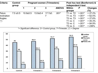
Table 4. The pelvic inclination angle changes during pregnancy
Criteria Control group
Pregnant women (Trimesters) Post hoc test (Bonferroni) & independent t-test
1 2 3 ANOVA Within groups
P Changes (%)
Pelvic Inclination Angles (°)
7.5 ±0.5 10.9±0.5 13.9±0.4 17.7±0. 7
.001* T1 vs. T2 >.001* + 27.2% T1 vs. T3 >.001* + 62.4% T2 vs. T3 >.001* + 27.6% T1 vs. C >.001* + 45.3% T2 vs. C >.001* + 84.9% T3 vs. C >.001* + 236% *= Significant difference, C= Control group, T=Trimester, (°) = Degree
Fig. 3. Changes of the lumbar and thoracic curvatures and pelvic tilt in each trimester of
pregnancy and in control group
Table 4 revealed a huge increased anterior pelvic tilt during pregnancy between the control and experimental groups and among all trimesters in the experimental group (P<.001) (Fig.3).
4. DISCUSSION
The marked postural changes following pregnancy may affect on the musculo-skeletal system and functional abilities of pregnant women [1]. The mal-alignments of the spinal column during pregnancy resulting from physiological and biomechanical changes may be associated with pain and disabilities [31]. However, a deliberate review of the literature showed that a controversy still exist regarding the exact spinal curvature changes as pregnancy advances.
The current study intended to track the postural changes during the first, second and third trimesters in primigravid pregnant women. The
The findings of the current study was in agreement with some previous research [1,2,5,8,14,19,32], but disagreed with some others [4,6,7,9]. lumbar lordosis is the key postural component in maintaining sagittal balance. Affection of lumbar lordotic curve often results in sagittal spinal imbalance causing low back pain that represents one of the leading causes of disability [33]. Increased lumbar and thoracic curvatures and pelvic inclination angles during pregnancy might be due to the some physiologic changes. It seems that, initially, pregnancy does not alter any curve shapes and all increased masses are suffered by the muscles located around the lumbar area (core stabilizers). After 12 weeks pregnancy, the uterus moves to the anterior, outward and upper part of the pelvis. Up to sixteen kilograms increased mass of both fetus and mother at the front of the abdomen will produce an anterior pelvic tilt [1,3]. This will, in turn, shifts the center of body mass and the line of gravity to the anterior and will pressurize more on the lumbar muscles. To compensate this force and to prevent falling in front, the lumbar curvature increases to let the line of gravity passes through its behind. As this study showed, in early pregnancy, the pregnant women compensate this anterior abdominal shift only by anterior pelvic tilt without any significant changes at the lumbar or thoracic curvatures [34]. As pregnancy progresses and the lumbar lordosis increases, the line of gravity will be forwarding more and the risk of falling increases again. To keep the line of gravity in its correct position, the thoracic curvature starts increasing in opposite direction (kyphosis) to return the line of gravity to the back of the lumbar curve to reduce pressures on the lumbar discs, places the head in front and reduces the compression and irritation of the dura matter and the synovial membrane of the facet joints in lumbar area [8,29]. With regards to this point that the literature has already showed a positive relationship between the lumbar and thoracic changes and vice versa [35] in human body, it can be deduced that usually the position of pelvis might be representing the global position of the whole spinal column [16]. In addition, due to the hormonal release, some positional changes occur in ribs before any enlargement of the uterus. These include increased sub-costal angle forcing the ribs to the upper and lateral sides of the thoracic cage, thus increasing about 2 Cm anterior-posterior diameter of the cage, elevating the diaphragm up to 4 Cm and a total 5-7 Cm increased thoracic cage enlargement are other reasons to have an
increased thoracic curvature [36]. The huge increased thoracic curvature angle at the third trimester of pregnancy (26% in this study) might be due to the enlarged breasts to prepare mothers for caring babies and breast feeding. This is associated with an increased scapular protraction, internal rotation of the upper limbs and rounding shoulders, which is so-called “mother posture” [36].
The pelvic tilt is controlled by contraction of the abdominals, hip flexors, hip extensors and spinal extensor muscles (two paired muscle groups). Any changes in strength and endurance of these muscles will result in pelvic tilt changes. This, in turn, may result in lumbar lordosis angle changes. For instance, over stretching and weakness of the abdominal muscles will result in increased lumbo-sacral angle. This over stretching will result in shortness of the hip flexors and lumbar extensor muscles [5,14]. Some researchers have also shown the increased length of the rectus abdominis muscle and its diastasis during pregnancy that have reduced functional abilities of pelvic stabilizer muscles with advancement of pregnancy. This weakness has even lasted for 8 weeks after the delivery [37].
The results of a study in the Queensland University introduce the length of the abdominal and lumbar extensor muscles as the best indicators for lumbar curvature angles. In details, the lumbar lordosis showed a significant positive correlation with the length of abdominal muscles (r=0.209, p<.05), but had a negative correlation with the lumbar extensor muscles (r= -0.24, p<.05). This means that the shortest lumbar extensor muscle length, the most lumbar curvature angle [38].
musculo-skeletal systems to facilitate the delivery process [1]. Since these two hormones released during pregnancy remain in the body for at least six months post delivery, the hormonal changes might also be responsible for remaining increased spinal curvatures and pelvic inclination following the delivery [5,40]. The role of hormones producing joint laxities from previous deliveries was omitted in this study by studying only primigravid women. Marques et al reported different joint laxities in multiparus women after the first pregnancy [31].
In addition to all mentioned above, Collition et al. [43] stated that all postural changes during pregnancy including increased lumbar lordosis help pregnant women to be in a more stable position during pregnancy [2]. It can be deduced that all postural changes take place in pregnant women intend to reduce her postural sways via increased lumbar curve and anterior pelvic tilt as well as shifting her head to back [5]. Although the balance index was not studied in the current study, one can assume that the significant increased thoracic curvature angle at the end of pregnancy, when the weight of fetus and mother reaches to its highest level, occurs to compensate the severe increased lumbar lordosis helping her for not falling to the front [16].
As brief, the postural changes occur during pregnancy can be divided into the real and compensatory changes. The real changes happen to counteract with the increased weight of the fetus and mother, while the compensatory curvatures happen to return the balance to the spinal column [8,44]. In an interesting anthropometric study, Whitcome et al. [45] showed that these postural changes happen to make bipedal standing and walking available for pregnant women.
5. CONCLUSION
The current study clearly showed the obvious spinal curvature changes during all trimesters of pregnancy. In this study, all the lumbar, thoracic and pelvic tilt angles increased as the pregnancy advanced. The results of the current study encourages pregnant women to have postural cares including suitable exercises and possible using suitable supports to keep their spinal column in best positions during pregnancy. It can be inferred that although the increased curvatures occurred during pregnancy are involuntarily, keeping the spinal curves as
minimal as possible is recommended to have easier return to its normal pre-pregnancy position.
CONSENT
All authors declare that ‘written informed consent was obtained from the patient for publication of this cohort study.
ETHICAL APPROVAL
All authors hereby declare that all experiments have been examined and approved by the appropriate ethics committee and have therefore been performed in accordance with the ethical standards laid down by Shahid Beheshti Medical University’s Ethical Committee.
ACKNOWLEDGEMENTS
The authors would like to sincerely thank all the subjects for their participation and the medical staff of Milad Hospital in Tehran for their cooperation. They also thank to the Physiotherapy Research Center of Shahid Beheshti University of Medical Sciences for its financial supports.
COMPETING INTERESTS
Authors have declared that no competing interests exist.
REFERENCES
1. Yousef AM, Hanfy HM, Elshamy FF, Awad MA, Kandil IM. Postural changes during normal pregnancy. Journal of American Science. 2011;7(6):1013-8.
2. Bullock JE, Jull GA, Bullock MI. The relationship of low back pain to postural changes during pregnancy. Aust J Physiother. 1987;33(1):10-7.
3. Gaymer C, Whalley H, Achten J, Vatish M, Costa ML. Midfoot plantar pressure significantly increases during late gestation. The Foot. 2009;19(2):114-6. 4. Gilleard WL, Crosbie J, Smith R. Static
trunk posture in sitting and standing during pregnancy and early postpartum. Archives of Physical Medicine and Rehabilitation. 2002;83(12):1739-44.
6. Okanishi N, Kito N, Akiyama M, Yamamoto M. Spinal curvature and characteristics of postural change in pregnant women. Acta Obstetriciaet Gynecologica Scandinavica. 2012;91(7):856-61.
7. Moore K, Dumas G, Reid J. Postural changes associated with pregnancy and their relationship with low-back pain. Clinical Biomechanics. 1990;5(3):169-74. 8. Dumas G, Reid J, Wolfe L, Griffin M,
McGrath M. Exercise, posture, and back pain during pregnancy: Part 1. Exercise and posture. Clinical Biomechanics. 1995;10(2):98-103.
9. Östgaard H, Andersson G, Schultz A, Miller J. Influence of some biomechanical factors on low-back pain in pregnancy. Spine. 1993;18(1):61-5.
10. Wang S-M, DeZinno P, Lin EC, Lin H, Yue JJ, Berman MR, et al. Auricular acupuncture as a treatment for pregnant women who have low back and posterior pelvic pain: a pilot study. American journal of obstetrics and gynecology. 2009; 201(3):271. e1-e9.
11. Wang S-M, Dezinno P, Maranets I, Berman MR, Caldwell-Andrews AA, Kain ZN. Low back pain during pregnancy: prevalence, risk factors, and outcomes. Obstetrics & Gynecology. 2004;104(1):65-70.
12. Kelly-Jones A, McDonald G. Assessing musculoskeletal back pain during pregnancy. Primary Care Update for OB/GYNS. 1997;4(5):205-10.
13. Ee CC, Manheimer E, Pirotta MV, White AR. Acupuncture for pelvic and back pain in pregnancy: A systematic review. American journal of obstetrics and gynecology. 2008;198(3):254-9.
14. Otman AS, Beksaç MS, Başgöze O. The
importance of ‘lumbar lordosis
measurement device’ application during pregnancy, and post-partum isometric exercise. European Journal of Obstetrics & Gynecology and Reproductive Biology. 1989;31(2):155-62.
15. Dunning K, LeMasters G, Levin L, Bhattacharya A, Alterman T, Lordo K. Falls in workers during pregnancy: risk factors, job hazards, and high risk occupations. American Journal of Industrial Medicine. 2003;44(6):664-72.
16. Le Huec J, Saddiki R, Franke J, Rigal J, Aunoble S. Equilibrium of the human body and the gravity line: The basics. European Spine Journal. 2011;20(5):558-63.
17. Vullo VJ, Richardson JK, Hurvitz EA. Hip, knee, and foot pain during pregnancy and the postpartum period. The Journal of Family Practice. 1996;43(1):63-8.
18. Ponnapula P, Boberg JS. Lower extremity changes experienced during pregnancy. The Journal of Foot and Ankle Surgery. 2010;49(5):452-8.
19. Bullock-Saxton JE. Changes in posture associated with pregnancy and the early post-natal period measured in standing. Physiotherapy Theory and Practice. 1991; 7(2):103-9.
20. Peterson ML, Bertram S, Neelly K, Ausili A, Atterberry B. A comparison of posture and sit-to-stand biomechanics of pregnant women in the third trimester with and without a maternity support: A pilot study. Journal of Women’s Health Physical Therapy. 2010;34(1):3-9.
21. Hart D, Rose S. Reliability of a noninvasive method for measuring the lumbar curve*. The Journal of Orthopaedic and Sports Physical therapy. 1986;8(4):180.
22. Walker ML, Rothstein JM, Finucane SD, Lamb RL. Relationships between lumbar lordosis, pelvic tilt, and abdominal muscle performance. Physical Therapy. 1987;67 (4):512-6.
23. Norbakhsh M, Mosavi S. An investigation into reliability and validity of flexible ruler in lumbar lordosis measurment. Journal of Mazandaran University of Medical Scienc. 2002;12(36):46-51.
24. Teixeira F, Carvalho G. Reliability and validity of thoracic kyphosis measurements using flexicurve method. Revista Brasileira de Fisioterapia. 2007;11(3):199-204. 25. Post R, Leferink V. Spinal mobility: Sagital
range of motion measured with the spinal mouse, a new noninvasive device. Arch Orthop Trauma Surg. 2004;124(3):187-92. 26. Youdas JW, Hollman JH, Krause DA. The
effects of gender, age, and body mass index on standing lumbar curvature in persons without current low back pain. Physiotherapy Theory and Practice. 2006;22(5):229-37.
27. Hoppenfeld S. Physical examination of the spine and extremities. New York: NY:Appleton-Century-Croft; 1976.
ISBN-10: 0838578454 • ISBN-13:
9780838578452, ©1976 • Pearson.
28. Magee D. Orthopedic physical
29. Herrington L. Assessment of the degree of pelvic tilt within a normal asymptomatic population. Manual Therapy. 2011;16(6): 646-8.
30. Eftekhar H, Khalkhali M. The designe and implementation of two instrument for mesearing pure hip flexion and pelvic tilt. Informative Scientific Journal of Shahed University. 1994;1(4):48-51.
31. Marques A, Gonçalves P, Santos R, Vilas-Boas J. Comfort and Functionality of Pregnant Women’s Feet Study of kinetic parameters with silicon insoles. Brazilian Journal of Biomechanics. 2005;6(10):9-15. 32. Ibrahim F. Changes of lumbar curvature
and back muscles activity as early predictor of low back pain in normal pregnant women. Unpublished Thesis, Faculty of Physical Therapy, Cairo University. 2002;64-70.
33. Hegazy AA, Hegazy RA. Midsagittal Anatomy of Lumbar Lordosis in Adult Egyptians: MRI Study. Anatomy research international. 2014;2014:1-12.
34. Vismara L, Menegoni F, Zaina F, Galli M, Negrini S, Capodaglio P. Effect of obesity and low back pain on spinal mobility: A cross sectional study in women. J Neuroeng Rehabil. 2010;7(3):1-8.
35. Uetake T, Ohtsuki F. Sagittal configuration of spinal curvature line in sportsmen using Moire technique. Okajimas folia anatomica Japonica. 1993;70(2-3):91-103.
36. Kisner C, Colby L. Therapeutic exercise, foundations and techniques. Textbook. 3, editor: F. A. Davis Co., Philadelphia. 1990; 210:800-11.
37. Gilleard WL, Brown JMM. Structure and function of the abdominal muscles in primigravid subjects during pregnancy and
the immediate post birth period. Physical Therapy. 1996;76(7):750-62.
38. Toppenberg RM, Bullock MI. The interrelation of spinal curves, pelvic tilt and muscle lengths in the adolescent female. Australian Journal of Physiotherapy. 1986; 32(1):6-12.
39. Dumas G, Reid J. Laxity of knee cruciate ligaments during pregnancy. Journal of Orthopaedic & Sports Physical Therapy. 1997;26(1):2-6.
40. Forczek W, Staszkiewicz R. Changes of kinematic gait parameters due to pregnancy. Acta of Bioengineering & Biomechanics. 2012;14(4).
41. Huang TH, Lin SC, Ho CS, Yu CY, Chou YL. The gait analysis of pregnant women. Biomedical Engineering: Applications, Basis and Communications. 2002;14 (02):67-70.
42. Carpes F, Griebeler D, Kleinpaul J, Mann L, Mota C. women able-bodied gait kinematics during and post pregnancy period. Revista Brasileira de Biomecânica. 2008;9(16):33-40.
43. Collition J. Back pain in pregnancy: active management strategies. Physician and Sports Medicine & Science in Sports & Exercise. 1996;24:1-8.
44. Cunningham; F, Levano; K, MacDonald; P, Bloom; S, Hauth; J, Rouse; D, et al. Williams Obstetrics, Ghazijahani, B.; Ghotbi, R (Persian translator). 20 st ed. Tehran: Golban Publication; 2010;6(174): 245-7.
45. Whitcome KK, Shapiro LJ, Lieberman DE. Fetal load and the evolution of lumbar lordosis in bipedal hominins. Nature. 2007;450(7172):1075-8.
© 2015 Kouhkan et al.; This is an Open Access article distributed under the terms of the Creative Commons Attribution License
(http://creativecommons.org/licenses/by/4.0), which permits unrestricted use, distribution, and reproduction in any medium,
provided the original work is properly cited.
Peer-review history: