Spiralian model systems
JONATHAN Q. HENRY*
Department of Cell & Developmental Biology, University of Illinois, Urbana, IL, USA
ABSTRACT The “Spiralia” represent one of the three major clades of bilaterian metazoans. Though members of this clade exhibit tremendous diversity in terms of their larval and adult body plans, many share a highly conserved early pattern of development involving a stereotypic cleavage program referred to as spiral cleavage. This group therefore represents an excellent one in which to undertake comparative studies to understand the origins of such diversity from a seemingly common ground plan. These organisms also present varied and diverse modes in terms of their ecology, development and life history strategies. A number of well established and emerging model systems have been developed to undertake studies at the molecular, genetic, cell and organismal levels. The Special Issue of the Int. J. Dev. Biol. entitled “Spiralian Model Systems” focuses on these organisms and here, I introduce this clade, pointing out different types of studies being undertaken with representative spiralian model systems.
KEY WORDS:
bilaterian metazoan, spiral cleavage, life history strategy
The Spiralia (Lophotrochozoa)
Of the three major clades of bilaterians, the Spiralia (Lophotro-chozoa) comprise nearly half of the extant metazoan phyla (see Fig. 1). Despite this fact, the group has received relatively little attention compared to the other two clades, the deuterostomes and ecdysozoans, notably in the areas of genetics, as well as molecular, cellular and developmental biology. This is due in part to the long standing predominance of key experimental models positioned within the Ecdysozoa, (e.g., the fruit fly Drosophila and the nematode, C. elegans), and the Deuterostomia, (e.g., chordates such as the Zebrafish and mouse, as well as a few invertebrate representatives from the Echinodermata).
The Spiralia include 14 of roughly 36 metazoan phyla (Fig. 1). The Spiralia include the Lophotrochozoa, and sometimes these terms have been used synonomously. The clade “Lophotrochozoa” was first recognized by Halanych et al., (1995, see also Giribet et al., 2007; Helmkampf et al., 2008a,b; Dunn et al., 2008; Hejnol et al., 2009; Edgecomb et al., 2011) who showed that the Lophophorata (consisting of groups possessing characteristic ciliated feeding structures, such as brachiopods and phoronids), are clearly united with other protostome phyla that include annelids, molluscs, and nemerteans. The Spiralia, however, encompass an even larger group of metazoans, and the exact relationships amongst the Spi-ralia are, however, not fully resolved. The consensus from recent analyses suggest that there are two large sub-groups (clades). One group is the “Trochozoa,” (Roule, 1891), which include Annelida,
www.intjdevbiol.com
*Address correspondence to: Jonathan Q. Henry. Department of Cell & Developmental Biology, University of Illinois, 601 S. Goodwin, Ave., Urbana, IL 61801, USA.
e-mail: [email protected]
Accepted: 18 September 2014.
ISSN: Online 1696-3547, Print 0214-6282
© 2014 UBC Press Printed in Spain
Mollusca, Nemertea, as the “Eutrochozoa”, together with the “Bra-chiozoa” (see Cavalier-Smith, 1998), comprised of Brachiopoda and Phoronida, and the “Polyzoa” (Funch and Kristensen 1995; Passamaneck and Halanych 2006; Helmkampf et al., 2008a,b; Edgecombe et al., 2011) consisting of the Bryozoa, Entoprocta, and Cycliophora. The other group includes the Platyzoa (Cavalier-Smith, 1998; Giribet et al., 2000), which include Gastrotricha, Platyhelminthes, and the groups comprising the “Gnathozoa” or “Gnathifera” (Gnathostomulida, Micrognathozoa and Rotifera (Syndermata)). More recently, however, an analysis by Struck et al., (2014), which included additional species, suggests that the Platyzoa are paraphyletic. Their data suggest that, with the exclusion of the Gnathifera, the Gastrotricha and Platyhelminthes comprise a monophylum, which they term the “Rouphozoa.” They argue that the Rouphozoa together with the other spiralians comprise a monophyletic group called the “Platytrochozoa.” They argue that the Rouphozoa should not be included in the Lophotrochozoa, and that the terms Lophotrochozoa should not be used synony-mously with the larger encompassing clade, the Spiralia. Additional lophotrochozoan taxa, with more uncertain affiliations, include the parasitic Acanthocephala (closely related to rotifers), Myzostomida (likely highly derived annelids), and a unique group referred to as the Mesozoa, which includes the Orthinectida and Rhombozoa (see Giribet, 2002, 2008, Hejnol et al., 2009; Edgecombe, 2011). These phyla are listed in Table 1 and a recent view of their phylogenetic relationships is depicted in Fig. 1.
prin-ciple morphological characters displayed by different members of this clade. One trait is the ciliated feeding and respiratory tentacles of the lophophore (found in brachiopods and phoronids See Fig. 2L,P) while the other is the ciliated trochophore larva that possesses a prominent ciliated band involved in feeding and locomotion (called the “prototroch” see Fig. 3A), as seen in some annelids and molluscs, and possibly Entoprocts (Nielsen, 2001), nemerteans (Maslakova et al., 2004a,b) and Cycliophora (Funch, 1996). However, the underlying synapomorphic trait exhibited by more members of this clade is the highly conserved pattern of early development characterized by a stereotypical cleavage pat-tern, termed “spiral cleavage” (Fig. 4) At least seven phyla have members that exhibit spiral cleavage including the Annelida, Mol-lusca, Nemertea, Entoprocta, Gnathostomulida, Platyhelminthes (Polycladida) and dycemid Mesozoa (i.e., Rhombozoa, see Fig. 1, Table 1). Pennerstorfer and Scholtz (2012) also claim that a phoronid (Phoronis muelleri) exhibits spiral cleavage (though see Temereva and Malakhov, 2000; and Malakhov and Temereva, 2007). Because of the wider presence of this unifying developmental trait, Giribet (2002, see also Dunn et al., 2008, and Hejnol, 2010) argue the lophotrochozoan clade should be referred to as the “Spiralia.”
The Spiralia exhibit diverse body plans and life history
strategies
Remarkably, the Spiralia have exploited most habitats on earth and exhibit the greatest diversity of body plans compared to any other clade of multicellular organisms (see Fig. 2 and Table 1). In fact, all fundamental grades of organization can be found (Brusca and Brusca, 2003; Ruppert et al., 2003). For instance, groups
such as the annelids, and molluscs, exhibit mesodermally-lined true coelomic cavities, while others such as the Platyhelminthes, and entoprocts lack these cavities and possess acoelomate or pseudocoelomate body plans. Members of one phyla, the Annelida exhibit overtly segmented bodies along their anterior-posterior axes (Balavoine, 2014; Weisblat and Kuo, 2014, in this issue). Some groups posses skeletal elements such as the external or internal mineralized shells of molluscs and brachiopods or the hardened exoskeletons found amongst the bryozoans. Others possess specialized external or internal cuticular structures, such as those found in entoprocts, annelids, and gnathostomulids, while many representatives have no skeletal elements at all (e.g., nemerteans, phoronids, Platyhelminthes).
Likewise, different groups exhibit varied modes of development, including many with diverse larval body plans (Fig. 3, see papers by Rockman and Zakas, 2014, Arenas-Mena and Li, 2014, Helm et al., 2014; Boyle and Rice, 2014, Lesoway et al., 2014; Maslakova and Hiebert, 2014, Rockman and Zakas, 2014, all in this issue). As mentioned briefly above, one striking characteristic shared by some members of the annelids and molluscs and possibly also certain nemerteans, bryozoans and cycliophorans is the formation of a trochophore or trochophore-like larvae that possesses a distinct circumferential ciliated band, the prototroch (Fig. 3A,D-E, G). In contrasts, some members of the Nemertea display maximal indirect development via the formation of a feeding larva that contains in-ternal sets of imaginal disks, from which the adult emerges through a radical process of metamorphosis (e.g., heteronemerteans, such as Cerebratulus lacteus, or C. montgomeryi, Fig. 3F, see review by Maslakova and Hiebert, 2014, in this issue).
Other representatives exhibit direct development without the
formation of an intervening larval stage (Fig. 3I). Even within the same genus one can find species with dramatically different modes of development. For instance, the genus of calyptraeid snails, Crepidula contains at least 60 recognized species (Collin, 2003a,b). Some species, such as C. fornicata, C. lingulata and C. plana exhibit indirect development with a planktotrophic feeding veliger larvae (Fig. 3D, Conklin, 1897; Werner, 1955; Fretter, 1972; Collin, 2000). On the other hand, species such as C. adunca, and C. convexa exhibit direct development leading to the formation of crawl-away juvenile snails (Conklin, 1897; Moritz, 1939). Yet others such as Crepipetella dilatata (formerly Crepidula dilatata) and Crepidula cf. onyx form adelphophagic embryos that ingest aborted sibling nurse eggs contained within the same egg capsules (Gallardo, 1977; Chaparro et al., 2002; see paper by Lesoway et al., 2014 in this issue).
Clearly the spiralian “developmental program” represents a highly flexible platform that supported the explosive radiation of these metazoan phyla. As such, the Spiralia provide an excellent group for studies aimed at understanding the developmental mechanisms that underlie the genesis of such diversity. Obviously, they represent a pivotal group in terms of the emergence of the Bilateria. Though currently lacking, a better understanding of the precise phylogenetic relationships amongst these groups will be critical for deciphering the evolutionary trajectory of those fundamental developmental processes that generated such diverse metazoan adult and larval body plans (see Figs. 2-3). The truly remarkable point is that such
vastly different body plans originated from an ancestral pattern of early development that involved spiral cleavage.
Spiral cleavage
The highly stereotyped spiral cleavage pattern exhibited by many members of the Spiralia is characterized by alternating sets of oblique cell divisions that generate staggered quartets of micromeres located towards the animal pole. The basic pattern is illustrated in Fig. 4. Beginning with the fertilized egg, the first two cell divisions occur along the animal-vegetal axis and are nearly orthogonal to one another. These divisions generate four cells (“blastomeres”) that establish the four basic embryonic quadrants, which are termed A, B, C, and D following the conventional no-menclature refined by Edwin Grant Conklin (1897, see Figs. 4). In many species symmetric divisions generate these four cells, which are all of roughly the same size (Fig. 4A-D, I). In other species asymmetric divisions generate these cells and typically one cell ends up being larger than the others, the so-called D blastomere (Fig. 4A’-D’). In either case, each of these four cells subsequently generates a series of animal daughter cells (called “micromeres”), which are formed in alternating clockwise and counterclockwise orientations around the animal-vegetal axis (Fig. 4E-H, E’-H’, J-M). These animal cells are typically smaller and therefore are termed “micromeres,” whereas the four vegetal-most cells are larger and termed “macromeres.” In some cases, such as in nemerteans,
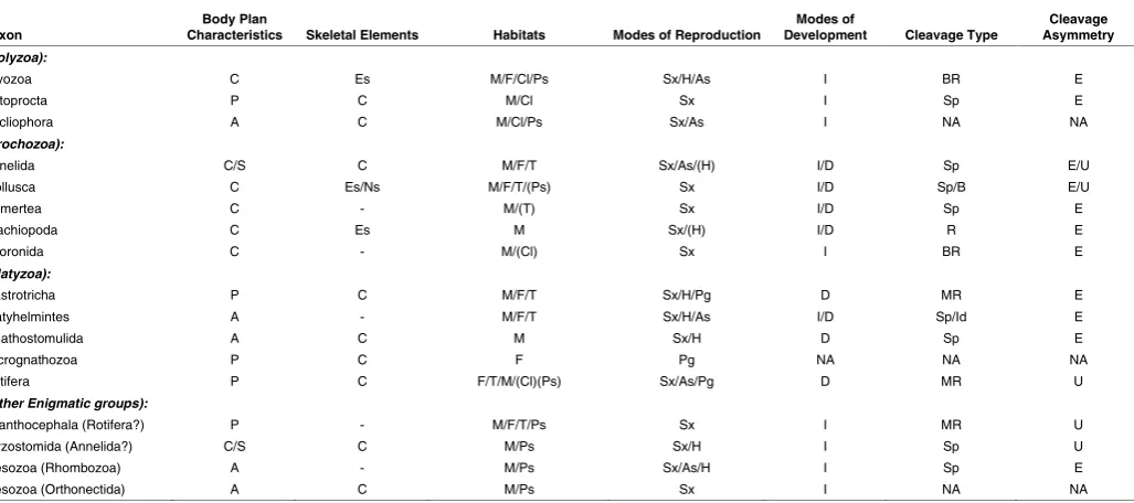
Taxon Characteristics Body Plan Skeletal Elements Habitats Modes of Reproduction Development Modes of Cleavage Type Asymmetry Cleavage
(Polyzoa):
Bryozoa C Es M/F/Cl/Ps Sx/H/As I BR E
Entoprocta P C M/Cl Sx I Sp E
Cycliophora A C M/Cl/Ps Sx/As I NA NA
(Trochozoa):
Annelida C/S C M/F/T Sx/As/(H) I/D Sp E/U
Mollusca C Es/Ns M/F/T/(Ps) Sx I/D Sp/B E/U
Nemertea C - M/(T) Sx I/D Sp E
Brachiopoda C Es M Sx/(H) I/D R E
Phoronida C - M/(Cl) Sx I BR E
(Platyzoa):
Gastrotricha P C M/F/T Sx/H/Pg D MR E
Platyhelmintes A - M/F/T Sx/H/As I/D Sp/Id E
Gnathostomulida A C M Sx/H D Sp E
Micrognathozoa P C F Pg NA NA NA
Rotifera P C F/T/M/(Cl)(Ps) Sx/As/Pg D MR U
(Other Enigmatic groups):
Acanthocephala (Rotifera?) P - M/F/T/Ps Sx I MR U
Myzostomida (Annelida?) C/S C M/Ps Sx/H I Sp U
Mesozoa (Rhombozoa) A - M/Ps Sx/As/H I Sp E
Mesozoa (Orthonectida) A C M/Ps Sx I NA NA
TABLE 1
GENERAL CHARACTERISTICS OF VARIOUS SPIRALIAN PHYLA
Listing of Lophotrochozoan phyla with details related to which ones exhibit spiral cleavage, different modes of development, larval forms, presence or absence of internal or external skeletal elements, and basic body plan organization.
the animal micromeres of the first quartet may actually be larger than the macromeres. Animal micromeres are designated with lower case letters, while the vegetal macromeres are designated with uppercase letters. Hence, the first quartet of micromeres is named 1a, 1b, 1c, and 1d, while the corresponding macromeres are named 1A, 1B, 1C, and 1D. (see Fig. 4E-F, E’F’, J). While the third cleavage division appears to occur at right angles to those of the first and second divisions the cleavage spindles are actually canted such that the micromeres are usually born with a slight clockwise (dextral) twist relative the macromeres when one views the embryos from the animal pole (see Fig. 4E-F, E’F’, J). Subsequently, a second quartet of animal micromeres (2a, 2b, 2c, 2d) is formed by the vegetal macromeres. During this division the spindles become shifted in the opposite direction, such that the second quartet micromeres become situated with a slight counterclockwise twist relative to the four macromeres (2A, 2B, 2C, 2D, Fig. 4G-H, G’H’, K). Typically a total of four micromere quartets (collectively referred to as 1q, 2q, 3q, and 4q) are formed and each set is formed with opposing chirality (Fig. 4L-M), though
in some species an additional fifth quartet of micromeres may be generated. Of course the individual micromeres belonging to each quartet also undergo further divisions as successive quartets are born and early on these divisions also follow the same alternating oblique orientations. These daughter cells are distinguished from one another by a system of successive superscript numbers (see Conklin, 1897). Typically those daughters born towards the animal pole receive a superscript of 1 while those towards the vegetal pole receive a 2 (e.g., 1b1 and 1b2, Fig. 34-M), and with successive
divisions additional superscripts are added (e.g., 1b11 and 1b12).
At some point, the spiral cleavage pattern is interrupted by the occurrence of bilateral sets of cell divisions. Those events represent a key transition in terms of establishing the bilaterian body plan, which is characteristic of both larvae and adults. In most cases the first sign of bilaterality is apparent in the symmetric divisions of cells located in the dorsal D quadrant. For instance, a daughter cell of 1d, 1d121, which is located at the base of the dorsal arm of the
“molluscan cross” divides bilaterally to form cells 1d1212 (to the right
of the midline) and 1d1211 (to the left of the midline) in the pulmonate
Fig. 2. Representative lophotrochozoans, illustrating some of the tremendous diversity of adult body plans.(A) The sipunculid, Themiste alu-tacea. (B) The nemertean, Cerebratulus Montgomeryi. (C) A gastropod mollusc, the cowry, Cypraea vitellus. (D) The mollusc Dentalium pilsbryi. (E)
A polyplacophoran mollusc, the chiton, Chaetopleura apiculata. (F) The polychaete annelid Platyneriesdumerilii. (G) The bryozoan, Bugulaneritina. (H)
snail Lymnaea stagnalis. In many cases 2d also exhibits and early bilaterally symmetric pattern of cell divisions (Dohle, 1999).
In the snail Crepidula 4d is the first cell to divide bilaterally to form the ML (left side) and MR (right side) mesendodermal teloblasts well before any of the other fourth quartet micromeres are even born (i.e., 4a, 4b and 4c, see Lyons et al., 2012). These teloblasts form bilaterally symmetrical bands of mesendodermal cells (see Lyons et al., 2012). These cells also appear to generate the primordial germ cells in all cases in which this has been carefully examined (see reviewed by Rebscher, 2014 in this issue). As development continues, the individual germ layers arise from specific cells and the tissues become organized via the processes of gastrulation (see review by Lyons and Henry, 2014, in this issue), organogen-esis and morphogenorganogen-esis to ultimately generate the larval and/or adult body plans.
It should be noted that there are some species in which alternating micromere quartets are formed with the opposite handedness (i.e., the first quartet micromeres are formed in the counter-clockwise direction, etc.), such as in the snail Biomphalaria or even amongst different populations of the same species (e.g., the pond snail Lymnaea peregra, Boycott et al., 1923, 1930; Sturtevant, 1923; Freeman and Lundelius, 1982; Abe et al., 2014, in this issue). Such differences have a profound effect on development, as the early cleavage patterns set up the adult body plan. For instance, in the case of gastropod molluscs such as Lymnaea), the chirality of the adult shell (i.e., right- vs. left-handed coiling) is directly related to the chirality of the early cleavage pattern (i.e., whether the first quartet formed via dextral vs. sinistral cleavages, respectively). The mechanisms that underlay the establishment of left-right asymmetry and changes in shell coiling are described further by
other authors contributing to this issue (Abe et al., 2014; Grande et al., 2014, in this issue).
A recent study in annelids (using the leech, Helobdella auste-nensis) suggests that the key transition to bilateral cleavage may be controlled by zygotic gene expression regulated by members of the Pax family of transcription factors, either PaxB1 and/or Pax2/5/8 (Schmerer, et al., 2013). This transcription factor appears to be necessary for the DNOPQ’” ectodermal proteloblast (equivalent to 2d111) and DM” mesodermal proteloblast (equivalent to 4d) to
undergo their transitions to bilateral cleavage. The fascinating development of the Clitellata, or Oligochaeta, including leeches and the sludge worm, Tubifex are described further by Shimizu and Nakamoto (2014, this issue) and Weisblat and Kuo, (2014, this issue). Continued studies of the Spiralia will inform us greatly as to key developmental-evolutionary transitions that have occurred to generate bilaterally symmetrical body plans.
Establishment of the D quadrant
As described above, and depending on the species under consideration, one of two main variations of the spiral cleavage pattern may be observed. In some cases the first two cell divisions are unequal, while in others they are equal. The identity of the D quadrant can be ascertained as soon as the four-cell stage is reached in the former, where the D blastomere is typically much larger then the other cells (Fig. 4A’-D’). On the other hand, the four quadrants cannot be distinguished in the case of the latter (Fig. 4A-D). These differences are closely tied to fundamental differ-ences in the timing and mechanism by which the cell quadrants actually become specified. Multistep models have emerged from
experimental data examining these systems that leads to the establishment of the D quadrant and its subsequent activity as an organizer of development (Fig. 5). In the case of species with asymmetric (unequal) cell divisions the larger D quadrant blasto-mere becomes specified autonomously by virtue of its inheriting specific vegetal determinants (Figs. 4, 5; van den Biggelaar and Guerrier, 1983; Verdonk and Cather, 1983). A specific cell or cells derived from the D quadrant subsequently serve as a key orga-nizer of development to establish the dorso-ventral axis and the fates of adjacent cells (Fig. 5A’-D’; see below). These asymmetric cleavages may take place as a consequence of the asymmetric shifting of the cleavage spindle that dictates where cytokinesis oc-curs or via the production of vegetal cytoplasmic lobes (so called “polar lobes”) that ultimately become shunted into the D quadrant blastomere during each of these divisions (Guerrier et al., 1978; Verdonk and Cather, 1983; Henry and Martindale, 1999). These determinants for the D quadrant are located in the vegetal region, which are also packaged within polar lobes. On the other hand, in those cases that exhibit symmetric (equal) cell divisions, the D quadrant is not specified until later during development and this occurs conditionally by virtue of cell-cell inductive interactions.
These inductive interaction take place between daughters of the animal first quartet micromeres (the 1q1s) and one of the vegetal
macromeres (e.g., the future 3D), typically early during the interval between fifth and sixth cleavage (Fig. 5A-D). Some data suggests that the distinction between these two forms of spiral cleavage may be closer than had been previously appreciated, as there is evidence that animal-vegetal interactions may also be important for the specification of the D quadrant even in the case of unequal cleavers (e.g., in Ilyanassa, Wandelt and Nagy, unpublished data; see Lambert, 2009a,b; Fig. 5B). The nature of these inductive signals is not understood.
The D quadrant organizer
In spiralians, one cell, or in some cases two cells derived from the D quadrant, serve as key embryonic organizers that set up the dorso-ventral axis and direct the development of adjacent cells via inductive interactions (Fig. 5). These cells are set aside relatively early during development. Although these organizer cells reside within the D quadrant, there is a fair degree of heterotopic and heterochronic variation in terms of which particular cell(s) serves
as the organizer and when this signaling may take place during early development. In the gastropods Ilyanassa, and Lymnaea, for example, this cell is the macromere 3D, which provides organizer signals during the interval between fifth and sixth cleavage (just before the birth of 4d). In Ilyanassa, the activity of 3D is continued somewhat by its daughter 4d (Lambert, 2009a). In Crepidula the micromere 4d serves as the principle organizer beginning at the 25-cell stage, prior to the time it divides to form the ML and MR teloblasts, and well before the birth of the other fourth quartet micromeres 4a, 4b, and 4c (Henry et al., 2006). In the clitellate annelid Tubifex, organizer activity appears to involve two cells, a daughter of 2d, (i.e., 2d11) and 4d, and their signaling takes place
at the 22-cell stage (see review by Shimizu and Nakamoto, 2014, in this issue). In another annelid, the polychaete Capitella teleta, the organizer is represented by 2d and its signaling occurs at a much earlier stage of development, prior to the birth of the third quartet micromeres (i.e., the 16-cell stage; Amiel et al., 2013).
The nature of these inductive signals is not fully understood. However, in some cases MAPK activation (likely as an intermediate in an unidentified signaling cascade) plays a role in establishing the identity of the dorsal D quadrant and possibly in controlling its activity as an organizer (e.g., in Ilyanassa and Crepidula, Lambert and Nagy, 2001, Koop et al., 2007; Henry and Perry, 2008; Lam-bert, 2009a,b). Application of an inhibitor of MAPK phosphorylation (U0126) leads to radialized forms of development, though in the case of Crepidula, MAPK activation is not required specifically in the organizer (4d) itself, but rather for the establishment of the D quadrant macromere 3D or within the animal micromeres that induce this cell to become the D quadrant macromere (Henry and Perry, 2008). On the other hand, activated MAPK does not seem to be important for any of these events in the annelid Capitella (Amiel
et al., 2013). In that species MAPK is first detected in cells located around the blastopore, and MAPK activation does not appear to be critical for normal development. This is in contrast with another annelid, Hydroides, where MAPK appears to be activated only in the 4d cell, though the function of MAPK in that system has not been determined (Lambert and Nagy, 2003). We are just beginning to understand the molecular level events that control the processes of cell fate and axis specification during spiralian development (see papers by Gharbiah et al., 2014, Pruitt et al., 2014, Grande et al., 2014, Kenny et al., 2014, in this issue).
Cell lineage fate maps
Not only is the cleavage pattern highly conserved, but so to are the general fates of the individual blastomeres. These obser-vations first became apparent from comparative analyses of cell lineages compiled by investigators working at the Marine Biological Laboratory in Woods Hole, MA. The very first of these was car-ried out by Charles Otis Whitman, who examined development of the leech Clepsine (Whitman, 1878, 1887). Leeches, like other oligochaetes, exhibit a modified form of spiral cleavage involving the formation of germinal bandlets that generate most of the adult ectoderm, endoderm and mesoderm (see review by Weisblat and Shankland, 1985; Weisblat and Kuo, 2014 in this issue). In fact, Whitman may be regarded as the “father” of cell lineage analysis. His student Frank Rattray Lillie and other individuals including Edmund Beecher Wilson and Edwin Grant Conklin, subsequently assembled cell lineage fate maps for a number of different spiralians including various annelids Nereis, Arica foetida, Spio fulginosus, the polyclad Leptoplana (Wilson, 1892; Mead, 1897), molluscs, such as the slipper snail Crepidula fornicata (Conklin, 1897) the
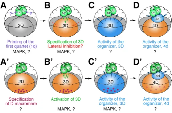
Fig. 5. Models summarizing basic mechanisms involved in specifying the dorsal D quadrant and subsequent D quadrant organizer activity during early developing in spiralians. A-D highlights these processes in equal-cleaving spiralians. A’-D’ illustrates these processes in unequal-cleaving spiralians. (A-B) In the case of equal-cleaving embryos, the D quadrant is established conditionally, as a result of animal-vegetal inductive interactions that involve the animal-most progeny of the first quartet micromeres (1q1 cells). Typically
this occurs during the interval between fifth and sixth cleav-age when animal cells come into contact with one of the four vegetal macromeres and transmit an unknown signal that triggers this cell to become 3D. This series of events appears to trigger MAPK activation within the 3D macromere (Henry and Perry, 2008). On the basis of observations made in one species, Crepidula, there may also be an earlier signal that primes the animal micromeres that also involves the activation of MAPK in those cells (Henry and Perry, 2008). (B) Though all four macromeres are capable of becoming 3D, only one emerges, and this could potentially involve some form of lateral inhibition. (C) Once the 3D macromere is specified, in some
species it becomes a key organizer of developing that sets up the dorso-ventral axis and directs the development of adjacent cells in the other quad-rants. The nature of those signals is not clear, though it appears to trigger the activation of MAPK in a subset of animal micromeres. (D) In some cases 4d may serve as the key organizer (i.e., Crepidula, Henry et al., 2006). (A’-B’) In the case of unequal-cleaving embryos, the D quadrant is established autonomously as a result of the initial asymmetric cell divisions. The first two cell divisions ultimately segregate vegetal determinants (of unknown nature illustrated here as purple dots) into the D blastomere. (B’) Some evidence suggests that the ultimate fate of the 3D macromere may also require inductive interactions from the animal micromeres, similar to the situation encountered in equal-cleaving embryos (Waldelt and Nagy, unpublished data).
(C’) Subsequently, the 3D macromere serves as the key organizer of development, in the same fashion described above for equal cleaving embryos.
freshwater bivalve Unio (Lillie, 1895). Additional work was carried out by their counterparts in Europe (e.g., Heymons, 1893; Wierze-jski, 1905). That early work has been extended in recent decades using modern cell-autonomous lineage tracers for a number of species (i.e., the gastropod molluscs Crepidula fornicata and C. convexa, Hejnol et al., 2007; Lyons et al., 2012, and Ilyanassa obsoleta, Render 1991, 1997; Chan and Lambert 2014; the poly-placophoran mollusc, Chaetopleura apiculata, Henry et al., 2004; the nemerteans, Cerebratulus lacteus, Henry and Martindale, 1998, and Carinoma tremaphoros, Maslakova et al., 2004a,b; the polyclad turbellarian Hoploplana inquilina, Boyer et al., 1996, 1998; and the annelids Capitella teleta, Meyer et al., 2010, Meyer and Seaver, 2009, 2010, and Platynereis dumerilii, Ackerman et al., 2005; Fischer and Arendt, 2013). Together, this body of work has revealed that the ultimate fates of these quadrants are, to a large extent, homologous across the embryos of different spiralian phyla. Generally speaking, the first three quartets of macromeres give rise to ectodermal tissues, and components of the nervous
system, including the photoreceptors (typically derived from 1a1
and 1c1). Specific combinations of cells derived from the second
and/or third quartets also generate mesodermal tissues that con-tribute to the larval and adult body plans (the so-called “ectome-soderm”, see review by Lyons and Henry in this issue). The cells of the fourth quartet typically generate endodermal tissues of the digestive tract, though one cell, the mesentoblast 4d, also serves as a mesodermal progenitor (the so-called “endomesoderm”). In many, but not all cases this cell contributes to the formation of the hindgut intestine. The fourth quartet macromeres may or may not form endodermal tissues, depending on the species being exam-ined. As mentioned previously, the D quadrant is the first one to be specified in the embryo and its organizing activity subsequently directs the development of the other cell quadrants.
The positions of the four embryonic quadrants bear a specific relationship to the future dorso-ventral and left-right axes. Some authors have stated that the A, B, C, and D quadrants generally correspond to the right, ventral, left and dorsal sides of the embryo,
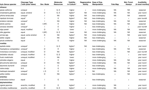
Phyla Genus species Cleavage Type (*with polar lobes) Dev. Mode Resources Molecular in Culture^ Viability Regenerative Ability Ease of Exp. Manipulation Fate Map Mol. Funct. Assays (# most months) Availibility Mollusca:
Aplysia california unequal I G, E higher + more challenging NA NA seasonal
Biomphalaria glabrata equal, sinistral D G, E higher^ NA more challenging NA NA year round
Bythinia tentaculata unequal* I NA higher NA less challenging NA NA seasonal
Crepidula fornicata equal* I E higher^ NA less challenging + + year round#
Dentalium dentale unequal* I NA higher NA less challenging NA NA seasonal
Haliotis asinina equal I (NF) E higher NA less challenging NA NA seasonal
Ilyanassa obsoleta unequal* I E higher + less challenging + + year round#
Loligo pealei equal, modified D E lower + more challenging NA NA seasonal
Lottia gigantea equal I (NF) G, E lower NA more challenging NA NA seasonal
Lymnea spp. equal, some sinistral D NA higher^ NA more challenging + NA year round
Patella vulgata equal I NA higher NA less challenging + + seasonal
Spisula solidisima equal I E higher NA more challenging NA NA seasonal
Annelida:
Capitella teleta unequal* I G, E higher^ NA less challenging + + year round
Cheatopterus variopedatus unequal* D E higher + less challenging NA NA seasonal
Enchytraeus coronatus unequal, modified D NA higher^ + less challenging + NA year round#
Helobdella robusta unequal, modified D G, E higher - less challenging + + year round
Hirudo medicinalis unequal, modified I NA lower - more challenging + - year round
Hydroides elegans equal I E higher + more challenging NA NA year round
Ophryotrocha labronica unequal D NA higher^ + more challenging NA NA year round
Platynereis dumerilii unequal I E higher^ + less challenging + +* year round
Pristina leidyi N.A. D E higher + more challenging NA NA year round
Streblospio benedicti unequal* I, D E higher^ + NA NA NA year round
Tubifex tubifex unequal D NA higher^ + less challenging + NA year round
Nemertea:
Cerebratulus lacteus equal I E lower - less challenging + + seasonal
Platyhelminthes:
Dugesia japonica anarchic, modified D E higher^ + more challenging NA + year round
Schmidtea mediteranea anarchic, modified D G, E higher^ + more challenging NA + year round
TABLE 2
COMPARISON OF SEVERAL SPIRALIAN SYSTEMS USED IN VARIOUS STUDIES
Commonly used spiralian model systems compared on the basis of several features, as listed.
respectively, but those relationships are oversimplified. Because individual micromeres within each quadrant are generated with an alternating clockwise/counterclockwise direction, they occupy slightly different positions relative the dorsoventral and left right axes at the completion of these cleavage divisions. Thus, in many cases, the progeny of the first quartet, 1a, 1b, 1c, 1d, occupy left-ventral, right-ventral, right-dorsal and left-dorsal positions, respectively (Henry and Martindale, 1999). The second quartet micromeres occupy left (2a), ventral (2b), right,(2c), and dorsal (2d) positions. The third quartet micromeres exhibit axial relationships similar to those of the first. Finally, the fourth quartet micromeres exhibits axial relationships similar to those of the second. Of course, these are generalities and, in fact, there has been some significant modifica-tion of these cleavage patterns and cell fates over the course of metazoan evolutionary history, as described below (see Henry and Martindale, 1999, and the review by Seaver, 2014, in this issue).
Differential localization of mRNAs: specification of the
micromere quartets
Elegant work by Lambert and Nagy (2002, see also Kingsley et al., 2007) showed that specific mRNAs are localized to particular cells during early cleavage in the snail Ilyanassa obsoleta, and subsequently that some of these mRNAs actually play a role in specifying the fates of the various micromeres (see Swartz et al., 2008, Rabinowitz, et al., 2008; Rabinowitz and Lambert, 2010; Chan and Lambert, 2011). These mRNAs become shuttled between the cytoplasm, centrosomes and the cell cortex to ultimately become differentially localized to specific daughter cells during cleavage. These localized mRNAs are thought to play key roles in establish-ing an animal-vegetal pre-pattern that distestablish-inguishes the different tiers of micromeres within these embryos (see Lambert, 2009a,b, 2010). Subsequent inductive interactions from the dorsal D quadrant organizer then refine this pattern to impart further complexity within each micromere quartet. Similar patterns of localized mRNAs also appear in the gastropod Crepidula fornicata (Henry et al., 2010c), and this could be a universal mechanism to distinguish cell fates in the Spiralia.
Evolution of the spiral cleavage program
Although spiral cleavage appears to represent a key aspect of the ancestral mode of development in this group of organisms, it has clearly undergone tremendous modifications, being completely lost in several groups such as the bryozoans, brachiopods and phoronids, in which cleavage appears to be radial. Even within the same phyla there are some representatives that exhibit spiral cleavage such as the polyclad turbellarians, while other platyhel-minthes exhibit radically different modes of cleavage (e.g., anarchic cleavage). Another striking example is found in the cephalopod molluscs, which form very large yolky eggs that initially undergo meroblastic bilateral cleavages resembling those seen in avian embryos (Watase, 1888; Arnold, 1965, 1971). Edmund Beecher Wilson (1898) was one of the first to recognize the tremendous degree of conservation between the cleavage patterns and the ultimate fates of identifiable blastomeres in those cases that do undergo spiral cleavage. He referred to these presumed homologies as a form of “ancestral reminiscence.” Frank Rattray Lillie (1895, 1899, see Maienschein, 1978) on the other hand noticed interesting
differences that led him to understand how specific changes are adaptive to the needs of the organism as it fills a particular niche. For instance, two cells in the embryo of the freshwater clam Unio are very large (2d and 2a) and consequently these cells contribute to substantially larger structures in the specialized glocidium larvae (which include the hooked larval valves (shell), and the adductor muscle, respectively). Furthermore, these two cells undergo a more rapid and increased number of cell divisions to form these structures when compared to those of the other embryonic quadrants, 2b and 2c, which are born as much smaller cells. Lillie (1899) referred to these changes as a form of evolutionary “adaptation in cleavage” (see also Seaver, 2014, in this issue).
More recently, Freeman and Lundelius (1992) observed that cleavage involving early equal patterns of cell division is more widely represented in the Spiralia, including the more basal members of this clade (see Tables 1-2 and Figs. 1,4-5). On this basis they argued that this mode of development, which involves epigenetic specification of the D quadrant, represents the ances-tral condition amongst this clade. Thus, forms with unequal spiral cleavage are derived, and they argue that unequal cleavage with precocious specification of the D quadrant may permit certain selective advantages that could, for instance, support accelerated development to the larval or juvenile stage. This also implies that embryos that undergo unequal cleavage divisions that involve the formation of polar lobes must have also arisen independently. Though the hypothesis of Freeman and Lundelius (1992) is more widely accepted, Dohle (1999) argued that equal cleavage and late specification of the D quadrant must be a derived condition in the annelids. He based that conclusion on comparisons of the cleavage patterns of the 2d lineage in a number of clitellate and some polychaete annelids, which he argued are too regular.
Spiralian model systems
studies examining the origins and mechanisms of segmentation (see review by Balavoine, 2014; and Weisblat and Kuo, 2014, in this issue). In fact spiralians have contributed greatly to the recent resurgence of the field of development and evolution and to our understanding of metazoan phylogeny.
The Spiralia contain many systems that are excellent for un-derstanding life history strategies related to transitions between different developmental modes, as well as the process of meta-morphosis. As mentioned above, members of the calyoptraeid snails (e.g., species in the genus Crepidula) exhibit a tremendous array of developmental modes including forms with direct develop-ment and others with planktotrophic feeding larval developdevelop-ment or yet others with intermediate forms of development (Henry et al., 2010a,b; Lesoway, et al., 2014 in this issue). Several species exhibit protandric hermaphroditism, like various Ophryotrocha and Crepidula species. For instance different members of the genus Ophryotrocha exhibit different modes including those with separate sexes (gonochoristic) while others exhibit different forms of her-maphroditism (Åkesson, 1973, 1975, 1994; Paxton and Åkesson, 2010), making them excellent systems for understanding factors that influence sexual development.
Various spiralians exhibit remarkable abilities to undergo asexual reproduction and many can regenerate missing body parts. Numer-ous studies focusing on regeneration have been carried out using the flatworms Schmidtea mediterannia and Dugesia japonica (cov-ered extensively in an earlier issue of this journal (IJDB, volume 56, 2012). Annelids such as Ophryotrocha and Pristina can regenerate missing posterior segments and represent excellent systems to study these phenomena (Pfannenstiel, 1974; see articles by Bely, 2014, and Szabó and Ferrier, 2014, in this issue).
Certain systems have been used extensively for studies of neurobiology, such as those with large, easily accessible neurons and relatively simple nervous systems that support complex be-haviors, like the squid Loligo and Aplysia (Abbott et al.,1995) and leeches such as Hirudo (Muller et al., 1981). Several have been used in behavioral studies of learning, memory and behavior, such as Lymnaea, (Benjamin and Kemenes, 2009, Feng et al., 2009), the limpet, Lottia gigantea, (Stimson, 1970, 1973) and the leech (Stent, et al., 1984; Muller et al., 1981). Annelids such as the leech and the polychaetes Capitella and Platynereis have also been used to study the development of the nervous system (Stent, 1984; Meyer and Seaver, 2009; see paper by Helm et al., 2014 in this issue). Due to the ease with which one can obtain large quantities of gametes, many cell biological, molecular and biochemical studies have been carried out using species such as the surf clam Spisula. The oligochaete Tubifex (the “sludge worm”) and the soil oligochaete Enchytraeus coronatus have served as models for studies of toxicology, as well as in developmental biol-ogy (see paper by Shimizu and Nakamoto, 2014 in this issue), and serve as important environmental water quality indicator species or in soil toxicity tests, respectively. Studies examining the biol-ogy of bio-fouling organisms have examined different organisms such as the calcareous tube dwelling annelid Hydroides (Nedved and Hadfield, 2009) and the encrusting bryozoan Bugula neritina (Callow and Callow, 2002; Mukaki et al., 1997). The Bobtail Squid (Euprymna scolopes) has served as a model for understanding the nature of eukaryote-prokaryote mutualism (Lee et al., 2009). The freshwater snail Biomphalaria, which represents the aquatic host for a key human parasite Schistosoma has been studied in
order to understand these host-parasite interactions, as a potential means to control this debilitating disease (Morgan et al., 2001).
Though the Spiralia currently lack a well established genetic model system, their tremendous strength lies in the rather broad understanding we have of their biology and, as mentioned above, their diverse body plans provide us with excellent subjects in which to undertake comparative studies aimed at understanding the evo-lution of triplobast bilaterian metazoans. It is only a matter of time before we develop tractable systems in which to undertake genetic analyses and, in fact, some labs are already working towards this end using species, such as the annelids Streblospio (see Rock-man and Zakas, 2014, in this issue) and Platynereis (e.g., lab of Dr. Detlev Arendt, EMBL, Heidelberg, Germany).
The papers featured in this issue highlight many of these tre-mendous systems and provide examples of the remarkable work that is being carried out by investigators from around the globe. This body of work concentrates mainly on those groups that exhibit the ancestral mode of development that involves spiral cleavage.
Acknowledgements
The author would like to thank Drs. Deirdre Lyons and Marty Shank-land for helpful comments concerning this manuscript. He thanks Drs. Richard Behringer and Juan Aréchaga for inviting him to serve as the editor of this special issue of I.J.D.B. on spiralian model systems. The author also thanks, Drs. Michael LaBarbera, Aldine Amiel, Eric Röddinger, Sveta Maslakova, Mark Martindale, and Michael Boyle for the use of their beautiful photographs that appear in Figures 2 and 3. Finally, the author thanks Nickelodeon Animation Studios for permission to use the image of Sponge Bob that appears in Figure 1. The author (J.J.H.) is supported by NSF grant number 1121268.
References
ABBOTT, J. N., WILLIAMSON, R. and MADDOCK, L. (1995). Cephalopod Neurobiol-ogy: Neuroscience Studies in Squid, Octopus and Cuttlefish. Oxford Univ. Press. ABE, N., TAKAHASHI and KURODA, R. (2014). Spiral cleavages in gastropods
determine the left-right body plan by regulating Nodal pathway. Int J Dev Biol 58: 513-520.
ACKERMANN C, DORRESTEIJN A, FISCHER A. (2005). Clonal domains in postlarval
Platynereis dumerilii (Annelida: Polychaeta). J Morphol. 266: 258-80.
ÅKESSON, B. (1973) Reproduction and larval morphology of five Ophryotrocha species (Polychaeta, Dorvilleidae). Zoologica Scripta 2. 145-155.
ÅKESSON, B. (1975) Reproduction in the genus Ophryotrocha (Polychaeta).
Pub-blicazioni della Stazione Zoologica di Napoli, 39 Suppl: 377-398.
ÅKESSON, B., B. (1994) Evolution of viviparity in the genus Ophryotrocha (Polychaeta, Dorvilleidae). In ‘Actes de la 4ème conférence internationale des polychètes’ (ed. J.-C. Dauvin, L. Laubier & D.J. Reish). Memoires du Muséum National d’Histoire
Naturelle 162: 29–35.
AMIEL, A., HENRY, J. Q. and SEAVER, E. C. (2013). An organizing activity is re-quired for head patterning and cell fate specification in the polychaete annelid
Capitella teleta: New insights into cell-cell signaling in Lophotrochozoa. Dev. Biol. 379: 107-122.
ARENAS-MENA, C. and LI, A. (2014), The feeding trochophore of the polychaete
Hydroides elegans and the evolution of indirect development. Int J Dev Biol
58: 575-583.
ARNOLD, J. M. (1965). Normal embryonic stages of the squid Loligo pealii. Biol.
Bull. 123: 53-57.
ARNOLD, J. M. (1971). Cephalopods. In G. Reverberi (ed.), “Experimental embryology of marine and freshwater invertebrates”. Chapter 10. North Holland Publishing. Co. BALAVOINE, G. (2014). Segment formation in annelids: patterns, processes and
evolution. Int J Dev Biol 58: 469-483.
processes. Int J Dev Biol 58: 623-634.
BENJAMIN, P.R. & KEMENES, G. (2009) Invertebrate models to study learning and memory: Lymnaea. Encycloped. Neurosci. 5: 197-204.
BOYCOTT, A. E. and DIVER, C. (1923). On the inheritance of sinistrality in Limnaea
peregra. Proc. Roy. Soc. London. B 95: 207-213.
BOYCOTT, A. E. DIVER, C. AND GARSTANG, S.L. and TURNER, F. M. (1930). The inheritance of sinistrality in Limnaea peregra (Mollusca, Pulmonata). Phil Trans.
Roy Soc. Ser B 219: 51-131.
BOYER, B. C., HENRY, J. Q. and MARTINDALE, M. Q. (1996). Dual origins of mesoderm in a basal member of the spiralian clade: cell lineage analyses in the polyclad turbellarian Hoploplana inquilina. Dev. Biol. 179: 329-338.
BOYER, B. C., HENRY, J. Q. and MARTINDALE, M. Q. (1998). The cell lineage of a polyclad turbellarian embryo reveals close similarity to coelomate spiralians.
Dev. Biol. 204: 111-123.
BOYLE, M. J. and RICE, M. E. (2014). Sipuncula: an emerging model of spiralian development and evolution. Int J Dev Biol 58: 485-499.
BRUSCA, R. C. and BRUSCA, G. J. (2003). “Invertebrates.” Sinauer. Sunderland, MA. 936 p.
CALLOW ME, CALLOW JA (2002) Marine biofouling: a sticky problem. Biologist 49: 10-14.
CAVALIER-SMITH, T. (1998). A revised six-kingdom system of life. Biol. Rev. 73: 203-266.
CHAN, XY and LAMBERT JD. (2011). Patterning a spiralian embryo: a segregated RNA for a Tis11 ortholog is required in the 3a and 3b cells of the Ilyanassa embryo.
Dev. Biol. 349: 102-112.
CHAN, X. Y. and LAMBERT, J. D. (2014). Development and larval contribution of blastomere clones in the Ilyanassa embryo: transformation of the spiralian blastula into the trochophore. Dev Genes Evol. 224: 159-174.
CHAPARRO, O. R., J. L. CHARPENTIER and R. COLLIN. (2002). Embryonic velar structure and function of two sibling apecies of Crepidula with different modes of development. Biol. Bull. 203: 80-86.
COLLIN, R. (2000). Sex change, reproduction and development of Crepidula adunca and C. lingulata (Gastropoda: Calyptraeidae) Veliger 43: 24 -33.
COLLIN, R. (2003a). Phylogenetic relationships among calyptraeid gastro- pods and their implications for the biogeography of speciation. Syst. Biol. 52: 618-640. COLLIN, R. (2003b). The utility of morphological characters in gastropod phylogenetics:
an example from the Calyptraeidae. Biol. J. Linn. Soc. 78: 541-593. CONKLIN, EG. (1897). The embryology of Crepidula. J. Morphol. 13: 1-226. DOHLE, W. (1999). The ancestral cleavage pattern of the clitellates and its
phyloge-netic deviations. Hydrobiologia 402: 267-283.
DUNN CW, HEJNOL A, MATUS DQ, PANG K, BROWNE WE, SMITH SA, SEAVER E, ROUSE GW, OBST M, EDGECOMBE GD, SØRENSEN MV, HADDOCK SH, SCHMIDT-RHAESA A, OKUSU A, KRISTENSEN RM, WHEELER WC, MARTIN-DALE MQ, GIRIBET G. (2008). Broad phylogenomic sampling improves resolution of the animal tree of life. Nature 452: 745-749.
EDGECOMBE, G. D., G. GIRIBET, C. W. DUNN, A. HEJNOL, R. M. KRISTENSEN, R. C. NEVES, G. W. ROUSE, K. WORSAAE and M. V. SORENSEN. (2011). Higher-level metazoan relationships: recent progress and remaining questions.
Organisms Divers. Evol. 11: 151-172.
FENG ZP, ZHANG Z, VAN KESTEREN RE, STRAUB VA, VAN NIEROP P, JIN K, NEJATBAKHSH, N, GOLDBERG JI, SPENCER GE, YEOMAN MS, WILDER-ING W, COORSSEN JR, CROLL RP, BUCK, LT, SYED NI, SMIT AB. (2009). Transcriptome analysis of the central nervous system of the mollusc Lymnaea
stagnalis. BMC Genomics. 10: 451.
FISCHER, A H. L. THORSTEN, H. and ARENDT, D. (2010). The normal development of Platynereis dumerilii, (Nereididae, Annelida) Front. Zoology 7: 31.
FISCHER AH, ARENDT D. (2013). Mesoteloblast-like mesodermal stem cells in the polychaete annelid Platynereis dumerilii (Nereididae). J Exp Zool B Mol Dev
Evol. 320: 94-104.
FREEMAN G. and LUNDELIUS, J. W. (1982). The development of dextrality and sinistrality in the gastropod Lymnaea peregra. Roux’s Arch. Dev. Biol. 191: 69-83. FREEMAN, G. and LUNDELIUS, J. W. (1992). Evolutionary implications of the
mode of D quadrant specification in coelomates with spiral cleavage. J. Evol.
Biol. 5: 205-247.
FRETTER, V. (1972). Metamorphic changes in the velar musculature, head and shell of some prosobranch veligers. J. Mar. Biol. Assoc. UK 52: 161-177.
FUNCH, P. (1996). The chordoid larva of Symbion pandora (Cycliophora) is a modi-fied trochophore. J. Morphol. 230: 231-263.
FUNCH, P. and KRISTENSEN, R. M. (1995). Cycliophora is a new phylum with af-finities to Endoprocta and Ectoprocta. Nature. 378: 711-714.
GALLARDO, C. S. (1977). Two modes of development in the morphospecies Crepidula
dilatata (Gastropoda: Calyptraeidae) from southern Chile. Mar. Biol. 39: 241-251.
GHARBIAH, M., COOLEY, J., LEISE, E. M., NAKAMOTO, A., RABINOWITZ, J. S., LAMBERT, J. D., AND. NAGY, L. M. (2009). The Snail Ilyanassa: A Reemerging Model for Studies in Development, In: “Emerging Model Organisms” Cold Spring Harbor Protocols. doi:10.1101/pdb.emo120
GIRIBET, G. (2002). Current advances in the phylogenetic reconstruction of meta-zoan evolution. a new paradigm for the Cambrian Explosion? Mol Phylogenet
Evol. 24:345-357.
GIRIBET, G. (2008). Assembling the lophotrochozoan (=spiralian) tree of life. Phil.
Trans. R. Soc. B. 363: 1513-1522.
GIRIBET, G., DISTEL, D. L., POLZ, M., STERRER, W., WHEELER, W. C. (2000). Triploblastic relationships with emphasis on the acoelomates and the position of Gnathostomulida, Cycliophora, Platyhelminthes, and Chaetognatha: A combined approach of 18S rDNA sequences and morphology. Syst. Biol. 49, 539-562. GIRIBET, G., C. W. DUNN, G. D. EDGECOMBE and G. W. ROUSE. (2007). A modern
look at the Animal Tree of Life. Zootaxa. 1668: 61-79.
GRANDE, C., MARTÍN-DURÁN, J.M., KENNY, N.J., TRUCHADO-GARCÍA, M. and HEJNOL, A. (2014). Evolution, divergence and loss of the Nodal signalling path-way: new data and a synthesis across the Bilateria. Int J Dev Biol 58: 52-532. GUERRIER, P., VAN DEN BIGGELAAR, J. A. M., VAN DONGEN, C. A. M. and
VERDONK, N. H. (1978) Significance of the polar lobe for the determination of dorsoventral polarity in Dentalium vulgare (da Costa). Dev. Biol. 63: 233-242. HALANYCH, K. M., J. D. BACHELLER, A. M. A. AGUINALDO, S. M. LIVA, D. M.
HILLIS and J. A. LAKE. (1995). Evidence from 18S ribosomal DNA that the lophophorates are protostome animals. Science 267: 1641-1643.
HEJNOL, A., OBST, M., STAMATAKIS, A., OTT, M., ROUSE, G. W., EDGECOMBE, G. D., MARTINEZ, P., BAGUÑÀ, J., BAILLY, X., JONDELIUS, U., WIENS, M., MÜLLER, W.E.G., SEAVER, E., WHEELER, W. W. MARTINDALE, M. Q., GIRI-BET, G. and DUNN C. A. (2009). Assessing the root of bilaterian animals using scalable phylogenomic methods. Proc Roy Soc. B. 276: 4261-4270.
HEJNOL, A (2010). A Twist in Time--The Evolution of Spiral Cleavage in the Light of Animal Phylogeny. Integr. Comp. Biol. 50: 695-706.
HEJNOL, A., MARTINDALE, M. Q. and HENRY J. Q. (2007). High resolution fate map of the gastropod snail Crepidula fornicata. Origins of ciliary bands, nervous and musculature elements. Dev. Biol. 305: 63-76.
HELM, C., ADAMO, H., HOURDEZ, S. and BLEIDORN, C. (2014). Immunohisto-chemical investigations of the development of Platynereis massiliensis (Annelida, Nereididae). Int J Dev Biol 58: 613-622.
HELMKAMPF, M., BRUCHHAUS, I. and B. HAUSDORF, B. (2008a). Phylogenomic analyses of lophophorates (brachiopods, phoronids and bryozoans) confirm the Lophotrochozoa concept. Proc. Royal Soc. B. 275: 1927-1933.
HELMKAMPF M., BRUCHHAUS I. and HAUSDORF B. (2008b). Multigene analysis of lophophorate and chaetognath phylogenetic relationships. Mol. Phylogenet.
Evol. 46: 206-214.
HENRY, J. J. (2002). Conserved mechanisms of dorsoventral axis determination in equal-cleaving spiralians. Dev. Biol. 248: 343-355.
HENRY, J. Q., COLLIN, R. and PERRY, K. J. (2010a). The Slipper Snail, Crepidula: An Emerging Lophotrochozoan Model System. Biological Bull. 218: 211-229. HENRY, J. Q., COLLIN, R. and PERRY, K. J. (2010b). Methods for Working with the
Slipper snail, Crepidula: An Emerging Lophotrochozoan Model System.
Biologi-cal Bull. (On-line, Peer-reviewed companion to the paper listed above). http://
biogeodb.stri.si.edu/bioinformatics/dfm/metas/view/38301
HENRY. J. and MARTINDALE, M. Q. (1998). Conservation of the spiralian devel-opmental program: Cell lineage of the nemertean, Cerebratulus lacteus. Dev.
Biol. 201: 253-269.
HENRY, J. and MARTINDALE, M. Q. (1999). Conservation and innovation in the Spiralian Developmental Program. Hydrobiologia 402: 255-265.
poly-placophoran, Chaetopleura apiculata: Variation in the spiralian program and implications for molluscan evolution. Dev. Biol. 272: 145-160.
HENRY, J. J. and PERRY, K. J. (2008). MAPK activation and the specification of the D quadrant in the gastropod mollusc Crepidula fornicata. Dev. Biol. 313: 181-195. HENRY, J. J., PERRY, K. J., FUKUI, L. and ALVI, N. (2010c). Differential Localization
of mRNAs During Early Development in the Mollusc, Crepidula fornicata. Integrat.
Compar. Biology 50: 720-733.
HENRY, J. Q. PERRY, K. J. and MARTINDALE, M. Q. (2006). Cell specification and the role of the polar lobe in the gastropod mollusc, Crepidula fornicata. Dev.
Biol. 297: 295-307.
HEYMONS, R. (1893). Zur Entwicklungsgeschichte von Umbrella mediterranea. Z.
Wiss. Zool. 56: 245-298.
KENNY, N. J., NAMIGAI, E. K. O., DEARDEN, P. K., HUI, J. H. L., GRANDE, C. and SHIMELD, S. (2014). The Lophotrochozoan TGF b Signalling Cassette: Diversifica-tion and ConservaDiversifica-tion in a Key Signalling Pathway. Int J Dev Biol 58: 533-549. KINGSLEY, E. P., CHAN, X. Y., DUAN, Y. and LAMBERT, J. D. (2007). Widespread
RNA segregation in a spiralian embryo. Evol. Dev. 9: 527-539.
KOOP, D., RICHARDS, G. S., WANNINGER, A., GUNTER, H. M. and DEGNEN, B. M. (2007). The role of MAPK signaling in patterning and establishing axial sym-metry in the gastropod Haliotis asinina. Dev. Biol. 31: 200-212.
LAMBERT, J. D. (2008). Mesoderm in Spiralians: the Organizer and the 4d Cell. J.
Exp. Zool. (Mol. Dev. Evol). 310B: 15-23.
LAMBERT JD. (2009a). Patterning the spiralian embryo: insights from Ilyanassa. In: Jeffery WR, editor. Current topics in developmental biology. Vol. 86. Burlington: Academic Press; 2009. p. 107-33.
LAMBERT, JD. (2009b). Developmental patterns in Spiralian embryos. Curr. Biol. 20: R27-R77.
LAMBERT, J. D. and NAGY, L. M. (2001). MAPK signaling by the D quadrant em-bryonic organizer of the mollusc Ilyanassa obsoleta. Development 128: 45-56. LAMBERT, J. D. and NAGY, L. M. (2002). Asymmetric inheritance of centrosomally
localized mRNAs during embryonic cleavages. Nature 420: 682-686.
LAMBERT, JD and NAGY, LM. (2003). The MAPK cascade in equally cleaving spiralian embryos. Dev Biol. 263: 231-241.
LEE, P N, MCFALL-NGAI, M J. CALLAERTS, P. and GERT-DE COUET, H. (2009). The Hawaiian Bobtail Squid (Euprymna scolopes): A Model to Study the Molecular Basis of Eukaryote-Prokaryote Mutualism and the Development and Evolution of Morphological Novelties in Cephalopod. In: “Emerging Model Organisms” Cold Spring Harbor Protocols.
LESOWAY M., ABOUHEIF E. and COLLIN, R., (2014). The Development of Viable and Nutritive Embryos in the Direct Developing Gastropod Crepidula navicella.
Int J Dev Biol 58: 601-611.
LILLIE, F. R. (1895). The embryology of the Unionidae. J. Morphol. 10: 1-100. LILLIE, F. R. (1899). Adaptation in cleavage. Biol. Lects., MBL, summers of 1897
and 1898. Ginn, Boston.
LIU, M., DAVEY, J. W. JACKSON, D. J., BLAXTER, M. L. and DAVISON, A. (2014). A conserved set of maternal genes? Insights from a molluscan transcriptome.
Int J Dev Biol 58: 501-511.
LYONS, D. C. and HENRY, J. Q., (2014). The Ins and Outs of Spiralian Gastrulation.
Int J Dev Biol 58: 413-428.
LYONS, D. C. PERRY, K. J., LESOWAY, M. P. and HENRY, J. Q. (2012). Cleavage pattern and fate map of the mesentoblast, 4d, in the gastropod Crepidula: A hallmark of spiralian development. EvoDevo 3: 21.
MAIENSCHEIN, J. (1978). Cell Lineage, Ancestral Reminiscence, and the Biogenetic Law,” J. Hist. Biol. 11: 129-158
MASLAKOVA S. A., MARTINDALE, M. Q. and NORENBURG, J. L. (2004a), Vesti-gial prototroch in a basal nemertean Carinoma tremaphoros (Palaeonemertea, Nemertea). Evol. Dev. 6: 219-226.
MASLAKOVA S. A., MARTINDALE, M. Q. and NORENBURG, J. L. (2004b). Fun-damental properties of spiralian developmental program are displayed by the basal nemertean Carinoma tremaphoros (Palaeonemertea, Nemertea). Dev.
Biol. 267: 342-360.
MALAKHOV, V. V. and TEMEREVA, E. N. (2000). Embryonic development of the phoronid Phoronis ijimai. Russ. J. Mar. Biol. 26: 412-421.
MASLAKOVA, S. A. and VON DASSOW, G. (2012). A non-feeding pilidium with
ap-parent prototroch and telotroch. J. Exp. Zool. Part B: Molec. Dev. Evol. 9999B: 1-5 MASLAKOVA, S. A. and HIEBERT, T. C. (2014). From trochophore to pilidium and
back again - a larva’s journey. Int J Dev Biol 58: 585-591.
MEAD, A. (1897) The early development of marine annelids. J. Morph. 13: 227-326. MEYER, N. P., BOYLE, M. J., MARTINDALE, M. Q. and SEAVER, E. C. (2010). A
comprehensive fate map by intracellular injection of identified blastomeres in the marine polychaete Capitella teleta. Evo Devo 1: 8.
MEYER, N. P. and SEAVER, E. C. (2009) Neurogenesis in an annelid: characterization of neural progenitors in the polychaete Capitella sp. I. Dev. Biol. 335: 237-252. MEYER, N. P. and SEAVER, E. C. (2010). Cell lineage and fate map of the primary
somatoblast of the polychaete annelid Capitella teleta. Int Comp Biology. 50: 756-767.
MORGAN JA, DEJONG RJ, SNYDER SD, MKOJI GM, LOKER ES. (2001).
Schis-tosoma mansoni and Biomphalaria: past history and future trends. Parasitology.
23 Suppl: S211-S28.
MORITZ, C. E. (1939). Organogenesis in the gastropod Crepidula adunca Sowerby.
Univ. Calif. Publ. Zool. 43: 217-248.
MUKAI H, TERAKADO K, REED CG (1997) Bryozoa. In: Harrison FW, Woollacott RM, editors. Microscopic Anatomy of Invertebrates. New York: Wiley-Liss. 69-72. MULLER, K., NICHOLLS, J. and STENT, G. ED. (1981). Neurobiology of the Leech.
New York: Cold Spring Harbor Laboratory. New York, Pp 320.
GHARBIAH, M., NAKAMOTO, A., JOHNSON, A. B., LAMBERT, J. D. and NAGY, L. M. (2014). Ilyanassa Notch signaling implicated in dynamic signaling between all three germ layers. Int J Dev Biol 58: 551-562.
NEDVED, B. T. and M. G. HADFIELD. (2009). Hydroides elegans (Annelida: Poly-chaeta): a model for biofouling research. Pp. 203 - 217 in: Marine and Industrial Biofouling, H.C. Flemming, R. Venkatesan, S.P. Murthy, K. Cooksey, Eds. Springer Series on Biofilms, Springer-Vergla, Berlin.
NIELSEN, C. (2001). Animal Evolution: Interrelationships of the Living Phyla. 2nd Edition. Oxford University Press. Oxford.
PASSAMANECK, Y. and K. M. HALANYCH. (2006). Lophotrochozoan phylogeny assessed with LSU and SSU data: Evidence of lophophorate polyphyly. Molec. Phylogenet. Evol. 40: 20-28.
PFANNENSTIEL H. D. (1974). Regeneration in the gonochoristic polychaete
Ophryotrocha notoglandulata. Marine Biology 24: 269-272
PENNERSTORFER M, SCHOLTZ G. (2012). Early cleavage in Phoronis muelleri (Phoronida) displays spiral features. Evol Dev. 14: 484-500
PAXTON, H. and ÅKESSON, B. (2010). The Ophryotrocha labronica group (Annelida: Dorvilleidae) - with the description of seven new species. Zootaxa 2713: 1-24. PRUITT, M. M., LETCHER, E. J., CHOU, H-C., BASTIN, B. R. and SCHNEIDER,
S. Q. (2014). Temporal and spatial expression of the wnt gene complement in a spiral-cleaving embryo and trochophore larva. Int J Dev Biol 58: 563-573. RABINOWITZ, J. S. and LAMBERT, J.D. (2010). Spiralian quartet developmental
potential is regulated by specific localization elements that mediate asymmetric RNA segregation. Development. 137: 4039-4049.
RABINOWITZ, J. S., CHAN, X Y., KINGSLEY, E. P., DUAN, Y. and LAMBERT, J. D. (2008). Nanos is required in somatic blast cell lineages in the posterior of the mollusk embryo. Curr. Biol.18: 331-336.
RENDER, J. A. (1991). Fate maps of the first quartet of micromeres in the gastropod
Ilyanassa obsoleta Development. 113: 495-501.
RENDER, J. A. (1997). Cell fate maps in the Ilyanassa obsoleta embryo beyond the third division. Dev. Biol. 89: 301-310.
REBSCHER, N. (2014). Establishing the germline in spiralian embyos. Int J Dev
Biol 58: 403-411.
ROCKMAN M. and C. ZAKAS (2014). Dimorphic development in Streblospio
bene-dicti: genetic analysis of morphological differences between larval types. Int J Dev Biol 58: 593-599.
RUPPERT, E. E. FOX, R. F. and BARNES R. D. (2003). “Invertebrate Zoology.” Cengage Learning. 1008 p.
SEAVER, E. (2014). Variation in Spiralian Development; Insights from Polychaetes.
Int J Dev Biol 58: 457-467.
SHIMIZU, T. and NAKAMOTO, A. (2014). Developmental significance of D quadrant micromeres 2d and 4d in the oligochaete annelid Tubifex tubifex. Int J Dev Biol 58: 445-456.
STENT, G.S., WEISBLAT, D.A., BLAIR, S.S. and ZACKSON, S.L. (1982). Cell lineage in the development of the leech nervous system. In Neuronal Development (Ed. N. Spitzer). Plenum, New York, pp. 1-44.
STENT, G. S., KRISTAN JR., W.B., FRIESEN, O. W., ORT, C. A., POON, M., CAL-ABRESE, R. L. (1984). Neuronal Generation of the Leech Swimming Movement.
Science 200: 1348-1357.
STIMSON J (1970) Territorial behavior of the owl limpet, Lottia gigantea. Ecology 51: 113-118.
STIMSON J (1973) The role of the territory in the ecology of the intertidal limpet Lottia
gigantea (Gray). Ecology 54: 1020-1030.
STURTEVANT, A. H. (1923). Inheritance of direction of coiling in Lymnaea. Science 58: 269-270.
STRUCK TH, WEY-FABRIZIUS AR, GOLOMBEK A, HERING L, WEIGERT A, BLEI-DORN C, KLEBOW S, IAKOVENKO N, HAUSDORF B, PETERSEN M, KÜCK P, HERLYN H, HANKELN T. (2014). Platyzoan paraphyly based on phylogenomic data supports a non-coelomate ancestry of Spiralia. Mol Biol Evol. 31: 1833-1849. SWARTZ, S.Z., CHAN, X.Y. and LAMBERT, J.D. (2008). Localization of Vasa mRNA during early cleavage of the snail Ilyanassa. Dev. Genes Evol. 218: 107-113. SZABÓ, R. and FERRIER, D. E. K. (2014). The dynamics of alkaline phosphatase
activity during operculum regeneration and biomineralization in the polychaete
Pomatoceros lamarckii Int J Dev Biol 58: 635-642.
TEMEREVA, E. N. and MALAKHOV, V. V. (2007). Embryogenesis and larval develop-ment of Phoronopsis harmeri Pixell, 1912 (Phoronida): Dual origin of the coelomic mesoderm. Invert. Reprod. Dev. 50: 57- 66.
ROULE, L. (1891). Considerations sur l’embranchement des Trochozoaires. Annales
des Sciences Naturelle (Zoologie). 7e série 11: 121-178.
VAN DEN BIGGELAAR, J. A. M. and GUERRIER, P. (1983). Origin of spatial informa-tion. In: “The Mollusca.” eds N. H. Verdonk, J. A. M. van den Biggelaar, and A. S. Tompa. Academic Press, New York. pp 179-213.
VERDONK, N. H. and CATHER, J. N. (1983) Morphogenetic determination and dif-ferentiation. In: “The Mollusca.” eds N. H. Verdonk, J. A. M. van den Biggelaar, and A. S. Tompa. Academic Press, New York. pp. 215-252.
WATASE, S. (1891). Observations on the development of Cephalopods; homology of the germ layer. Stud. John Hopkins Biol. Lab. 4: 165-183.
WERNER, B. (1955). Über die Anatomi, die Entwicklung und Biologie des Veligers und der Viliconcha von Crepidula fornicata L. (Gastropoda, Prosobranchia). Helgol.
Wiss. Meeresunters. 5: 169-217.
WEISBLAT, D and KUO, D-H. (2014). Developmental Biology of the Leech Helobdella”. Int J Dev Biol 58: 429-443.
WEISBLAT, D. A. and SHNAKLAND, M. (1985). Cell lineage and segmentation in the leech. Phil. Trans Roy. Soc. London. 312: 39-56.
WEISBLAT, D. A. and KUO, D-H. (2009). Helobdella (Leech): A Model for Develop-mental Studies. Emerging Model Organisms: A Laboratory Manual, Vol. 1. CSHL Press, Cold Spring Harbor, NY, USA,.
WHITMANN C. O. (1978). The embryology of Clepsine. Quart. J. Mic. Sci. 18. WHITMANN, C. O. (1887). A contribution to the history of the germ-layers in Clepsine.
J. Morphol. 1: 105-182.
WIERZEJSKI, A. (1905). Embryologie von Physa fontinalis. L. Z. Wiss, Zool. 83: 502-706.
WILSON, E. B. (1892) The cell-lineage of Nereis. J. Morphol. 6: 361-480. WILSON, E. B. (1898). Considerations in cell lineage and ancestral reminiscence.
Brachyury, Tbx2/3 and sall expression during embryogenesis of the indirectly developing polychaete Hydroides elegans
Cesar Arenas-Mena
Int. J. Dev. Biol. (2013) 57: 73-83
Hydra, a model system to trace the emergence of boundaries in developing eumetazoans
Angelika Böttger and Monika Hassel Int. J. Dev. Biol. (2012) 56: 583-591
The head organizer in Hydra
Hans R. Bode
Int. J. Dev. Biol. (2012) 56: 473-478
Planarian embryology in the era of comparative developmental biology
José M. Martín-Durán, Francisco Monjo and Rafael Romero Int. J. Dev. Biol. (2012) 56: 39-48
Planarian regeneration: a classic topic claiming new attention
Emili Saló and Kiyokazu Agata Int. J. Dev. Biol. (2012) 56: 1-4
5 yr ISI Impact Factor (2011) = 2.959 Function and specificity of Hox genes
David Foronda, Luis F. de Navas, Daniel L. Garaulet and Ernesto Sánchez-Herrero Int. J. Dev. Biol. (2009) 53: 1409-1419
Segmentation, metamerism and the Cambrian explosion
Juan Pablo Couso
Int. J. Dev. Biol. (2009) 53: 1305-1316
A history of Evo-Devo research in Spain
Jaume Baguñà
Int. J. Dev. Biol. (2009) 53: 1205-1217
The first bilaterian organisms: simple or complex? New molecular evidence
J Baguna, I Ruiz-Trillo, J Paps, M Loukota, C Ribera, U Jondelius, M Riutort Int. J. Dev. Biol. (2001) 45: S133-S134
Characterization of novel F-actin envelopes surrounding nuclei during cleavage of a polychaete worm.
S Jacobsohn