O R I G I N A L R E S E A R C H
Molecular Characterization Of
Vancomycin-Resistant
Enterococcus faecalis
Among Inpatients At Iranian University
Hospitals: Clonal Dissemination Of ST6 And
ST422
This article was published in the following Dove Press journal: Infection and Drug Resistance
Mehrdad Zalipour1
Bahram Nasr Esfahani1
Mehrdad Halaji1
Amir Azimian 2
Seyed Asghar Havaei1,3
1Department of Microbiology, School of
Medicine, Isfahan University of Medical
Sciences, Isfahan, Iran;2Department of
Pathobiology and Laboratory Sciences, North Khorasan University of Medical
Sciences, Bojnurd, Iran;3Infectious
Diseases and Tropical Medicine Research Center, Isfahan University of Medical Sciences, Isfahan, Iran
Purpose: Over the past two decades, enterococci have emerged as an important opportu-nistic pathogen causing life-threatening infections in hospitals. The purpose of the present study was to examine the prevalence of genes encoding virulence factor and molecular characterization of vancomycin-resistant E. faecalis strains isolated from hospitalized patients in Isfahan, the central city of Iran.
Patients and methods: A total of 53 vancomycin-resistant E. faecalis isolates (VRE) obtained from clinical samples of hospitalized patients were characterized by phenotypic and genotypic methods, and 25 selected VRE isolates from internal and ICU wards were typed by multilocus sequence typing.
Results:Theefawas the most prevalent virulence gene (100%) among isolates, followed by
gelE (92.45%), asa1 (90.56%), ace (86.79%), esp (75.47%), cylA (39.62%), and hyl
(18.86%). More than 80% of the isolates were HLGR. Multilocus sequence typing showed eight different sequence types including ST6, ST422, ST28, ST448, ST531, ST328, ST421, and ST495. STs were grouped into two clonal complex (CC) including CCA (ST6, ST422, ST448, ST531) and CCF (ST28, ST421) and two singletons (ST328, ST495).
Conclusion:Our data indicated a high prevalence of virulence genes among STs described in this study. In addition, the molecular analysis demonstrated a relatively high genetic diversity among selected VRE strains from the ICU in comparison with the internal ward. Therefore, in order to prevent the colonization of virulent strains in the hospital environment, infection control procedures should be performed.
Keywords: Enterococcus faecalis, virulence factors, vancomycin resistant, multilocus sequence typing, Iran
Introduction
Nosocomial infections (NIs) are a widespread problem in the current clinical setting
and occurs in approximately 4% to 10% of hospitalized patients annually.1,2Previous
studies indicate that the most common bacterial isolates from hospitalized patients are
methicillin-resistant Staphylococcus aureus (MRSA), vancomycin-resistant
entero-cocci (VRE), multidrug-resistant gram-negative bacteria, and Clostridium difficile.3
Enterococci, in particular, VRE, is one of the most important nosocomial pathogens,
Correspondence: Seyed Asghar Havaei Department of Microbiology, School of Medicine, Isfahan University of Medical Sciences, Hezar-Jerib Avenue, Isfahan 81746 73461, Iran
Tel +983137922478 Email [email protected]
Infection and Drug Resistance
Dove
press
open access to scientific and medical research
Open Access Full Text Article
Infection and Drug Resistance downloaded from https://www.dovepress.com/ by 118.70.13.36 on 22-Aug-2020
that causes serious infections such as bloodstream infections
(BSIs), catheter-associated urinary tract infections
(CAUTIs), and intra-abdominal and intra-pelvic
abscesses.-4,5
According to previous reports, prolonged hospitalization,
immunodeficiency, and uncontrolled antibiotic
administra-tion as a high risk of condiadministra-tions are responsible for
acquir-ing enterococcal infections in patients.6 Moreover, their
ability to acquire specific genetic traits, such as virulence
and antibiotic resistance determinants, plays an important role in the success and survival of enterococci in the
hospi-tal environment.7 Virulence factors through the ability to
adhere to a range of extracellular matrix proteins and fol-lowed by colonization and invasion into the host tissues lead to modulation of the host immunity and production of pathological changes directly through the production of enzymes and toxins or indirectly through induction
of inflammation, contributing to pathogenesis and severity
of enterococcal infections.8
Therefore, characterization of adhesions and invasion factors such as enterococcal surface protein (Esp), aggre-gation substance (AS) proteins (Asa1), collagen-binding protein (Ace), gelatinase (GelE), hyaluronidase (Hyl) and cytolysin (CylA) can be useful to improve our understand-ing and assessment of the pathogenicity of enterococcal
infections.9,10,11 Different molecular typing methods have
been developed for the epidemiological investigations of
enterococci.12 Among the known enterococcal molecular
typing methods, pulsed-field gel electrophoresis (PFGE)
was approved as a standard and efficient typing method
with a high degree of discrimination,13but PFGE remains
difficult to standardize between laboratories due to its
unsuitability for long-term epidemiology studies or for phylogenetic or population structure studies; this method
is more susceptible to small genetic changes.14,15
However, the most appropriate technique for global and long-term epidemiology studies is multilocus sequence
typing (MLST).15,16In addition, MLST provides an
unam-biguous nomenclature for genotypes, and clones and data are easily stored in databases that can be exchanged
between different laboratories via the Internet.16,17,18 The
emergence of antimicrobial resistant virulent enterococci is a serious problem for hospital infection control
practi-tioners and clinicians treating infected patients.19,20
Previous studies have demonstrated that Enteroccus
fae-calisis an important and virulent pathogen causing various
infections.21 Although there are several reports on the
endemicity of vancomycin-resistant enterococci (VRE) in Iran, limited information is available in relation to the
virulence determinants and molecular relatedness of
van-comycin-resistant E. faecalis isolates in Isfahan (Center
city of Iran). Therefore, in the present study, we investi-gated the molecular characterization of vancomycin-resis-tant E. faecalis isolates among Iranian hospitalized
patients as afirst study using MLST technique.
Materials And Methods
Study Design And Samples
This cross-sectional study was performed during the 7-month period from April 2017 to October 2017 at four
teaching hospitals affiliated to Isfahan University of
Medical Science, Isfahan, Iran. The study was approved by the Ethics Committee and was in accordance with the declaration of Helsinki [IR.MUI.REC.1396.3.066].
A total of 53 vancomycin-resistantE. faecalisisolates
(VRE) were obtained from true infections including urin-ary tract infections (UTIs) (n = 35), respiratory tract infec-tions (n = 5), bloodstream infecinfec-tions (n = 4), wound infections (n=4), abdominal infections (n = 3), eye infec-tion (n = 1), and meningitis (n=1).
The samples were cultured on blood agar (Merck, Germany) and then were incubated at 37°C for 24 hrs.
Enterococcal isolates were identified according to
conven-tional microbiological tests such as Gram staining (FARA CO.) (Gram positive), catalase reaction (catalase negative), growth on brain heart Infusion agar (Conda, Madrid, Spain) with 6.5% NaCl, and bile-esculin test (positive) (Merck,
Germany). Furthermore, E. faecaliswas confirmed by
amplification of the ddlE gene using species-specific
pri-mers (ddl E. faecalisF- 5ʹ- ATCAAGTACAGTTAGTCT-3ʹ
and R-5ʹ-ACGATTCAAAGCTAACTG-3ʹ).22 The PCR
protocol consisted of a pre-denaturation step at 95°C for 5 mins, followed by 30 cycles of 60 s at 95°C, 45 s at 48°C
and 1 min at 72°C. Afinal extension step was performed at
72°C for 5 mins. However, high levels of vancomycin resistance and gentamicin resistance were detected by the
E-test strips (Liofilchem, Italy) and gentamicin (120 μg)
disk (Mast Group Ltd., UK.), respectively, on the
Mueller–Hinton agar (Merck, Germany) in accordance
with Clinical and Laboratory Standards Institute (CLSI)
guidelines.23
DNA Extraction And Detection Of
Virulence Genes
Genomic DNA was extracted from fresh colonies as
described previously.21 PCR was performed for detecting
Infection and Drug Resistance downloaded from https://www.dovepress.com/ by 118.70.13.36 on 22-Aug-2020
seven of the genes encoding virulence factors including
enterococcal surface protein (esp), gelatinase (gelE),
aggregation substance (asa1), collagen-binding protein
(ace), Enterococcus faecalis endocarditis antigen (efa),
hyaluronidase (hyl), and cytolysin (cylA). Primers were
synthesized as previously described and were as follows:
esp, hyl, gelE, asa1,24 cylA,25 ace,26 andefa.27 The PCR products were separated by electrophoresis in 1% agarose gels with 1× TAE (Tris/Acetate/EDTA) buffer, stained with safe stain load dye (CinnaGen Co., Tehran, Iran) and visualized under ultraviolet illumination.
Multilocus Sequence Typing (MLST)
A total of 25 vancomycin-resistant E. faecalis isolates
(VRE) with high levels of gentamicin resistance from internal (n=12) and intensive care units (ICU) (n=13)
wards were subjected to MLST. Seven of E. faecalis
housekeeping genes including gdh (glucose-6-phosphate
dehydrogenase), gyd (glyceraldehyde-3-phosphate
dehy-drogenase), pstS (phosphate ATP-binding cassette
trans-porter), gki (glucokinase, putative), aroE (shikimate
5-dehydrogenase), xpt (xanthine phosphoribosyl
transfer-ase), and yqil (acetyl-CoA acetyltransferase) were
ampli-fied by PCR method. Information of theE. faecalisMLST
scheme, sequences of PCR primers, and the PCR
condi-tions are available at MLST website (https://pubmlst.org/
efaecalis/). STs were analyzed to determine clonal com-plex (CCs), single-locus variants (SLV), double-locus var-iants (DLV), and singletons using the eBURST algorithm (http://eburst.mlst.net/). Sequence types that were not
grouped into a CC were defined as singletons. Also, the
genetic relationship among strains was analyzed by con-structing a dendrogram using eBURST program.
Statistical Analysis
The analysis was performed by using SPSSTM software,
version 21.0 (IBM Corp., USA). The results are presented as descriptive statistics in terms of relative frequency.
Results
During 6 months of the study, a total of 53 VRE isolates were collected randomly with the possibility of true
infec-tions. Totally, of 53 confirmed VRE isolates, 56.6% and
43.4% isolates were obtained from female and male
sub-jects, respectively.28 The most frequent isolates 35 (66%)
were obtained from UTIs and followed that from respira-tory infections and bloodstream infections. Meanwhile, 32% (17/53) and 26.4% (14/53) isolates were obtained
from hospitalized patients in ICUs and internal wards,
respectively. According to MDR definition, all of 53
E. faecalis strains were MDR (100%) and more than 80% of the isolates were high-level gentamicin resistance
(HLGR) (Table 1).28
Distribution Of Virulence Genes
According to the results of PCR assay, theefa was most
prevalent virulence gene (100%), followed by gelE
(92.4%), asa1(90.6%), ace (86.8%), esp (75.5%), cylA
(39.6%), andhyl (18.9%). However, none of the isolates
contained all the virulent genes. The distribution of genes encoding virulence factors among VRE isolates and
dif-ferent STs is shown inTable 1.
Clonal Lineages Identi
fi
ed By MLST
MLST analysis was performed for 25 selected VRE
iso-lates from internal and ICU wards that were classified into
eight different STs: ST6, ST422, ST28, ST448, ST531, ST328, ST421, and ST495. Using the BURST v3 algo-rithm, STs were grouped into two CCs including CCA (ST6, ST422, ST448, ST531) and CCF (ST28, ST421) and two singletons (ST328, ST495). Two STs were found to be highly prevalent and comprised more than half of the isolates; 40% (10 isolates) of the isolates were ST6 and 24% (six isolates) were ST422, followed by ST28, ST448, and ST531 (two isolates), whereas other three STs were represented by a single isolate (ST328, ST421, and ST495). 91.6% (11/12) of the selected strains from internal wards were ST6 (seven isolates) and ST422 (four isolates), while selected isolates of ICU ward showed a higher diversity.
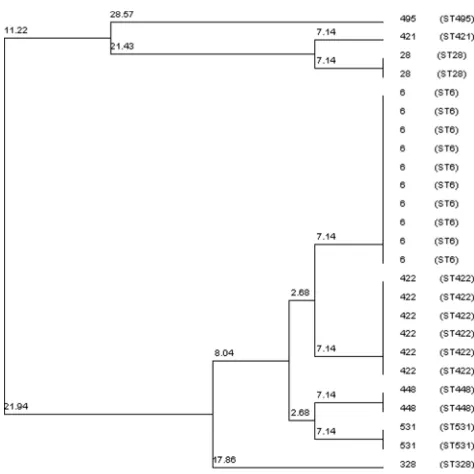
Phylogenetic analysis of the concatenated sequences, including all seven MLST genes, from the 25 VRE isolates
examined in this study is demonstrated in Figure 1. The
phylogenetic tree showed that there is a close phylogenetic relationship between most of the strains in our study.
Overall, the distribution of virulence genes indicated a high frequency of those among different STs, so that the
asa1, efa, gelE, and acegenes were present in all STs and
the prevalence of esp, cylA and hyl genes was 84.0%,
44.0% and 28.0%, respectively.
Discussion
Despite the fact that many studies have investigated anti-biotic resistance patterns and the virulence factors of
Enterococcusspp. isolates, only few studies have
system-atically investigated vancomycin-resistant E. faecalis
Infection and Drug Resistance downloaded from https://www.dovepress.com/ by 118.70.13.36 on 22-Aug-2020
T able 1 The Phenotypic And Genotypic Characteristics Of The 53 V ancom ycin-r esistant E. faecalis Isolates Fr om Clinical Samples Number Of Isolates Gender Infections W ar d Virulence Genes HLGR/Non-HLGR ST 1 M UTI Infectious diseases ef a, gelE, ace HLGR 2 M UTI ICU asa1, ef a, gelE, ace , esp,c ylA, HLGR 421 3 M BSI ICU asa1, ef a, gelE, ace , esp,c ylA, HLGR 6 4 F UTI Emergency asa1, ef a, gelE, esp HLGR 5 F UTI Outpatient ef a, gelE, ace , esp, HLGR 6 F WI Surger y asa1, ef a, gelE, ace , esp,c ylA, HLGR 7 M BSI Infectious diseases asa1, ef a, gelE, ace , h yl HLGR 8 M UTI Emergency asa1, ef a, gelE, ace , esp,c ylA, HLGR 9 M UTI Internal asa1, ef a, gelE, ace ,, HLGR 6 10 F UTI Emergency asa1, ef a, gelE, ace , esp Non-HLGR 11 F BSI Respirator y asa1, ef a, gelE, esp HLGR 12 F UTI Internal asa1, ef a, gelE, ace , esp HLGR 6 13 F UTI Internal asa1, ef a, gelE, ace , esp,c ylA, HLGR 422 14 M UTI Internal asa1, ef a, gelE, ace , esp HLGR 422 15 F UTI ICU asa1, ef a, gelE, ace , esp,c ylA, Non-HLGR 16 F UTI Emergency asa1, ef a, ace HLGR 17 F UTI Surger y asa1, ef a, gelE, esp,c ylA,h yl HLGR 18 F UTI Rheumatology asa1, ef a, gelE, ace , cylA Non-HLGR 19 M UTI ICU asa1, ef a, gelE, ace ,esp, cylA HLGR 422 20 M UTI Surger y asa1, ef a, gelE, ace , esp,h yl HLGR 21 F R TI ICU asa1, ef a, gelE, ace , cylA HLGR 28 22 F WI Rheumatology asa1, ef a, ace , esp Non-HLGR 23 F UTI Ur ology asa1, ef a, ace ,,c ylA HLGR 24 M WI Internal asa1, ef a, gelE, ace Non-HLGR 25 M UTI ICU asa1, ef a, gelE,ace , cylA, HLGR 531 26 F UTI NICU asa1, ef a, gelE, ace , esp,c ylA, HLGR 448 27 M WI Infectious diseases ef a, esp HLGR 28 F EI NICU asa1, ef a, gelE, ace Non-HLGR 29 M R TI ICU asa1, ef a, gelE, ace , esp,c ylA, HLGR 422 30 F UTI Internal ef a, gelE, esp Non-HLGR 31 F R TI ICU asa1, ef a, gelE, ace , esp HLGR 28 32 F UTI Respirator y asa1, ef a, gelE, ace , esp,c ylA, HLGR 33 F UTI ICU asa1, ef a, gelE, ace , esp HLGR 328 ( Continued )
Infection and Drug Resistance downloaded from https://www.dovepress.com/ by 118.70.13.36 on 22-Aug-2020
T able 1 (Continued). Number Of Isolates Gender Infections W ar d Virulence Genes HLGR/Non-HLGR ST 34 M R TI Surger y ef a, gelE HLGR 35 M UTI ICU asa1, ef a, gelE, ace , esp,c ylA, HLGR 6 36 M UTI Internal asa1, ef a, gelE, ace , cylA, h yl HLGR 422 37 M AI Internal asa1, ef a, gelE, ace , esp,h yl HLGR 6 38 M AI ICU asa1, ef a, gelE, ace , esp,h yl HLGR 531 39 F R TI Internal asa1, ef a, gelE, ace , esp,c ylA, HLGR 422 40 F UTI ICU asa1, ef a, gelE, esp,c ylA Non-HLGR 41 M UTI Emergency asa1, ef a, gelE, ace , esp,c ylA, HLGR 42 F UTI Internal asa1, ef a, gelE, ace , esp,h yl HLGR 6 43 F UTI ICU asa1, ef a, gelE, ace , esp,c ylA, HLGR 495 44 F Me CCU asa1, ef a, gelE, ace , esp HLGR 45 F UTI CCU asa1, ef a, gelE, esp, cylA, HLGR 46 F AI Internal asa1, ef a, gelE, ace , esp, hyl HLGR 448 47 M UTI ICU asa1, ef a, gelE, ace , esp,c ylA, HLGR 6 48 M UTI Internal asa1, ef a, gelE, ace , esp HLGR 6 49 F BSI ICU asa1, ef a, gelE, ace , esp Non-HLGR 50 F UTI Internal asa1, ef a, gelE, ace , esp, hyl HLGR 6 51 M UTI Surger y asa1, ef a, gelE, ace Non-HLGR 52 F UTI Internal asa1, ef a, gelE, ace , esp,h yl HLGR 6 53 M UTI Emergency asa1, ef a, gelE, ace , esp,c ylA HLGR Abbre viations: F, female; M, male; UTI, urinar y tract infection; BSI, bloodstr eam infection; WI, woun d infection; R TI, respirator y tract infection; EI, e ye infect ion; AI, abdominal infection; Me, meningitis; ST , sequence type.
Infection and Drug Resistance downloaded from https://www.dovepress.com/ by 118.70.13.36 on 22-Aug-2020
isolates originating from clinical specimens in our country. Therefore, we characterized the vancomycin-resistant
E. faecalis (VRE) collected from different clinical speci-mens during a 7-month period in 2017 from various teach-ing hospitals in Isfahan city, Iran. Similar to other reports from Iran, resistance to multiple classes of antibiotics was
common in E. faecalis strains as observed in the current
study.21,29,30 In this study, 81% of VRE strains were
HLGR. Resistance to high concentrations of gentamicin
among E. faecalis isolates has been showed in previous
researches.30,31 High-level resistance to gentamicin is
caused by the aminoglycoside-modifying enzymes,
redu-cing the effect of aminoglycosides except for
streptomycin.32 Antibiotic resistance is not sufficient for
the pathogenesis of enterococcal infections and the pre-sence of virulence factors plays an important role in the
severity of infection.10The VRE isolates characterized in
this study showed a high prevalence of virulence genes.
Ourfindings indicated that all of the isolates harboredefa
gene and similar frequencies of the efaAwere reported in
various studies.21,26,33 It seems that the efaA gene is
always present inclinical E. faecalis strains, whereas the
strains isolated from other sources possessed the less
fre-quency of EfaA determinant.26 As reported in other
stu-dies, gelE was identified as the second most common
virulence factor among the isolates in the current study
(92.45%).31,33 GelE seems to mediate virulence through
effects such as degradation of host tissues and participates in the activation of autolysin, leading to the release of
extracellular DNA and formation of a biofilm.11 The
aggregation substance gene (asa1) was present in 90.56%
of all isolates. These results are in agreement with those obtained by Choi et al and Nasaj et al who showed a high
incidence of asa1 genes in clinical isolates of
E. faecalis.31,34 AS proteins encoded by asa1 cause
clumping of E. faecalis cells and mediate the
high-fre-quency transfer of plasmid DNA between donor and
reci-pient bacteria.8These high rates ofasa1gene among all of
our isolates can facilitate the exchange of resistance and
virulence-associated genes via pheromone-responsive
plasmids in a hospital setting. The results of our study
indicated that the ace gene was present in 86.79% of
isolates. Similarly, a high incidence of this gene in E.
faecalis has been reported in previous studies.21,35,36
E. faecalis Ace is a collagen and laminin adhesin, which
seems to be effective in endocarditis and UTIs.11 In the
current study, the prevalence of the esp gene was also
75.47% in VRE strains. This finding in accordance with
other studies indicate a possible role of the Esp as a
colonization factor in UTI.10,26,34 The frequency of cylA
and hyl genes was 39.62% and 18.86%, respectively.
Previous studies by Heidari et al in Tehran and Shiraz
showed that the virulence genescylAandhylwere present
at varying levels inE. faecalisisolates.21,33Thesefindings
are in agreement with the results of Wanxiang et al, who
showed thatcylAandhylgenes were 19.4% and 19.6% in
the Enterococcus isolates, respectively.37 During this
study, we also identified that hyl-positive strains were
HLGR, and this may be due to the co-presence of thehyl
andaac(60)-Ie-aph(200)-Iagenes in a common transmis-sible plasmid.
Molecular typing of 25 strains of VRE isolated in
Isfahan using MLST provided thefirst data for the
knowl-edge on the genetic population structure for this species in Iran, allowing us to compare it to those obtained
world-wide, and to know the spread of someE. faecalisclones to
Iran. In the current study, eight different STs were found among the 25 VRE isolates, and distributed into two CCs, including CCA (ST6, ST422, ST448, and ST531), CCF (ST28, ST421), and two singletons (ST328, ST495). ST422 and ST531 represented an SLV of ST6 and ST448, respectively. ST531 is a DLV of ST422 belonging
to CCA and is also reported in Denmark.38 Furthermore,
ST531 represented SLV of ST6 belonging to CCA. ST28 is
Figure 1Cluster tree of sequence types (ST) of VRE strains. The tree constructed with the neighbor-joining method. In each branch is shown the corresponding bootstrap NJ values, taken over 1000 replicates,which assign confidence values for the groupings in the tree.
Infection and Drug Resistance downloaded from https://www.dovepress.com/ by 118.70.13.36 on 22-Aug-2020
an SLV of ST421 belonging to CCF. However, our study results showed that most of the strains examined had a common genetic origin. ST6, which is the ancestor of CCA, was the most frequently found ST compared to other STs (40%), and ST422 was the second most detected ST among the isolates (24%). Furthermore, it was found
that CCA contained the greatest number of STs identified
in this study (20/25; 80%).E. faecalisstrains identified as
ST6 showed similar characteristics to ST6 clones isolated
in Portugal and Poland39,40 and were characterized by
high-level resistance to aminoglycosides, and vancomycin, but differed from the strains described in Cuba and Spain
where they were vancomycin susceptible.12,41 ST6 has
been reported worldwide, and it is probably the major lineage in CCA, which is often associated with invasive infections and resistance to multiple drugs, owing to its adaptability to hospital environment as a result of the acquisition of pathogenicity islands and antimicrobial resistance genes by recombination and horizontal gene
transfer.14,42,43,44,45 Notably, 6 of the 25 isolates were
found to be ST422 (Table 1). According to a review of
studies worldwide, there are few reports of ST422 in E.
faecalis, so that Hammerum et al report ST422 as HLGR,
agreeing with our results.38 Other STs reported in this
study were also found previously from different parts of
the world.12,31,38,42,46 A high prevalence of virulence
genes was also detected among STs described in this
study, and there was no significant difference in the
dis-tribution of virulence factors between HLGR and non-HLGR isolates. There were some limitations related to the present study. First, a small number of VRE isolates
were typed by MLST. Second, identification of the source
of infection, taking preventive measures in the hospital setting, and molecular analysis of environmental speci-mens were required.
Conclusion
In summary, this research provided thefirst insight into the
population structure ofE. faecalisin Isfahan, Iran, and most
of the strains examined in this study were related to European strains, and CCA was evidently circulating in Isfahan hospi-tals, being associated with MDR and virulence traits. In
addition, our data indicate that E. faecalis strains isolated
from clinical samples possess distinctive patterns of potential virulence factors with a high incidence of genes encoding virulence factors among isolates. It is recommended that some programs be performed to prevent the colonization of such virulent strains in the hospital environment, including
better stewardship of antimicrobial agents and better aware-ness of the source for pathogen transmission in the hospital environment.
Ethics approval
The study protocol was approved by the Ethics Committee of Isfahan University of Medical Sciences (IR.MUI. REC.1396.3.066). However, we did not have human par-ticipants. The study used bacteria isolated from clinical samples in the clinical microbiology laboratory.
Acknowledgment
We are thankful to all members of the Department of Microbiology, School of Medicine, at Isfahan University of Medical Sciences. This was an original research paper from a Ph.D thesis.
Funding
This study was funded, in part, by a grant from the Isfahan University of Medical Sciences, [Grant no 396066].
Disclosure
The authors declare that they have no competing interests in this work.
References
1. Sligl WI, Dragan T, Smith SW. Nosocomial gram-negative bacteremia in intensive care: epidemiology, antimicrobial susceptibilities, and outcomes. Int J Infect Dis.2015;37:129–134. doi:10.1016/j.ijid.2015.06.024 2. Schabrun S, Chipchase L. Healthcare equipment as a source of
noso-comial infection: a systematic review.J Hosp Infect.2006;63(3):239– 245. doi:10.1016/j.jhin.2005.10.013
3. Bereket W, Hemalatha K, Getenet B, et al. Update on bacterial noso-comial infections. Eur Rev Med Pharmacol Sci. 2012;16(8):1039– 1044.
4. Kafil HS, Asgharzadeh M. Vancomycin-resistant Enterococcus
fae-ciumand Enterococcus faecalisisolated from education hospital of
iran.Maedica.2014;9(4):323.
5. Oravcova V, Mihalcin M, Zakova J, Pospisilova L, Masarikova M, Literak I. Vancomycin-resistant enterococci withvanAgene in treated municipal wastewater and their association with human hospital strains. Sci Total Environ. 2017;609:633–643. doi:10.1016/j. scitotenv.2017.07.121
6. O’Driscoll T, Crank CW. Vancomycin-resistant enterococcal infec-tions: epidemiology, clinical manifestations, and optimal management.
Infect Drug Resist.2015;8:217.
7. Sabouni F, Movahedi Z, Mahmoudi S, Pourakbari B, Keshavarz Valian S, Mamishi S. High frequency of vancomycin resistantEnterococcus
faecalisin children: an alarming concern.J Prev Med Hyg.2016;57
(4):E201–e204.
8. Vu J, Carvalho J. Enterococcus: review of its physiology, pathogen-esis, diseases and the challenges it poses for clinical microbiology.
Front Biol.2011;6(5):357. doi:10.1007/s11515-011-1167-x
9. Upadhyaya PG, Ravikumar K, Umapathy B. Review of virulence factors of enterococcus: an emerging nosocomial pathogen.Indian J
Med Microbiol.2009;27(4):301. doi:10.4103/0255-0857.55443
Infection and Drug Resistance downloaded from https://www.dovepress.com/ by 118.70.13.36 on 22-Aug-2020
10. Arbabi L, Boustanshenas M, Rahbar M, et al. Antibiotic susceptibil-ity pattern and virulence genes in Enterococcus spp. isolated from clinical samples of Milad hospital of Tehran, Iran.Arch Clin Infect Dis.2016;11:3. doi:10.5812/archcid.36260
11. Arias CA, Murray BE. The rise of the Enterococcus: beyond vanco-mycin resistance. Nat Rev Microbiol.2012;10(4):266. doi:10.1038/ nrmicro2761
12. Quinones D, Kobayashi N, Nagashima S. Molecular epidemiologic analysis of Enterococcus faecalis isolates in Cuba by multilocus sequence typing. Microb Drug Resist. 2009;15(4):287–293. doi: 10.1089/mdr.2009.0028
13. Chuang YC, Wang JT, Chen ML, Chen YC. Comparison of an automated repetitive-sequence-based PCR microbial typing system with pulsed-field gel electrophoresis for molecular typing of vanco-mycin-resistantEnterococcus faecium. J Clin Microbiol. 2010;48 (8):2897–2901. doi:10.1128/JCM.00136-10
14. Ruiz-Garbajosa P, Bonten MJ, Robinson DA, et al. Multilocus sequence typing scheme forEnterococcus faecalisreveals hospital-adapted genetic complexes in a background of high rates of recom-bination.J Clin Microbiol.2006;44(6):2220–2228. doi:10.1128/JCM. 02596-05
15. Tong SY, Xie S, Richardson LJ, et al. High-resolution melting geno-typing ofEnterococcus faeciumbased on multilocus sequence typing derived single nucleotide polymorphisms. PloS One. 2011;6(12): e29189. doi:10.1371/journal.pone.0029189
16. Lytsy B, Engstrand L, Gustafsson A, Kaden R. Time to review the gold standard for genotyping vancomycin-resistant enterococci in epidemiology: comparing whole-genome sequencing with PFGE and MLST in three suspected outbreaks in Sweden during 2013– 2015.Infect Genet Evol.2017;54:74–80. doi:10.1016/j.meegid.2017. 06.010
17. Werner G. Molecular typing of enterococci/VRE. J Bacteriol
Parasitol S5–001.2013;10:2155–9597.
18. Aanensen DM, Spratt BG. The multilocus sequence typing network: mlst. net.Nucleic Acids Res.2005;33(suppl_2):W728–W733. doi:10. 1093/nar/gki415
19. Miller WR, Munita JM, Arias CA. Mechanisms of antibiotic resis-tance in enterococci.Expert Rev Anti Infect Ther.2014;12(10):1221– 1236. doi:10.1586/14787210.2014.956092
20. Raza T, Ullah SR, Mehmood K, Andleeb S. Vancomycin resistant Enterococci: A brief review.J Pak Med Assoc.2018;68(5):768–772. 21. Heidari H, Emaneini M, Dabiri H, Jabalameli F. Virulence factors,
antimicrobial resistance pattern and molecular analysis of
Enterococcal strains isolated from burn patients. Microb Pathog.
2016;90:93–97. doi:10.1016/j.micpath.2015.11.017
22. Dutka-Malen S, Evers S, Courvalin P. Detection of glycopeptide resistance genotypes and identification to the species level of clini-cally relevant enterococci by PCR.J Clin Microbiol.1995;33(1):24– 27.
23. Wayne P.CLSI. Performance Standards For Antimicrobial Susceptibility
Testing; 26th Informational Supplement. CLSI Document M100. Clinical
and Laboratory Standards Institute (CLSI);2016.
24. Vankerckhoven V, Van Autgaerden T, Vael C, et al. Development of a multiplex PCR for the detection ofasa1, gelE, cylA, esp, andhyl genes in enterococci and survey for virulence determinants among
European hospital isolates of Enterococcus faecium. J Clin
Microbiol. 2004;42(10):4473–4479.
doi:10.1128/JCM.42.10.4473-4479.2004
25. Coque TM, Patterson JE, Steckelberg JM, Murray BE. Incidence of hemolysin, gelatinase, and aggregation substance among enterococci isolated from patients with endocarditis and other infections and from feces of hospitalized and community-based persons. J Infect Dis.
1995;171(5):1223–1229. doi:10.1093/infdis/171.5.1223
26. Creti R, Imperi M, Bertuccini L, et al. Survey for virulence determi-nants amongEnterococcus faecalisisolated from different sources.J Clin Microbiol.2004;53(1):13–20.
27. Dupre I, Zanetti S, Schito AM, Fadda G, Sechi LA. Incidence of virulence determinants in clinicalEnterococcus faeciumandEnterococcus faecalis isolates collected in Sardinia (Italy).J Med Microbiol.2003;52(Pt 6):491– 498. doi:10.1099/jmm.0.05038-0
28. Zalipour M, Esfahani BN, Havaei SA. Phenotypic and genotypic characterization of glycopeptide, aminoglycoside and macrolide resistance among clinical isolates ofEnterococcus faecalis: a multi-center based study. BMC Res Notes.2019;12(1):292. doi:10.1186/ s13104-019-4339-4
29. Asadollahi P, Razavi S, Asadollahi K, Pourshafie M, Talebi M. Rise of antibiotic resistance in clinical enterococcal isolates during 2001– 2016 in Iran: a review.New Microbes New Infect.2018;26:92–99. doi:10.1016/j.nmni.2018.08.018
30. Mohammadi F, Ghafourian S, Mohebi R, et al.Enterococcus faecalisas multidrug resistance strains in clinical isolates in Imam Reza Hospital in Kermanshah, Iran.Br J Biomed Sci.2015;72(4):182–184.
31. Choi J-M, Woo G-J. Molecular characterization of high-level gentamicin-resistantEnterococcus faecalisfrom chicken meat in Korea.Int J Food Microbiol.2013;165(1):1–6. doi:10.1016/j.ijfoodmicro.2013.02.016 32. Emaneini M, Aligholi M, Aminshahi M. Characterization of
gly-copeptides, aminoglycosides and macrolide resistance among
Enterococcus faecalis and Enterococcus faecium isolates from
hospitals in Tehran.Pol J Microbiol.2008;57(2):173–178. 33. Heidari H, Hasanpour S, Ebrahim-Saraie HS, Motamedifar M. High
incidence of virulence factors among clinicalEnterococcus faecalis isolates in Southwestern Iran.J Infect Chemother.2017;49(1):51–56. doi:10.3947/ic.2017.49.1.51
34. Nasaj M, Mousavi SM, Hosseini SM, Arabestani MR. Prevalence of virulence factors and vancomycin-resistant genes among
Enterococcus faecalis and Enterococcus faecium isolated from
clinical specimens.Iran J Public Health.2016;45(6):806–813. 35. Iweriebor BC, Obi LC, Okoh AI. Virulence and antimicrobial
resis-tance factors of Enterococcus spp. isolated from fecal samples from piggery farms in Eastern Cape, South Africa. BMC Microbiol.
2015;15(1):136. doi:10.1186/s12866-015-0468-7
36. Strateva T, Atanasova D, Savov E, Petrova G, Mitov I. Incidence of virulence determinants in clinical Enterococcus faecalis and
Enterococcus faecium isolates collected in Bulgaria. Braz J Infect
Dis.2016;20(2):127–133. doi:10.1016/j.bjid.2015.11.011
37. Li W, Li J, Wei Q, et al. Characterization of aminoglycoside resistance and virulence genes among Enterococcus spp. isolated from a hospital in China.Int J Environ Res Public Health.2015;12 (3):3014–3025. doi:10.3390/ijerph120303014
38. Hammerum AM, Lester CH, Olsen SS, Holm A, Hansen DS, Schønheyder HC. Molecular characterisation of high-level gentami-cin-resistant enterococci from bloodstream infections in Denmark: first description of clonal spread of aph (2′′)-Ib.Int J Antimicrob
Agents.2012;39(3):266–268. doi:10.1016/j.ijantimicag.2011.11.007
39. Freitas AR, Novais C, Ruiz-Garbajosa P, Coque TM, Peixe L. Clonal expansion within clonal complex 2 and spread of vancomycin-resis-tant plasmids among different genetic lineages ofEnterococcus
fae-calisfrom Portugal.J Antimicrob Chemother.2009;63(6):1104–1111.
doi:10.1093/jac/dkp103
40. Kawalec M, Pietras Z, Daniłowicz E, et al. Clonal structure of
Enterococcus faecalisisolated from Polish hospitals: characterization
of epidemic clones. J Clin Microbiol. 2007;45(1):147–153. doi:10.1128/JCM.01704-06
41. Muruzábal-Lecumberri I, Girbau C, Canut A, Alonso R,
Fernández-Astorga A. Spread of an Enterococcus faecalis
sequence type 6 (CC 2) clone in patients undergoing selective decontamination of the digestive tract.Apmis. 2015;123(3):245– 251. doi:10.1111/apm.12336
42. Bourafa N, Abat C, Loucif L, et al. Identification of vancomycin-susceptible major clones of clinical Enterococcus from Algeria. J
Glob Antimicrob Resist. 2016;6:78–83. doi:10.1016/j.jgar.2016.03.
009
Infection and Drug Resistance downloaded from https://www.dovepress.com/ by 118.70.13.36 on 22-Aug-2020
43. Weng PL, Ramli R, Shamsudin MN, Cheah Y-K, Hamat RA. High genetic diversity ofEnterococcus faeciumandEnterococcus faecalis clinical isolates by pulsed-field gel electrophoresis and multilocus sequence typing from a hospital in Malaysia. Biomed Res Int.
2013;2013.
44. Santos BA, Oliveira JS, Cardoso NT, et al. Major globally dis-seminated clonal complexes of antimicrobial resistant enterococci associated with infections in cancer patients in Brazil. Infect
Genet Evol.2017;55:56–62. doi:10.1016/j.meegid.2017.08.027
45. Zheng JX, Wu Y, Lin ZW, et al. Characteristics of and virulence factors associated with biofilm formation in clinical Enterococcus
faecalisisolates in China.Front Microbiol.2017;8:2338. doi:10.33
89/fmicb.2017.02338
46. Said LB, Klibi N, Lozano C, et al. Diversity of enterococcal species and characterization of high-level aminoglycoside resis-tant enterococci of samples of wastewater and surface water in Tunisia. Sci Total Environ. 2015;530:11–17. doi:10.1016/j. scitotenv.2015.05.091
Infection and Drug Resistance
Dove
press
Publish your work in this journal
Infection and Drug Resistance is an international, peer-reviewed open-access journal that focuses on the optimal treatment of infection (bacterial, fungal and viral) and the development and institution of preventive strategies to minimize the development and spread of resis-tance. The journal is specifically concerned with the epidemiology of
antibiotic resistance and the mechanisms of resistance development and diffusion in both hospitals and the community. The manuscript manage-ment system is completely online and includes a very quick and fair peer-review system, which is all easy to use. Visit http://www.dovepress.com/ testimonials.php to read real quotes from published authors.
Submit your manuscript here:https://www.dovepress.com/infection-and-drug-resistance-journal
Infection and Drug Resistance downloaded from https://www.dovepress.com/ by 118.70.13.36 on 22-Aug-2020