Electronic Thesis and Dissertation Repository
6-21-2017 12:00 AM
A Naturalistic Paradigm to Probe Conscious Information
A Naturalistic Paradigm to Probe Conscious Information
Processing During Sleep
Processing During Sleep
Max M. Silverbrook
The University of Western Ontario
Supervisor Dr. Adrian Owen
The University of Western Ontario Joint Supervisor Dr. Lorina Naci
The University of Western Ontario Graduate Program in Neuroscience
A thesis submitted in partial fulfillment of the requirements for the degree in Master of Science © Max M. Silverbrook 2017
Follow this and additional works at: https://ir.lib.uwo.ca/etd
Part of the Cognitive Neuroscience Commons
Recommended Citation Recommended Citation
Silverbrook, Max M., "A Naturalistic Paradigm to Probe Conscious Information Processing During Sleep" (2017). Electronic Thesis and Dissertation Repository. 4610.
https://ir.lib.uwo.ca/etd/4610
This Dissertation/Thesis is brought to you for free and open access by Scholarship@Western. It has been accepted for inclusion in Electronic Thesis and Dissertation Repository by an authorized administrator of
Abstract
Sleep was long considered a passive mental state. The extent to which external
information is integrated in, and consciously processed during sleep remains unknown. Here, simultaneous electroencephalographic (EEG) and functional magnetic resonance imaging (fMRI) data were collected from sleeping participants. First, the stimulus elicited significantly correlated fMRI activity in the auditory and fronto-parietal networks of awake participants. Behavioural testing found individuals to perceive the story’s suspense similarly. Then neural activity related to high-level processing of the story was
investigated in 5 individuals who slept through it. Fronto-parietal activity in 1 individual in rapid eye movement (REM) sleep followed that of the wakeful individuals and was also predicted by the suspense ratings. This activity was not observed in non-REM
individuals. REM is a known substrate for vibrant dreams, but these results suggest that it may also allow for high-level processing of exogenous auditory information.
Acknowledgements
This thesis could not have been completed without the guidance, collaboration, and support that I have received throughout this project. First, I wish to extend many thanks to my supervisors, Adrian Owen and Lorina Naci. Lorina’s clear vision and communication helped me to organize the various compenents of this study. Her
generous feedback helped to strengthen my presentation skills and writing. And of course, participation in such exciting and advanced research would not have been possible without Adrian’s support. He always asked the most important questions and provided great perspective.
The sleep lab was an instrumental part of this project. I would like to thank Stuart Fogel for his leadership of our team, for guiding me through the sleep literature, and for providing academic and personal mentorship. The sleep lab’s organization and sleep scoring of our EEG data would not have been possible without our lab manager and registered polysomnographic technologist Laura Ray.
I must express my sincerest gratitude to my colleagues who spent countless nights tirelessly collecting sleep data. Lydia, Mazen, Evan, and Heather, thank you for sharing in the overnight delirium – hope you have finally recovered! I would also like to thank the many graduate students in the Brain and Mind Institute for providing valuable feedback and much needed coffee breaks.
Table of Contents
Abstract... ii
Acknowledgements... iii
Chapter 1: Introduction... 1
Sleep... 1
Responsiveness to auditory stimulation in sleep... 2
Naturalistic paradigm... 6
Hypotheses... 8
Thesis Objectives... 9
Chapter 2: Awake Control Group and Behavioural Testing... 11
Introduction... 11
Method... 11
Experiment 1: Behavioral measure of subjective experience of suspense... 14
Participants... 14
Procedure... 14
Experiment 2: Awake audio listening inside the fMRI scanner... 15
Participants... 16
Procedure... 16
Results... 20
Discussion... 29
Chapter 3: Sleep Study... 35
Introduction... 35
Method... 39
Participants... 39
Procedure... 41
Results... 49
Discussion... 61
Chapter 4: General Discussion... 66
References... 77
Appendix A - Ethics... 86
Table of Figures
Figure 1a: Whole brain inter-subject correlation elicited by Taken audio-narrative Figure 1b: Whole brain inter-subject correlation for auditory envelope of Taken. Figure 3: Group ICA results.
Figure 4: Estimation of activity in the auditory network for each awake individual. Figure 5: Estimation of activity in the fronto-parietal network for each awake individual. Figure 6: Suspense ratings for Taken.
Figure 7: Group-level inter-subject correlation estimated by suspense ratings.
Figure 8: Estimation of activity modeled by suspense ratings. for each awake individual. Figure 9: Average functional connectivity in stimulus-free states.
Figure 10: Within-network connectivity differences per sleep stage. Figure 11: Between-network connectivity differences per sleep stage. Figure 12: Sleep collected during stimulus presentation.
Figure 13: Individual hypnograms during Taken.
Figure 14: Taken-specific auditory activity in each sleeping individual. Figure 15: Taken-specific fronto-parietal activity in each sleeping individual.
Chapter 1: Introduction
Sleep.Sleep is no longer considered a passive mental state; the brain is known to be very active and much debate about its function remains (Frank, 2006). During sleep only the
most relevant external stimuli, such as loud noises, can evoke a behavioural response, the
most extreme of which is awakening. The notable importance of processing external
information in such as way as to preserve sleep has generated many questions about how
this information is processed or inhibited. Though sleep has been an active area of study
for over a century, questions about how external information is gated, and about the
extent of its meaningful processing during sleep remain active areas of research.
Although sleep is accompanied by a visible absence of behaviour, brain activity is
very dynamic and is characterized by a sequence of different electrical rhythms, as
observed by electroencephalography (EEG; Hobson & Pace-Schott, 2002). These
patterns are used to divide sleep into four distinct stages. Based on the presence or
absence of rapid eye movement, sleep stages are broadly categorized into Rapid Eye
Movement (REM) or non-REM (NREM) and further classified by distinct EEG features
into NREM-1, NREM-2, and NREM-3. The cyclical progression through NREM and
then REM is a defining feature of natural sleep but shows significant individual variability
(Ohayon, Carskadon, Guilleminault, & Vitiello, 2004). In addition to differences in sleep
architecture, inter-individual differences in sleep EEG, as measured by EEG power maps,
are distinct enough to reliably identify individual sleepers (Buckelmüller, Landolt, Stassen,
& Achermann, 2006; Finelli, Ackermann, & Borbély, 2001). A meta-analysis of EEG data
and found the percentage of time spent in each stage and the transitions between stages
changed with age (Ohayon et al., 2004).
The presence of dreams during sleep demonstrates that the brain is very active
and can generate endogenous content. Awakenings during REM sleep are associated with
the most vivid and animated recollections of dreams (Hobson & Pace-Schott, 2002). Early
research found EEG activity in REM to be most similar to that of wakefulness and
proposed REM to be the exclusive substrate of dreams (Dement & Kleitman, 1957), but it
is now well accepted that dreams occur across all stages of sleep (Hobson, Pace-Schott, &
Stickgold, 2000; Nielsen, 2000; Siclari, LaRocque, Postle, & Tononi, 2013). Using a
serial-awakening paradigm, reports of recent mental activity were collected at regular
intervals during the night and phenomenal experience was reported across all sleep stages
(Siclari et al., 2013). Each sleep stage was shown to provide dreamers with unique
features of experience. Hobson and Pace-Schott (2002) recorded dream reports from
sleepers awakened during different stages and found different profiles of sensation,
perception, and organization of thought for each stage. This research suggests that REM
is accompanied with the recovery of rich mentation and wake-like EEG activity. Thus,
REM sleep should provide an optimal state for processing external information, but
studies find that external stimuli are rarely integrated into dream content (Nir & Tononi,
2010). It remains unclear when during information-processing this disconnection from
the environment arises.
Responsiveness to auditory stimulation in sleep. The dynamic nature of sleep may influence the processing of incoming information. In awake individuals, the
and visual regions prior to stimulus presentation (Boly et al., 2007; Hanslmayr, Aslan,
Staudigl, Klimesch, Herrmann, and Bauml, 2007; Hesselmann, Kell, Eger, &
Kleinschmidt, 2008). Changes in brain activity during sleep have also been found to
influence the processing of external information. A dynamic profile of cortical
responsiveness to auditory stimulation in sleep has been observed, and further research
has helped elucidate the EEG correlates associated with the varying levels of processing.
Studies show that the presentation of simple auditory information can result in low-level
sensory processing during sleep (Dang-Vu, Bonjean, Schabus, Boly, & Darsaud, 2011;
Portas et al., 2000; Schabus et al., 2012; Sela, Vyazovskiy, Cirelli, Tononi, & Nir, 2016).
Technological advancements make it possible to record EEG data and functional
magnetic resonance images (fMRI) simultaneously. This technology has provided the
means to acquire sleep data in the MRI environment. The first combined EEG/fMRI
sleep study found that the pattern of brain activation elicited by a simple tone (beep) did
not differ significantly between wake and NREM sleep, suggesting that basic auditory
processing remains intact in NREM (Portas et al., 2000). In that study, 12 sleep-deprived
participants heard beeps during NREM sleep which resulted in bilateral activation of the
auditory cortex, thalamus, and caudate with reduced activity in frontal and parietal
regions, as compared to stimulus-driven responses while awake.
By contrast, due to the decreased responsiveness to beeps in frontal and parietal
cortices, regions known to support executive function (Barbey et al., 2012; Duncan, 2010;
Hampshire & Owen, 2005; Ptak, 2012; Sauseng, Klimesch, Schabus, & Doppelmayr,
2005), Portas et al., 2000) proposed that sensory processing in the sleeping brain occurs in
More recently, low-level auditory processing has been found to vary during sleep.
Specific sleep-related EEG features, such as sleep spindles (spontaneous, high frequency
oscillations in electrical activity; 11–15 Hz, 0.5 sec duration) and slow wave activity
(SWA; <4.5 Hz oscillations), characteristics of NREM-2 and NREM-3 sleep stages
respectively, have been determined to influence the fate of incoming auditory stimuli. By
presenting simple auditory beeps during NREM-2, Dang-Vu et al. (2011) demonstrated
the interaction between spontaneous sleep-specific and stimulus-induced brain activity.
Greater auditory-cortex activation was found for beeps (compared to silence) during
NREM-2, except in the presence of sleep spindles. Accordingly, they proposed that
spindles serve to isolate the cortex from the environment during sleep.
In addition to the effect of spindles on suppressing processing in the auditory
cortex, Schabus et al. (2012) showed brain responses to be phase-dependent with respect
to NREM-3 oscillations in SWA. They presented simple tones during NREM-3 phases.
The oscillation phase is a label for the positive or negative EEG waveform slopes and
corresponds to higher and lower energy states. SWA did not alter brain responses in the
primary auditory cortex; rather, the oscillations modulated responses in higher cortical
areas. During the negative phase of SWA (cellular down state), a lack of propagation was
observed beyond the primary sensory cortex. Again, these findings highlight the
importance of pre-stimulus sleep-specific cortical activity on the processing of simple
auditory stimuli and support the notion that the temporal window of stimulus arrival,
during NREM, determines its level of downstream processing. More complex paradigms
are beginning to be applied to assess the extent of processing during sleep beyond
low-level sensory-driven activity (Ibáñez, López, & Cornejo, 2006; Kouider, Andrillon,
Recently, evidence has been provided for limited semantic processing during
sleep. In a study by Andrillon et al. (2016), participants were trained to categorize words
and provide a lateralized motor response (left or right button press depending on
semantic category) while awake. A reliable EEG response (LRP; lateralized readiness
potential) accompanied the motor planning associated with each category. Thus, the LRP
was determined to be a suitable marker of flexible task-processing to be investigated
further in sleeping individuals. This marker of semantic processing persisted during novel
word presentation in NREM-2, but was absent in NREM-3 and REM. However, a
preserved LRP response was found for previously trained words presented in REM. This
is the first study to identify task-specific processing indicative of preserved binary
semantic-differentiation during NREM-2 sleep.
The imaging studies, especially those combining EEG and fMRI, provide an
important step in examining exogenous information-processing during sleep, but are
limited in their approach. They have established that sensory information can be
processed to varying degrees in sleeping subjects. However, their use of simple auditory
stimuli and binary decision tasks limits assessment of complex and continuous
information-processing, the semantics of which evolve over time, akin to our processing of
information from the natural environment. To investigate the extent to which complex
and naturalistic information can be processed during sleep, a previously reported
paradigm (Naci, Cusack, Anello, & Owen, 2014) was applied during sleep. The paradigm
consists of task-free and eyes-closed listening to an auditory stimulus that constructs a rich
Naturalistic paradigm. In neuroscience research, there is an increasing use of paradigms that present natural, “real-life,” stimuli unconstrained by any task-specific
behavioural output (Zaki & Ochsner, 2009). Studies which employ this approach are
categorized as naturalistic paradigms. The use of these paradigms, with their rich and
continuous information, allow researchers to measure cognition over longer periods and
in a natural context.
Whether audio-visual movies or auditory-only stories, dynamic narratives engage
audience members in similar ways. As they experience the events of a story over the same
time-span, brain activity across individuals has been found to be correlated. When
presented with the same evocative stimulus, Hasson and colleagues (2004, 2008, 2010)
observed inter-subject correlation (ISC) of the blood-oxygen-level dependent (BOLD)
signal across much of the brain. Importantly, this synchronous activity was not observed
in participants at rest, which suggested the synchronization was driven by the rich
“real-life” stimulus. To understand the narratives, Naci et al. (2014, 2015, 2017) showed that
viewers must recruit extra-modal high-level brain regions in the frontal and parietal
cortices to integrate observations while filtering out irrelevant detail. Movies are designed
to give their viewers rich experiences and synchronous brain activity demonstrates that
successful movies engage viewers’ brains in similar ways, leading them to a common
understanding of the narrative.
Naci et al. (2014) were the first to show that whole-brain ISC could be divided
into particular networks which also exhibited synchronized activity across individuals.
Specifically, they focused on sensory-driven (auditory, visual) and the extra-modal,
fronto-parietal networks, which are important for perceiving stimuli and for understanding their
Naci et al. (2014) linked the fronto-parietal network activity to the behavioural
manifestation of plot comprehension, specifically the executive load and the participants’
perception of suspense during the narrative. They developed a neural index of executive
function for a short suspenseful movie by Alfred Hitchcock, which they used as a template
to successfully assess covert awareness in individual participants.
In a different set of studies, Naci et al. (2015, 2017, under review) developed an
auditory-only paradigm where participants freely listened to a 5-minute, 12-second
excerpt from the movie “Taken” (2008) with their eyes closed, in order to test covert
awareness and preserved cognition in behaviourally nonresponsive brain-injured patients
who had impaired vision, as well as in healthy individuals under deep propofol sedation
(Naci et al., under review). Notably, in the deeply anesthetized group in whom conscious
processing was abolished, the fronto-parietal network no longer synchronized across
subjects. This study provided additional evidence that processing of executive demands in
the fronto-parietal network is associated with the conscious processing of the narrative.
The stimulus-specific brain activity reflective of executive processes can be used as
a marker for executive function in the absence of behaviour. Movies are designed to give
viewers a shared conscious experience; an experience generated by captivating them with
complex narratives demanding the recruitment of similar executive processes across
individuals. The cognitive demands of a movie’s narrative are considered to be
“executive”, as they require integrative brain processes beyond the simple planning and
execution of motor commands. Viewers are required to continuously integrate and
analyze details of the story while forming predictions and filtering out distractions.Both
the suspenseful Hitchcock movie and excerpt from Taken elicited synchronous activity in
2015). These synchronized fluctuations of brain activity have been proposed to reflect
similar executive processes across audience members as each individual continuously
tried to understand the narrative. These findings support using a naturalistic paradigm to
assess high-order executive function from brain activity alone.
In summary, naturalistic paradigms evoke similar brain activity across different
healthy individuals, and this can provide a unique and reliable signature of high-level
cognition at the single-subject level. Importantly, the processing of an auditory-only
narrative requires neither eye-opening nor behavioural response, and thus is highly
suitable for use in sleep research. For these reasons, this paradigm will be used to
investigate the potential processing of executive demands from a story presented during
sleep.
Hypotheses. In past studies of sleep-related changes in information-processing, direct
access to mental content has proven difficult. As previously mentioned, recent technical
and conceptual advances are enabling, for the first time, the investigation of mental
content during sleep from brain activity alone. In this thesis, the synchronization-based
(ISC) analytical approach from Naci et al. (2014, 2015, 2017) is combined with
state-of-the-art simultaneous EEG/fMRI acquisitions, to investigate the extent to which complex,
“real-world” information is meaningfully integrated and understood in different sleep
states.
In Chapter 2 brain responses to Taken are investigated in awake individuals. It was
hypothesized that to understand the story, participants would recruit executive processes
in similar ways, leading to synchronous activity across all participants’ fronto-parietal
the unfolding of suspense in the story, as modelled by an independent group’s subjective
ratings of suspense.
In Chapter 3, the Taken stimulus was used to examine complex
information-processing during sleep. Knowing that sleep is a dynamic process, it was hypothesized
that the inherent organization of functional networks would differ by sleep stage.
Moreover, given previous evidence that low-level auditory processing varies during sleep,
altered Taken-specific responses in the auditory cortex of sleepers was expected. Lastly, it
was hypothesized that the integration of continuous information during sleep (if any were
to be found) would be dependent on sleep stage.
Thesis objectives.
Chapter 2.
I. The sleep-specific MRI scanning parameters required for the simultaneous
EEG/fMRI will be used to test for the similar recruitment of specific networks of
interest across individuals during Taken, to replicate the previous results from this
paradigm in wakeful individuals (Naci et al., 2016, 2017). I will test whether the
time-course of the fronto-parietal network in individuals can be predicted from
that of the group, so as to allow investigation of fronto-parietal activity in sleeping
individuals from the awake group.
II. Acquire a behavioural measure of suspense throughout the story in an
III. Test the relationship between the behavioural measure of suspense and the
fronto-parietal activity elicited by the story in awake individuals. If the fronto-fronto-parietal
network responds significantly to the measure of suspense during Taken in the
awake control group, this would suggest that the fronto-parietal activity supports
the perception of suspense throughout the story. I will also test whether any such
results are reliable at the single-subject level, which is necessary for investigating
sleeping individuals. In turn, this would enable the interpretation of wake-like
activity in the fronto-parietal network of sleeping individuals as a proxy for
wake-like perception of suspense during sleep.
Chapter 3.
IV. Examine network organization in each stage of stimulus-free sleep to determine
whether sleep stages should be considered together or separately for further
investigation of processing during Taken.
V. Investigate information-processing in the auditory and fronto-parietal networks of
individual sleepers based on the time-course of these networks during Taken in
awake individuals.
VI. Examine whether any sleeping individuals show brain-based evidence of
Chapter 2:
Awake Control Group and Behavioural
Testing
Introduction
The aim of this chapter is to confirm the ability of using an auditory-only,
naturalistic paradigm to assess high-level processing in sleeping individuals. The goal was
to replicate Naci et al. (2017) while using the scanning parameters required for the sleep
study. To accomplish this, an awake healthy group heard an audio-story in the MR
scanner. Based on a previous study, significant inter-subject correlation (ISC) of BOLD
activity was expected to reflect high-order cognition (Naci et al., 2017).
Sleep is a dynamic process with significant inter-individual variability in
architecture (Ohayon et al., 2004) and EEG power (Finelli et al., 2001). It is difficult to
sleep in the foreign and constrained environment of the MR scanner, and individuals’
sleep profiles will likely vary further there. For example, individual participants are likely
to hear the auditory stimulus while they are in different sleep stages. To account for this
anticipated inconsistency, the proposed paradigm must engage participants in similar
ways and the analytical approach must be able to detect the expected synchronous
activity in individual participants.
Naturalistic stimuli drive similar activity in a wide set of brain regions, including
those known to support auditory processing and executive function (Hasson et al., 2010).
Naci and colleagues (2014) used the ISC elicited by the movie Bang! You’re Dead (1954) to
for testing information integration in individual participants. The significant similarity of
different individuals’ brain activity in the fronto-parietal (FP) network was used to probe
high-order cognition (Naci et al., 2014). The FP network is known to support the
executive demands of other stimuli (Duncan, 2010; Naci et al., 2014). The FP activity
observed when participants watched the multisensory movie was determined to support
the perception of suspense. To apply this synchronization-based approach in sleep, an
auditory-only stimulus must be selected.
Recently, an auditory clip was shown to produce synchronous brain activity across
participants (Naci et al., 2017). The stimulus, hereafter referred to as Taken, was a
5-minute, 12-second auditory clip selected from the thrilling movie released in 2008. The
excerpt is a conversation between a father and his teenaged daughter that climaxes with
the girl’s abduction. The father then communicates with the kidnappers and delivers a
famously threatening speech. Suspense is developed through the narrative, background
score, and sound effects. Naci et al. (2017) did not investigate the relationship between the
fronto-parietal activity during Taken and the subjective experience of listeners, as
determined by a behavioural measure of cognition.
In this chapter the aim was to demonstrate the feasibility of using auditory-only
stimulus to investigate the potential integration of exogenous information in sleeping
individuals. The sleep study required specific scanning parameters to minimize noise in
specific EEG frequency bands. These parameters differed from those used in the previous
Taken study (Naci et al., 2017). Therefore, an awake group was presented with Taken while
scanning with the sleep-specific MRI parameters. To examine if Taken engaged viewers
recruitment of the auditory and fronto-parietal networks was then investigated. To
spatially resolve these networks, an independent component analysis (ICA) was performed
on the data from the awake group who heard Taken. ICA is a data-driven method used to
extract functional networks from fMRI data (Huettel, Song, & McCarthy, 2009). This
method will produce a template of similar brain activity across individuals processing
Taken.
The reliability of network activity driven by Taken was then tested in each
individual. If a reliable signal in each individual could be demonstrated, this method
could be later applied to assess activity in individual sleepers. The group-ICA derived
time-courses of activity in auditory and fronto-parietal networks were used to estimate the
corresponding network activity in each awake individual.
Lastly, to assess Taken-specific cognition in the absence of behaviour, an aim was
to establish a relationship between fronto-parietal activity and a direct measure of
cognition. In the previous study using the audio-visual movie (Naci et al., 2014) a
relationship was established between activity in the fronto-parietal network and a
behavioural measure of participants who shared suspenseful experience. This behavioural
measure has not been previously quantified for the Taken stimulus. Therefore, I aim to
test whether participants perceive suspense during Taken in a similar manner and, if so,
discover if this experience is supported by similar brain activity.
To summarize, the overall aim of this chapter is to support the use of this
naturalistic paradigm in the assessment of information-processing during sleep.
reports to corroborate the presence of meaningful processing. Being able to correctly
interpret the activity in the fronto-partietal network of each sleeper and link it to cognitive
experience is of importance to the interpretation of high-order cognition from brain
activity alone.
Method
Experiment 1 – Behavioural measure of subjective experience of suspense
Participants. Ethics approval was obtained from the Psychology Research Ethics Board
of Western University. Participants were 18 years of age or older, right-handed, native
English-speakers, and had no history of psychiatric or neurological disorders. An
independent group of 20 healthy participants – 12 females and 8 males between the ages
of 18-35 years old (M = 19.9, SD = 3.90) -- completed behavioural testing. Data from all
participants were included in the final analysis.
Procedure
Suspense Ratings. To provide a subjective measure of suspense, participants listened
to the audio narrative and rated how “suspenseful” it was every 2.16 seconds. Participants
heard the stimulus through over-ear headphones in sound-isolated rooms within the
Brain and Mind Institute, London, Ontario, Canada. Laboratory computers were used to
present the stimulus, cue, and collect responses. Participants were instructed to provide
ratings on a 9-point Likert scale from 1 (least suspenseful) to 9 (most suspenseful).The
the repetition time (TR, 2.16 sec) used in the MRI. After every clip, participants were
given up to 3 seconds to respond using a key-press. Once a response was made, the next
sequential clip would begin immediately. At the end of the experiment, participants
completed a self-administered feedback form and indicated that the brief interruptions
did not disrupt the ability to understand the story.
Suspense Scoring. A set of suspense ratings was calculated by averaging participants’
ratings at each time-point. In a leave-one-out set of Pearson correlation analyses, each
participant’s ratings were compared to those of the rest of the group (n-1). To get an
average correlation value, first each correlation coefficient was normalized (Fisher’s r-to-z
transformation), then average of these values was computed and then back-transformed
into a correlation coefficient.
Experiment 2- Awake audio listening inside the fMRI scanner
Participants. Ethics approval was obtained from the Psychology Research Ethics Board
of Western University. Participants were 18 years of age or older, right-handed, native
English-speakers and had no history of psychiatric or neurological disorders. They
provided informed written consent and were financially compensated for their time. An
awake control group of 16 healthy participants was scanned for the study at the Robarts
Research Institute in London, Ontario, Canada. One individual experienced a
headphone malfunction and was excluded from final analyses. The final sample used in
analysis was 15 participants (8 females) between the ages of 19-47 years old (M = 24.9,
Procedure
Design.The scanning parameters to be used in the simultaneous EEG/fMRI
acquisition in the sleep study were also used to establish baseline BOLD responses to the
audio narrative (Taken, 2008) in an awake control group. A short excerpt from the movie
Taken (5 min 12 sec), previously shown to elicit a stereotypical BOLD response in
individual brain networks, including sensory and higher-order, fronto-parietal systems
(Naci et al., 2017), was presented to participants via in-ear pneumatic headphones.
Volume was individually set to a comfortable level to be heard over scanner noise. After
structural imaging, two different functional scans were acquired, a resting-state scan and
the free listening to the audio narrative. For the resting-state scan, participants were asked
to lie still for 8 minutes, awake but with their eyes closed, in order to keep this acquisition
as similar as possible to that from the auditory-only condition. For the free listening
session, participants were asked to listen to the audio excerpt, follow the story, and keep
their eyes closed (Taken). The two functional scanning conditions were counterbalanced
across participants.
fMRI Data Acquisition. Although no EEG hardware was used in the awake control
group, the imaging parameters were kept consistent with those used in the EEG/fMRI
sleep-scanning. Participants were scanned in a 3 Tesla Siemens Tim Trio MRI scanner
using a 12-channel radiofrequency (RF) head coil. A T1-weighted 3D magnetization
prepared rapid acquisition gradient echo (MPRAGE) sequence was used for anatomical
scans [voxel size = 1 × 1 × 1 mm3, TR =2300ms, TE = 2.98ms, matrix size = 256 × 256
seconds. Functional images during Resting State (220 scans) and Taken (150 scans) were
acquired by a T2*-weighted echo-planar imaging (EPI) sequence [40 slices, voxel size =
3.44 × 3.44 × 3 mm3, TR = 2160ms, TE = 30ms, inter-slice gap = 10%, matrix size =
64 × 64 × 40, FA= 90 degrees].
fMRI Preprocessing.fMRI data were analyzed using SPM8 (2009) and Automatic
Analysis pipeline software (AA; Cusack et al., 2014). To minimize effects of T1 saturation
and allow participants time to adjust to scanner noise, the first five volumes per scanning
run were discarded. Data preprocessing included: fMRI motion correction, slice-timing
correction, coregistration with structural, normalization to Montreal Neurological
Institute (MNI) space, and smoothing. The data were smoothed with a Gaussian
smoothing kernel of 10mm full width at half maximum. Spatial normalization was
performed using SPM8’s segment-and-normalize procedure, whereby the T1 structural
was segmented into grey and white matter and normalized to the segmented MNI-152
template and then applied to all EPIs. A temporal high-pass filter with a 1/128 Hz cut-off
was then applied at each voxel to remove low-frequency noise. To avoid the calculation
of artificial anti-correlations in later functional connectivity analyses, global signal
regression was not performed. Anderson et al. (2011) and Murphy, Birn, Handwerker,
Jones, & Bandettini, (2009), reported a confounding effect of global signal regression on
connectivity analyses.)
Group ICA. Independent component analysis (ICA) was employed to separate the
activity of distinct brain networks involved in the processing of the naturalistic stimulus.
(Huettel, Song, & McCarthy, 2009). ICA does not require any prior assumptions or
modelling; instead, it identifies spatial sets of voxels that express similar temporal
activations and are maximally distinct to other sets. The sets of voxels, or independent
components (ICs), can be visually inspected and classified into either known functional
networks or noise according to certain profile features. Neuronal components were
identified as their respective functional network through visual inspection. Components
were classified as non-neuronal based on the distribution of power across the frequency
spectrum (e.g., high frequency signals which cannot correspond to the BOLD signal) and
spatial location (signals originating outside the head).
Most components originated from neuronal sources, but non-neuronal
components can explain artifacts driven from breathing, movement, and vascular
pulsation. Each independent component of a neuronal origin has a unique time-course
and respective spatial map in the brain. As all participants received the same stimulus, the
group ICA method, tensor-ICA was performed (Beckmann & Smith, 2005). Tensor-ICA
assumes the temporal BOLD response pattern to be the same across individuals and
provides a single decomposition where each component is characterized by the BOLD
signal variation across the temporal, spatial, and participant domains (Beckmann &
Smith, 2005). This is the most appropriate group ICA method, because the spatial
distribution of networks is known to be consistent across participant and task conditions
(Damoiseaux et al., 2006). The MELODIC software (2013) was used to perform the ICA,
and a 20-component cut-off (Smith et al., 2009) was implemented for group data.
fMRI Regression Analysis.To assess the suitability of using group-level independent
during the Taken presentation, this method was tested to see if it could reliably predict
network activity at the individual level in wakeful individuals, as shown by Naci et al.
(2017). A previous set of leave-one-out analyses using the tensor ICA method and the
general linear model (GLM) in 15 individuals listening to Taken demonstrated reliable
prediction of each participant’s auditory and frontoparietal activity from the group (n-1)
time-course (Naci et al., 2017). To test whether these results could be replicated in the
present data acquired with a different set of scanning parameters, the auditory and
fronto-parietal time-courses obtained from the awake control group ICA were used as
regressors in the GLM of data from each awake individual.
To examine the neural correlate of high-order cognitive processes that support the
experience of suspense during Taken, the suspense ratings were used to estimate brain
activity. The group-averaged suspense ratings were convolved with a canonical
hemodynamic response function (HRF) and then used as a parametric regressor in the
GLM of the awake group data. To determine if suspense ratings reliably predicted the
activity observed in the group, the suspense regressor was used in the GLM for data from
each awake individual.
Movement parameters in the three directions of motion and three degrees of
rotation, as well as session mean BOLD signals were modelled as nuisance variables.
Linear contrasts were used to obtain participant-specific estimates for each of the
independent analyses investigating the involvement of the auditory/fronto-parietal
networks and for the networks which may support the subjective experience of suspense.
Linear contrast coefficients, derived for each participant, were entered into the second
level random-effects analysis. Only clusters or voxels that reached significance (p < .05
(FWE) (Worsley et al., 1996) were reported.
Results
Widespread synchronization of brain activity across awake participants
during the Taken narrative. The extent of similar brain activity evoked by the Taken
stimulus across wakeful participants was examined. Whole-brain analysis revealed
inter-subject correlation of neural activity (p < .0001, FWE corr.) across a wide set of brain
regions (Figure 1A). The spatial extent of this significant and widespread synchronization
included the fronto-parietal network, known from neuropsychological lesion research and
functional-imaging studies to support executive function (Barbey et al., 2012; Duncan,
2010; Hampshire & Owen, 2005; Naci et al., 2014; Owen, Downes, Sahakian, Polkey, &
Robbins, 1990; Ptak, 2012; Sauseng et al., 2005; Woolgar et al., 2010). To show that this
synchronous activity was not merely the result of the same low-level auditory stimulation
across different individuals, a regression analysis was run using the auditory envelope
from the Taken clip. This analysis revealed correlated activity localized to the auditory
cortex (Figure 1B). Together, these findings replicated the findings from Naci et al. (2017)
and demonstrated that the Taken narrative reliably elicited significantly similar brain
activity across participants in sensory-driven (e.g., auditory) and higher-order
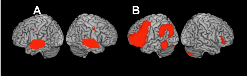
Figure 1. A: Whole brain inter-subject correlation for Taken audio-narrative (p < .0001,
FWE corr.). B: Correlated activity estimated by the auditory envelope for Taken (p < .05,
FWE corr.).
To extract network-specific activity, particularly to do with the auditory and
fronto-parietal, from the groups’ Taken fMRI data, a 20-component tensor ICA was
performed. For each network, the IC which best fitted the expected spatial extent in the
previously described brain regions and explained the most variance relative to other
versions of the same network across the awake group were selected as the representative
for the subsequence analyses (Figure 3). For the fronto-parietal network, the spatial map
of the fronto-parital IC showed dominant left distribution in the superior, middle, and
inferior frontal gyri, as well as in the right inferior parietal lobule and inferior temporal
gyrus. The auditory IC showed bilateral distribution in the superior temporal gyrus and
superior temporal sulci.
Figure 3. Group ICA results. A: Auditory network. B: Fronto-parietal.
In the next analysis, first, the ICA-derived time-course for the auditory
component was used as a regressor in the GLM analysis and successfully estimated
activity in the auditory network of each awake participant (15-of-15; p < .05, FWE corr.);
suggesting similar perceptual processing across wakeful individuals (Figure 4). Next, the
time-course of the fronto-parietal component was used in the GLM analyses of individual
participants. Significant activity was estimated (p < .05, FWE corr.) and the expected
fronto-parietal network distribution was observed in 12-of-15 individuals (Figure 5). Two
additional participants showed sub-threshold activity in the frontoparietal network that
did not need the criterion for statistical significance (as denoted by ‘*’ in Figure). This
result is consistent with previous research that showed higher inter-individual variability
(lower ISC) in regions supporting high-order cognition as compared to sensory-driven
brain areas (Hasson et al., 2004).
Figure 4. This figure shows the significant (p < 0.05, FWE corr.) activity estimated by the
time-course of the auditory IC derived from the awake group Taken data in individual
participants. The observed significant activity demonstrates that processing in the
auditory regions at the single-subject can be predicted from the group in 100% of
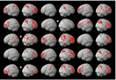
Figure 5. This figure shows the significant activity estimated by the time-course of the
fronto-parietal IC of the awake group Taken data (p < 0.05, FWE corr.) in individual
participants. The observed significant activity demonstrates that processing in the frontal
and parietal regions at the single-subject can be predicted from the group in the majority
(80%) of participants (12/15). 13% of participants (2/15) showed appropriate, but weak
(sub-threshold) activity in the fronto-parietal regions, which may be expected for
single-subject datasets. “*” denotes results prior to FWE correction; these clusters do not meet
significance once corrected.
Shared experience of suspense during the Taken narrative. An attempt was
made to explicitly connect the synchronized processing in the fronto-parietal network to
qualitative reports of cognition, in particular the subjective experience of suspense, to
this, a group of participants independent of the group that listened to Taken in the scanner
performed a behavioural experiment in the laboratory in which they rated their
experience of suspense during the Taken narrative, every 2.16 seconds. Figure 7 shows
each individual’s ratings of suspense over the duration of the stimulus and the
group-averaged rating. In a leave-on-out analysis, each individual’s sequential suspense ratings
were compared to the group-averaged ratings of all other participants, which showed
highly similar suspense ratings across different participants [r = .89, t(18) = 8.28, p <
.001]. These results suggested that different participants experienced suspense in a highly
similar manner throughout the story on a moment-by-moment basis.
Figure 6. Suspense ratings over the duration of the Taken audio-narrative (N=20).
Suspense was rated on a Likert scale from 1 (least suspenseful) to 9 (most suspenseful). Ratings
were collected every 2.16 seconds to correspond with the repetition time (TR) used in the
independent fMRI control group. Each thin coloured line displays a participant’s
suspense ratings over time. The thick red line represents the group-average suspense
rating. -5 -2 1 4 7 10
1 16 31 46 61 76 91 106 121 136
Su
sp
en
se
R
at
in
g
(1
-9)
As the experience of suspense was highly similar across individual participants in
the behavioural group, this suggested that it may be similar across different participants
or participant groups regardless of testing conditions. This result motivated the use of the
behavioural measure of suspense, which was obtained in a behavioural group, as a
regressor in the fMRI data of the independent group that listened to the narrative in the
scanner, in order to investigate the brain regions that supported the shared experience of
suspense.
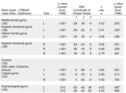
The group-averaged suspense ratings were used as a regressor in the GLM of the
fMRI awake group. This model significantly estimated activity (p < .05, FWE corr.) in a
set of regions including the left middle frontal gyrus, inferior frontal gyrus, right superior
temporal gyrus, calcarine sulcus, bilateral lingual gyrus, and left supramarginal gyrus
(Table 1). These regions comprise the left fronto-parietal functional network and a set of
Figure 7. The ratings of suspense obtained from the independent behavioural group
predicted brain activity in fronto-parietal and auditory regions of the participants who
heard the Taken narrative inside the scanner. (p < 0.05, FWE corr.).
Table 1. Group activation predicted by suspense rating.
Brain areas - LPBA40
Label (Max. Likelihood) Side
p value (cluster-level, FWE) MNI Coordinate of Cluster Peaks z valu e p value (peak-level, FWE)
x y z
Middle frontal gyrus
(.90) L <.001 -26 38 -4 5.52 .003
Superior temporal gyrus
(.99) L <.001 -46 -22 2 5.07 .029
Inferior frontal gyrus
(.97) L <.001 -40 40 -2 4.95 .046
Superior temporal gyrus
(.99) R <.001 62 -24 8 5.05 .031
R <.001 60 -16 6 4.99 .039 R <.001 46 -16 6 4.79 .081
Cuneus (.52) (AAL atlas: Calcarine
sulcus) L <.001 -2 -86 4 4.92 .051
Lingual gyrus
(.83) L <.001 -8 -78 4 4.50 .213
R <.001 4 -82 -4 4.00 .700
Supramarginal gyrus
(.94) L L .010 .010 -52 -42 38 4.03 -50 -44 50 3.45 .667 .996
Significant whole-brain results for the group data modeled by the group-averaged
suspense ratings (p<0.001, uncorrected). Significance is displayed for the cluster and peak
voxel. LPBA40; LONI Probabilistic Brain Atlas (Shattuck et al., 2007). MNI coordinates
of each cluster’s peaks were referenced to maximum likelihood maps which identify the
In summary, the previous result showed that suspense ratings obtained from an
independent group predicted activity in a network of regions known to support the
experience of suspense, including fronto-parietal cortices in the awake group. This
suggests that it can be used to probe the experience of suspense during the narrative in
independent participants who undergo fMRI scanning in the absence of their subjective
reports of suspense. To determine the suitability of this measure for the assessment of the
potential experience of suspense in individual sleepers in the sleep experiment, a test was
conducted to investigate whether the group-level activity could be reliably observed in
each wakeful individual.
For each individual participant who heard Taken in the scanner, the
group-averaged suspense rating was used in the GLM of their individual data. These
single-subject models demonstrated that significant brain activity in the frontal, parietal and
temporal regions could be estimated by the independent behavioural measure (p < .05,
FWE corr.) for the majority (80%) of awake participants (12 of the 15) (Figure 8). These
findings suggest that the behavioural ratings of suspense provide a robust measure for
investigating the experience of suspense in the absence of report in individual
participants. Therefore, the group-averaged suspense ratings provide a suitable measure
for testing the potential experience of suspense in individual sleepers whom are unable to
Figure 8. This figure displays the significant brain activity estimated in awake single
individuals, who listened to Taken inside the scanner, by the average suspense rating of the
independent behavioural group (p < 0.05, FWE corr.). The auditory and fronto-parietal
regions in the majority (80%) of individuals (12/15) responded significantly to suspense
throughout the narrative. In 13% (2/15) of participants only auditory regions respond to
suspense, but these exhibit weak and sub-threshold activity that does not meet the
significance criterion. Results prior to FWE correction are denoted with ‘*’; these clusters
do not meet significance once corrected.
Discussion
In this chapter two experiments in independent groups of awake and healthy
paradigm consisting of unconstrained and eyes-closed listening to an auditory narrative
(Naci et al., 2017), in the assessment of individual participant’s understanding of the story.
First, an fMRI imaging study was performed to investigate if listeners similarly processed
Taken, and if a template of expected brain activity could be developed to assess processing
in individual participants. Although the previous study (Naci et al., 2017) that used the
same paradigm in awake individuals and which informs this work suggested that this
might be the case, here the aim was to replicate the results and test the suitability of this
paradigm with a different set of scanning parameters required for the simultaneous
EEG/fMRI sleep acquisition. Second, a behavioural measure of high-level cognition
based on direct report was developed to investigate whether high-order cognition during
the story could be detected based on brain activity alone and in the absence of any
behavioural measures in individual sleeping participants.
Strong ISC of brain activity was found during the Taken narrative in a wide set of
brain regions across different awake individuals, replicating the findings from Naci et al.
(2017). Further, these results agreed with the findings of previous studies which have also
shown movie-viewers to have highly correlated brain activity (Hasson et al., 2010, 2004;
Naci et al., 2014). Next, it was important to demonstrate that this broad synchronous
activity spanning different functional cortical areas was related to the understanding of
the story and was not driven simply by the low-level auditory features.
Synchronized activity only in the primary auditory cortex was found to be driven
by low-level auditory features. This suggests that higher level features of the stimulus must
be responsible for the extra-modal neuronal engagement. In a similar attempt to show the
response to an acoustically scrambled version of Taken. This meaningless audio stimulated
activity in sensory-driven perceptual regions and did not elicit synchronous activity in
frontal and parietal regions. This suggests that the cognitive demands of plot-following,
namely the integration of the continuous stream of information into a coherent narrative,
drove similar brain activity across individuals.
An ICA approach was then used to investigate the specific recruitment of
functional networks known to be important for processing sensory-specific and
higher-order aspects of the complex auditory narrative that evolved over time, in particular the
auditory and fronto-parietal networks. Awake listeners displayed a stereotypical
distribution of activity in superior temporal gyrus and superior temporal sulcus,
sensory-driven auditory regions known to respond to incoming auditory stimuli regardless of
complexity (Binder et al., 1994). The spatial map of the fronto-parietal IC displayed
activity in areas known to support executive functioning (Barbey et al., 2012; Duncan,
2010; Hampshire & Owen, 2005; Naci et al., 2014; Owen et al., 1990; Ptak, 2012;
Sauseng et al., 2005; Woolgar et al., 2010). However, this component was mostly
lateralized to the left hemisphere. As this activation is the neural response from
auditory-only information, with most meaning conveyed through speech, the strong
left-lateralization may not be too surprising. Speech processing is known to be supported by
brain regions in the left hemisphere (Poldrack et al., 1999; Zatorre, Evans, Meyer, &
Gjedde, 1992) so the left-lateralized frontal and parietal synchronization may indicate the
propagation of this speech information. The greater integration of this information in the
left hemisphere may explain the emergence of a lateralized FP component.
reliability of brain activity in the auditory and fronto-parietal networks of each awake
individual was investigated. Importantly, the activity in the auditory and fronto-parietal
networks were successfully estimated at the individual level, which suggests that this
method can be used to assess processing in individual sleepers. Taken-specific auditory
processing was robustly estimated in all 15 out of 15 participants. The fronto-parietal
activity was estimated in 12 of 15 participants; however 2 participants showed
sub-threshold activity in the right regions. Inter-individual variability in brain activity is a
known property of high-order information-processing (Geerligs, Rubinov, Cam-CAN, &
Henson, 2015; Rypma & D’Esposito, 1999). Therefore, it is sensible that the
group-derived time-course for fronto-parietal activity does not perfectly estimate brain activity in
each individual. Additionally, the fronto-parietal IC is in itself not a direct measure of
high-order information-processing and may not best model for the meaningful processing
of the narrative across all individuals. To address this, a continuous measure of the
perceived suspense during Taken was developed and used it as a proxy for high-level
cognition.
To determine if Taken provided participants with a shared conscious experience
which could be used as a template to investigate the experience of individual sleepers in
the next study, in the second experiment the relationship between each individual’s
suspense ratings and those of the other participants was examined. Suspense ratings were
strikingly similar across the independent behavioural group. This suggested that the
conscious experience of suspense was similar for all awake participants. A large body of
research has demonstrated that suspenseful stimuli such as reading suspenseful sentences,
functional brain networks (Kober et al., 2008). Therefore, the ability for the
group-averaged suspense ratings for Taken to reliably estimate a neural correlate of executive
function in each individual was tested.
The suspense ratings were used as a regressor in a GLM of the awake Taken group
data. This modelled activity in the fronto-parietal network and a set of auditory and
speech related regions. This suggests that the experience of suspense was supported by the
fronto-parietal network. Importantly, the group-averaged suspense ratings robustly
estimated similar fronto-parietal activity at the individual level, suggesting that this is
indeed a good measure for investigating information-processing in individual sleepers.
The fronto-parietal network has been previously implicated in processing
suspense. When participants viewed a clip from the movie Bang! You’re Dead (1954), Naci
and colleagues (2014) also found that common fronto-parietal activity across participants
was driven by the perception of suspense. In another study, when participants viewed a
suspenseful movie, fronto-parietal activity increased in moments of suspense (Bezdek et
al., 2015). To reiterate from Chapter 1, the executive processes required to understand a
complex narrative are known to be supported by a fronto-parietal network; as a result,
Taken-driven activity in this network can be used as a marker for executive processing
related to the contents of the Taken movie in the absence of behaviour.
More research has found suspense to reliably drive audiences’ shared experiences.
By studying the emission of specific chemicals by audiences watching different genres of
movies, researchers were able to blindly predict suspenseful moments in a film (Williams
reliably produced for a film, providing further evidence that suspenseful content drives
synchronized activity.
In summary, the results suggest that the suspense ratings are an effective
measurement of a shared experience and can be used to evaluate this experience in the
absence of behaviour. The auditory and fronto-parietal networks were found to
synchronize across awake participants who listened to Taken. Lastly, the time-courses of
this network activity, as well as the suspense measure, could estimate fronto-parietal
activity in each individual. This establishes that the Taken stimulus and analytical
Chapter 3: Sleep Study
Introduction
The aim of this chapter is to examine if the sleeping brain is able to respond to the
high-order cognitive demands of complex and continuous information from the external
environment. To accomplish this, a group of participants attempted to sleep in the MR
scanner and were presented Taken once sleep was observed to be stable. Sleeping
individuals were unable to provide subjective feedback to corroborate their experience
associated with the Taken stimulus. For this reason, a test was completed to see if any
individuals showed BOLD activity similar to that of the independent awake group who
heard Taken in the scanner and secondly, tested whether their brain activity responded
specifically to the measure of suspense throughout the story.
Complex information-processing may be disrupted by the neurophysiological
changes that occur during sleep (Hobson & Pace-Schott, 2002). Specifically, the
communication within and between functional networks known to support the
understanding of a story (Naci et al., under review) may inherently vary across sleep
stages. Thus, the first aim was to determine if each sleep stage is accompanied by inherent
changes in the organization of functional networks.
While participants are engaged in typical sensory, cognitive, or resting-state
paradigms, correlations of slow (<0.1 Hz) spontaneous BOLD signal fluctuations have
been found to reliably establish a number of widely distributed functional networks (Fox
& Raichle, 2007). Plot-following during audio-visual and auditory-only movies is known
awake resting state (Raichle, 2011).
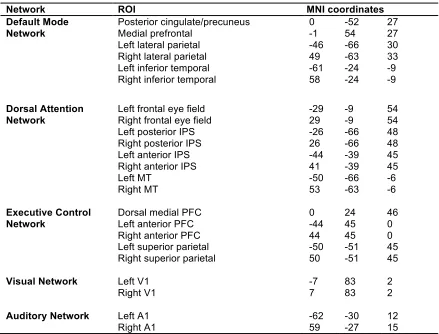
Five key networks important for auditory processing that have previously been
investigated for fMRI data of the Taken story used here (Naci et al., under review) were
focused on: three higher order and two primary sensory systems. The higher order
networks included: the dorsal attention network (DAN; Corbetta & Shulman, 2002),
which focuses attention on important environmental features; the executive control
network (ECN; Boly et al., 2008), which regulates overt responses demanded by complex
situations; and the default-mode network (DMN; Buckner, Andrews-Hanna, & Schacter,
2008; Raichle et al., 2001), which is often deactivated during externally oriented tasks but
theorized to play an active role in autobiographical memory, social cognition, and mental
simulations of the future. Notably, and a little surprisingly, the auditory-only excerpt of
Taken has been shown to drive activity in the visual cortex (Naci et al., 2017), possibly due
to the visually evocative information presented. As such, the primary auditory and visual
networks were also included in our functional connectivity (FC) analysis.
During sleep, the potential functional re-organization of these five key networks
may well influence the fate of incoming information. Using fMRI, functional connectivity
is often measured as the correlation strength between BOLD activity in nodes of a
network (within-network) and between networks (between-network; Fox et al., 2005).
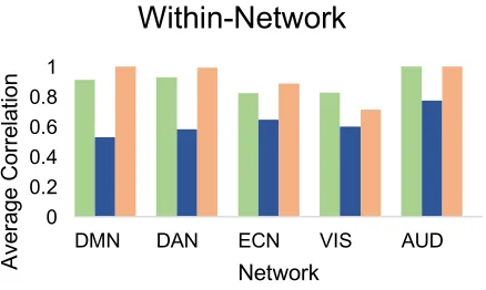
Within-network connectivity of the DAN, ECN, DMN, AUD, and VIS networks have
been found to persist during NREM-1 and NREM-2 sleep (Fukunaga et al., 2006;
Picchioni, Duyn, & Horovitz, 2013) and during propofol-induced loss of consciousness
(Boveroux et al., 2010). A lack of signal propagation between networks has been observed
propagation of a transcranial magnetic stimulation (TMS) evoked signal, Massimini et al.
(2005) observed a breakdown in connectivity beyond the stimulation site during NREM-2
and NREM-3 sleep. A breakdown of inter-cortical propagation was also found by Esser et
al. (2009). Hierarchical clustering analyses have also shown a breakdown of the
fronto-parietal network during NREM sleep (Larson-Prior et al., 2011; Spoormaker et al., 2010).
The breakdown of connectivity in NREM-2 and NREM-3 may restrict the brain’s ability
to process complex and continuous information during sleep.
Further, global and local changes in the brain’s metabolism may help to inform
predictions about the potential processing of complex information during sleep. Using
positron emission tomography (PET), global glucose metabolism has been found to
decrease progressively through NREM sleep (Maquet, 1995) and increase above waking
levels during REM (Maquet, 2000). Primary sensory networks have shown similar
reductions in metabolic demand and blood-flow during NREM-2 as compared to when
awake (Braun et al., 1997) with intact FC (Larson-Prior et al., 2009). However, the
transition to NREM-3 shows decreased blood flow in the lateral and medial prefrontal
cortex (Braun et al., 1997; Maquet, 2000). The prefrontal cortex (PFC) includes
non-motor regions of the frontal lobe that support the complex processes of the DAN, ECN
and anterior DMN. The decreased metabolism in the PFC during NREM-3 may limit
the ability for sleepers to process the high-order demands of Taken.
Because of a lack of activity in the fronto-parietal network during REM and
NREM-3, executive function may be impaired. The decreased activity in the PFC
observed during NREM sleep was shown to persist during REM (Maquet et al., 1996).
inability to reflect on, or attend to, the contents of consciousness during sleep (Hobson et
al., 2000). Executive deficiencies during sleep as observed in dream mentation, and the
decreased metabolism in the PFC, may likely restrict the ability for sleeping participants
to integrate information over time so as to process the meaning of complex narratives.
Participants were expected to experience difficulty sleeping and remaining asleep
through Taken in the restrictive scanner environment. Additionally, if participants were to
sleep, the sleep stage/s present during Taken were expected to vary by individual because
sleep is a very dynamic process with high inter-individual variability in architecture
(Ohayon et al., 2004) as shown by EEG activity (Buckelmüller et al., 2006). Thus, to
understand the inherent inter-individual variability in functional connectivity between
sleep stages, which may underlie inter-individual variability in stimulus-driven responses
during sleep, the functional connectivity of aforementioned functional networks was
investigated in each stage of stimulus-free sleep.
Beyond the assessment of functional connectivity changes inherent to different
sleep stages, the focal aim of this chapter was to apply a naturalistic paradigm to
investigate if the brain is capable of meaningfully integrating information from the
environment while in a state of sleep. Support for the application of the Taken paradigm
was described in Chapter 2. As described in the previous chapter, of specific interest to
the application of the Taken paradigm to investigating sleep was its ability to reliably elicit
similar activity in the auditory and fronto-parietal networks of individual awake listeners.
Here, the functional time-courses of these networks were used to test for the presence of
activity in individual sleeping participants that was similar to that of awake individuals.
processing, whereas activity in the fronto-parietal network would suggest preserved
high-order cognition. Importantly, as described in Chapter 2, awake participants in an
independent behavioural experiment experienced suspense during Taken highly similarly
to one another, an experience that was supported by engaging the fronto-parietal
network. Therefore, the brain activity reliably estimated by an awake group’s suspense
ratings (see Chapter 2) could be used to infer if individual sleepers were able to follow the
narrative and had a cognitive experience of suspense that was similar to awake
individuals.
To summarize, first, a functional connectivity analysis was applied to data from
stimulus-free sleep to see if inherent brain connectivity differed in different sleep stages,
and therefore warranted separate analyses of Taken data from different sleep stages.
Secondly, the time-courses of the auditory and fronto-parietal ICs from the awake group,
who listened to Taken inside the scanner, were used to investigate stimulus-driven
processing in individual sleepers. Thirdly, the measure of suspense from the independent
awake group was used to investigate the potential meaningful integration of external
information during sleep.
Method
Participants. Ethics approval was obtained from the Psychology Research Ethics Board
history of psychiatric or neurological disorders, and passed sleep-screening criteria. To
increase the likelihood of collecting normal sleep in the evening, participants were
excluded if they worked night shifts, had taken a trans-meridian trip in the last three
months, or were categorized as extreme morning or evening types (Horne-Ostberg
Morningness-Eveningness Scale; Horne & Ostberg, 1976). Participants were reportedly
non-smokers, medication-free, and were asked to abstain from consuming alcohol,
caffeine, and nicotine for the day of the experimental night. To be included in this study,
participants had to score less than 10 on the Beck Depression (Beck, Rial, & Rickels,
1974) and the Beck Anxiety (Beck, Epstein, Brown, & Steer, 1988) inventories. This
criterion was used as depression is the most common psychiatric disorder associated with
abnormal sleep (Tsuno, Besset, & Ritchie, 2005). Participants also had to have no history
or signs of sleep disorders as indicated by the Sleep Disorders Questionnaire (Douglass et
al., 1994).
Participants were required to keep a regular sleep-wake cycle (asleep from 22h00 -
01h00, awake from 07h00 - 10h00) and abstain from napping for a week prior to the
experiment. They were not sleep deprived. Sleep schedule adherence was assessed using
both sleep-log and wrist actigraphy (Actiwatch 2, Philips Respironics). Participants were
given a letter of information and provided informed written consent prior to study
participation, and were monetarily compensated. A group of 30 healthy participants, who
met the inclusion criteria, were scanned at night (21h30 – 24h00) while attempting to
sleep at the Robarts Research Institute in London, Ontario, Canada. Data from 26
participants were used in the final analyses (15 females) between the ages of 18-34 years