DISCOVERY OF MATRIPTASE
Matriptase (MT-SP1, TADG-15, ep-ithin, ST14) was first described in 1993 as a new gelatinolytic activity in cul-tured breast cancer cells (1). It was mol-ecularly cloned by four different groups at the turn of the millennium (2-5) and was shown to be the sixth member of the then-emerging family of type II transmembrane serine proteases (TTSPs), which today comprises 17 members in humans and 20 in mice (6-8). Two close cousins of matriptase, matriptase-2 and matriptase-3, were identified recently (7,9,10). Orthologs of matriptase are present in all nine vertebrate genomes examined to date (human, chimpanzee, dog, mouse, rat, chicken, zebrafish, spotted green pufferfish, and tiger pufferfish), which suggests a conserved evolution-ary function (R. Szabo and T. Bugge, unpublished data). This review summa-rizes the progress over the past half-dozen years in unraveling the biochem-istry, physiology, and pathology of this complex and fascinating cell surface serine protease.
THE COMPLICATED LIFE CYCLE OF MATRIPTASE: INHIBITOR-ASSISTED AUTOACTIVATION, INHIBITION, AND SHEDDING
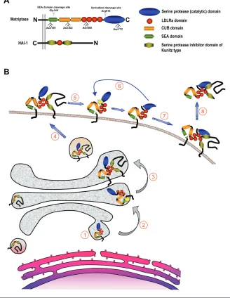
Matriptase is a 80- to 90-kDa cell sur-face glycoprotein with a complex modu-lar structure that is common to all matrip-tases (Figure 1A). It lacks a classical signal peptide, and the N-terminal signal an-chor, which is not removed during syn-thesis, functions as a single-span trans-membrane domain that orients the protease in the plasma membrane as a type II integral membrane protein with a cytoplasmic N-terminus and an extracel-lular C-terminus (11). The function of the intracellular domain of matriptase (residues 1-54) is presently unknown, al-though it is plausible to speculate that it binds cytosolic proteins to regulate enzy-matic activity and cellular distribution of the protease. Indeed, a recent report by Kim et al. (12) suggests that the direct in-teraction between matriptase and the actin-binding protein filamin anchors the protease to the actin cytoskeleton. This would be consistent with the observation that the exposure of immortalized human
mammary epithelial cells to sphingosine-1-phosphate (see below) leads to the rapid translocation of matriptase to the cell sur-face and a subsequent activation in an actin cytoskeleton remodeling–dependent manner (13). The extracellular stem region of matriptase consists of a single SEA (residues 86-201), 2 CUB (residues 214-334 and 340-447), and 4 LDLRA (residues 452-486, 487-523, 524-561, and 566-604) do-mains. These noncatalytic modules ap-pear to play an essential role in the cellular localization, activation, inhibition, and, likely, the substrate specificity of ma-triptase. The C-terminal serine protease domain (residues 614-855) is structurally highly similar to that of the other TTSPs and other members of the S1 clan of trypsin-like serine proteases (6).
Matriptase is synthesized as an inac-tive, single-chain zymogen. The activation of the matriptase zymogen is extraordi-narily complex, unique among all serine proteases studied to date, and incom-pletely understood. Matriptase activation requires two sequential endoproteolytic cleavages and involves the transient inter-action with its cognate inhibitor, hepato-cyte growth factor activator inhibitor (HAI)-1 (Figure 1B). The mature single-chain proenzyme is first cleaved after Gly149 located in a conserved GSVIA motif in the N-terminal SEA domain by an unknown proteolytic activity or possi-bly by nonenzymatic hydrolysis of the Address correspondence and reprint requests toThomas H. Bugge, Proteases and Tissue
Remodeling Unit, Oral and Pharyngeal Cancer Branch, National Institute of Dental and Craniofacial Research, National Institutes of Health, 30 Convent Dr, Room 211, Bethesda, MD 20892. Phone: (301) 435-1840; fax: (301) 402-0823; e-mail: [email protected]. Submitted March 22, 2006; accepted for publication April 4, 2006.
Karin List, Thomas H Bugge, and Roman Szabo
Proteases and Tissue Remodeling Unit, National Institute of Dental and Craniofacial Research, National Institutes of Health, Bethesda, MD, USA
Matriptase is a type II transmembrane serine protease expressed in most human epithelia, where it is coexpressed with its cog-nate transmembrane inhibitor, hepatocyte growth factor activator inhibitor (HAI)-1. Activation of the matriptase zymogen re-quires sequential N-terminal cleavage, activation site autocleavage, and transient association with HAI-1. Matriptase has an es-sential physiological role in profilaggrin processing, corneocyte maturation, and lipid matrix formation associated with terminal differentiation of the oral epithelium and the epidermis, and is also critical for hair follicle growth. Matriptase and HAI expression are frequently dysregulated in human cancer, and matriptase expression that is unopposed by HAI-1 potently promotes car-cinogenesis and metastatic dissemination in animal models.
peptide bond (12,14). Although this severs the covalent link to the signal anchor, ma-triptase remains tightly attached to the cell surface, possibly via noncovalent in-teractions within the cleaved SEA
do-main. SEA-domain cleavage appears to take place already in the secretory path-way, as only the N-terminally cleaved form of the enzyme is present on the sur-face of cells (14). Subsequent to and
de-pendent on SEA domain cleavage, matrip-tase next is converted into its active con-formation by proteolytic cleavage after Arg614 within the highly conserved acti-vation cleavage site R-VVGG in the serine protease domain. The activation site cleavage appears to require the prote-olytic activity of matriptase, as mutations in any of the residues of the catalytic triad render matriptase unable to undergo acti-vation site cleavage. This obseracti-vation led to a proposed transactivation mechanism in which a weak intrinsic proteolytic ac-tivity of SEA domain–cleaved matriptase zymogen activates neighboring SEA do-main–cleaved matriptase molecules (15).
The proteolytic autoactivation of ma-triptase appears to be controlled by the stem region, posttranslational modifica-tions, and the cellular localization of the protease. Inactivating mutations in the Ca2+-binding motifs of any or all of the four LDLRA domains prevents the activa-tion of matriptase. Interestingly, however, the complete deletion of all four LDLRA domains allows efficient activation of the enzyme, suggesting an autoinhibitory role of the LDLRA modules that may prevent premature activation of matriptase in the absence of appropriate activation stimuli (15). Glycosylation also appears to be crit-ical to activation. Matriptase contains four functional N-glycosylation sites, Asn109, 302, 485, and 772. Whereas the inactiva-tion of Asn109 and Asn485 had no effect on the activation of matriptase, glycosyla-tion of the first CUB domain (Asn302) and the catalytic domain (Asn772) was re-quired for zymogen activation in cultured breast cancer cells (15).
The specific mechanisms that trigger the activation of matriptase are incom-pletely understood. In a pioneering study, Benaud et al. (16) showed that matriptase translocates to the cell surface and is acti-vated within minutes after exposure of breast cancer cells to sphingosine-1-phosphate, a serum-derived lipid that sig-nals through specific G-protein–coupled receptors. Interestingly, this activation process was shown to require actin cy-toskeleton remodeling (17). Other mole-cules shown to induce matriptase activa-Figure 1.Structure of matriptase and HAI-1 and proposed life cycle of matriptase. (A) Domain
tion linked to spatial redistribution in-clude suramin and androgens in prostate cancer cells (18). The ability of the matrip-tase zymogen to become activated and presented on the cell surface may depend on the direct physical interaction with its cognate Kunitz-type serine protease in-hibitor HAI-1 (Figure 1B). Thus, in the ab-sence of HAI-1, matriptase accumulates in the Golgi compartment (19). Interestingly, a point mutation in the single LDLRA do-main or the Kunitz dodo-main 1 of HAI-1 prevented both cell surface translocation and activation of matriptase (15,19). In contrast to the wild-type enzyme, catalyti-cally inactive matriptase mutants were readily deposited on the cell surface even in the absence of HAI-1 (19).
The inhibition of activated matriptase by HAI-1 was first documented by the identification of matriptase/HAI-1 com-plexes in human milk and conditioned medium of cultured mammary epithelial cells and in a number of cancer cell lines (20). Recently, the functional relevance of HAI-1 inhibition of matriptase was con-firmed in a transgenic mouse model in which matriptase-induced skin tumorige-nesis was completely prevented by the overexpression of HAI-1 in the same tis-sue (21) (see below). Kunitz domain 1, but not Kunitz domain 2, of HAI-1 is re-sponsible for the inhibition of matriptase (15). Interestingly, the interaction also re-quires a functional LDLRA domain of HAI-1 (22). The N-terminal cleavage of the SEA domain makes matriptase sus-ceptible to the shedding of its extracellu-lar part. Indeed, the original isolation of matriptase from human milk suggests that a significant shedding of the protease takes place from the cell surface in vivo. Data obtained from cell culture systems indicate that most of matriptase released from cells is in a two-chain form, and, in fact, the deletion of the fourth LDLRA domain or the activation cleavage site, which stabilizes the zymogen form, pre-vents cell surface shedding, without in-terfering with N-terminal processing (23). The shedding of matriptase also appears to require the presence of HAI-1, as only HAI-1–complexed, but never HAI-1–free,
active matriptase is detected in milk or conditioned medium (24,25). The specific molecular events that lead to matrip-tase/HAI-1 shedding as well as the ulti-mate fate of the complex are unknown.
PHYSIOLOGICAL FUNCTIONS OF MATRIPTASE
Matriptase is a strictly epithelial pro-tease with a fairly widespread, but not ubiquitous, expression in human and mouse tissues. Expression has been docu-mented in epidermis, cornea, salivary gland, oral and nasal cavities, thyroid, thymus, esophagus, trachea, bronchioles, alveoli, stomach, pancreas, gallbladder, duodenum, small intestine, colon, rectum, kidney, adrenals, urinary bladder, ureter, seminal vesicles, epididymis, prostate, ovaries, uterus, and vagina (26,27, K. List and T. Bugge, unpublished data). Mutations in the matriptase gene have not been identified to date in humans or other animal species. However, a gene-targeting study in mice has revealed an essential role of the membrane protease in oral epithelium, epidermis, hair folli-cles, and thymic epithelium (27-29). Matriptase-deficient mice develop to term but uniformly die within 48 hours of birth as a consequence of seriously compro-mised epidermal barrier function, which leads to rapid and fatal dehydration. Detailed analysis of this barrier defect un-covered an essential role of matriptase in the formation of the two physical struc-tures that form the epidermal barrier: the stratum corneum lipid matrix and the cornified envelope of corneocytes.
Interestingly, at the molecular level, ma-triptase deficiency completely abrogates the proteolytic processing of the polyprotein profilaggrin into filaggrin monomer units and an N-terminal filaggrin S-100 regula-tory protein, the latter of which translocates to the nucleus to promote terminal epider-mal differentiation. As partial loss-of-func-tion mutaloss-of-func-tions in both the mouse and human profilaggrin genes are associated with epidermal barrier defects, this matrip-tase-dependent profilaggrin processing pathway may define one key step in the initiation of terminal epidermal
differentia-tion and acquisidifferentia-tion of epidermal barrier function (30,31). In this regard, it should be noted that deletion of the SPINK-5gene, which encodes a Kazal-type serine protease inhibitor that is coexpressed with matrip-tase in the epidermis, leads to accelerated profilaggrin processing and premature dif-ferentiation of human and mouse skin (32,33). Loss of matriptase also impairs the growth of hair follicles and prevents vibris-sae eruption owing to absence of formation of vibrissal hair canals. Furthermore, ma-triptase dramatically increases apoptosis of immature thymocytes in the thymus, lead-ing to thymocyte depletion.
Detailed localization studies, using en-zymatic gene trapping combined with im-munohistochemical and ultrastructural analysis, have revealed a close association between matriptase expression and func-tion (Figure 2A,B). In the epidermis, kera-tinized oral epithelium, and thymic ep-ithelium, matriptase is exclusively expressed in postmitotic transitional-layer keratinocytes in the process of undergo-ing terminal differentiation. In all three tissues, matriptase colocalizes with profi-laggrin, frequently displaying close prox-imity to profilaggrin-containing granules, as revealed by ultrastructural analysis (27). Likewise, in accordance with the im-portant function of matriptase in hair fol-licle growth, matriptase is specifically ex-pressed in growth phase (anagen) hair follicles and is located in undifferentiated and rapidly proliferating hair matrix cells, precortex, and cortex cells of the cycling portion of the hair follicle (27). The explo-ration of potential physiological functions of matriptase in nonepidermal tissues is complicated by the perinatal mortality of matriptase-deficient mice and awaits the identification of spontaneous matriptase mutations in humans or other animal species, or the generation of conditional matriptase knockout mice.
MATRIPTASE IN EPITHELIAL CARCINOGENESIS
matriptase is expressed in carcinomas of the head and neck, mesothelium, breast, ovary, cervix, prostate, lung, and gastroin-testinal tract, as well as in cell lines derived from these tumors. Matriptase does not
ap-pear to be expressed in tumors of mes-enchymal origin, suggesting a specific function of the protease in epithelial car-cinogenesis (34-38, K. List and T. Bugge, unpublished data). Multiple studies have
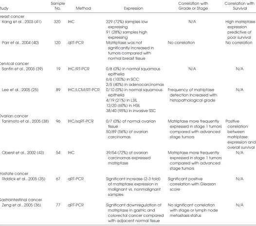
assessed the level of expression of matrip-tase during malignant progression and the potential value of matriptase as a prognos-tic marker in various human cancers. The findings from these studies, which are summarized in part in Table 1, do not paint a consistent picture. In prostate and cervical cancer, matriptase mRNA and pro-tein are upregulated in cancerous lesions compared with normal tissue, and there is a positive correlation between matriptase expression and histopathological grade of the tumor (25,35,39). In contrast, in the gas-trointestinal tract, a significant downregu-lation of matriptase mRNA compared with normal tissue, as well as a decrease of ma-triptase mRNA levels with increasing tumor grade, has been reported (36). Matriptase is expressed at very low levels in the normal ovary, becomes highly ex-pressed in early-stage ovarian carcinoma, and is then downregulated in advanced-stage tumors (38). In breast cancer, one study concluded that matriptase mRNA was not significantly increased in tumors compared with normal breast tissue (40), whereas another study reported that high matriptase expression is predictive of poor survival as assessed by immunohistochem-ical detection of matriptase (41).
Although variations in cancer-associated matriptase expression between different human cancers may reflect, in part, dif-ferences in quantitation methodology, tis-sue sampling, tistis-sue composition, and tumor staging, it is likely that potential diverging roles of matriptase in different epithelia—under both normal physiologi-cal conditions and tumor progression— also contribute to the complexity of interpreting the relationship between ma-triptase expression and human cancer. Furthermore, whereas expression studies give valuable information regarding ma-triptase localization and total levels of matriptase mRNA or protein, the activity level and regulation of matriptase are not measured. As for most extracellular pro-teases, the specific detection of matriptase activity in vivo and in situ is an impor-tant, but as yet unmet, challenge (42).
Interestingly, studies of tumor and can-cer cell extracts, where matriptase can be Figure 2.Expression and function of matriptase in normal epidermal structures and
detected in an active free form or in a high-affinity complex with HAI-1, suggest that a larger proportion of total matrip-tase exists in its inhibitor-free form in can-cer cells, compared with nontumorigenic epithelial cells (2,20,34). Matriptase and HAI-1 are coexpressed with remarkable consistency in normal epithelium, sug-gesting that the proteolytic activity of
ma-triptase is strictly regulated (34). It there-fore has been proposed that an imbalance in the matriptase–HAI-1 ratio, rather than absolute matriptase levels, may be indica-tive of a cancer-associated dysregulation of matriptase-mediated proteolysis. In support of this proposition, an increased matriptase–HAI-1 mRNA ratio that corre-lated with tumor grade has been reported
in studies of gastrointestinal (36) and ovarian cancer, where advanced-stage tu-mors that expressed matriptase protein were more likely to do so in the absence of HAI-1 (43).
A recent animal study detailing the pat-tern of matriptase expression in epidermis during chemically induced multistage car-cinogenesis yielded valuable insights into Table 1.Matriptase expression in human cancer
Sample Correlation with Correlation with
Study No. Method Expression Grade or Stage Survival
Breast cancer
Kang et al., 2003 (41) 320 IHC 229 (72%) samples low N/A High matriptase
expressing expression
91 (28%) samples high predictive of
expressing poor survival
Parr et al., 2004 (40) 120 qRT-PCR Matriptase was not No correlation No correlation significantly increased in
tumors compared with normal breast tissue Cervical cancer
Santin et al., 2003 (39) 19 IHC/RT-PCR 0/8 (0%) in normal squamous N/A N/A epithelia
6/6 (100%) in SCC
2/5 (40%) in adenocarcinomas
Lee et al., 2005 (25) 89 IHC/LCM/RT-PCR 0/10 (0%) in normal squamous Frequency of matriptase N/A epithelia detection increased with
4/19 (21%) in LSIL histopathological grade 12/20 (60%) in HSIL
38/40 (95%) in invasive SSC Ovarian cancer
Tanimoto et al., 2005 (38) 96 IHC/sqRT-PCR 0/7 (0%) of normal ovarian Matriptase more frequently Positive tissue expressed in stage 1 tumors correlation 50/89 (56%) of ovarian compared with advanced between
carcinomas stage tumors matriptase
expression and overall survival Oberst et al., 2002 (43) 54 IHC 39/54 (72%) of ovarian Matriptase more frequently N/A
carcinomas expressed expressed in stage 1 tumors matriptase compared with advanced
stage tumors Prostate cancer
Riddick et al., 2005 (35) 67 qRT-PCR Significant increase (2-3 fold) Significant positive N/A of matriptase expression in correlation with Gleason
malignant vs. nonmalignant score samples
Gastrointestinal cancer
Zeng et al., 2005 (36) 77 qRT-PCR Significant downregulation of No significant correlation N/A matriptase in gastric and with stage or lymph node
colorectal cancer compared metastasis status with adjacent normal tissue
the expression of the membrane protease during carcinogenesis (27). The study re-vealed that matriptase was expressed in all stages of carcinogenesis at comparable levels, but underwent a dramatic spatial redistribution during the transition of epidermal lesions from hyperplasia to dysplasia (Figure 2C). Thus, whereas ma-triptase expression in normal and hyper-plastic epidermis is narrowly confined to highly differentiated, nonproliferating ker-atinocytes with no potential for malignant transformation, dysplastic and malignant lesions presented expression of matriptase in a much broader subset of keratinocytes. This subset included proliferating, keratin-5–expressing basal keratinocytes with high self-renewal capacity, which include epi-dermal stem cells believed to be the pri-mary target cells in epithelial carcinogene-sis. That this carcinogen-induced spatial dysregulation of matriptase indeed may be functionally relevant to epithelial car-cinogenesis has gained strong support from a transgenic mouse study showing that expression of matriptase even at modest levels in keratin-5–positive ker-atinocytes of the skin sufficed to both cause spontaneous epithelial malignancies and dramatically potentiate the effect of carcinogen exposure (21).
Several studies have addressed the role of matriptase in later stages of car-cinogenesis by inhibiting the protease in established tumor cell lines from prostate, colon, and ovarian cancer using small interfering RNAs, antisense ma-triptase oligodeoxyribonucleotides, or synthetic active-site matriptase in-hibitors. In all cases, matriptase inhibi-tion did not affect cancer cell prolifera-tion in vitro or in xenografted tumors, whereas tumor cell invasion was im-paired (44-46). How matriptase promotes malignant progression in these model systems is unknown. Dysregulated ma-triptase activity may directly affect the cell environment by altered processing of extracellular components and cell-matrix adhesion proteins. Matriptase may also act through the activation or inactivation of downstream effector molecules, in-cluding growth factors and receptors,
chemokines, and protease zymogens. In this respect, it is noteworthy that most of the candidate substrates for matriptase are implicated in malignant progression, including protease-activated receptor-2 (11), prohepatocyte growth factor activa-tor/scatter factor (11,47), receptor-bound pro-urokinase plasminogen activator (11,47), and the src-associated transmem-brane protein SIMA135/CDCP1 (48).
PERSPECTIVES
Twenty-first-century bioinformatics, proteomics, and mouse genetics have provided rapid insight into the biochem-istry, physiology, and pathology of ma-triptase, and an epithelial transmem-brane serine protease with a fascinating biochemistry and biology has been un-veiled. The unprecedentedly complex posttranslational regulation of matriptase defines a new paradigm for serine pro-tease zymogen activation and inhibition. Gene inactivation of matriptase provided the first demonstration of a cell surface serine proteolytic activity essential to oral epithelial and epidermal barrier for-mation, hair growth, and thymic epithe-lial function. Matriptase was found to be expressed in a curiously high proportion of human epithelial tumors and to pro-mote malignant progression in a multi-tude of animal models. As quickly as knowledge about matriptase is gained, however, pertinent new questions arise. As a conclusion to this review, we list just a few of these questions.
• What transcriptional and posttranscrip-tional regulatory networks govern the intricate regulation of matriptase expres-sion, activation, and inhibition? Studies of matriptase regulation and dysregula-tion in cells, tissues, and animals are well-deserving, given the interesting bi-ology and unique expression and regu-lation of the membrane serine protease. • Is dysregulation of matriptase causally
involved in the genesis or progression of cancer in humans? In other words, is the potent tumor-promoting potential of matriptase that is so convincingly demonstrated in xenograft and
trans-genic animal models unleashed during human carcinogenesis? The prognostic significance of matriptase and HAI-1 expression in several human carcino-mas suggests that this could be so. A final verdict may come from future clinical cancer trials using specific ma-triptase inhibitors.
• What are the overall physiological functions of matriptase beyond the epi-dermis? The evolutionarily conserved and intricate expression pattern of ma-triptase in nonepidermal tissues cer-tainly would suggest a generalized function of the protease in epithelial bi-ology. Tissue-specific matriptase gene ablation to overcome the neonatal lethality would be excellently suited to provide answers to this question. • Which substrates are cleaved by
ma-triptase to promote epidermal differen-tiation, hair growth, thymocyte sur-vival, and malignant transformation? • Do matriptase-2 and -3 have the same
molecular functions as matriptase in tissues where matriptase is not ex-pressed, or did the three members of the matriptase family evolve to per-form independent molecular tasks?
Future studies will provide answers to these and many other questions.
ACKNOWLEDGMENTS
We thank Drs. Robert Angerer, Silvio Gutkind, and Mary Jo Danton for critically reviewing this manuscript. This work was supported by the NIH Intramural pro-gram and by a grant from the Department of Defense (DAMD-17-02-1-0693) to T.H.B.
REFERENCES
1. Shi YE et al. (1993) Identification and characteri-zation of a novel matrix-degrading protease from hormone-dependent human breast cancer cells.
Cancer Res.53:1409-15.
2. Lin CY, Anders J, Johnson M, Sang QA, Dickson RB. (1999) Molecular cloning of cDNA for ma-triptase, a matrix-degrading serine protease with trypsin-like activity. J. Biol. Chem274:18231-6. 3. Tanimoto H et al. (2001) Ovarian tumor cells
ex-press a transmembrane serine protease: a poten-tial candidate for early diagnosis and therapeutic intervention. Tumour Biol.22:104-14.
mapping of a gene isolated from thymic stromal cells encoding a new mouse type II membrane serine protease, epithin, containing four LDL re-ceptor modules and two CUB domains.
Immunogenetics49:420-8.
5. Takeuchi T, Shuman MA, Craik CS. (1999) Reverse biochemistry: use of macromolecular protease inhibitors to dissect complex biological processes and identify a membrane-type serine protease in epithelial cancer and normal tissue.
Proc. Natl. Acad. Sci. U.S.A. 96:11054-61. 6. Netzel-Arnett S et al. (2003) Membrane anchored
serine proteases: a rapidly expanding group of cell surface proteolytic enzymes with potential roles in cancer. Cancer Metastasis Rev.22:237-58. 7. Szabo R, Netzel-Arnett S, Hobson JP, Antalis TM,
Bugge TH. (2005) Matriptase-3 is a novel phyloge-netically preserved membrane-anchored serine protease with broad serpin reactivity. Biochem. J.
390:231-42.
8. Hobson JP et al. (2004) Mouse DESC1 is located within a cluster of seven DESC1-like genes and encodes a type II transmembrane serine protease that forms serpin inhibitory complexes. J. Biol. Chem. 279:46981-94.
9. Hooper JD, Campagnolo L, Goodarzi G, Truong TN, Stuhlmann H, Quigley JP. (2003) Mouse ma-triptase-2: identification, characterization and comparative mRNA expression analysis with mouse hepsin in adult and embryonic tissues.
Biochem. J.373:689-702.
10. Velasco G, Cal S, Quesada V, Sanchez LM, Lopez-Otin C. (2002) Matriptase-2, a membrane-bound mosaic serine proteinase predominantly expressed in human liver and showing degrad-ing activity against extracellular matrix proteins.
J. Biol. Chem.277:37637-46.
11. Takeuchi T et al. (2000) Cellular localization of membrane-type serine protease 1 and identifica-tion of protease-activated receptor-2 and single-chain urokinase-type plasminogen activator as substrates. J. Biol. Chem.275:26333-42. 12. Kim C et al. (2005) Filamin is essential for
shed-ding of the transmembrane serine protease, ep-ithin. EMBO Rep.6:1045-51.
13. Lee MS, Kiyomiya K, Benaud C, Dickson RB, Lin CY. (2005) Simultaneous activation and hepato-cyte growth factor activator inhibitor 1-mediated inhibition of matriptase induced at activation foci in human mammary epithelial cells. Am. J. Physiol. Cell Physiol.288:C932-41.
14. Cho EG et al. (2001) N-terminal processing is es-sential for release of epithin, a mouse type II mem-brane serine protease. J. Biol. Chem.276:44581-9. 15. Oberst MD, Williams CA, Dickson RB, Johnson MD,
Lin CY. (2003) The activation of matriptase requires its noncatalytic domains, serine protease domain, and its cognate inhibitor. J. Biol. Chem.278:26773-9. 16. Benaud C et al. (2002) Sphingosine 1-phosphate,
present in serum-derived lipoproteins, activates matriptase. J. Biol. Chem.277:10539-46. 17. Hung RJ et al. (2004) Assembly of adherens
junc-tions is required for sphingosine
1-phosphate-induced matriptase accumulation and activation at mammary epithelial cell-cell contacts. Am. J. Physiol. Cell Physiol.286:C1159-69.
18. Kiyomiya KI et al. (2006) Matriptase activation and subsequent shedding with HAI-1 is induced by steroid sex hormones in human prostate can-cer cells, but not in breast cancan-cer cells. Am. J. Physiol. Cell Physiol.Feb 8 [Epub ahead of print]. 19. Oberst MD et al. (2005) HAI-1 regulates
activa-tion and expression of matriptase, a membrane-bound serine protease. Am. J. Physiol. Cell Physiol.
289:C462-70.
20. Benaud CM et al. (2002) Deregulated activation of matriptase in breast cancer cells. Clin. Exp. Metastasis19:639-49.
21. List K et al. (2005) Deregulated matriptase causes ras-independent multistage carcinogenesis and promotes ras-mediated malignant transforma-tion. Genes Dev.19:1934-50.
22. Kirchhofer D et al. (2003) Tissue expression, pro-tease specificity, and Kunitz domain functions of hepatocyte growth factor activator inhibitor-1B (HAI-1B), a new splice variant of HAI-1. J. Biol. Chem.278:36341-9.
23. Cho EG, Schwartz RH, Kim MG. (2005) Shedding of membrane epithin is blocked without LDLRA4 and its protease activation site. Biochem. Biophys. Res. Commun.327:328-34.
24. Lin CY, Anders J, Johnson M, Dickson RB. (1999) Purification and characterization of a complex containing matriptase and a Kunitz-type serine protease inhibitor from human milk. J. Biol. Chem.274:18237-42.
25. Lee JW et al. (2005) Increased expression of ma-triptase is associated with histopathologic grades of cervical neoplasia. Hum. Pathol.36:626-33. 26. Oberst MD et al. (2003) Characterization of
matriptase expression in normal human tissues.
J. Histochem. Cytochem.51:1017-25.
27. List K, Szabo R, Molinolo A, Nielsen BS, Bugge TH. (2006) Delineation of matriptase protein ex-pression by enzymatic gene trapping suggests diverging roles in barrier function, hair forma-tion, and squamous cell carcinogenesis. Am. J. Pathol.168:1513-25.
28. List K et al. (2002) Matriptase/MT-SP1 is re-quired for postnatal survival, epidermal barrier function, hair follicle development, and thymic homeostasis. Oncogene21:3765-79.
29. List K et al. (2003) Loss of proteolytically processed filaggrin caused by epidermal deletion of matriptase/MT-SP1. J. Cell. Biol.163:901-10. 30. Smith FJ et al. (2006) Loss-of-function mutations
in the gene encoding filaggrin cause ichthyosis vulgaris. Nat. Genet.38:337-42.
31. Presland RB et al. (2000) Loss of normal profi-laggrin and fiprofi-laggrin in flaky tail (ft/ft) mice: an animal model for the filaggrin-deficient skin dis-ease ichthyosis vulgaris. J. Invest. Dermatol.
115:1072-81.
32. Chavanas S et al. (2000) Mutations in SPINK5, encoding a serine protease inhibitor, cause Netherton syndrome. Nat. Genet.25:141-2.
33. Descargues P et al. (2005) Spink5-deficient mice mimic Netherton syndrome through degradation of desmoglein 1 by epidermal protease hyperac-tivity. Nat. Genet.37:56-65.
34. Oberst M et al. (2001) Matriptase and HAI-1 are expressed by normal and malignant epithelial cells in vitro and in vivo. Am. J. Pathol.158:1301-11. 35. Riddick AC et al. (2005) Identification of de-gradome components associated with prostate cancer progression by expression analysis of human prostatic tissues. Br. J. Cancer.92:2171-80. 36. Zeng L, Cao J, Zhang X. (2005) Expression of
ser-ine protease SNC19/matriptase and its inhibitor hepatocyte growth factor activator inhibitor type 1 in normal and malignant tissues of gastroin-testinal tract. World J. Gastroenterol.11:6202-7. 37. Hoang CD et al. (2004) Gene expression profiling
identifies matriptase overexpression in malignant mesothelioma. Chest125:1843-52.
38. Tanimoto H et al. (2005) Transmembrane serine protease TADG-15 (ST14/matriptase/MT-SP1): expression and prognostic value in ovarian can-cer. Br. J. Cancer92:278-83.
39. Santin AD et al. (2003) The novel serine protease tumor-associated differentially expressed gene-15 (matriptase/MT-SP1) is highly overexpressed in cervical carcinoma. Cancer98:1898-904. 40. Parr C, Watkins G, Mansel RE, Jiang WG. (2004)
The hepatocyte growth factor regulatory factors in human breast cancer. Clin. Cancer Res.10:202-11. 41. Kang JY et al. (2003) Tissue microarray analysis of hepatocyte growth factor/Met pathway com-ponents reveals a role for Met, matriptase, and hepatocyte growth factor activator inhibitor 1 in the progression of node-negative breast cancer.
Cancer Res.63:1101-5.
42. Sloane BF et al. (2006) Functional imaging of tumor proteolysis. Ann. Rev. Pharmacol. Toxicol.46:301-15. 43. Oberst MD et al. (2002) Expression of the serine protease matriptase and its inhibitor HAI-1 in epithelial ovarian cancer: correlation with clinical outcome and tumor clinicopathological parame-ters. Clin. Cancer Res.8:1101-7.
44. Forbs D et al. (2005) In vitro inhibition of matriptase prevents invasive growth of cell lines of prostate and colon carcinoma. Int. J. Oncol.27:1061-70. 45. Suzuki M et al. (2004) Inhibition of tumor
inva-sion by genomic down-regulation of matrip-tase through suppression of activation of re-ceptor-bound pro-urokinase. J. Biol. Chem.
279:14899-908.
46. Galkin AV et al. (2004) CVS-3983, a selective ma-triptase inhibitor, suppresses the growth of an-drogen independent prostate tumor xenografts.
Prostate61:228.
47. Lee SL, Dickson RB, Lin CY. (2000) Activation of he-patocyte growth factor and urokinase/plasminogen activator by matriptase, an epithelial membrane ser-ine protease. J. Biol. Chem.275:36720-5.
48. Bhatt AS, Erdjument-Bromage H, Tempst P, Craik CS, Moasser MM. (2005) Adhesion signaling by a novel mitotic substrate of src kinases. Oncogene