R E S E A R C H P A P E R
Anthocyanins protect human endothelial cells from mild
hyperoxia damage through modulation of Nrf2 pathway
Francesco Cimino•Antonio Speciale•Sirajudheen Anwar • Raffaella Canali• Elisabetta Ricciardi•Fabio Virgili• Domenico Trombetta•Antonina Saija
Received: 11 September 2012 / Accepted: 18 October 2012 / Published online: 16 November 2012
ÓSpringer-Verlag Berlin Heidelberg 2012
Abstract The detrimental effects of high oxygen supple-mentation have been widely reported. Conversely, few is known about the effects of exposure to mild hyperoxic conditions, an interesting issue since the use of oxygen-enriched mixture is now increasingly used in clinical practice and especially for professional and recreational reasons. Our study investigated if in vitro exposure of human umbilical vein endothelial cells (HUVECs) to moderate hyperoxia (O232 %) induces cellular alterations,
measured as changes in cell signaling pathways. Further-more, by means of an ex vivo experimental model where human volunteers were used as bioreactors, we studied whether anthocyanin metabolites are able to protect HUVECs against mild hyperoxia-induced damage. We observed that the cytotoxic effect of mild hyperoxia came along with a significant decrease in nuclear accumulation of the transcription factor Nrf2, as well as in the expression of Nrf2-regulated antioxidant and cytoprotective genes. Fur-thermore, under normoxic conditions, anthocyanin metab-olites appeared able to activate the Nrf2 pathway, through the involvement of specific kinases (ERK1/2); this adaptive effect may explain the protective effect observed in mild hyperoxia-exposed HUVECs following anthocyanin pre-treatment. This study confirms that dietary anthocyanins
and/or their metabolites can protect endothelial cells against mild hyperoxia-induced alterations acting as cell signaling modulators.
Keywords Mild hyperoxiaAnthocyanins Endothelial cellsCellular adaptive responseNrf2/ARE pathway Abbreviations
ARE Antioxidant responsive element AS Anthocyanin serum
ERK Extracellular signal-regulated kinases FRAP Ferric reducing/antioxidant power FS Fasting serum
GAPDH Glyceraldehyde-3-phosphate dehydrogenase HO-1 Heme oxygenase-1
Keap1 Kelch-like ECH-associated protein 1 MAPKs Mitogen-activated protein kinases MAPKK Mitogen-activated protein kinase kinase NQO-1 NAD(P)H:quinone oxidoreductase 1 Nrf2 NF-E2-related factor-2
ROS Reactive oxygen species
TEAC Trolox equivalent antioxidant capacity
Introduction
High oxygen concentrations (hyperoxia) can cause cell injury and death, through not-completely understood mechanisms associated with reactive oxygen species (ROS) formation and inflammatory responses (Gore et al.
2010). In fact, an overly produced ROS is detrimental because it significantly damages cell structures such as lipids, proteins and nucleic acids resulting in relevant alteration of health status (Wells et al.2009). On the other
Cimino F. and Speciale A. contributed equally to this work.
F. CiminoA. SpecialeS. AnwarE. Ricciardi
D. Trombetta (&)A. Saija
Department Farmaco-Biologico, University of Messina, Viale Annunziata, 98168 Messina, Italy
e-mail: [email protected]
R. CanaliF. Virgili
National Research Institute for Food and Nutrition, Via Ardeatina 546, 00178 Rome, Italy
hand, slightly increased ROS may be beneficial because ROS may also act as secondary messengers in the intra-cellular signal transduction as well as in a variety of cell processes in different biological systems (Dro¨ge 2002; Thom 2009) and in particular those related to the physi-ological handling of oxygen bioavailability (Genestra
2007).
The detrimental effects of ventilation with pure oxygen or very high oxygen concentration have been well reported (Altemeier and Sinclair 2007). Although respiratory dis-eases causing hypoxemia, such as pneumonia and neonatal surfactant deficiency, often necessitate the use of supple-mental oxygen as part of their management, it is known that administering high oxygen concentrations for pro-longed periods may contribute to cause lung injury (such as acute respiratory distress syndrome in adults and bron-chopulmonary dysplasia in preterm infants), targeting especially the pulmonary vasculature and producing endothelial injury and eventually loss of vascular integrity (Pagano and Barazzone-Argiroffo2003; Phillips and Tsan
1988).
Conversely, few is known about the effects of exposure to mild hyperoxic conditions. There is a large interest in this issue since the use of oxygen-enriched mixture, such as EAN (Enriched Air Nitrox), containing 32 % O2and also
N2, is now increasingly used in clinical practice and
especially for professional and recreational reasons (i.e., scuba diving).
The main aim of our study was to investigate the cellular alterations induced by in vitro exposure of human umbili-cal vein endothelial cells (HUVECs) to moderate hyper-oxia (O2 32 %), measured as changes in cell signaling
pathways, in comparison with normoxia conditions (O2
21 %). Furthermore, we wanted to demonstrate that, through dietary supplementation, anthocyanins or their metabolites are able to protect HUVECs against mild hyperoxia-induced damage. In fact, several scientific studies suggest that some polyphenols contained in Medi-terranean plant food and generally referred to as antioxi-dants may inhibit the oxidative damage and restore endothelial function in order to slowdown the development of atherogenic injury. Among these compounds, anthocy-anins are known for their effectiveness, including cyanidin-3-O-glucoside (C3G) that is probably the most studied among the others. However, phenolic phytochemicals may exert their effects independently from the antioxidant capacity, interacting at different levels with cellular sig-naling pathways (Speciale et al. 2011b). In our experi-ments, we employed an experimental model mimicking the complex human metabolic system, in which cells were fed with human serum taken after supplementation with the anthocyanins mixture and then containing only absorbed compounds and/or metabolites (Canali et al.2010).
Materials and methods
Reagents
Methanol, ethanol, acetonitrile, chloroform and isopropyl alcohol were obtained from CarloErba Reagent (Milan, Italy) in their highest commercially available purity grade. All other reagents, if not specified, were purchased from Sigma-Aldrich (Milan, Italy).
Dietary supplementation for ex vivo experiments
To obtain the serum to be used in the following ex vivo experiments, a dietary anthocyanin supplement was given to 4 healthy men volunteers (aged 35–40 years). The sub-jects were asked to consume, in fasting conditions, a unique dose (net wt 390 mg) of MedoxÒ(Biolink Group AS, Sandnes, Norway), a dietary supplement whose 41 % (160 mg) is represented by purified anthocyanins isolated from bilberries (Vaccinium myrtillus) and black currant (Ribes nigrum). The total anthocyanin content consisted of 17 different natural purified anthocyanins (Qin et al.2009). The amount of anthocyanins was determined based on a previous human study (Qin et al.2009).
To determine changes in serum antioxidant activity, blood samples were collected immediately before (fasting serum, FS) and 2 h after MedoxÒ administration (AS, anthocyanin serum). In fact, this time was chosen because preliminary experiments showed that the maximum change in serum antioxidant activity appears 2 h following MedoxÒadministration (data not shown).
Serum was isolated by centrifugation at 1,8009g for 10 min. Both AS and FS serums were then used to feed human umbilical vein endothelial cells. This study has been conducted in full accordance with the Declaration of Helsinki, and a written informed consent was obtained from each volunteer.
Human serum Trolox equivalent antioxidant capacity (TEAC)
L of serum (TEAC mmoles/L serum). Each analysis was carried out in triplicate.
Human serum ferric reducing/antioxidant power (FRAP)
The principle of this method is based on the reduction of a ferric–tripyridyltriazine complex to its colored ferrous form in the presence of antioxidants (Morabito et al.2010). Briefly, the FRAP reagent contained 2.5 mL of a 10 mM TPTZ (2,4,6-tripyridyl-s-triazine, Fluka) solution in 40 mM HCl, 2.5 mL of 20 mM FeCl3and 25 mL of 0.3 M
acetate buffer, pH 3.6. It was prepared freshly and warmed at 37°C. The absorbance of the reaction mixture at 593 nm was measured spectrophotometrically after incubation at 37°C for 10 min. FeSO4 1 mM was used as standard
solution. The final result was expressed as the concentra-tion of antioxidants having a ferric reducing ability equivalent to that of 1 mM FeSO4.
Cell culture and hyperoxic conditions
Human umbilical vein endothelial cells (HUVECs) were isolated from freshly obtained human umbilical cords by collagenase digestion of the interior of the umbilical vein as described elsewhere (Speciale et al. 2011a) and were cultured in medium 199, supplemented with 20 % of fetal bovine serum (FBS), 1 %L-glutamine, 20 mM hepes, 100 units/mL penicillin/streptomycin, 50 mg/ml endothelial cell growth factor and 10lg/mL of heparin, in gelatin-pretreated flasks. Cells were maintained at 37 °C in an incubator with a humidified atmosphere containing 5 % CO2. Cells used in this study were from the second to
fourth passage.
Afterward cells were incubated in hyperoxic (32 % O2)
or normoxic (21 % O2) conditions at 37°C. Mild
hyper-oxia was produced using a modular incubator gas chamber (M.I.C.101 modular-incubator, Billups-Rothenberg Co.). The chamber was purged with 32 % O2for 4 min at a flow
rate of 20 L/min and re-flushed after 1 h according Billups-Rothenberg Co. protocol. All the reagents used to manage cells treated in hyperoxic conditions were also flushed with 32 % O2.
In a preliminary series of experiments, HUVECs were incubated under mild hyperoxic conditions for different times (0, 16, 24, 48 h). On the basis of cell viability under these experimental conditions, we have choosen 24 h time exposure for all the other experiments.
Pharmacological treatments
To investigate the possible protective effect of poly-phenolic compounds/metabolites contained in the AS,
HUVECs were washed with DPBS and pretreated for 24 h with medium supplemented with 20 % AS or FS.
In some experiments carried out to investigate the role of two protein kinases belonging to the mitogen-activated protein kinases (MAPK) family, ERK1 and ERK2 (extra-cellular signal-regulated kinases), in the observed results, the selective pharmacological inhibitor of mitogen-acti-vated protein kinase kinase (MAPKK), PD98059, was used. PD98059, dissolved in DMSO, was added to cell culture media, at a final concentration of 25 lM, 1 h before the addition of AS or FS.
Cytotoxicity assay
The cytotoxic effect of hyperoxia was evaluated by the trypan blue dye exclusion test. Briefly, 10ll of cell sus-pension was mixed with 30 ll trypan blue isotonic solution (0.4 % w/v) and loaded into a hemocytometer for both live and dead cell counting. To avoid interferences, the same test was also used to evaluate human serum cytotoxicity in HUVECs.
Quantitative RT-PCR
Total cellular RNA was isolated according to the TRIzol protocol. The quality of the RNA was tested in 1 % form-aldehyde–agarose gel stained with ethidium bromide (EtBr), and spectrophotometrically quantified. After reverse transcription (RT) with oligo (dT)15 primers, polymerase
chain reaction (PCR) was performed for identification of HO-1 and NQO-1 mRNA levels. Glyceraldehyde-3-phos-phate dehydrogenase (GAPDH) was used as housekeeping gene for normalization.
(Livak and Schmittgen2001). Primer efficiencies for the test genes were comparable to those for GAPDH (reference gene). Nuclear and cytosolic lysate preparation
Following appropriate treatment, nuclear and cytosolic lysates were prepared as follows. Briefly, cells were lysed for 10 min at 2°C in a hypotonic buffer (10 mM Hepes, 1.5 mM MgCl2, 10 mM KCl and 5 % glycerol, pH 7.8),
containing a cocktail of protease inhibitors (2lg/ml apro-tinin, 1lg/ml leupeptin and 1 mM benzamidine) and 1 mM DTT, and treated with 0.65 % Igepal (Sigma) for 5 min. Nuclei were recovered by centrifugation at 20,000g for 1 min at 4°C, and the supernatant was kept as the cyto-plasmic extract. Nuclear proteins were obtained by incu-bating with a hypertonic buffer (20 mM Hepes, 400 mM NaCl, 1 mM MgCl2, 0.1 mM EDTA, 1 mM EGTA, 10 %
glycerol, pH 7.8) also containing a cocktail of protease inhibitors (2lg/ml aprotinine, 1lg/ml leupeptine and 1 mM benzamidine) and 1 mM DTT. All the protein frac-tions were stored at-70°C until use. Protein concentration in lysates was determined using Bradford reagent.
Western blotting analysis
For immunoblot analyses, 40lg of protein lysates per sample were denatured in 49 SDS-PAGE sample buffer (Tris–HCl 260 mM, pH 8.0, 40 % (v/v) glycerol, 9.2 % (w/v) SDS, 0.04 % bromophenol blue and 2-mercap-toethanol as reducing agent) and subjected to SDS-PAGE on 10 % acrilamide/bisacrilamide gels.
Separated proteins were transferred to nitrocellulose membrane (Hybond-P PVDF, Amersham Bioscience). Residual binding sites on the membrane were blocked by incubation in TBST (10 mM Tris, 100 mM NaCl, 0.1 % Tween 20) with 5 % (w/v) nonfat milk powder overnight at 4°C. Membranes were then probed with specific primary antibodies: rabbit anti-Nrf2 polyclonal antibody (Santa Cruz Biotechnology) (1:200); rabbit anti-p-ERK1/2 poly-clonal antibody (Santa Cruz Biotechnology) (1:150); mouse anti-Lamin B monoclonal antibody (Santa Cruz Biotechnology) (1:200); rabbit anti-cytoskeletal actin (Bethyl Laboratories) (1:5,000), followed by peroxidase-conjugated secondary antibody HRP-labeled goat anti-rabbit Ig (BD Pharmigen) (1:5,000) and visualized with an ECL plus detection system (Amersham Biosciences). The equivalent loading of proteins in each well was confirmed by Ponceau staining and actin or lamin B control. Statistical analysis
All the experiments were performed in triplicate and repe-ated three times. Results are expressed as mean±SD from
three experiments and statistically analyzed by a one-way ANOVA test, followed by Tukey’s HSD, using the statis-tical software ezANOVA (http://www.sph.sc.edu/comd/ rorden/ezanova/home.html). Differences in groups and treatments were considered significant forP\0.05.
Results
Human serum antioxidant activity
In order to evaluate the changes in serum antioxidant power following anthocyanins-enriched mixture intake, serum antioxidant activity was analyzed by means of TEAC and FRAP tests. Higher levels of both TEAC and FRAP were detected in serum obtained from blood samples withdrawn 2 h after MedoxÒintake (AS), when compared with serum from blood collected immediately before MedoxÒadministration (FS) (Table1).
Time-dependent effect of 32 % O2on HUVECs
viability
As specific information addressing the sensitivity of HUVECs to mild hyperoxic conditions is not available in the literature, we sought to assess the cytotoxic effects of mild hyperoxia (32 % O2) on this primary cell line. At this
aim, HUVECs were exposed to hyperoxic stress for 16, 24 and 48 h. Mild hyperoxia induced a time-related cytotoxic effect (as evaluated by the trypan blue dye exclusion test), with a significant decrease in cell viability at 16 h from the beginning of hyperoxia and steadily increasing at 24 and 48 h. The 24-h exposure time point, inducing a mild cell damage (30 % of cell death), was, therefore, chosen for following experiments (Fig. 1).
Effects on mild hyperoxia-induced cytotoxicity
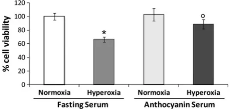
The cytotoxic effects of mild hyperoxia was evaluated, by means of the trypan blue dye exclusion test, on HUVECs pretreated for 24 h with medium containing 20 % AS or FS and then exposed for 24 h to 32 % O2 (mild hyperoxic
conditions) or 21 % O2 (normoxic conditions). The
pre-treatment with both AS or FS did not affected cell viability of HUVECs exposed to normoxic conditions (Fig.2). On the contrary, only AS pretreatment was able to significantly protect cells from death induced by mild hyperoxia (Fig.2). Activation of the Nrf2/ARE pathway
in these toxic conditions, we evaluated under these experimental settings the level of Nrf2 (NF-E2-related factor-2) nuclear localization and the gene expression of two well-known antioxidant enzymes containing ARE
sequences within the promoter, namely heme oxygenase-1 (HO-1) and NAD(P)H:quinone oxidoreductase 1 (NQO-1). Mild hyperoxia induced a significant decrease in the nuclear levels of the transcription factor Nrf2. Interest-ingly, pretreatment for 24 h with AS significantly increased Nrf2 nuclear levels in HUVECs subsequently exposed to normoxic conditions (Fig.3). The alteration induced by 32 % O2was partially prevented by cells pretreatment with
AS; in fact, the increase in Nrf2 nuclear localization induced by AS treatment still remains evident in cells later exposed for 24 h to mild hyperoxia (Fig.3).
A similar trend was observed as to the expression of both HO-1 and NQO-1 (Fig.4). In fact, higher intracellular mRNA levels of the two genes were measured in AS-pretreated cells under normoxic conditions, in com-parison with those treated with FS (Fig.4). Similarly to that found for Nrf2 nuclear localization (Fig.3), HUVECs exposure to mild hyperoxia induced a significant decrease in the expression of the both HO-1 and NQO-1; this effect was partially but significantly prevented by cells pretreat-ment with AS but not with FS (Fig.4).
These findings are in agreement with literature data indicating that dietary polyphenols can induce the expres-sion of genes encoding for antioxidant and detoxifying proteins through the activation of the Nrf2/ARE pathway (Speciale et al. 2010).
Effects on MAPKs phosphorylation
In basal conditions, Nrf2 translocation is inhibited by the cytoplasmic repressor Kelch-like ECH-associated protein 1 (Keap1). Phase II enzyme inducers and pro-oxidants have been reported to cause modification directly on Nrf2– Keap1 complex resulting in Nrf2 release from Keap1, or indirectly causing ERK 1/2 (extracellular signal-regulated kinases 1/2) phosphorylation that in turn activates Nrf2 and its subsequent translocation into the nucleus.
Thus, in order to investigate the upstream mechanism by which hyperoxic conditions and/or polyphenol compounds/ metabolites can modulate Nrf2/ARE pathway in HUVECs, the activation of two protein kinases belonging to the mitogen-activated protein kinases (MAPK) family, ERK1
Table 1 Serum TEAC and FRAP before and after administration of a unique dose (390 mg) of MedoxÒ, containing 160 mg of purified anthocyanins
TEAC (mmoles/L serum) FRAP (lM Fe2?/L serum)
Fasting serum 2.88±0.05 629.5±23.2
Anthocyanin serum 3.34±0.02* 706.1±25.1* Results are expressed as mean±SD from three independent experiments
AShuman serum obtained after MedoxÒcapsules consumption,FShuman serum obtained immediately before MedoxÒcapsules consumption *P\0.05 versus FS
Fig. 1 HUVECs viability after 16-, 24- or 48-h exposure to 32 % O2,
as evaluated by the trypan blue assay. The results were compared with those obtained at time 0 h (control). Results are expressed as mean±SD of at least three experiments. Bars represent the percentage of viable treated cells (mean percentage) versus control. *P\0.05 versus 0 h. °P\0.05 versus cells exposed to mild hyperoxic conditions for 16 h. §P\0.05 versus cells exposed to mild hyperoxic conditions for 24 h
Fig. 2 Effects of 24-h pretreatment with AS or FS on viability of HUVECs exposed for 24 h to 32 % O2(mild hyperoxic conditions) or
to 21 % O2(normoxic conditions), as evaluated by the trypan blue
and ERK2, was studied. These two protein kinases, usually localized into the cytoplasm in an inactive form under physiological conditions, are considered as the main effectors for Nrf2 nuclear translocation. Cytoplasmic levels of p-ERK1/2 were assessed by means of an antibody rec-ognizing Thr 202 phosphorylated ERK 1 and Thr 184 phosphorylated ERK 2.
We found ERK1/2 phosphorylation (i.e., activation) was induced by AS in cells under normoxic conditions, thus supporting a direct involvement of MAPK cascade in Nrf2/ ARE pathway activation. Furthermore, ERK1/2 phos-phorylation appeared to be inhibited by mild hyperoxia (Fig.5); as seen above for the induction of Nrf2 nuclear localization (Fig.3) and for the expression of genes con-taining ARE sequences (Fig.4), this effect was partially prevented by pretreatment with AS.
To further investigate the relevance of Nrf2 activation pathway on the effects of AS in HUVECs, the MAPKK pharmacological inhibitor PD98059 was used at a con-centration (25lM) not cytotoxic by itself under normoxic conditions (data not shown). PD98059 induced a further inhibition of Nrf2 nuclear translocation (without increasing cytotoxicity) in hyperoxia-exposed HUVECs; furthermore, PD98059 heavily hampered AS-induced protective effects against hyperoxia-induced cell death (Fig.6) and Nrf2 nuclear localization (Fig.7).
These results clearly indicate that the activation of the Nrf2–ARE pathway and the modulation of the ERK1/2 phosphorylation is one of the main mechanisms by which polyphenolic compounds/metabolites can exert their ben-eficial effects against hyperoxia-induced endothelial tox-icity, also supporting the essential role of cell adaptive responses elicited by these compounds in their capability to protect cells against stressor agents.
Discussion
Redox cellular homeostasis is a complex phenomenon crucial for cellular well-being. A large number of experi-mental results support the involvement of free radicals and oxidative stress in development and progression of numerous diseases, while an enhanced antioxidant status is associated with reduced risk of several diseases. In fact, ROS cause damage to cellular proteins, lipids and nucleic acids, etc., often leading to cumulative organ injury (Wells
Fig. 3 Effects of 24-h pretreatment with AS or FS on Nrf2 nuclear localization in HUVECs exposed for 24 h to 32 % O2(mild hyperoxic
conditions) or to 21 % O2(normoxic conditions). The figure shows a
representative image from three independent experiments. Nrf2 bands intensity values were normalized to the corresponding Lamin B values. Results by densitometry are reported as fold change against FS in normoxia and expressed as mean±SD of at least three independent experiments. *P\0.05 versus FS-treated cells exposed to normoxic conditions.°P\0.05 versus FS-treated cells exposed to hyperoxic conditions. §P\0.05 versus AS-treated cells exposed to normoxic conditions. AS, human serum obtained after MedoxÒ capsules consumption; FS, human serum obtained immediately before MedoxÒcapsules consumption
Fig. 4 Effect of 24-h pretreatment with AS or FS on HO-1 and NQO-1 gene expression in HUVECs exposed for 24 h to 32 % O2(mild
hyperoxic conditions) or to 21 % O2 (normoxic conditions). Cells
et al. 2009). In order to protect against oxidative stress, organisms have developed antioxidant systems consisting of thiol containing small molecules, such as glutathione and thioredoxin or enzymes like catalase, glutathione
peroxidase and peroxiredoxins (Oktyabrsky and Smirnova
2007). Various studies demonstrated the importance of the activation of Nrf2-regulated transcriptional response in the protection against hyperoxia, a condition which can result in transient oxidative stress. Nrf2 binds to the consensus sequence called antioxidant responsive element commonly found in the regulatory regions (promoter and/or enhanc-ers) of genes encoding several cytoprotective enzymes and proteins and up-regulates their transcription in response to a variety of stimuli (Speciale et al. 2011b). In unstressed cells, Nrf2 is predominantly localized in the cytoplasm in association with Keap1, which promotes proteasomal degradation of Nrf2. Oxidant or toxic insults may perturb the tertiary structure of Keap1, thereby impeding Nrf2 degradation and facilitating Nrf2 nuclear accumulation and subsequent ARE-mediated transcription (Speciale et al.
2011b). The role of the activation of Nrf2-regulated tran-scriptional response in the protection against hyperoxia ([95 % of O2)-induced injury was investigated in mice
lacking Nrf2 expression and activity (Cho et al. 2002; Reddy et al. 2009). In these studies, 95–99 % of O2
exposure enhanced expression of Nrf2 mRNA and func-tionally activated nuclear Nrf2 in the lungs of normal (wt) mice, contributing to the protection against hyperoxic lung injury, while a significant attenuation in basal and/or hyperoxia-induced mRNA expression of NQO-1 and HO-1 was observed in the lungs of mice deficient in Nrf2, sug-gesting that these enzyme genes are downstream effector molecules transcriptionally activated by Nrf2 in the lungs of mice. Furthermore, exposure of human lung microvas-cular endothelial cells to hyperoxia (95 % O2) stimulates
Nrf2 translocation from the cytoplasm to the nucleus and increases Nox4 expression (Pendyala et al. 2011). These results suggest that the use of Nrf2 activators may represent a new therapeutic strategy to prevent and attenuate hyperoxia insult through up-regulation of Nrf2 signaling.
However, all studies reported in literature on this subject have been conducted in experimental conditions using pure oxygen or high concentrations of oxygen ([95 %), but few is known about the effects of exposure to mild hyperoxia, and on the cellular signaling pathways which may be critical in the development of such effects. For this reason, the goal of our study was, instead, to investigate the alterations induced in HUVECs by exposure to mild hyperoxia (O2 32 %). Interestingly, the cytotoxic effect
following this experimental condition (Fig. 1) came along with a significant decrease in the nuclear levels of the transcription factor Nrf2 (Fig.3), as well as with a con-sequent decrease in the expression of both the investigated ARE-genes, HO-1 and NQO-1 (Fig.4). Thus, this appears to be a good model to investigate if protection against hyperoxia endothelial damage may be elicited by Nrf2 activators, including selected dietary plant phenolics
Fig. 5 Effect of 24-h pretreatment with AS or FS on ERK1 and ERK2 phosphorylation in HUVECs exposed for 24 h to 32 % O2
(mild hyperoxic conditions) or to 21 % O2(normoxic conditions).
Cells treated with FS and exposed to normoxic conditions were used as negative controls. The figure shows a representative image from three independent experiments. Results by densitometry are reported as fold change against control and expressed as mean±SD of three experiments. p-ERK1/2 bands intensity values were normalized to the corresponding b-actin value. *P\0.05 versus FS-treated cells exposed to normoxic conditions.°P\0.05 versus FS-treated cells exposed to mild hyperoxic conditions.§P\0.05 versus AS-treated
cells exposed to normoxic conditions. AS, human serum obtained after MedoxÒ capsules consumption; FS, human serum obtained immediately before MedoxÒcapsules consumption
Fig. 6 Effect of MAPKK inhibition on HUVEC viability pretreated for 24 h with AS or FS and then exposed for 24 h to 32 % O2(mild
hyperoxic conditions) or to 21 % O2 (normoxic conditions), as
known to have beneficial health effects not only for anti-oxidant activity but also as modulators of the Nrf2 system (Rahman et al.2006; Speciale et al.2011a). However, many currently used in vitro experimental models are able to identify, at cellular and molecular levels, responses induced by phytocomplexes or individual phytochemicals but are usually conducted by adding them directly to the culture medium. In our study, we used a new ex vivo experimental protocol, representing an improvement to current in vitro cellular models, with a more physiological approach (Canali et al.2010). In fact, taking into account that healthy subjects may be used as ‘‘bioreactors,’’ we fed endothelial cells with the human serum (AS) taken after supplementa-tion with the anthocyanins mixture and then containing only absorbed compounds and/or metabolites. In normoxic conditions, HUVEC treatment with AS induces a significant increase in Nrf2 nuclear accumulation activation and in HO-1 and NQO-1 expression (Figs.3,4), when compared with cells fed with FS. On the other hand, pretreatment for 24 h with AS partially prevented the decrease in Nrf2 nuclear accumulation and in HO-1 and NQO-1 gene expression, observed in HUVECs subsequently exposed to mild hyperoxia (Figs.3,4). These findings confirm that the intake of MedoxÒ is able to produce serum levels of anthocyanin compounds/metabolites high enough to per-form a pharmacological effect, as observed also by anti-oxidant assays (Table1). Furthermore, the observed protective effect of AS against mild hyperoxia-induced endothelial damage can be ascribed to the activation of the
Nrf2/ARE pathway. Our findings are in agreement with those reported by several papers which report that plant phenols can protect endothelial cell against different stres-sor conditions by pharmacological activation of the Nrf2/ ARE pathway (Speciale et al.2011b; Surh et al.2008).
Nrf2 nuclear translocation requires the activation of several signal transduction pathways as well as MAPKs (Shen et al. 2004). MAPKs (ERKs, JNKs and p38) are downstream effectors in antioxidant responses, and their activities are manifested in activation of many transcription factors, including Nrf2 (Limo´n-Pacheco et al.2007). Pre-vious studies have shown that some MAPKs, like ERK1/2, play a role in the induction of Nrf2 (Filomeni et al.2012; Speciale et al.2011a). However, the exact role of MAPKs in Nrf2 activation, as well as the underlying molecular mechanism, remains poorly understood. In our experi-mental conditions, manipulation of the catalytic activity of ERK1 and ERK2, through the use of PD98059 (a selective pharmacological inhibitor of ERK pathway), markedly abolished Nrf2 nuclear translocation induced by AS (Fig.5). These results demonstrated that the ERK1/2 pathway is involved in AS-induced Nrf2/ARE pathway activation and in the protective effect against mild hyper-oxia-induced endothelial damage although we cannot exclude the involvement of other mechanisms.
In conclusion, this study shows that anthocyanins and/or their metabolites, present in human serum after oral administration of a dietary plant-derived supplementation, can protect HUVECs against mild hyperoxia-induced
Fig. 7 Effect of MAPKK inhibition on the Nrf2 activation pathway in HUVECs pretreated for 24 h with AS or FS and then exposed for 24 h to 32 % O2(mild hyperoxic conditions) or to 21 % O2 (normoxic
conditions), as evaluated by Western blot analysis. Cells were incubated with the pharmacological MAPKK inhibitor PD98059 (25lM) for 1 h before AS or FS addition. Cultures treated with FS and exposed to normoxic conditions were used as negative controls. Each point represents mean±SD of three experiments. The figure shows a representative image from three independent experiments. Results by densitometry are reported as fold change against controls and expressed as mean±SD of three experiments. Nrf2 bands intensity
values were normalized to the corresponding Lamin B values. *P\0.05 versus FS-treated cells exposed to normoxic conditions.
°P\0.05 versus FS-treated cells exposed to hyperoxic conditions.
@P\0.05 versus FS-treated cells exposed to hyperoxic conditions and
alterations acting as cell signaling modulators and inducing activation of the Nrf2/ARE pathway. Thus, the present findings contribute to support the hypothesis that plant polyphenols, including anthocyanins, can play an important role in the prevention of diseases associated with oxidative stress, not due to their putative antioxidant properties but through activities and roles totally independent on such capacity and interacting with cell functions at different levels. A significant contribution is also given by the ‘‘bio-reactor’’ model used to obtain these results, because it let to take into account the possible problems related to the drug oral availability and metabolism, as well as the possible synergisms between different components. Finally, the present results suggest the importance to plan dietary intervention based on plant-derived supplements which may efficiently prevent endothelial damage due to occupational or therapeutic exposure to mild hyperoxic conditions.
References
Altemeier WA, Sinclair SE (2007) Hyperoxia in the intensive care unit: why more is not always better. Curr Opin Crit Care 13:73–78
Canali R, Ambra R, Stelitano C, Mattivi F, Scaccini C, Virgili F (2007) A novel model to study the biological effects of red wine at the molecular level. British J Nutr 97:1053–1058
Canali R, Comitato R, Ambra R, Virgili F (2010) Red wine metabolites modulate NF-kappaB, activator protein-1 and cAMP response element-binding proteins in human endothelial cells. Br J Nutr 103:807–814
Cho HY, Jedlicka AE, Reddy SP, Kensler TW, Yamamoto M, Zhang LY, Kleeberger SR (2002) Role of NRF2 in protection against hyperoxic lung injury in mice. Am J Respir Cell Mol Biol 26:175–182
Dro¨ge W (2002) Free radicals in the physiological control of cell function. Physiol Rev 82:47–95
Filomeni G, Piccirillo S, Rotilio G, Ciriolo MR (2012) p38(MAPK) and ERK1/2 dictate cell death/survival response to different pro-oxidant stimuli via p53 and Nrf2 in neuroblastoma cells SH-SY5Y. Biochem Pharmacol 83:1349–1357
Genestra M (2007) Oxyl radicals, redox-sensitive signalling cascades and antioxidants. Cell Signal 19:1807–1819
Gore A, Muralidhar M, Espey MG, Degenhardt K, Mantell LL (2010) Hyperoxia sensing: from molecular mechanisms to significance in disease. J Immunotoxicol 7:239–254
Limo´n-Pacheco JH, Herna´ndez NA, Fanjul-Moles ML, Gonsebatt ME (2007) Glutathione depletion activates mitogen-activated protein kinase (MAPK) pathways that display organ-specific responses and brain protection in mice. Free Radic Biol Med 43:1335–1347 Livak KJ, Schmittgen TD (2001) Analysis of relative gene expression data using real-time quantitative PCR and the 2(-Delta Delta C(T)) Method. Methods 25:402–408
Morabito G, Trombetta D, Singh Brajendra K, Prasad Ashok K, Parmar Virinder S, Naccari C, Mancari F, Saija A, Cristani M, Firuzi O, Saso L (2010) Antioxidant properties of 4-methylcoumarins in in vitro cell-free systems. Biochimie 92:1101–1107
Oktyabrsky ON, Smirnova GV (2007) Redox regulation of cellular functions. Biochemistry 72:132–145
Pagano A, Barazzone-Argiroffo C (2003) Alveolar cell death in hyperoxia-induced lung injury. Ann NY Acad Sci 1010:405–416 Pendyala S, Moitra J, Kalari S, Kleeberger SR, Zhao Y, Reddy SP, Garcia JG, Natarajan V (2011) Nrf2 regulates hyperoxia-induced Nox4 expression in human lung endothelium: identification of functional antioxidant response elements on the Nox4 promoter. Free Radic Biol Med 50:1749–1759
Phillips PG, Tsan MF (1988) Hyperoxia causes increased albumin permeability of cultured endothelial monolayers. J Appl Physiol 64:1196–1202
Qin Y, Xia M, Ma J, Hao Y, Liu J, Mou H, Cao L, Ling W (2009) Anthocyanin supplementation improves serum LDL- and HDL-cholesterol concentrations associated with the inhibition of cholesteryl ester transfer protein in dyslipidemic subjects. Am J Clin Nutr 90:485–492
Rahman I, Biswas SK, Kirkham PA (2006) Regulation of inflamma-tion and redox signaling by dietary polyphenols. Biochem Pharmacol 72:1439–1452
Reddy NM, Kleeberger SR, Kensler TW, Yamamoto M, Hassoun PM, Reddy SP (2009) Disruption of Nrf2 impairs the resolution of hyperoxia-induced acute lung injury and inflammation in mice. J Immunol 182:7264–7271
Shen G, Hebbar V, Nair S, Xu C, Li W, Lin W, Keum YS, Han J, Gallo MA, Kong AN (2004) Regulation of Nrf2 transactivation domain activity. The differential effects of mitogen-activated protein kinase cascades and synergistic stimulatory effect of Raf and CREB-binding protein. J Biol Chem 279:23052–23060 Speciale A, Canali R, Chirafisi J, Saija A, Virgili F, Cimino F (2010)
Cyanidin-3-O-glucoside protection against TNF-a-induced endothelial dysfunction: involvement of nuclear factor-jB signaling. J Agric Food Chem 58:12048–12054
Speciale A, Anwar S, Ricciardi E, Chirafisi J, Saija A, Cimino F (2011a) Cellular adaptive response to glutathione depletion modulates endothelial dysfunction triggered by TNF-a. Toxicol Lett 207:291–297
Speciale A, Chirafisi J, Saija A, Cimino F (2011b) Nutritional antioxidants and adaptive cell responses: an update. Curr Mol Med 11:770–789
Surh YJ, Kundu JK, Na HK (2008) Nrf2 as a master redox switch in turning on the cellular signaling involved in the induction of cytoprotective genes by some chemopreventive phytochemicals. Planta Med 74:1526–1539
Thom SR (2009) Oxidative stress is fundamental to hyperbaric oxygen therapy. J Appl Physiol 106:988–995
Tomaino A, Martorana M, Arcoraci T, Monteleone D, Giovinazzo C, Saija A (2010) Antioxidant activity and phenolic profile of pistachio (Pistacia vera L., variety Bronte) seeds and skins. Biochimie 92:1115–1122