Open Access
Review
Drosophila
as a genetic and cellular model for studies on axonal
growth
Natalia Sánchez-Soriano
1, Guy Tear
2, Paul Whitington
3and
Andreas Prokop*
1Address: 1The Wellcome Trust Centre for Cell-Matrix Research, Faculty of Life Sciences, The University of Manchester, Manchester, UK, 2MRC Centre for Developmental Neurobiology, Guy's Campus, King's College, London, UK and 3Department of Anatomy and Cell Biology, University of Melbourne, Victoria, Australia
Email: Natalia Sánchez-Soriano - [email protected]; Guy Tear - [email protected];
Paul Whitington - [email protected]; Andreas Prokop* - [email protected] * Corresponding author
Abstract
One of the most fascinating processes during nervous system development is the establishment of stereotypic neuronal networks. An essential step in this process is the outgrowth and precise navigation (pathfinding) of axons and dendrites towards their synaptic partner cells. This phenomenon was first described more than a century ago and, over the past decades, increasing insights have been gained into the cellular and molecular mechanisms regulating neuronal growth and navigation. Progress in this area has been greatly assisted by the use of simple and genetically tractable invertebrate model systems, such as the fruit fly Drosophila melanogaster. This review is dedicated to Drosophila as a genetic and cellular model to study axonal growth and demonstrates how it can and has been used for this research. We describe the various cellular systems of
Drosophila used for such studies, insights into axonal growth cones and their cytoskeletal dynamics, and summarise identified molecular signalling pathways required for growth cone navigation, with particular focus on pathfinding decisions in the ventral nerve cord of Drosophila embryos. These
Drosophila-specific aspects are viewed in the general context of our current knowledge about neuronal growth.
Background
The function of a nervous system depends on the proper arrangement of its cellular elements, that is, neurons and glia cells. Amongst these, neurons bear axonal processes that establish synaptic contacts with other cells (neurons, muscles or gland cells) that can be a significant distance away. The transfer of information between these cells is the key feature of nervous system function and is usually mediated by action potentials that propagate along axons and are passed on to other cells at synapses. The wiring of
such a system has to be precise and reproducible from individual to individual, as was first highlighted by Ramón y Cajal for the nervous systems of humans, other vertebrates, and also invertebrates [1]. Such precision is achieved during development through the guided growth of axons along specific paths, a process clearly governed by genetic mechanisms [2,3].
Essential work contributing to our current understanding of axonal growth has been carried out in vertebrates and
Published: 2 May 2007
Neural Development 2007, 2:9 doi:10.1186/1749-8104-2-9
Received: 28 January 2007 Accepted: 2 May 2007
This article is available from: http://www.neuraldevelopment.com/content/2/1/9
© 2007 Sánchez-Soriano et al; licensee BioMed Central Ltd.
invertebrates alike, in many instances demonstrating the conservation of principal mechanisms across the animal kingdom. One strategy towards improving molecular insights into axonal growth is the use of genetically tracta-ble invertebrate model organisms, such as the worm
Caenorhabditis elegans or the fruit fly Drosophila mela-nogaster. The strength of these species lies in their amena-bility to genetic manipulation (see below) and the fact that their nervous systems are composed of relatively low numbers of cellular elements. Whereas specific neuronal connections in vertebrates are usually formed by larger groups of neurons that develop and act in parallel, these connections are mostly represented by unique, individu-ally recognisable neurons in invertebrates. Studies capital-ising on such identifiable neurons, for example in insects, have helped to unravel principles of neuronal circuit for-mation. For example, the initial observation of guidepost cells as stepping stones for axonal growth came from stud-ies of the grasshopper limb bud [4]. The concept of pio-neer guidance, which proposes that axonal tracts are established by single pioneer neurons that are subse-quently used as guidance cues for follower neurons, was discovered in vertebrates [5]. However, work on insects has helped to refine these concepts ('selective fascicula-tion' and 'labelled pathways' hypotheses) [6-8] and to contribute molecular players [9].
Once it was revealed that Drosophila embryos were experi-mentally accessible and largely homologous to grasshop-pers at the single cell level [10], insights that had to that date mostly been gained on larger insects could be trans-ferred into a genetically tractable model system. Such work in Drosophila was further fuelled by the development of new technologies, strategies and molecular tools, such as specific anatomical antibody probes [11,12] and the development of ever more sophisticated genetic screening strategies [13]. Furthermore, such research has profited enormously from the increasingly well organised research environment of Drosophila, including the systematic gen-eration and provision at a large scale of new genetic tools and mutant fly stocks, and the improvement of access to research-relevant information [14-18].
Undoubtedly, the use of Drosophila as a model system for the study of axonal growth has been a prolific endeavour, providing a plethora of novel and/or refined insights into relevant molecular mechanisms, many of which have been shown to be conserved in higher organisms. This review will provide an overview of the major cellular model systems established for the fly, our current insights into Drosophila growth cones (the key structure executing axonal growth), the housekeeping machinery regulating the cytoskeletal dynamics required for axonal growth, and the signalling events involved in axon guidance, focussing
Models for axonal growth in
Drosophila
One of the major strengths of Drosophila lies in its use as a model where axonal growth can be studied in situ. A widely used strategy for the unbiased discovery of neuro-nal growth mechanisms in Drosophila is the search for mutations that cause morphological aberrations of axonal tracts or neuronal connections in situ. Subsequently, the genes associated with such mutations can be identified, and the molecular nature, function and type of interac-tions of their products can be studied. To facilitate such genetic screens and functional studies of gene action, descriptions of axonal pathways and neuronal connec-tions in wild-type animals have been provided for a number of different neural systems in Drosophila. A selec-tion of these cellular models is illustrated in Figure 1, and some of their principal features will be discussed below.
Figure 1
10-&;
QHUYHV
QH
UYH
FRQQHFWLYHV
$
7
7
7
13
EUDLQ
2/$
2/
2/
$/
0%
DQWHQQD
RFHOOL
WUDFKHD
PXVFOH
770
H\H
GLVF
DQWHQQDO
GLVF
KHDGPRXWK
DSSDUDWXV
%ROZLJRUJDQ
FRPSRXQG
H\H
Y1&
Y1&
$
%
&
'
VHQVRU\QHXURQ
LQWHUQHXURQ
PRWRUQHXURQ
ODUYD
DGXOW
IO\
Neurons used for studies on neuronal growth at different stages of Drosophila development
Figure 1
Comparing principal features of neuronal organisation and growth in Drosophila (larva) and vertebrates (human)
Figure 2
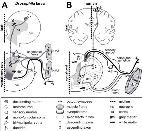
Comparing principal features of neuronal organisation and growth in Drosophila (larva) and vertebrates (human). (a) Saggital section (one body half; dotted line is midline) through the larval brain and ventral nerve cord (compare Figure 1a,b). (b) Sagg-ital section through the adult human brain and one half of the spinal cord. Symbols are explained in the box below. Whereas axons of unipolar inter- and motorneurons in Drosophila have to grow into the synaptic area where they form dendrites, com-parable neurons in vertebrates are multipolar and locate themselves in the synaptic area. All Drosophila motorneurons locate their dendrites in the dorsal neuropile, regardless of their soma position (see 'i' versus 'ii') [236]. Vice versa, sensory somata in
Drosophila are located next to their dendrites, whereas cell bodies of most sensory neurons in vertebrates are grouped together in the dorsal root ganglia. Sensory output (dark grey) and motor input areas (bright grey) are inverted in both phyla, which might be explained through a general dorsoventral body axis inversion between vertebrates and arthropods [328], that is, not represent an organisational difference between their CNS. Ascending/descending axons in Drosophila are non-myeli-nated and project through the synaptic area (compare neurons 2 and 13 in Figure 1) where they take on characteristic posi-tions [236,329]. In vertebrates, ascending/descending axons are myelinated and positioned outside the synaptic area, grouping into characteristic tracts in defined positions of the white matter; examples named here: fasciculus cuneatus (1), tractus corticos-pinalis lateralis (2; pyramidal tract; only descending), and tractus spinothalamicus lateralis (3).
$
YHQWUDOQHUYHFRUG
EUDLQ
QS
L
LL
QS
F[
F[
%
VSLQDOFRUG
EUDLQ
JP ZP
ZP
PRWRU QHUYH VHQVRU\
QHUYH VHJPHQWDO
QHUYH
GRUVDOURRW JDQJOLRQ
10-V
'URVRSKLOD
ODUYD
KXPDQ
PRWRUQHXURQ
GHVFHQGLQJQHXURQ
VHQVRU\QHXURQ
PRQRXQLSRODUVRPD
ELPXOWLSRODUVRPD
GHQGULWH
RXWSXWV\QDSVHV
PXVFOHILEUHV
V\QDSWLFDUHD
D[RQWUDFWVLQZP
GHVFHQGLQJD[RQ
DVFHQGLQJD[RQ
QS F[ JP ZP
expression must differ considerably between vertebrates and arthropods.
Second, Drosophila is a holometabolous insect that exists as a maggot during larval life but is substantially reorgan-ised into the adult fly during metamorphosis at the inter-mediate pupal stage. This process is not dissimilar to metamorphic events known from lower vertebrates, such as amphibians. One phase of de novo axonal growth takes place in the embryo, and a vast number of these cellular elements are maintained into the adult stage, albeit being remodelled during the pupal period [22]. However, to adapt to the far more complex behavioural repertoires of the adult, a second phase of de novo neurogenesis and axonal growth is initiated during larval life and completed during the process of metamorphosis. There is abundant evidence that common cellular and molecular mecha-nisms for axon guidance apply at both stages, allowing us, in many instances, to study the same molecules with the multiple techniques and cellular systems available for both stages of axonal growth in Drosophila [3].
Cellular models from both developmental phases (Figure 1a,b versus 1c,d) have been used to screen for genes involved in neuronal growth regulation. At embryonic/ larval stages, genetic screens have mostly been based on analyses of animals bearing mutations that were induced by chemical treatment, radiation or via transposable ele-ments. Morphological read-outs used in these screens have included motornerves, projections in the CNS, or sensory projections in the trunk or head (for example, Bolwig's organ; 1–4 in Figure 1) [23-32]. Some screens have been carried out on adult flies using as read-outs the giant fibre system (13 and 14 in Figure 1) or general brain morphology [33,34]. However, since many mutations cause embryonic or larval lethality, the utility of adult ani-mals for genetic screens is limited. To overcome this prob-lem, mosaic strategies have been used in which only specific tissues express the mutant phenotype and can be maintained by an otherwise normal body [13]. Such mosaic screens have been successfully carried out using adult photoreceptor axons as read-outs (8 in Figure 1) [35,36]. A further refined mosaic strategy is the MARCM technique, in which homozygous mutant neurons can be visualised at the single cell level, surrounded by hetero-zygous, non-mutant cells [37]. This technique has been used to study and screen for growth aberrations of mush-room body, olfactory and photoreceptor neurons (8, 10 and 12 in Figure 1) [38-40]. Based on these various screens in embryonic, larval or adult individuals, many mutations that result in defective axon growth have been identified [3]. The gene functions underlying a number of these mutations will be discussed below (see 'Regulators of actin dynamics in Drosophila growth cones' below).
Studies of neurons in situ have the essential advantage that they address the relevance of genetic mechanisms for developmental processes in a natural context. However, the ability to utilise precise and well-controlled pharma-cological, physiological or imaging methods in such in situ systems is limited. To address these shortcomings, cul-tures of primary neurons, obtained from embryonic or larval tissues, have been established in Drosophila. These primary culture systems have been used successfully to demonstrate and/or analyse various phenomena, such as promotion of axonal growth by the neural cell adhesion molecule (N-CAM) homologue Fasciclin 2 or the extracel-lular matrix molecule laminin [41-43], induction of axonal fasciculation through neuronal activity [44], dependence of axonal growth on endocytosis [45,46], or cAMP-dependent neurotransmitter release from growing neurons [47]. All of these phenomena are likely to be of relevance to neuronal growth in situ [9,21,48], and the established culture systems provide valuable additional means for their study.
Taken together, the Drosophila system provides a wide range of cellular models to be used for studies of axonal growth. In addition, a number of models exist for other forms of neuronal growth, such as dendritic arborisation or the branching and plasticity of synaptic terminals [49-52]. However, these systems will not be considered here.
Growth cone dynamics in
Drosophila
Principal structure and function of growth conesThe elongation of an axon is led at its tip by a highly dynamic structure, the growth cone, first described and named by S Ramón y Cajal ("cono de crecimiento") and then confirmed in live studies by RG Harrison [53-55]. Growth cones navigate along stereotypical paths, steadily elongating the axon by adding new structural components such as microtubules and membrane to its tip [56]. Just like the leading front of migrating cells, growth cones dis-play actomyosin-containing filopodia and lamellipodia, focal contacts, a dynamic population of microtubules, and cell polarity markers such as Par-3 and -6 [57-62]. In contrast to migrating cells, where stress fibres pull the entire cell forward, neuronal cell bodies generally remain behind when growth cones advance, remaining con-nected by the steadily elongating axons, the core of which contains bundled, stable microtubules representing the highways for axonal transport.
actin-bound ATP to ADP, and hydrolysis or severing at their pointed ends (pointing towards the growth cone's centre) [64-66]. This process of actin assembly and disas-sembly and its organisation into lamellipodial networks and filopodial bundles is regulated by a complex molecu-lar machinery [65,67-69], which we detail below in the context of Drosophila components (see 'Regulators of actin dynamics in Drosophila growth cones').
Whereas growth cone guidance crucially depends on actin dynamics, growth cone advance depends on microtu-bules. This is clearly demonstrated by the persistence of axonal growth in the presence of actin polymerisation blockers, in vertebrates as well as in Drosophila [55,70-72], whereas pharmacological destabilisation of microtubules causes growth cone retraction [55,73]. Furthermore, extending axons can even be induced to turn in the absence of filamentous actin if they are exposed to an elec-trical field [74]. Microtubules in the axon shaft and central zone of growth cones are stable and bundled, whereas sin-gle unstable microtubules elongate into and retract from the peripheral actin-rich zone in a highly dynamic fash-ion. These microtubules can be trapped or stabilised through signals in the growth cone periphery, thus deter-mining the direction in which microtubules of the axon shaft will extend [55,58-60,63]. Microtubule growth and shrinkage is, per se, an autocatalytic process [75-77]. How-ever, a number of cellular factors regulate microtubule dynamics, as detailed below in the context of Drosophila
components (see 'Regulators of microtubule dynamics in
Drosophila growth cones').
Growth cones in Drosophila
Initial work on Drosophila growth cones dates back more than 20 years when growth cones were first described inci-dentally in a study on the role of the activity regulator Maleless/Nap in primary neurons cultured from the larval nervous systems [78]. In vivo observations carried out on growth cones of primary neurons in culture demonstrated classifiable shape differences (longer filopodia of ventral ganglion-derived neurons versus those derived from brain), and also established strategies to measure their dynamics [79]. These strategies were later used to demon-strate that genetic or pharmacological alterations of cAMP levels influence growth cone dynamics [80]. Analyses of dye-filled motorneurons in Drosophila embryos demon-strated that their growth cones make characteristic and reproducible directional decisions when advancing in the CNS and periphery [81]. In agreement with this finding, ultrastructural studies in the developing embryonic Dro-sophila CNS showed that filopodia of pioneering growth cones establish close contacts with characteristic sets of cells in their immediate environment [82]. In vivo studies of the identified RP2 motorneuron in the same context revealed that these growth cones grow at rates comparable
to other animal model systems and carry out a set of char-acteristic shape changes along their reproducible paths [83]. Follow-up studies using this system showed that genetic removal of the Roundabout receptor (see 'Medi-olateral patterning of longitudinal fascicles' and 'Growth cone guidance at the Drosophila CNS midline' sections below) in these growth cones led to an increase in the length and persistence of filopodia [84]. In vivo observa-tions on growth cones of a different set of motorneurons (ISNb) demonstrated that their filopodial dynamics and guidance are regulated through GTPases of the Rho-family and their downstream effectors [85,86]. Further studies on embryonic motorneurons have shown that growth cones of pioneer neurons display a significantly more complex shape than their follower axons [9], a finding that is consistent with descriptions of the growth cones of pioneer/follower neurons in vertebrates [87,88]. In the
Drosophila system, this behaviour is regulated by the N-CAM homologue Fasciclin 2 [9]. Motorneuronal growth cones have also been studied in their transition phase into a characteristically shaped neuromuscular junction. Such analyses have revealed how growth cones, which initially explore a wider muscle field, transform into precisely tar-geted neuromuscular terminals [89,90]. Ultrastructural analyses of such growth cones have shown how filopodia of identified motorneurons establish intimate contacts only with their specific target muscles, and that this proc-ess is regulated through the presentation of specific cell recognition molecules [91]. Consequently, ablation of such target muscles changes the behaviour of growth cones in their target area [92].
Drosophila growth cones display the characteristic hall-marks of those in other systems, such as highly dynamic filopodia, enrichment of microtubules at their core and the presence of filamentous actin in their periphery (Fig-ure 3). Yet, to date, subcellular studies of Drosophila
growth cones are fairly sparse. Most of the insights into axonal growth mechanisms in Drosophila (as reviewed below) have been derived from analyses of the axonal projection defects found in the mature embryonic or adult nervous system in mutant animals or under experimen-tally manipulated conditions. There are only few descrip-tions of the behaviours of growth cones during the axon elongation phase in these animals.
Regulators of actin dynamics in Drosophila growth cones
Figure 3
$EO
(QD
&DSW
&KE
&KLF
DFWLQ
PW
'RFN
6RV
3DN
5KR*$3%5HSXOVLRQ
5DF
RWKHU HIIHFWRUV &D0 *()&
&
%
%µ
'
(
'
JURZWK FRQH
D[RQ
$
D&&
53
D&&
Drosophila growth cones and the (potential) factors regulating their cytoskeletal dynamics
Figure 3
Drosophila growth cones and the (potential) factors regulating their cytoskeletal dynamics. (a) Growth cones of aCC (arrows) and RP2 motorneurons (double chevrons; cell bodies named) in two consecutive segments of the trunk of a Drosophila embryo, stained with a cell-specifically expressed membrane marker. (b,b') Cultured Drosophila growth cone stained for microtubules (green) and filamentous actin (magenta); some filopodia lack microtubules (curved arrows), whereas others are deeply invaded (arrow heads indicate microtubule tips). (c) Schematic representation of the cytoskeletal organisation in Drosophila growth cones as extrapolated from work on growth cones in other species (detailed in the section 'Principal structure and function of growth cones'): veil-like lamellipodia (black arrowhead) contain mesh-like networks of actin filaments (randomly oriented red lines), whereas pointed filopodia (white arrowhead) contain bundled actin filaments (parallel red lines); microtubules (blue lines) are bundled in the axon, but single splayed microtubules extend into the periphery of the growth cone (curved white arrows indicate splayed microtubule tips), reaching into filopodia, as was similarly reported for growth cones of other species or migrating cells [63,330]. (d) Details of the boxed area in (c); circled numbers correlate with the numbers in Table 1 and rep-resent the following molecular activities: 1, actin filament nucleation by Arp2/3 (which subsequently stays with the pointed ends); 2, actin filament nucleation and elongation by formins (which stay with barbed ends); 3, actin monomer binding; 4, barbed-end capping; 5, pointed end-depolymerisation/severing; 6, actin filament bundling; 7, retrograde flow of actin cytoskele-ton; 8, microtubule plus end binding; 9, microtubule stabilising; 10, actin-microtubule linkage. Black straight arrows indicate growth of actin filaments or microtubules, grey straight arrows shrinkage, black curved arrows addition of actin monomers, grey curved arrows removal of actin monomers or filamentous fragments, hatched arrows indicate direction of retrograde actin flow, and the grey dashed curved double arrow linkage of actin and microtubules. (e) Current view of the effectors downstream of the Slit receptor Robo mediating repulsion from the midline of the ventral nerve cord. Robo (top right) habours five immunoglobulin domains (half elipses) and three fibronectin type III domains (blue boxes) extracellularly, and four conserved cytoplasmic (CC) domains (light to dark green) intracellularly. Robo induces growth cone repulsion by controlling cytoskeletal dynamics via either Abelson kinase (Abl) and Enabled (Ena), or Rac activity. Ena binds at CC2 and acts most likely through Chickadee/Profilin on actin dynamics. Abl binding to Robo at CC3 influences actin dynamics via Capulet and microtu-bule dynamics via the +TIP protein Chromosome Bows (Chb/Orbit/MAST). Simultaneously, Abl phosphorylates CC1 to antag-onise Robo function. The regulation of Rac activity through Robo occurs through CC2/3 recruitment of the SH3-SH2 adaptor molecule Dreadlocks (Dock) which, in turn, activates Rac through both Pak and the GEF Sos. In parallel, active Robo can influ-ence Rac activity via the binding of RhoGAP93B (vilse/CrGAP) to CC2, but it remains unclear whether RhoGAP93B is posi-tively or negaposi-tively regulated by Robo. Paradoxically, both decrease and increase of Rac activation levels can cause midline crossing, suggesting that: Rac might influence other effectors to cause repulsion; a precise Rac activation level is required to mediate Slit-induced repulsion; or a sequential modification of Rac in response to Robo activation has to occur, such as an ini-tial role to prevent extension towards the source of the repellent and another role to encourage extension away from the Slit source. Calmodulin and GEF64C have additionally been identified as modifiers of Robo activity, although it is not clear yet how they influence Robo signalling (calmodulin possibly through Chic).
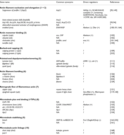
Table 1.
Several factors have been reported to be able to seed new actin filaments, a process called 'nucleation'. One of these factors is the Arp2/3 (actin-related protein) complex, which is composed of seven subunits. The genes encoding all its subunits have been identified in Drosophila and some of them have been functionally assessed [93,94]. Loss of function of the genes Suppressor of profilin 2
(ArpC1) and/or Arp66B (Arp3) causes severe axonal pro-jection defects when analysed at late embryonic stages (Figure 4, xxxix) [95]. Another independent nucleator of actin filaments, Spire, has been described for Drosophila
[96] but seems not to be localised in the nervous system [14]. As opposed to Arp2/3-complex or Spire, members of the Formin family of proteins are nucleators and, in addi-tion, effective barbed end-binding elongators of actin fila-ments [97]. Of the six Drosophila Formins [98], DAAM (dishevelled associated activator of morphogenesis) is expressed pan-neurally while two others, CG32030 and
CG32138, are specifically expressed within midline glia (the AA142 transposon insertion, a classic midline glia marker, is inserted within CG32030; Richard Tuxworth and GT, unpublished observations). The other Formins,
fam-Table 1: Direct regulators of the cytoskeleton with (potential) functions at growth cones
Gene name Common synonyms Direct regulators References
Actin filament nucleation and elongation (1 + 2)
Suppressor of profilin 2 (Sop2) ArpC1 WASp (+), SCAR/WAVE complex (+; containing: SCAR/WAVE, Hem/kette, Sra-1/CYFIP, Abi, SIP1/HSPC300)
[95,149]
Actin-related protein 66B (Arp66B) Arp3
Arp14D, Arc-p34, Arpc3A/3B, Arc-p20, p16-Arc Arp2, ArpC2-C5 [93,94] dishevelled associated activator of morphogenesis (DAAM) NA ? *
enabled (ena) ENA/VASP Abelson (-), Dlar (+) [100,101,164]
Actin monomer binding (3)
capulet (capt) acu, CAP Abelson (+) [103]
ciboulot (cib) NA ? [102]
chickadee (chic) profilin, sand ena (+) [101,164]
twinfilin (twf) NA ? [104]
Barbed-end capping (4)
capping protein α(cpa) NA ? [109]
capping protein β(cpb) NA ? [333]
Pointed-end depolymerisation/severing (5)
twinstar (tsr) ADF/cofilin LIMK1 (-), ssh (+) [111]
flightless I (fliI) (gelsolin family) ? [115]
quail (qua) villin-related (gelsolin family) ? [116]
Actin filament bundling (6)
singed (sn) fascin ? [126]
α Actinin (Actn) flightless A ? [128]
Fimbrin (Fim) NA ? [334]
cheerio (cher) filamin ? [129]
Retrograde flow of filamentous actin (7)
zipper (zip) myosin heavy chain ? [121]
spaghetti squash (sqh) myosin II light chain Strn-Mlck (+), Mbs/myosin phosphatase (-)
[119,120]
Microtubule plus end binding (+TIPs) (8)
CLIP-190 NA ? [152]
chromosome bows (chb) CLASP, Orbit/MAST Abelson (+) [153]
eb1, CG18190, CG32371 NA ? [155]
APC-like (Apc) Apc1 ? [154]
Apc2 NA ? [154]
Microtubule stabilising (9)
futsch MAP1B, mAB22C10 Fmr1/fragileX/fmrp (-) [163,335]
tau NA ? [161]
Microtubule-actin linkage (10)
short stop (shot) kakapo, groovin ? [168]
pod-1 NA ? [169]
The table lists official gene names according to FlyBase [14] (first column), examples of commonly used synonyms (second column; NA, not applicable), molecules reported to regulate the respective gene/protein directly (third column; '+', activating; '-', down-regulating), and
ily of proteins, which is a direct target of the cytoplasmic Abelson tyrosine kinase in the context of neuronal growth [100,101]. Further factors regulating actin assembly are molecules that bind monomeric actin, such as Capulet, Ciboulot and Profilin/Chickadee, all of which are essen-tial for neuronal growth regulation in Drosophila [101-103]. A further actin monomer-binding molecule, Twinfi-lin, is likely to be expressed in the nervous system, but no data exist concerning its cellular function [104].
Profilin/Chickadee has been shown to interact with Cal-modulin [105], suggesting [Ca2+]
i may influence its
activ-ity. Consistent with this observation, growth cones of
Drosophila, like those of other species, show measurable changes in local calcium concentration [106-108]. Barbed-end capping proteins are typical inhibitors of actin polymerisation. For Drosophila, the capping proteins Cpa and Cpb have been reported, but their role in the nervous system has yet to be addressed [109].
While actin filaments elongate at their barbed ends, they are severed and/or depolymerised at their pointed ends, and a number of factors have been assigned to this process [110]. One of these factors is ADF/cofilin (Twinstar in
Drosophila), the activity of which is directly regulated by LIM kinase and the Slingshot phosphatase in vertebrate growth cones and the developing Drosophila nervous sys-tem alike [111-113]. Another group of potent filamentous actin severing molecules comprises members of the gelso-lin family [114], at least two of which have been identified in Drosophila, flightless I and quail [115,116]. However, nothing is known about their potential requirement for axonal growth.
Distal elongation and proximal shortening of actin fila-ments, generally referred to as 'treadmilling', would potentially lead to a constant outward movement of actin filaments. However, such movement is antagonised by myosin motor proteins, which produce a steady retro-grade/reaward flow of the actin network. This action of myosin II plays an important regulatory role downstream of guidance-related signalling events [58,117,118]. Accordingly, two subunits of non-muscle myosin, the heavy chain (Zipper) and the regulatory light chain (Spa-ghetti-squash), and the direct regulators myosin light chain kinase (MLCK) and myosin light chain phosphatase have been implicated in neuronal growth in Drosophila
[119-121].
Accumulating evidence suggests that the set of actin regu-lators involved in lamellipodia formation (producing branching networks of filamentous actin) is not congru-ent to those in filopodia (bundled linear actin filamcongru-ents) [69]. The typical parallel bundling of actin filaments in filopodia can be initiated by the actin polymerisation
fac-tor Enabled/VASP in collaboration with Formins [122,123], and the principle involvement of both these factors in axonal growth of Drosophila has been reported (see above). Another suggested mode of filopodium for-mation is through bundling of filamentous actin via the powerful cross-linker Fascin [124,125]. Singed, the Dro-sophila orthologue of Fascin, has been shown to influence neuronal growth [126]. Other factors cross-linking actin filaments, such as α Actinin, Fimbrin or Filamin (cheerio
in Drosophila) are able to substitute Fascin in the context of Listeria propulsion [127]. In Drosophila, α Actinin is expressed in the developing CNS [128], and cheerio has been associated with associative learning [129], but none of these factors has been studied in the context of neuro-nal growth so far [14].
The complex machinery mediating actin dynamics in growth cones is essentially orchestrated by Rho-family GTPases [130,131], six of which have been reported for
Drosophila. Of these, Rac1, Rac2 and Mtl have an overlap-ping requirement during the regulation of axon extension, branching and guidance (Figure 4, xxxvi and xxxvii) [132,133]. Cdc42 is similarly involved in growth cone guidance, but its functions appear to involve downstream effectors different from those of Rac1 (for example, Pak) [85,86,134]. RhoA has been shown to inhibit neuronal growth in Drosophila [135], whereas little is known about the other two Rho-like GTPases, RhoL and RhoBTB [136]. Many of the factors shown to act up- or downstream of Rho-GTPases in vertebrates have been identified in Dro-sophila and their functions in neuronal growth were revealed by their mutant phenotypes. Thus, GTPases are active in a GTP-bound state, promoted by RhoGEFs (gua-nine nucleotide exchange factors), and inactive in a GDP-bound state, catalysed by RhoGAPs (GTPase activating proteins) [131]. The Drosophila genome contains 20 pre-dicted RhoGAPs and 22 RhoGEFs [137-139]. At least three RhoGEFs, Still life, Gef64C and Trio, have been associated with neuronal growth regulation of the axons of motorneurons, photoreceptors, and neurons in the ven-tral nerve cord (Figure 4, xxxviii) [138,140-142]. Three of the 20 RhoGAPs, RhoGAP-71E1, RhoGAP-50C14 and RhoGAP-16B12/p190, show neuronal growth pheno-types when assayed in the mushroom body neuropile (MB in Figure 1d) [139], and RhoGAP93B/CrGAP/Vilse is involved in midline crossing of axons (see 'Growth cone guidance at the Drosophila CNS midline' below) [143,144]. An involvement in neuronal growth regulation has also been demonstrated in various cellular contexts for typical effectors of Rho-like GTPases, such as p21-acti-vated kinase (PAK) and its closely associated partner, the SH2/SH3 adaptor protein Nck (Dreadlocks = Dock in Dro-sophila), the RhoA effector Rho-kinase (Drok or Rock in
par-Representative embryonic mutant phenotypes of axonal projections in the ventral nerve cord
Figure 4
Representative embryonic mutant phenotypes of axonal projections in the ventral nerve cord. Images of ventral nerve cords in horizontal view (dorsal up) of embryos that are wild type, mutant or display targeted expression of genes in subsets of neu-rons. Genotypes are indicated in black at the top, antibody stainings in grey (abbreviations: Robo, Roundabout; Comm, Com-missureless; Fas2, Fasciclin 2; Drl, Derailed; Rep, reporter gene; eg, eagle; ftz, fushi tarazu; ap, apterous; unc5, uncoordinated 5;
alogue SCAR in itself is not an immediate effector of GTPases, but the WAVE/SCAR complex (containing SCAR, CYFIP, Kette, Abi and HSPC300) can be targeted by Rac through its direct interaction with CYFIP [147,148]. The WAVE/SCAR complex activates the Arp2/3 complex and has a clear impact on neuronal growth (Figure 4, xl) [95,149].
Taken together, most if not all classes of actin regulatory factors have been identified in Drosophila and many of them have been associated to varying degrees of certainty with neuronal growth.
Regulators of microtubule dynamics in Drosophila growth cones
The second cytoskeletal component essential for growth cone advance is the microtubule; factors regulating their dynamics are summarised in Figure 3 and Table 1. One important class of proteins regulating the elongation and shortening of microtubules comprises the plus-end-track-ing molecules (+TIPs) [150,151]. A number of these have been identified in Drosophila. CLIP-190 (orthologue of vertebrate CLIP-170) is strongly expressed in the nervous system [152] and has been shown to interact with the +TIP protein Chb (Chromosome bows/Orbit/MAST, an orthologue of vertebrate CLASP, CLIP-associated protein) [153]. Further +TIP molecules described for Drosophila are APC1 and APC2 (adenomatous polyposis coli), both of which are expressed in the brain (though mainly in neu-ronal somata) [154], and three possible homologues of EB1 (Endbinding protein 1; Eb1, CG18190, and
CG32371), the potential role of which has yet to be addressed in the nervous system [155]. A potential regula-tor of these molecules is the Par3/6-complex, which local-ises to growth cones in both mammals and Drosophila, and has been shown to organise the cytoskeleton in Dro-sophila motorneurons [19,57,156,157]. Another molecule required for neuronal growth in Drosophila is the lissen-cephaly-associated factor Lis1, a molecule shown to be capable of binding microtubules to reduce microtubule catastrophe events [158,159]. Characterisation of the lis1
mutant growth phenotype in Drosophila suggests Lis1 may function through an interaction with the motor protein Dynein [158,160]. Finally, an important class of microtu-bule-binding proteins, also reported for Drosophila, com-prises the microtubule associated proteins (MAPs), which stabilise and facilitate transport along microtubules [151]. MAP2 seems to be absent from the Drosophila genome, but one Drosophila Tau-like protein that localises within axons has been described, although no functional data have been reported to date [161,162]. A MAP1B-like mol-ecule called Futsch has been shown to regulate axonal growth in Drosophila, presumably through stabilisation and loop formation of microtubules [163].
Therefore, as for the actin cytoskeleton, many regulators of microtubule dynamics in the Drosophila nervous system have been identified. Interestingly, the proteins described here are regulated in part through the same factors described previously in relation to actin dynamics. For example, the activity of the +TIP protein Chb/CLASP is regulated by the tyrosine kinase Abelson during axon guidance at the CNS midline [153], which is also a regu-lator of the actin filament elongating factor Enabled/VASP [164]. Furthermore, GTPases of the Rho-family, which are major regulators of the actin cytoskeleton (see 'Regulators of actin dynamics in Drosophila growth cones' above), also influence a number of microtubule regulating molecules [150,156].
Regulators of actin-microtubule cross-talk
Cross-talk between microtubules and actin is an essential aspect of regulation of growth cone behaviour (see 'Prin-cipal structure and function of growth cones' above), and this process has long been known to be facilitated by microtubule-associated proteins [165]. Such a function may, in part, be carried out by MAPs, as suggested by the observation that MAP2 can bind both microtubules and actin [166]. In Drosophila, two molecules with dual actin-and microtubule-binding capabilities have been reported. Short stop/Kakapo is a member of the Spectraplakin fam-ily of cytoskeletal linker molecules [167], and its cytoskel-etal binding activity is required for axon extension [168]. Another actin-microtubule linker in Drosophila is the highly conserved DPod-1, which has been suggested to be required for growth cone guidance rather than axon exten-sion [169].
In conclusion, many (potential) components of the machinery regulating the cytoskeletal dynamics of growth cones have been identified and characterised to differing degrees in Drosophila. It is essential to extend the analysis of these factors both at the subcellular level and in combi-nation with genetic strategies, using the various growth cone models available in Drosophila to identify precisely how guidance cues are transduced to direct cytoskeletal re-organisation.
Signalling mechanisms involved in axonal
pathfinding in
Drosophila
Nature of axon guidance cues
be responsive to a broad spectrum of axon guidance cues and signalling pathways. Factors that have been shown to guide growing axons include simple structural barriers such as impenetrable tissues or gaps due to wounding [170], electric fields (galvanotropism) [171,172], light (phototropism) [173], or mechanical forces (stereotro-pism) that can be either applied externally [174,175] or generated via the growth cone's own pulling forces antag-onised by adhesive interactions with the environment [176]. However, the best understood form of axon guid-ance is through external chemical/molecular cues and their receptive machinery within growth cones. Many lig-and/receptor pathways involved in axon guidance have been identified [177-179]. Guidance molecules can be either diffusible, or attached to the extracellular matrix or cell surfaces. They are present in the environment of growth cones in an evenly distributed, graded or spatially restricted manner. Depending on its receptive machinery, a growth cone may either fail to respond to certain guid-ance molecules, or interpret them as growth-permissive, attractive or repulsive signals [2]. It is now a universally accepted idea that the same guidance molecules can act both as repellents and attractants on different axons, as was shown for ligands such as Unc-6/Netrin or Sema-phorins in invertebrates and vertebrates alike [180-183]. To add to this complexity, the behaviour of an individual growth cone can be modified over time, for example, through alterations in its electrical activity patterns [184,185].
In the following, we will focus on a subset of neuronal model systems studied in Drosophila to illustrate the com-plexity of the molecular signalling pathways discovered and analysed in this model organism.
Pathfinding in the trunk of the
Drosophila
embryo
Axons of motorneurons and sensory neurons
A variety of neuronal model systems have been used for studies of axonal growth in Drosophila (see 'Models for axonal growth in Drosophila' above; Figure 1). Of these, the most intensely studied are the systems in the trunk of the Drosophila embryo, in particular, motoraxons in the periphery and axons of motor-, inter-, and sensory neu-rons within the CNS (neuneu-rons 1–3 in Figure 1). From developmental and morphological analyses of these neu-rons, it has become clear that the identity of individual neurons is specified mainly by their stereotypic lineages [186-188]. Each neuron then makes a number of stereo-typic pathfinding decisions as they advance to their partic-ular target and the mature morphology of many of these axons have been characterised (Figure 5c,d) [189-193].
For example, motorneuronal growth cones make a series
to take the appropriate stepwise decisions that guarantee that their axons exit the CNS and join one of six possible principal nerve branches leading to their appropriate tar-get muscle fields. Second, they defasciculate from other axons in their nerve at precise points to approach their appropriate target muscles. Third, they establish adhesive contacts with their appropriate target muscles at precise positions. Fourth, the motorneuronal growth cones even-tually undergo drastic morphogenetic changes and differ-entiate into neuromuscular junctions. In this cellular system, we currently have insights into: the specification of motorneuron and muscle identities (which in turn determine motor axonal pathfinding behaviour); relevant cells and tissues used as guideposts by motor neurons; mechanisms of fasciculation, defasciculation and pioneer guidance; and the combination of permissive, attractive or repulsive molecular guidance cues governing motor axonal navigation. The systems utilised for studying motor axon guidance and the mechanisms they have revealed are discussed in detail elsewhere [49,194].
In contrast to the motoraxonal system, the sensory system of the trunk has been somewhat neglected, in spite of its obvious advantages, which include the following. First, each of the approximately 40 sensory neurons per hemisegment has been individually identified [195]. Sec-ond, sensory cell bodies are relatively accessible and can be visualised at the single cell level in the living embryo [192]. Third, the normal pattern of axon growth from sev-eral of these neurons, and the environment through which they navigate in the periphery and where they ter-minate in the CNS, have been characterised in detail [192,196-201], providing an ideal background for studies of the genetic control of sensory axon growth. While at least two morphological screens have been performed to uncover such genes [25,30], our understanding of the molecular basis for sensory axon guidance is still limited. Nonetheless, several factors that guide motor axons in the periphery and interneuronal axons in the CNS have been shown to play a role in sensory axon guidance, such as Delta and its receptor Notch [197], the actin-microtubule linker molecule Short stop/Kakapo [25,202], Slit and its cognate receptors Robo and Robo2 [203], and the secreted Semaphorin, Sema2a and its receptors Plexin B and Plexin A [204].
Principal axonal pathfinding decisions in the CNS
via genetically targeted markers (Figure 4, xxi-xxxi) [81,208], or ultrastructural analyses [8]. The level of preci-sion provided by each of these techniques varies signifi-cantly, which can make it difficult to compare studies obtained via different strategies. However, when combin-ing the various approaches, a very precise view of neuro-nal growth processes in the embryonic Drosophila ventral nerve cord has been obtained, as outlined below.
One obvious feature of axonal growth behaviour in the CNS is that projections of motor-, inter- and sensory neu-rons aggregate into a cell body-free region, the neuropile,
in which synaptic contacts will be established (Figure 2a). This behaviour of neurites to segregate away from cell bodies and constitute the neuropile seems to be driven by axo-axonal affinity, as illustrated by axons of larger groups of neurons in primary cell culture, which always associate into small neuropile-like arrangements [44]. The neuro-pile stereotypically forms in the dorsal plane of the ventral nerve cord (Figures 1b and 2a), where many of the pio-neer motor- or interneurons that initiate formation of the neuropile are located. In contrast, motor- and interneu-rons in ventral or lateral positions of the nerve cord, as well as sensory neurons entering through the segmental
Axonal pathfinding and fasciculation behaviour in the embryonic ventral nerve cord
Figure 5
Axonal pathfinding and fasciculation behaviour in the embryonic ventral nerve cord. (a) In the ventral nerve cord of stage 13/ 14 embryos, growth cones of identified neurons (aCC, curved arrow; pCC, arrow; RP2, arrow head) navigate in stereotypic positions (stippled line, midline; asterisk, somata of aCC and pCC). (b) Schematic representations of early growing neurons vMP2, dMP2, MP1, aCC and pCC (colour coded): at stage 13, their axons are partly guided by glia cells (grey circles) and Netrin A and B (light green) bound to lateral fields of Frazzled expression (wave pattern); MP1s and dMP2s grow jointly poste-riorward, whereas vMP2s and pCCs fasciculate and grow together anteriorward until all four neurons contact one another midway between adjacent neuromeres and establish a single longitudinal fascicle (stage 13) that splits (stage 14), re-fasciculates (stage 15) and splits again (stage 16), partly mediated by glia cells (pCCs, dMP2s, vMP2s form a common fascicle close to the midline, MP1s a distinct axon tract further lateral; grey stippled line represents the lateral Fas2 fascicle of unknown identity; compare Figure 4, ix). (c,d) The neuroblast lineage NB1-2 [190] illustrates the stereotypic pathfinding choices of individual neurons (curved arrow, ipsilateral longitudinal path; AC/PC, anterior/posterior commissure; arrow, medial contralateral longi-tudinal; double chevron, lateral contralateral longilongi-tudinal; arrow head, soma of identified TB neuron; CX, cortex; NP, neuro-pile). (e) Regulation of midline crossing and mediolateral longitudinal path choice: ipsilateral neurons don't express
Commissureless (Comm), and their combinatorial Robo receptor code determines the mediolateral positioning of their axons; contralateral neurons express Comm (black T), thus preventing transport of Robo receptors to the growth cone (curved red arrow); subsequent downregulation of Comm activity permits the Robo-mediated fascicle choice. (f) Choice of anterior versus posterior commissure during midline crossing is partly determined by posterior expression of Wnt5 (Figure 4, xx), which repels growth cones of Derailed expressing neurons. (c,d) Kindly provided by Janina Seibert, Christoph Rickert and Gerd Technau; (b) redrawn from Hidalgo and Booth [223].
'HUDLOHG 'HUDLOHG
:QW
$& 3& $& $& $& $&
&;
13
3& 3&
5RER 5RER 5RER
&RPPRII &RPPRII RQ
FRQWUD
LSVL
6OLW
D&&
S&&
03 G03Y03
VWDJH
VWDJH VWDJH VWDJH VWDJH
VWDJH VWDJH
$
%
&
'
(
)
nerves, have to grow through the cortex towards the neuropile. On their way towards the neuropile, axons of neurons derived from the same neuroblast lineage tend to fasciculate [189,190], a property that is also true for the larval nerve cord [209].
Within the (prospective) neuropile, growth cones are faced with a number of decisions (Figures 1b and 4c,d). They may first simply terminate shortly after entering the ipsilateral neuropile (some interneurons); second, grow towards a peripheral nerve root to exit the neuropile (ipsi-laterally projecting motorneurons); third, turn anteriorly or posteriorly to project along longitudinal fascicles in the connectives (see 'The formation of longitudinal fascicles in the embryonic ventral nerve cord' below; almost all sensory axons and some interneurons); or fourth, cross the midline (see 'Growth cone guidance at the Drosophila
CNS midline'; most interneurons and some motorneu-rons). Those axons that cross the midline are faced with the first and third choices again on the contralateral side. Those axons that do not leave the neuropile terminate their growth in stereotypic positions, presumably at points of future synapse formation [210].
Motoraxonal growth towards the CNS exit point
The third and fourth growth decisions outlined above (choice of longitudinal fascicles and midline crossing) have been studied extensively during the past two dec-ades, and insights gained from these studies will be reviewed in detail in the sections 'The formation of longi-tudinal fascicles in the embryonic ventral nerve cord' and 'Growth cone guidance at the Drosophila CNS midline' below. In contrast, virtually nothing is known about the mechanisms underlying axonal growth termination in the neuropile. In the context of the navigational capability of motorneuronal growth cones to exit the CNS, the involve-ment of only two signalling pathways has been suggested so far: the diffusible ligands Netrin A+B and their recep-tors Unc5 and Frazzled, and the receptor tyrosine kinase Eph and its ligand Ephrin. Targeted expression of Unc5 or of double stranded Eph or Ephrin RNA in interneurons causes their aberrant exit from the CNS (Figure 4, xxx) [211,212]. However, later analyses using unc5 and Eph
loss-of-function mutations failed to detect equivalent axonal phenotypes, calling the original findings into debate [213,214]. At the cellular level, detailed analyses of the contralaterally projecting motorneurons RP1 and RP3 have revealed that their growth cones depend on the pres-ence of the somata of their contralateral homologues, which are required as stepping stones to continue normal pathfinding towards the neuropile exit points. In contrast, ablation studies have revealed that intimate contacts established by these motorneurons with a number of glial guide post cells are not required [81,82,215]. If RP3
experimentally (axotomy) or genetically (commissureless
mutant background; see below), their growth cones will carry out the normal growth regime, however, in mirror image fashion on the ipsilateral side, demonstrating that midline crossing is not an essential prerequisite for their capability to exit the CNS [216,217]. Obviously, the RP3 cell body (ipsi- or contralaterally) lies in a strategic posi-tion from which the pathfinding cues towards the exit point can be reached. However, under normal conditions, early growth cones of these motorneurons seem first to be attracted exclusively towards the midline, which domi-nates over all other pathfinding cues.
The formation of longitudinal fascicles in the
embryonic ventral nerve cord
The formation of longitudinal fascicles
Detailed analyses of the developing ventral nerve cord have revealed the time course of initial antero-posterior axonal growth within the longitudinal connectives [8,82,218,219]. These investigations demonstrated that a small number of early ipsilateral interneurons, namely MP1, dMP2, vMP2 and pCC, pioneer a scaffold of longi-tudinal fascicles that are subsequently joined by other ipsi- and contralateral axons projecting in anterior or pos-terior directions (Figure 5a,b). Cell-specific ablations of MP1, dMP2, vMP2 and pCC in various combinations have demonstrated that these pioneer axons are indeed essential for the establishment of connectives as a whole (Figure 4, xxiv) [219,220].
In addition to responding to external cues such as glial surfaces and Netrins, the axons of the four pioneer neu-rons interact with each other in a stereotypical way, involving sequential steps of fasciculation, defasciculation and refasciculation (details in Figure 5b) [219,223]. Two factors seem essential for this complex fasciculation behaviour. First, defasciculation of axonal tracts depends crucially on longitudinal glial cells [223], potentially through physical intercalation of glial protrusions. Sec-ond, CAMs are expressed on longitudinal axons in a dynamic fashion and are believed to be the immediate factors regulating selective fasciculation processes. These CAMs comprise N-Cadherin [226], Neurotactin (bearing a catalytically inactive cholinesterase-like domain) [227,228], the GPI-anchored molecules Fasciclin 1 (com-posed of four Fasciclin 1 domains) [229], Connectin (containing leucin-rich repeats) [230,231], and the three immunoglobulin family members Neuroglian (ortho-logue of vertebrate L1), Fasciclin 2 (ortho(ortho-logue of verte-brate CAM) and Fasciclin 3 [232-235]. Of these, N-Cadherin is strongly expressed throughout the neuropile and its loss causes aberrations of longitudinal fascicles (Figure 4, xxxiv) [226]. Fasciclin 2 is initially expressed only in the pCC pioneer neuron (and its sibling aCC) [234], but its expression gradually spreads to further neu-rons, constituting a final number of at least 11 longitudi-nal Fasciclin 2-positive axon tracts in the mature embryonic ventral nerve cord (Figure 4, ix) [236]. In the absence of Fasciclin 2, the selective fasciculation behav-iour of the pioneer neurons is considerably impaired, although this does not have other obvious developmental consequences [8,206]. An explanation for this mild phe-notype seems to be redundancy of factors involved in axonal pathfinding, as best exemplified by Neurotactin. Neurotactin acts as a homophilic CAM in cell culture assays, but this interaction requires its heterophilic bind-ing to the secreted immunoglobulin protein Amalgam [237-240]. Both ligand and receptor are widely expressed in the developing nervous system, but their absence causes no serious detectable phenotypes. However, neuro-tactin mutant phenotypes emerge in combination with mutations in other neuronal growth-related genes [241]. Thus, embryos carrying loss-of-function mutations of both neurotactin and neuroglian display fasciculation defects of dMP2 and MP1 axons and disrupted connec-tives at high frequency (Figure 4, xxxv) [241]. Further strong neurotactin mutant phenotypes were revealed in combination with mutations of genes encoding the recep-tor tyrosine kinase Derailed, the cytoplasmic tyrosine kinase Abelson, or the CAM-like molecule Kekkon, but not with a number of other tested factors (Ptp69D, Ptp99A, Fasciclins 1–3, Pollux and Neuromusculin) [241,242].
As illustrated by neurotactin's genetic interactions, the range of molecules involved in longitudinal guidance of central axons in Drosophila clearly goes beyond adhesion factors, and this is supported by a number of further mutant phenotypes. For example, lack of the transmem-brane protein Semaphorin-1A and its receptor PlexinA causes disruption of a lateral Fasciclin 2 tract (Figure 4, xxxiii), whereas lack of PlexinB (the Semaphorin-2A receptor) causes defasciculation of a median Fasciclin 2 tract [243-245]. Selective manipulations of [Ca2+]
i
-dependent Calmodulin function in the four pioneer neu-rons affect both their growth ability and fasciculation behaviour [246]. Embryos carrying mutations in the genes
wnt5 or glaikit (encoding a phospholipase D superfamily protein) show severe disruption of connectives and strong defasciculation phenotypes [247,248]. Also, embryos lacking Kuzbanian, a secreted metalloprotease of the ADAM family, or the two receptor-linked protein tyrosine phosphatases Ptp10D and Ptp69D, show severe aberra-tions of the neuropile. However, in these latter two cases the aberrations are not caused through aberrant growth or fasciculation behaviours of the pioneer neurons, but rather due to the misguidance of neurons growing at sub-sequent stages [249,250].
The expression of these various neuronal receptors, adhe-sion molecules and their associated molecules involved in the neuronal response to environmental cues must be pre-cisely regulated by pioneer neuron-intrinsic gene regula-tory programmes. One candidate regulator is the neuronal transcription factor Longitudinals lacking (Lola). In the absence of Lola, longitudinal glia cells form normally, but pCC, vMP2 and MP1 growth cones stall dramatically and connectives fail to form [23,251]. Lola encodes a variety of isoforms that are expressed within subsets of neurons and these are likely to regulate the expression of many guid-ance molecules [252].
mediated by cell adhesive molecules to direct the neuro-nal fasciculation decisions necessary during the formation of longitudinal fascicles. These decisions are regulated by additional contact mediated signals and effected by the activation of cytoskeletal regulators.
Mediolateral patterning of longitudinal fascicles
Individual longitudinally projecting axons are positioned at particular locations within the connectives. This is mediated both by selective fasciculation behaviour and the use of protein gradients that establish positional infor-mation within the connectives to dictate where individual axons project. As explained above, longitudinal pioneer axons are initially grouped into one fascicle, but they sub-sequently separate into distinct fascicles in characteristic dorso-ventral and medio-lateral positions (Figure 5b) [219,223]. This may suggest a gradual increase in distinct positional values in the neuropile of the ventral nerve cord. In agreement with this hypothesis, the expression of the three paralogous Roundabout receptors (Robo, Robo3, Leak/Robo2) is largely overlapping at early stages of axonal growth. However, at later stages, these three receptors take on clearly distinguishable expression domains, that is, Leak/Robo2 becomes restricted to the lateral area, Robo3 spans from lateral to medial, and Robo covers the whole width of the neuropile (Figure 4, ii to iv) [253,254]. This provides a combinatorial code based on the number of these three Robo receptor types expressed by each particular axon, thus dictating their spatial arrangements within the connectives (Figure 5e). Experi-mental changes of the Robo receptor composition/dosage expressed by single neurons are sufficient to shift the posi-tion of their projecposi-tions in the medio-lateral plane (Figure 4, xxix) [205,254-256]. During normal development, the state of Robo receptor expression of individual neurons appears to be determined by their cell-autonomous pro-grammes. Thus, the expression or non-expression of the fate-determining transcription factor Atonal in chordo-tonal versus multidendritic sensory neurons differentially regulates their distinct medio-lateral fasciculation behav-iours by instructing the expression state of the Robo3 receptor in a cell type-specific manner [255]. Similarly, the Lola transcription factor has a role to regulate the expression of Robo within central neurons [257].
The ligand to all three Robo receptors is the secreted factor Slit, which is released from glia cells in the midline of the ventral nerve cord where it is present at its maximal con-centration (Figure 5e) [258]. It is also present, although at much lower levels, within the connectives, and this local-isation is partly mediated by the heparan sulfate prote-oglycan Syndecan (Figure 4, vi) [259]. Axons expressing a higher dosage or greater number of Robo receptor types are more sensitive to the Slit repellent and are positioned
tion, Robo function inactivates N-Cadherin-mediated adhesion [260]. Thus, higher Robo activity weakens the adhesive interactions driving fasciculation. This might allow growth cones with high Robo expression to progress further laterally and aid in regulating their mediolateral positioning. Consistent with this hypothesis, N-cadherin
mutant embryos display aberrations of longitudinal fasci-cle patterns (Figure 4, xxxiv) and, at the identified cell level (apterous-expressing neurons), growth cones alter their medio-lateral pathfinding and longitudinal fascicu-lation behaviour in stereotypical ways (Figure 4, xxxi) [226]. It remains to be seen whether additional adhesion molecules are regulated similarly through Robo-depend-ent pathways.
Interestingly, axons shifted in the medio-lateral plane through targeted manipulations of their Robo genes do not project randomly but seem to partition in an organ-ised way with a different group of axons, as suggested by their fasciculated appearance (Figure 4, xxix) [205,254,255]. A potential molecular explanation for this phenomenon is that the types and patterns of adhesion molecules is repeated within different mediolateral posi-tions, allowing shifted axons to find an appropriate fasci-cle even in an inappropriate Robo zone.
In conclusion, both short-range interactions as well as long range organiser activity contribute to the spatial arrangement of longitudinal fascicles along the medio-lat-eral axis of connectives.
Growth cone guidance at the Drosophila CNS midline
The majority of axons cross the midline within either of two commissures per segment, called anterior and poste-rior commissures (Figures 1b, 4c,d and 5i). The formation of these commissures begins at embryonic stage 12 and involves dynamic but reproducible interactions between pioneering growth cones, neuronal cell bodies and migrating glia cells [28], and a dynamic up- and downreg-ulation of cell adhesion molecules, such as Fasciclin 1, during the period of axonal invasion of these commis-sures [8,261]. Each neuron makes a stereotypic choice as to whether to cross the midline and, for those that cross, whether to grow through the anterior or posterior com-missure (Figure 5c,d). Within each comcom-missure axons are arranged in fascicles, as illustrated by the expression pat-tern of molecules such as the adhesion molecule Connec-tin, which is restricted to specific sub-fascicles within commissures [230,231].
rior commissure (Figure 4, xix) [208,262]. A ligand of Derailed that has been shown to mediate this commis-sure-specific repulsion is Wnt5, which is enriched in the area of the posterior commissure (Figure 4, xx) [248,263]. Absence of Derailed or Wnt5 causes random misrouting of axons from the anterior towards the posterior commis-sure (Figure 4, xviii), whereas Derailed gain-of-function repels axons from the posterior commissure (Figure 4, xxii and xxvi) [208,248,263]. Obvious candidate factors organising the intrasegmental distribution of guidance cues along the antero-posterior axis are the segment polar-ity genes. Consistent with this assumption, embryos mutant for the segment polarity gene gooseberry have a severely reduced posterior commissure (Figure 4, xvii) [23,264].
With respect to the axonal choice of whether to cross the midline or stay ipsilateral, two major regulatory pathways, the Netrin/Frazzled and Slit/Robo systems, have been identified. Cells at the midline of the ventral nerve cord express the secreted factors Netrin A and B, in the absence of which commissure formation is clearly reduced (Figure 4, vii and xv) [265,266]. Although the Netrin molecules can influence the growth of axons some distance from the midline, this does not appear to be necessary for midline crossing in Drosophila, as membrane tethered versions of Netrin that necessarily act over a short-range are sufficient to attract axons across the midline [267]. In contrast, long-range signalling activity of Netrin is necessary for commis-sural axons to traverse the floor plate of the vertebrate spi-nal cord [268]. Netrin A and B act as attractive ligands to CNS axons expressing the Frazzled receptor to bring them across the midline (Figure 4, viii and xvi) [225]. However, not all neurons are attracted by Netrins, and neurons expressing the alternative receptor Unc5 are repelled by them (Figure 4, xxx) [212]. First insights are being gained into the spatial regulation of Frazzled expression. Thus, in neurons of the posterior commissure, Frazzled (together with Trio and Enabled) is directly upregulated by the seg-mentation gene Engrailed [269].
Another major factor guiding axons at the CNS midline and opposing the effects of Netrins is the secreted repel-lent factor Slit, which is also expressed by the glia cells at the midline of the ventral nerve cord (Figure 4, vi) [258,270]. In the absence of slit function, all axons of the ventral nerve cord collapse towards the midline (Figure 4, xiv) [23]. Slit activity to control midline crossing is medi-ated by the two immunoglobulin domain containing receptors, Robo and Leak/Robo2 (see 'Mediolateral pat-terning of longitudinal fascicles' above). Absence of these two proteins phenocopies the slit mutant phenotype (Fig-ure 4, xi) [205,271,272]. Thus, Slit is required to prevent axons from inappropriately reaching the midline; it acts to maintain all ipsilateral growth cones away from the
mid-line, and also ensures that contralateral growth cones are prevented from re-crossing the midline. To avoid a grid-lock of advancing growth cones at the midline, the oppos-ing Slit/Robo and Netrin/Frazzled signalloppos-ing pathways are precisely regulated via a number of mechanisms, which may involve cross-pathway regulation of their antagonis-ing activities [273,274]. Also, the heterotrimeric G-protein AcGq3 has been suggested to assist in setting the balance of attractive versus repulsive cues in growth cones [275]. Alternatively, direct interactions between both signalling pathways may occur, as demonstrated in Xenopus where Robo receptors are capable of binding and inhibiting the Netrin receptor DCC [276]. The most crucial regulator of Robo activity during midline crossing in Drosophila is the short transmembrane protein Commissureless (Comm) (Figure 4, v and xiii) [23,277]. Comm acts in commissural axons to traffic Robo away from the growth cone surface before and during crossing of the midline (Figure 5e). Subsequently, growth cones are prevented from growing back across the midline by down-regulation of Comm activity within distal regions of commissural axons fol-lowing midline crossing [207,278]. This mechanism aids likewise in steering contralateral growth cones towards the correct medio-lateral position in the connective (Fig-ure 5e; see 'Mediolateral patterning of longitudinal fasci-cles' above). The Comm-dependent sorting of Robo away from the plasma membrane has been shown to involve Nedd4 ubiquitin ligase activity [279], although details of this regulation have recently been questioned [280]. Inter-estingly, no Comm homologue has been found so far in mammals. Instead, midline crossing seems to be regu-lated by cis-inhibition of Robo1 activity through another Slit ligand, Robo3/Rig-1 [281]. However, it has recently been identified that a Rab guanine nucleotide dissociation factor is required in chick for Robo1 surface expression (E Stoeckli, personal communication), suggesting that activ-ity of vertebrate Robo may, in part, be regulated via intra-cellular trafficking as in Drosophila. Furthermore, interactions between Robo receptors in Drosophila may influence each others activity (A Myat, personal commu-nication), suggesting further parallels to the vertebrate sys-tem.
In vivo observations in normal and mutant Drosophila
embryos have revealed that growth cones of the ipsilateral motorneuron RP2 form longer and more persistent filo-podia in robo mutant animals, and that filopodia reaching across the midline have the unusual tendency to persist and develop into contralateral branches [84]. This clearly suggests that Robo influences the cytoskeletal machinery of these growth cones. In agreement with this notion, robo
Furthermore, it was shown that both Enabled and Abel-son can bind and genetically interact with Robo, thereby providing a link from Robo to the actin cytoskeleton (Fig-ure 3e) [103,285-287]. This link is further strengthened by the demonstration that Abelson, in the context of midline crossing, interacts genetically with the actin momomer-binding molecule Capulet and the microtubule plus end-binding molecule Chb/Orbit/MAST (Figure 3e) [103,153]. In addition, Abelson links genetically to the Netrin/Frazzled pathway [288]. Thus, Abelson seems to be a major factor orchestrating cytoskeletal dynamics in growth cones crossing the midline. Further components of the cytoskeletal machinery described in the first part of this review (Figure 3, Table 1) have been implicated in midline crossing. These factors include the Rho-like GTPases, Pak, Myosin light chain kinase, the actin-micro-tubule linkers DPod-1 and Short stop, RhoGAP93B/Vilse, and the RhoGEFs Sos (Son of sevenless), Trio and GEF64C (Figures 3e and 5, xxxvi to xxxviii) [119,132,134,138,143,144,169,284,288-291]. A further 'housekeeping' factor with clear involvement at the mid-line is the Ca2+-binding molecule Calmodulin [289].
Apart from the three ligand receptor systems discussed above, a number of other genes have been implicated in the process of midline crossing and need to be incorpo-rated into any model that seeks to explain how midline crossing is regulated. Thus, Amalgam, a secreted protein with three immunoglobulin domains, and its binding partner Neurotactin, a transmembrane protein with a cat-alytically inactive cholinesterase domain, interact with Abelson in the context of midline crossing [242]. Another molecule interacting with Abelson in midline crossing is Fasciclin 1, a lipid-linked cell-surface glycoprotein that can act as a homophilic adhesion molecule [261]. Synde-can, a transmembrane heparan sulfate proteo-glycan (HSPG) is expressed on longitudinal and commissural axons, genetically interacts with and physically binds to both Slit and Robo, and promotes their signalling [259,292]. Its function at the midline is partially redun-dant with the GPI-anchored HSPG Dally-like [259]. Fur-thermore, Syndecan can bind the receptor tyrosine phosphatase (RPTP) DLAR, present on many axons in the embryonic CNS [293]. Besides DLAR, another three RPTPs are expressed in the Drosophila CNS, DPTP69D, DPTP99A, and DPTP10D [250,294]. Of these, DPTP10D and DPTP69D seem to promote Slit/Robo function, and combined loss of both causes a robo-like midline-crossing phenotype [250]. Robo signalling is also regulated by Kuzbanian, a member of the ADAM family of metallopro-teases, which may be involved in proteolytic activation of the Slit/Roundabout receptor complex [272], while Slit secretion is regulated by Schizo, an ARF6 GEF [295]. The translational inhibitor Krasavietz (Kra) is required for
nor-cally with short stop, slit and robo [291]