Keratocan Expression Is Increased in the Stroma of Keratoconus Corneas
Kelly Wentz-Hunter, E. Lillian Cheng, Jun Ueda, Joel Sugar, and Beatrice Y.J.T. Yue
Department of Ophthalmology and Visual Sciences, University of Illinois at Chicago, College of Medicine, Chicago, Illinois, USA
Accepted April 5, 2001
Abstract
Background:Keratoconus is a noninflammatory disease characterized by thinning and scarring of the central por-tion of the cornea. The etiology is unclear. In this study, we sought to identify mRNAs that are differentially expressed in the stroma of keratoconus corneas in com-parison to those of corneas from normal individuals and patients with other corneal diseases.
Materials and Methods: Total RNA was isolated from the stromal layer of normal human, keratoconus, and pseudophakic bullous keratopathy corneas. cDNA was synthesized and PCR-select subtractive hybridi-zation experiments were performed. The differen-tially expressed genes noted were verified by dot blot analysis, cloned, and sequenced. Immunohistochemical staining, in situ hybridization, and/or reverse transcrip-tion polymerase chain reactranscrip-tion were used to assess expression of the identified genes at protein and/or
Address correspondence and reprint requests to: Dr. Beatrice Y.J.T. Yue, Department of Ophthalmology and Visual Sciences, University of Illinois at Chicago, 1855 W. Taylor Street, Chicago, IL 60612. Phone: 312-996-6125; Fax 312-996-7773; e-mail: [email protected]
mRNA levels in normal, keratoconus, and other diseased corneas.
Results: A number of genes were found to be up-regulated in keratoconus specimens. These included heat shock protein 90, decorin, fibronectin, ferritin heavy chain, and keratocan. Among them, keratocan mRNA transcript and protein were demonstrated to be expressed at a higher level specifically in the keratoconus stroma.
Conclusions: Keratocan expression in the stoma was increased in keratoconus corneas. This up-regulation ap-pears to be keratoconus specific. Keratocan is one of the three keratan sulfate proteoglycans in the cornea specu-lated to be important for structure of the stromal matrix and maintenance of corneal transparency. The overex-pressed keratocan may conceivably alter the fibrillogen-esis in the stroma, leading to structural defects and contributing to the development of keratoconus.
Introduction
Keratoconus is a noninflammatory disease character-ized by thinning and scarring of the central portion of the cornea (1,2). Its exact cause is unclear, al-though the pathogenesis may involve genetic (3,4) as well as environmental and behavioral factors (5,6). Keratoconus usually is noted during the sec-ond decade of life. No specific treatment exists, ex-cept to replace the cornea by transplantation when the patient’s vision is beyond correction with contact lenses. This disease is one of the leading reasons for corneal transplantation (7).
The stroma, which comprises over 90% of the thickness of the cornea, is a highly specialized con-nective tissue. The stromal matrix is made up of tightly packed orthogonal layers of collagen fibrils and abundant keratan and dermatan sulfate pro-teoglycans. Cells called keratocytes are dispersed in the stromal lamellae. Collagens are believed to be essential for the strength of the cornea, and the interactions between collagen and proteoglycans contribute to the proper collagen spacing and, in turn, to corneal transparency (8–10). In keratoconus,
the stroma is the site where thinning and scarring occurs.
Corneas obtained from keratoconus patients have been shown to contain less total protein than normal ones (11,12). Studies in our laboratory have also revealed biochemical abnormalities in expres-sion levels of both degradative enzymes and pro-tease inhibitors in keratoconus corneas. Specifically, the levels of enzymes including acid esterase, acid phosphatase (13), and cathepsins B and G (14) are increased, whereas those of inhibitors such as ␣ 1-proteinase inhibitor and ␣2-macroglobulin are de-creased (15,16). The mRNA levels are also altered and changes paralleled those of the proteins (17). These findings have led to the hypothesis that the degradative process may be one of the mechanisms affected, leading ultimately to stromal thinning manifested in keratoconus (11).
the human cornea. It has recently been linked to one form of cornea plana, a condition with decreased re-fraction from a flattened cornea (18). Our data sug-gest that overexpression of keratocan may be in-volved in keratoconus.
Materials and Methods
PCR Select Subtractive Hybridization
Normal human eyes were obtained from the Na-tional Disease Research Interchange, Philadelphia, PA, and the Illinois Eye Bank, Chicago, IL. The donors ranged in age from 15–54 years. They did not have any known ocular disease and the corneas were clear. Corneal buttons from keratoconus pa-tients were obtained either at the time of transplan-tation from the Cornea Service of the University of Illinois at Chicago or within 24 hr of transplantation from Dr. Theodore Perl, Corneal Associates of New Jersey, through arrangement by the National Kerato-conus Foundation, Los Angles, CA. Patients ranged in age from 16–67 years at surgery. Corneal buttons from patients (ranging in age from 36–84 years at surgery) with PBK, aphakic bullous keratopathy, Fuchs’ corneal dystrophy, and corneal scar from trauma were obtained from the Cornea Service of the University of Illinois at Chicago to serve as controls.
The central region of normal human corneas was obtained using a 7.5-mm trephine. The endothelial and epithelial layers of normal (14- and 33-year-old), keratoconus (16- and 34-year-old), and PBK (81-year-old) corneas were removed and total RNA of the stro-mal layer was isolated using Trizol reagent (GIBCO/ BRL, Grand Island, NY, USA) as previously described
(14). The poly ARNA was purified using Oligotex
mRNA Mini Kit (Qiagen, Valencia, CA, USA) and quantified by absorbance at 260 nm. Approximately 0.1 g of mRNA of normal, keratoconus, or PBK was used to reverse transcribe cDNA by SMART PCR fol-lowing the manufacturer’s protocol (Clontech, Palo Alto, CA, USA).
PCR-select subtractive hybridization (Clontech) was performed. Briefly, each cDNA library was di-gested with RsaI to create blunt end fragments. For each experiment, one cDNA sample (e.g., normal control or PBK) was used as the driver and the other (e.g., keratoconus) as the tester. The PCR-select sub-tractive hybridization allows identification of cDNA up-regulated in the tester population in comparison to the driver. The tester population was divided in half and further processed by ligation to the blunt ends with either adapter 1 or adapter 2, resulting in two populations of tester cDNAs. These adapter 1 and 2 cDNAs were then hybridized individually with a 30-fold excess of driver cDNA. Each mixture was heat denatured and allowed to anneal. As com-mon cDNAs in the tester formed heterohybrids with the driver cDNAs, the differentially expressed or up-regulated ones in the tester remained single stranded and became enriched. A second hybridization was
subsequently performed by combining the adapter 1 and 2 hybridization reaction mixtures together with additional excess driver to further enrich the differ-entially expressed cDNA hybrids. Because these hy-brids contained adapter 1 at one end and adapter 2 at the opposite end, preferential amplification of this population was achieved by two rounds of suppres-sion PCR through the use of primers specific for the adapters. cDNAs containing the same adapter at both ends were not amplified in the PCR reaction. The PCR products were subsequently ligated into pGEM-T Easy vector (Promega, Madison, WI, USA), transformed into JM109, and selected through blue/ white screening. To identify genes down-regulated in keratoconus stroma, experiments were performed using the keratoconus cDNAs as the driver and nor-mal human or PBK samples as the tester.
Dot Blot Assay
The subtracted library was differentially screened by dot blot analysis to eliminate false-positive cDNAs. The subtracted library and a reverse subtracted li-brary were used as probes. PCR amplification of the inserts was completed using T7 and SP6 primers (Promega) located on either side of the pGEM-T easy multiple cloning site. The PCR products were spot-ted onto a nitrocellulose membrane and crosslinked by UV exposure. The cDNAs (100 ng) from the sub-tractive hybridization were labeled with ␣32P dCTP (Amersham, Arlington Heights, IL, USA) by random primed DNA labeling kit (Roche, Nutley, NJ, USA) and were used to hybridize (1106cpm) overnight
at 42C with the blots. The blots were washed and exposed to x-ray films. The clones confirmed to be differentially expressed were sequenced using the DNA Big Dye Sequencing ABI Prism kit (PE Applied Biochemicals, Foster City, CA, USA) and analyzed by the DNA sequencing facility at the University of Chicago. Sequences obtained were compared with those in the GenBank database using BLAST.
Immunostaining
Paraffin sections of normal, keratoconus, and other diseased corneas were blocked in 10% heat-inac-tivated normal goat serum and incubated with poly-clonal anti-decorin (1:250, US Biologicals, Swamp-scott, MA, USA), anti-ferritin heavy chain (1:200, gift from Dr. James Connor [19]), anti-fibronectin (1:100, Cappel, Aurora, OH, USA), or anti-keratocan (1:100) for 1 hr as described previously (20). Corneal sections serving as negative controls received the same dilu-tions of normal rabbit IgG.
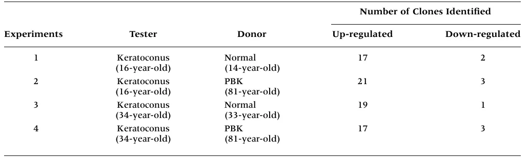
Table 1. Summary of subtraction experiments performed on corneal stromal tissue cDNA.
Number of Clones Identified
Experiments Tester Donor Up-regulated Down-regulated
1 Keratoconus Normal 17 2
(16-year-old) (14-year-old)
2 Keratoconus PBK 21 3
(16-year-old) (81-year-old)
3 Keratoconus Normal 19 1
(34-year-old) (33-year-old)
4 Keratoconus PBK 17 3
(34-year-old) (81-year-old)
NBT/BCIP (Sigma). The experiment was repeated at least three times.
Relative Quantitative Reverse Transcriptase PCR
Total stromal RNA (0.5 g) from corneas of normal in-dividuals (aged 20, 22, 35, 45, and 54 years), and pa-tients with keratoconus (25, 34, 36, 45, and 62 years), PBK (68 and 86 years), Fuchs’ corneal dystrophy (51 and 80 years), and corneal scar (36 years) was re-verse transcribed into cDNA using random hexamer primers and SuperScript II (Roche). Relative reverse transcriptase PCR (RT-PCR) was conducted using the QuantumRNA classical ribosomal 18S primer kit (Ambion, Austin, TX, USA) with keratocan specific
primers 5-GAATTGAAAAAGGAGCCCTAA-3and
5-CGGAGGTAGCGAAGATGAG-3and a 2:8 ratio of
18S primers to 18S copetimers. PCR was carried out as follows: one cycle 94C, 2 min followed by 27 cy-cles, 94C, 10 sec; 62C, 30 sec; 72C, 2 min followed by one cycle, 72C, 7 min. The expected sizes of the keratocan and 18S PCR products were 645- and 488-base pairs (bp), respectively. A sample containing only total RNA was used as a negative control. Sam-ples were run on ethidium bromide–stained agarose gels and quantified by densitometry. The level of ker-atocan expression was normalized to the 18S product in each specimen.
Results
Subtractive hybridization reactions were completed using cDNAs from stromal tissues of normal human, keratoconus, and PBK corneas. A total of four subtrac-tive hybridization reactions in two sets of experiments were done. In each set, keratoconus samples were ex-amined in two reactions against either normal human or PBK stroma. When a clone or a mRNA transcript was identified from both reactions, it was judged more likely to be keratoconus specific and further pursued. Table 1 summarizes the cDNA samples used and the number of clones that were found differentially expressed. A total of 252 clones were observed to be raised by Alpha Diagnostic International (San
An-tonio, TX, USA). Anti-keratocan was purified by af-finity column and the specificity was confirmed by enzyme-linked immunosorbant assay (ELISA). Western blot analysis using anti-keratocan against keratan sulfate proteoglycan extracted from human corneas (a generous gift of Dr. James Fundergurgh, University of Pittsburgh) yielded a 37-kD band not immunoreactive to anti-decorin (data not shown).
After primary antibody incubation, the sections were incubated sequentially with biotinylated goat anti-rabbit IgG (Jackson ImmunoResearch Labora-tories, Inc., West Grove, PA, USA), 0.3% H2O2
-methanol, and avidin-biotin-horseradish peroxidase complex (Vector, Burlingame, CA, USA) each for 30 min. Following color development with 3,3-diaminobenzidine tetrahydrochloride (Sigma Chemi-cals, St. Louis, MO, USA), the sections were dehy-drated and mounted in Permount (Fisher Scientific, Itasca, IL, USA). All tissue sections and negative controls were stained simultaneously under identi-cal conditions. Comparisons were made on only sec-tions stained in the same experiment and the im-munostaining was repeated at least three times to confirm the results.
In Situ Hybridization
Fig. 1. A representative differential screening dot blot of subtractive hybridization clones.Dot blot analysis was com-pleted on clones isolated from subtractive hybridization experi-ments. Blots were probed with the subtracted library “positive” and the reverse subtracted library “negative” (106cpm per 100 ng of subtracted cDNA probe) in ExpressHyb overnight at 65C. After washing, the membranes were autoradiographed overnight at70C. The positions of the clones are marked by numbers 1–10 and letters A–G. Clones identified through sequence analysis to encode portions of the keratocan gene that are up-regulated in the “positive” subtracted library (compared to the “negative”) are depicted by the arrows.
Fig. 2. Immunostaining for decorin.Corneal sections were from a 31-year-old normal human donor (A and B),two pa-tients with keratoconus (C and D,26-year-old; E,67-year-old), and a 77-year-old patient with pseudophakic bullous keratopa-thy (F). Sections were stained with anti-decorin (A, C, E and F) or normal sheep IgG (B and D,negative controls). Positive staining appearing as brown deposits is observed in the corneal stroma (St) as well as the epithelium (*). Note that the staining in the stroma is abnormally increased in diseased corneas (origi-nal magnification,40).
up-regulated when keratoconus samples were used as the tester and 142 to be down-regulated when ker-atoconus samples were used as the driver. Only 74 up-regulated and 9 down-regulated clones were sub-sequently confirmed by dot blot analyses (Fig. 1). The down-regulated clones were revealed by sequence analyses to be all mitochondrial genes. Of the up-regulated clones, 30 corresponded to novel sequences not found in the database. The remaining clones rep-resented 7 known genes that included heat shock pro-tein 90 (3 clones), decorin (8 clones), fibronectin (2 clones), ferritin heavy chain (2 clones), keratocan (16 clones), mitochondrial gene (7 clones), and ALU subfamily (6 clones). Because mitochondrial and ALU genes have been shown to be nonspecific artifacts fre-quently pulled out in differential display (21), sub-tractive hybridization (22) and yeast two-hybrid ex-periments (23), they were not investigated further.
Immunostaining experiments using antibodies for decorin, fibronectin, ferritin heavy chain, and ker-atocan were carried out. Figure 2 depicts the staining pattern for decorin in normal, keratoconus, and other disease corneas. Positive decorin immunoreactivity was observed in both keratocytes and matrices of normal human corneal stroma (Fig. 2A). The staining was increased especially in the stromal matrix in ker-atoconus (Figs. 2C and 2E). A similar increase was also observed in corneas of other diseases (Fig. 2F). This pattern was likewise noted for fibronectin and ferritin heavy chain (photographs not shown). The in-creased expression of these genes thus appeared to be related to disease conditions and was not keratoconus specific.
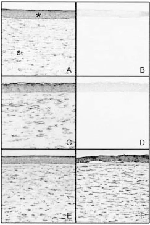
Positive keratocan staining (Fig. 3) was observed in the stroma of all normal and other diseased cor-neas. The staining in keratoconus corneas was much stronger throughout the stroma in both keratocytes and matrix lamella (Figs. 3C and 3D) than in normal (Fig. 3A) and disease controls (Figs. 3E and 3F).
tification of differentially expressed mRNAs. With this method, we detected a number of genes that may be up-regulated in the keratoconus stroma in comparison to normal human and PBK tissues. Apart from novel sequences, seven known genes were identified. Of them, mitochondrial gene and ALU subfamily genes are known to often be non-specific artifacts (21). Three others—heat shock protein 90, decorin, and fibronectin—have already been evaluated previously in keratoconus (24–26). Work in our laboratory has shown that heat shock protein 90, a member of the stress-response gene family, was not present in normal human, kerato-conus, or other diseased corneas (24). The expres-sion of other heat shock protein members was in-creased in keratoconus, but the increase was not keratoconus specific, and is most likely related to injury repair in disease conditions. Similarly, previous investigation demonstrated an increased decorin expression (25) in the stroma and an in-crease in fibronectin expression in the epithelium, basement membrane, and the stroma near defect ar-eas in keratoconus (26). Our immunostaining ex-periments corroborate these findings, and indicate moreover that the enhanced expression of ferritin For a more quantitative assessment of the
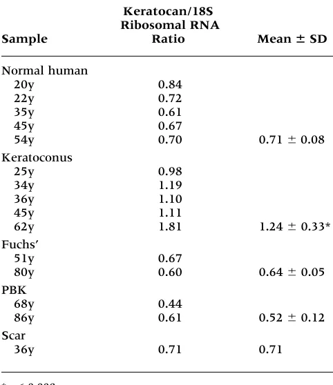
ker-atocan mRNA level, relative quantitative RT-PCR was completed using stromal RNAs from various corneas. The gel electrophoretogram (Fig. 5) after RT-PCR displayed two products, a 645-bp band for keratocan and a 488-bp band for 18S rRNA. The ratio from both band intensities were ob-tained via densitometric analyses. Results summa-rized in Table 2 indicate that the keratocan mRNA level in keratoconus stroma was approximately 1.8- to 2-fold higher than that in both normal
hu-man and other disease controls.
Discussion
PCR-select subtractive hybridization is a recently developed, powerful technique that allows for iden-Fig. 3. Immunostaining for keratocan in the corneal stroma.Sections were from a 16-year-old normal individual (A and B),two patients with keratoconus (C,29-year-old, and D,67-year-old), an 84-year-old patient with aphakic bullous keratopathy (E),and a 69-year-old patient with corneal scar (F).Sections were immunostained with an antibody against keratocan (A, C, D, E, and F)or normal rabbit IgG (B, nega-tive control). Posinega-tive staining appears as brown deposits in both keratocytes and stromal matrices. Note the increased stain-ing in keratoconus specimens (original magnification, 40).
heavy chain may also result from wound healing re-sponses seen in all corneal pathologies examined, including keratoconus.
Most significantly, keratocan was found to be up-regulated specifically in the stroma of kerato-conus corneas. This result was confirmed by inde-pendent methods including immunostaining, in situ hybridization, and relative quantitative RT-PCR. Fig. 5. Relative quantitative RT-PCR of keratocan.Total RNA samples were from 20-, 22-, 35-, 45-, and 54-year-old nor-mal human (NH) donors, 25-, 34-, 36-, 45-, and 62-year-old pa-tients with keratoconus (KC), 51- and 80-year-old papa-tients with Fuchs’ corneal dystrophy (FUCHS’), 68- and 86-year-old pa-tients with pseudophakic bullous keratopathy (PBK), and a 36-year-old patient with a corneal scar (SC). The keratocan and 18S rRNA bands are labeled. Total RNA was used as template instead of cDNA as a negative control for relative RT-PCR (lane not shown). No products were detected in the negative control.
Table 2. Relative quantitative RT-PCR for keratocan.
Keratocan/18S Ribosomal RNA
Sample Ratio Mean ⫾SD
Normal human
20y 0.84
22y 0.72
35y 0.61
45y 0.67
54y 0.70 0.71 0.08
Keratoconus
25y 0.98
34y 1.19
36y 1.10
45y 1.11
62y 1.81 1.24 0.33*
Fuchs’
51y 0.67
80y 0.60 0.64 0.05
PBK
68y 0.44
86y 0.61 0.52 0.12
Scar
36y 0.71 0.71
*p0.008.
Fuchs’, Fuchs’ corneal dystophy; PBK, pseudophakic bullous keratopathy; y, age of patients in years.
The increase in keratocan expression was not ob-served in other corneal disease such as PBK or Fuchs’ corneal dystrophy. With corneal scars, the levels of keratocan were either comparable to those in normals (Table 2) or slightly reduced (Fig. 3F). These data, consistent with previous results in chick corneas that keratocan protein levels remain unaltered during wound healing or in scars (27), argue against the possibility that the increased ker-atocan expression in keratoconus stroma is related to scarring.
Keratocan is a member of the small leucine rich proteoglycan (SLRP) gene family (28) expressed al-most exclusively by corneal keratocytes (29). Along with lumican and mimecan, it makes up the major proteoglycan, KSPG, in the cornea (30–32). The core proteoglycan proteins contain sulfated keratan sul-fate side chains in the cornea, whereas in noncorneal tissues, the core proteins lack sulfation (29). KSPGs have long been suggested to be important in corneal transparency and collagen fibrillogenesis (31,33–35). Developmental studies have determined that the emergence of keratan sulfate in the cornea correlates with transparency (30,36), and such a role for KSPG has recently been verified by results from lumi-can knockout mice that displayed bilateral corneal opacity (37,38). The collagen structure in the poste-rior stroma was also found altered in the lumican-deficient mice with both increased fibril diameter and abnormal lateral growth (39). Although the specifics have yet to be defined, it is generally be-lieved that keratocan, like lumican, may also take part in regulating both corneal transparency and collagen fibrillogenesis.
Earlier analyses of keratoconus samples have determined that although the amount of keratan sul-fate was decreased, the keratan sulsul-fate core protein content remained unaltered (40–42). The KSPG core protein was then considered as only one entity and was measured as such. Therefore, changes in indi-vidual KSPGs such as keratocan might have gone unnoticed.
supported by grants EY03890 and EY05628, core grant EY01792 from the National Eye Institute, Bethesda, MD, and by an award from Research to Prevent Blindness, Inc., New York, to B.Y.J.T.Y.
References
1. Krachmer JH, Feder RS, Belin MW. (1984) Keratoconus and related non-inflammatory corneal disorders. Surv. Ophthalmol.
28:293–322.
2. Rabinowitz YS. (1998) Keratoconus. Surv. Ophthalmol.42:297– 319.
3. Rabinowitz YS, Garbus J, McDonnell PJ. (1990) Computer-assisted corneal topography in family members of kerato-conus. Arch. Ophthalmol.108:365–371.
4. Jacobs DS, Dohlman CH. (1993) Is keratoconus genetic? Int. Ophthalmol. Clin.33:249–260.
5. Coyle JT. (1984) Keratoconus and eye rubbing. Am. J. Opthalmol.
97:527–528.
6. Macsai MS, Varley GA, Krachmer JH. (1990) Development of keratoconus after contact lens wear. Patient characteristics. Arch. Ophthalmol. 108:534–538.
7. Liu E, Slomovic AR. (1997) Indications for penetrating ker-atoplasty in Canada, 1986–1995. Cornea16:414–419. 8. Komai Y, Ushiki T. (1991) The three-dimensional
organiza-tion of collagen fibrils in the human cornea and sclera. Invest. Ophthalmol. Vis. Sci.32:2244–2258.
9. Axelsson I. (1984) Heterogeneity, polydispersity, and physi-ologic role of corneal proteoglycans. Acta Ophthalmol. 62: 25–38.
10. Hassell JR, Cintron C, Kublin C, Newsome DA. (1983) Pro-teoglycan changes during restoration of transparency in corneal scars. Arch. Biochem. Biophys.222:362–369.
11. Yue BYJT, Sugar J, Benveniste K. (1984) Heterogeneity in keratoconus: possible biochemical basis. Proc. Soc. Exp. Biol. Med.175:336–341.
12. Critchfield JW, Calandra AJ, Nesburn AB, Kenney MC. Ker-atoconus. I. Biochemical studies of normal and keratoconus corneas. Exp. Eye Res.46:953–963.
13. Sawaguchi S, Yue BYJT, Sugar J, Gilboy JE. (1989) Lysoso-mal enzyme abnorLysoso-malities in keratoconus. Arch. Ophthalmol.
107:1507–1510.
14. Zhou L, Sawaguchi S, Twining SS, Sugar J, Feder RS, Yue BYJT. (1998) Expression of degradative enzymes and pro-tease inhibitors in corneas with keratoconus. Invest. Ophthal-mol. Vis. Sci.39:1117–1124.
15. Sawaguchi S, Twining SS, Yue BYJT, Wilson PM, Sugar J, Chan S-K. (1990) ␣1-Proteinase inhibitor levels in kerato-conus. Exp. Eye Res.50:549–554.
16. Sawaguchi S, Twining SS, Yue BYJT, et al. (1994) ␣ 2-Macroglobulin levels in normal human and keratoconus corneas. Invest. Ophthalmol. Vis. Sci.35:4008–4014.
17. Whitelock RB, Fukuchi T, Zhou L, et al. (1997) Cathepsin G, Acid phophatase, and ␣1-proteinase inhibitor messenger RNA levels in keratoconus corneas. Invest. Ophthalmol. Vis. Sci.
38:529–534.
18. Pellegata NS, Diezuez-Lucena JL, Joensuu T, et al. (2000) Mutations in KERA, encoding keratocan, cause cornea plana. Nat. Genet.25:91–95.
19. Roskans AJI, Connor JR. (1994) Iron, transferrin, and ferritin in the rat brain during development and aging. J. Neurochem.
63:709–716.
20. Twining SS, Everse SJ, Wilson PM, Yue BYJT, Chan S-K. (1989) Localization and quantification of ␣1-proteinase in-hibitor in the human cornea. Curr. Eye Res.8:389–395. 21. Sompayrac L, Jane S, Burn TC, Tenen DG, Danna KJ. (1995)
Overcoming limitations of the mRNA differential display technique. Nucl. Acids Res.23:4738–4739.
22. Diatchenko L, Lukyanov S, Lau YF, Siebert PD. (1999) Sup-pression subtractive hybridization: a versatile method for identifying differentially expressed genes. Meth. Enzymol. 303: 349–380.
The current study also did not identify the trans-membrane phosphotyrosine (LAR) gene recently determined to be up-regulated in keratoconus by Chiplunkar et al. (44) in a differential display inves-tigation. LAR was one of the clones initially isolated in our subtractive hybridization, but was found not to be up-regulated in dot blot screening and was eliminated. In the differential display experiment (44), RNA isolated from corneal stromal cell cultures was used as the starting material. The gene expres-sion in corneal stromal cell cultures (45) may be considerably altered from that in tissue specimens used in our experiments. The differences in the start-ing material and the techniques may account, at least in part, for the disparity in the results.
To summarize, we used subtractive hybridiza-tion and suppression PCR techniques to identify differentially expressed transcripts in keratoco-nus stromal tissue. These experiments revealed that keratocan, uniquely expressed in the cornea stroma, is up-regulated in keratoconus corneas. Keratocan is one of the three KSPGs in the cornea speculated to be important for structure of the stromal matrix and maintenance of corneal trans-parency. The overexpressed keratocan may conceiv-ably alter the fibrillogenesis in the stroma, leading to structural defects and contributing to the devel-opment of keratoconus.
Recently, keratocan has been linked to another corneal disease, cornea plana (18). This disease is characterized by a flattened cornea and the loss of re-fraction. Two mutations were identified in the kera-tocan gene in patients with a severe, recessive form of cornea plana. One mutation replaces the single as-paragine residue with serine in the leucine-rich re-peat, a characteristic motif in SLRPs. The other mu-tation results in a stop codon at amino acid 174 and thus a truncated protein. The investigators hypothe-sized that both mutations would cause either a loss of function or absence of keratocan and a flattened cornea. It is hence of note that a connection of an in-creased keratocan expression with the corneal pro-trusion in keratoconus is suggested by the current study. These findings appear to be consistent with the role of keratocan in collagen structure and per-haps the mechanical architecture of the stroma. A keratocan knockout mouse model may ultimately provide insights in this regard.
Acknowledgments
23. Gyuris J, Golemis W, Chertov H, Brent R. (1993) Cid1, a hu-man G1 and S phase protein phosphatase that associates with Cdk2. Cell75:791–803.
24. Zhou L, Yue BYJT, Twining SS, Sugar J, Feder RS. (1996) Ex-pression of wound healing and stress-related proteins in ker-atoconus corneas. Curr. Eye Res.15:1124–1131.
25. Funderburgh JL, Hevelone ND, Roth MR, et al. (1998) Decorin and biglycan of normal and pathologic human corneas. Invest. Ophthalmol. Vis. Sci.39:1957–1964.
26. Tuori A, Virtanen I, Aine E, Uusitalo H. (1997) The expres-sion of tenascin and fibronectin in keratoconus, scarred and normal human corneas. Graefes Arch. Clin. Exp. Ophthalmol.235: 222–229.
27. Sundarraj N, Fite D, Belak R, et al. (1998) Proteoglycan dis-tribution during healing of corneal stromal wounds in chick. Exp. Eye Res.67: 433–442.
28. Iozzo RV. (1998) Matrix proteoglycans: from molecular de-sign to cellular function. Annu. Rev. Biochem.67:609–652. 29. Corpuz LM, Funderburgh JL, Funderburgh ML, Bottomley
GS, Prakash S, Conrad GW. (1996) Molecular cloning and tis-sue distribution of keratocan. Bovine corneal keratan sulfate proteoglycan. J. Biol. Chem.271:9759–9763.
30. Funderburgh JL, Funderburgh ML, Brown SJ, et al. (1993) Sequence and structural implications of a bovine corneal ker-atan sulfate proteoglycan core protein. Protein 37B represents bovine lumican and proteins 37A and 25 are unique. J. Biol. Chem. 268: 11874–11880.
31. Hassell JR, Kimura JH, Hascall VC. (1986) Proteoglycan core protein families. Annu. Rev. Biochem.55:539–567.
32. Funderburgh JL, Funderburgh ML, Mann MM, Conrad GW. (1991) Unique glycosylation of three keratan sulfate proteo-glycan isoforms. J. Biol. Chem.266: 14226–14231.
33. Scott JE (1988) Proteoglycan-fibrillar collagen interactions. Biochem. J.252:313–323.
34. Schonherr E, Hausser H, Beavan L, Kresse H. (1995) Decorin-type I collagen interaction. J. Biol. Chem.270:8877–8883. 35. Rada JA, Cornuet PK, Hassell JR. (1993) Regulation of
corneal collagen fibrillogenesis in vitro by corneal keratan
sulfate proteoglycan (lumican) and decorin core proteins. Exp. Eye Res.56:635–648.
36. Funderburgh JL, Caterson B, Conrad GW. (1986) Keratan sulfate proteoglycan during embronic development of the chicken cornea. Dev. Biol. 116:267–277.
37. Chakravarti S, Magnuson T, Lass JH, Jepsen KJ, LaMantia C, Carroll H. (1998) Lumican regulates collagen fibril assembly: skin fragility and corneal opacity in the absence of lumican. J. Cell. Biol.112:987–996.
38. Saika S, Shiraishi A, Saika S, et al. (2000) Role of lumican in the corneal epithelium during wound healing. J. Biol. Chem.
275:2607–2612.
39. Chakravarti S, Petroll WM, Hassell JR, et al. (2000) Corneal Opacity in lumican-null mice: defects in collagen fibril struc-ture and packing in the posterior stroma. Invest. Ophthalmol. Vis. Sci.41:3365–3373.
40. Funderburgh JL, Panjwani N, Conrad GW, Baum J. (1989) Altered keratan sulfate epitopes in keratoconus. Invest. Oph-thalmol. Vis. Sci.30:2278–2281.
41. Sawaguchi S, Yue BYJT, Chang I, Sugar J, Robin J. (1991) Proteoglycan molecules in keratoconus corneas. Invest. Oph-thalmol. Vis. Sci.32:1846–1853.
42. Funderburgh JL, Funderburgh ML, Rodrigues MM, Krachmer JH, Conrad GW. (1990) Altered antigenicity of keratan sulfate proteoglycan in selected corneal diseases. Invest. Ophthalmol. Vis. Sci. 31:419–425.
43. Whitelock RB, Li Y, Zhou L, Sugar J, Yue BYJT. (1997) Ex-pression of transcription factors in keratoconus, a cornea-thinning disease. Biochem. Biophys. Res. Commun. 235: 253– 258.
44. Chiplunkar S, Chamblis K, Chwa M, Rosenberg S, Kenney MC, Brown DJ. (1999) Enhanced expression of a transmem-brane phosphotyrosine phosphatase (LAR) in keratoconus cultures and corneas. Exp. Eye Res.68:283–293.