Volume 18 Number 3 pp. 281–289 C The Author(s) 2015 doi:10.1017/thg.2015.18
Genetic Influence on Left Ventricular Structure and
Function: A Korean Twin and Family Study
Hye-Mi Noh,1Sang Cheol Lee,2Seung Woo Park,2Joohon Sung,3,4and Yun-Mi Song5
1Department of Family Medicine, Hallym Sacred Heart Hospital, College of Medicine, Hallym University, Anyang, South Korea
2Department of Cardiology and Cardiovascular Imaging Center, Cardiac and Vascular Center, Samsung Medical Center, Sungkyunkwan University School of Medicine, Seoul, South Korea
3Department of Epidemiology, School of Public Health, Seoul National University 4Institute of Health Environment, Seoul National University
5Department of Family Medicine, Samsung Medical Center, Sungkyunkwan University School of Medicine, Seoul, South Korea
Genetic factors have been suggested to be one of the determinants of the variation of left ventricular (LV) structure and function. However, the heritability range of LV structure varies across studies and the influence of genetics on LV function is not well established, especially in Asian populations. Study subjects were 1,642 healthy Korean adults from 426 families, consisting of 298 pairs of monozygotic twins, 62 pairs of dizygotic twins, one set of triplets, 567 siblings, and 354 parents. LV structure and function were measured by M-mode and 2D echocardiography, and conventional and tissue Doppler imaging (TDI). Pairwise intra-class correlations for various familial relationships and heritability were estimated for LV structure and function. The heritability of LV mass, LV ejection fraction (LVEF), left atrial volume index, the ratio between early and late diastolic velocity of mitral inflow (E/A ratio), and the ratio between early diastolic velocity of mitral inflow and early diastolic mitral annular velocities (E/Ea ratio) was 0.44, 0.27, 0.44, 0.25, and 0.33, respectively. Bivariate genetic analysis showed that LV structural and functional traits had significant genetic correlations with cardiovascular risk factors. Additive genetic correlation (G)of LV mass with body mass index, systolic blood pressure, and high density lipoprotein cholesterol were 0.49, 0.42, and -0.15 respectively. LVEF (G=0.33) and left atrial volume index (G=0.24) also had a significant genetic correlation with systolic blood pressure. These findings support the theory that genetic factors have significant influence on these traits and necessitate further work to identify the specific genes involved.
Keywords:echocardiography, genetics, heart ventricles, Korean, twin study
Both LV mass and LV function are independent risk fac-tors for morbidity and mortality from cardiovascular dis-ease (Arnlov et al.,2005; Levy et al.,1990). LV mass varies markedly between individuals, and several factors, includ-ing age, ethnicity, sex, body size, blood pressure, and valvu-lar heart disease are associated with LV mass (Devereux et al.,1997; Gardin et al.,1995; Levy et al.,1988; Lorber et al.,2003), explaining 50–60% of the variance of LV mass (Devereux et al.,1997; Jones et al.,1997). On the other hand, twin and family studies, including two Asian studies, have reported significant heritability in LV mass (Assimes et al., 2007; Busjahn et al.,2009; Chien et al.,2006), which sug-gests that genetic factors are involved in the interindividual variation of LV structure. However, the reported heritability of LV mass varied markedly, with a wide range of heritability (15–84%) and it is not clear whether the genetic influence on LV structure is greater than the environmental influence.
Given that there were differences in study design (twin or family studies), the origin of study subjects (community-based or hospital-(community-based), adjusted covariates, and ethnicity between the previous studies, a well-designed study is still needed.
LV function is also suggested to be influenced signifi-cantly by genetics (Bielen et al.,1991; Fox et al.,2010; Jin et al., 2011; Swan et al.,2003). The heritability of LVEF
RECEIVED2 October 2014;ACCEPTED16 February 2015. First pub-lished online 14 April 2015.
H.-M. Noh et al.
for Belgian (Jin et al., 2011) and hypertensive African American families (Fox et al., 2010) was 0.48 and 0.40 respectively. Among the echocardiographic measures re-flecting LV diastolic function, the E peak was moderately heritable (0.37–0.49) in the Belgian twin study (Bielen et al., 1991), the Scottish twin study (Swan et al.,2003), and the hypertensive African American family study (Fox et al., 2010). In addition, the heritability of the A peak for Belgium and African American families was 0.26 and 0.45, respec-tively. However, the heritability of LV function has never been evaluated for Asian populations.
We conducted a Korean twin and family study in a com-munity setting to evaluate the genetic influence on variation in LV structure and function while considering major car-diovascular risk factors. Using data from both twins and their family members provides a more accurate estimation of heritability because comparison of the phenotypic cor-relations between various family cor-relationships that reflect different genetic correlations is possible. In addition, we sought to disentangle the genetic and environmental effects on LV structure and function by examining the change in estimated heritability with a stepwise adjustment for covari-ates and estimating additive genetic correlations between each echocardiographic measurement and each cardiovas-cular risk factor.
Methods
Subjects and Study Design
Study participants were twins and their siblings or parents who underwent echocardiography during a health exami-nation for the Healthy Twin Study between March 2009 and February 2013. Details of the methodology of the Healthy Twin Study have been reported previously (Sung et al., 2006). In brief, the Healthy Twin Study is a nationwide cohort study recruiting Korean same-sex adult twins (ࣙ30 years of age) and their first-degree adult family members from the general population. Among the initial 1,718 sub-jects, we excluded 76 subjects for the following reasons: insufficient echocardiogram quality (n=9), previous his-tory of ischemic heart disease (n= 44), congenital heart disease (n=8), clinically significant heart disease such as moderate-to-severe valvular heart disease or a prosthetic valve (n=5), and regional wall-motion abnormality (n= 10). Thus, 1,642 subjects from 426 families were included in the study, which was comprised of 298 pairs (n=596) of monozygotic twins, 62 pairs (n=125; including 1 set of triplets) of dizygotic twins, 567 siblings, and 354 parents. Written informed consent was obtained from all partici-pants.
Measurement of Echocardiography and Covariates A standard echocardiographic examination was performed according to the guidelines of the American Society of Echocardiography using echocardiography machines
(Vivid 7 and Vivid E9, GE Medical System, Horten, Nor-way, and EKO 7, Medison, Seoul, Korea). All of the echocar-diographers who were involved in the present study were registered diagnostic cardiac sonographers accredited by the American Registry for Diagnostic Medical Sonography. All members of the same family underwent echocardio-graphic measurements taken by a single sonographer with the same machine, while they were lying on their left side. Study participants refrained from smoking, heavy phys-ical activity, and intake of caffeine containing drinks or alcohol for at least 3 hours prior to echocardiographic ex-amination. The following components were measured by M-mode and 2-D echocardiography, and conventional and TDI: left ventricular internal diameter (LVID), interven-tricular septum (IVS), posterior wall thickness (PWT), left atrial diameter, aortic root diameter, mitral inflow veloc-ities, and mitral annular velocities (Supplementary Table S1). LVID, IVS, and PWT were measured at the end of diastole and at the end of systole, according to the Amer-ican Society of Echocardiography guidelines (Gottdiener et al.,2004). LVEF was calculated using the biplane Simp-son’s method. We computed LV mass in grams as 0.8 × (1.04×[{LVID+IVS+PWT}3- LVID3])+0.6 (Gottdi-ener et al.,2004). Mitral inflow and annular velocities were measured using conventional Doppler and TDI techniques. Peak early diastolic velocities (E peak) and peak late diastolic velocities (A peak) of mitral inflow were measured using pulsed wave Doppler at the tip of the mitral valve leaflets. Peak early diastolic mitral annular velocities (Ea peak) and peak late diastolic mitral annular velocities (Aa peak) were acquired on TDI at the septum in apical four-chamber view.
We evaluated the reliability of echocardiographic measurements between different machines (GE and Medison) and between different echocardiographers. The inter-machine intra-class correlation coefficients were esti-mated in 21 randomly selected persons and were between 0.72–0.92 for LV structural and functional measurements, including ID, IVS, left atrial diameter, aortic root diame-ter, LVEF, LV mass, E peak, A peak, Ea peak, and Aa peak. The inter-observer intra-class correlation coefficients in 21 randomly selected subjects ranged between 0.64–0.99 for the same echocardiographic measurements (data shown in Supplementary Table S2).
Body weight and height for each participant was mea-sured twice by trained research assistants according to the standardized protocol, and the average value of the re-peated measurements was used for calculating body mass index (weight divided by height squared; kg/m2). A trained research assistant measured blood pressure twice using a mercury sphygmomanometer, and the average value of the repeated measurements was used for analysis.
A blood sample was drawn after a 12-hour overnight fast on the same day that the echocardiographic measure-ment was taken. Serum concentrations of total cholesterol,
282
TWIN RESEARCH AND HUMAN GENETICShttps://www.cambridge.org/core/terms. https://doi.org/10.1017/thg.2015.18
high-density lipoprotein (HDL) cholesterol, triglyceride, and low-density lipoprotein (LDL) cholesterol were enzymatically assayed in fresh sera using commercial kits in a designated central laboratory.
We collected information about pre-existing diseases (myocardial infarction, angina, hypertension, diabetes mel-litus and dyslipidemia), current treatment for hyperten-sion, diabetes mellitus, and dyslipidemia, smoking habits, alcohol consumption and physical exercise using a self-administered questionnaire. We categorized smoking habits into three groups: never smokers, former smokers, and current smokers. Alcohol consumption was categorized into two groups: ever drinkers and never drinkers. Phys-ical exercise was defined as moderate or vigorous in-tensity of regular exercise activity and was categorized into three groups by the frequency of exercise per week.
The study protocol was approved by the Institutional Review Board of the Samsung Medical Center.
Statistical Analysis
We estimated the intra-class correlation coefficient for residual variances of echocardiographic measurements after adjusting for age and sex with respect to family relationship using the Statistical Analysis System (SAS Institute, Cary, NC, USA).
We estimated heritability using the Sequential Oligogenic Linkage Analysis Routines (SOLAR, Version 4.2.0; South-west Foundation for Biomedical Research, San Antonio, TX, USA; Almasy & Blangero,1998) with adjustment for age, sex, smoking habits, alcohol consumption, physical activ-ity, height, body mass index, systolic blood pressure, fasting glucose, HDL-cholesterol, LDL-cholesterol, antihyperten-sive medication, diabetes medication and lipid-lowering medication as covariates. Total phenotypic variation (бp2) in echocardiographic measurements after consideration of the measured covariates was partitioned into additive ge-netic variation (бa2), a shared environmental component within a family (бc2), and an individual specific unique en-vironmental component (бe2), according to the variance decomposition model. For the non-normally distributed variables, we performed normalization by inverse normal transformation. This model assumed that the effects of envi-ronmental factors are common to the members of a family, and that the three factors (бa2,бc2, andбe2) have indepen-dent and additive effects on trait variance. The total residual variance is the sum of the additive and individual specific variance components (бp2=бa2+бc2+бe2). The her-itability (h2) indicates the proportion of residual variance attributed to additive genetic factors. It was calculated as the ratio of the additive component and the total variance (бa2/бp2). In order to ascertain whether the echocardio-graphic measurement and cardiovascular risk factors are concurrently determined by shared genes and the environ-ment, we also conducted bivariate
variance-component-based genetic analysis that allows partitioning of pheno-typic correlations into genetic (G) and environmental correlations (E).
Results
Characteristics and Echocardiographic Measurements of the Study Subjects
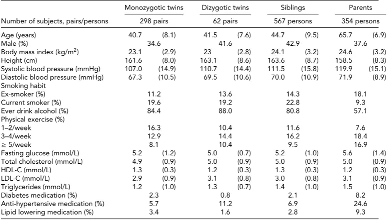
Table 1shows demographic and clinical characteristics of the study subjects. The mean ages (SD) of monozygotic twins, dizygotic twins, siblings, and parents were 40.7 (8.1), 41.5 (7.6), 44.7 (9.5), and 65.7 (6.9) years, respectively. As compared with offspring, parents had higher levels of body mass index, systolic and diastolic blood pressure, fasting glucose and triglycerides. Parents were less likely to be cur-rent smokers or to drink alcohol.Table 2shows the distribu-tion of M-mode and Doppler echocardiographic measure-ments. Compared with offspring, parents had higher mean levels for LV mass, left atrial diameter, aortic diameter, left atrial volume index and E/Ea ratio, and lower mean levels for Ea peak and E/A ratio.
Intra-Class Correlations Between Intrafamilial Pairs and Heritability for Left Ventricular Structure and Function Table 3summarizes the intra-class correlations within each pair of diverse familial relationships and heritability for various measurements of LV structure and function. After considering age and sex, intra-class correlation coefficients were highest within monozygotic twin pairs for all echocar-diographic measurements. The intra-class correlation co-efficients were lowest within spouse pairs for LV mass, LV internal diameter, IVS, left atrial diameter, aortic root diam-eter, E/A ratio and Ea peak, while the intra-class correlation coefficients for ejection fraction (EF), left atrial volume in-dex and E/Ea ratio were lowest within parent-child pairs. The age and sex-adjusted heritability for all LV structural and functional traits were low to moderate, ranging from 0.25 for IVS and E/A ratio to 0.58 for aortic root diameter. When we made further adjustments for other measured co-variates, the level of heritability did not materially change. All measured covariates explained about 8.3% to 63.2% of the total variance for most LV structural and functional traits.
H.-M. Noh et al.
TABLE 1
Characteristics of Study Subjects
Monozygotic twins Dizygotic twins Siblings Parents
Number of subjects, pairs/persons 298 pairs 62 pairs 567 persons 354 persons
Age (years) 40.7 (8.1) 41.5 (7.6) 44.7 (9.5) 65.7 (6.9)
Male (%) 34.6 41.6 42.9 37.6
Body mass index (kg/m2) 23.1 (2.9) 23 (2.8) 24.1 (3.2) 24.6 (3.2)
Height (cm) 161.6 (8.0) 163.1 (8.6) 163.6 (8.7) 158.5 (8.3)
Systolic blood pressure (mmHg) 107.0 (14.9) 110.7 (14.4) 111.5 (15.8) 119.9 (15.1) Diastolic blood pressure (mmHg) 67.3 (10.5) 69.5 (10.6) 70.0 (10.9) 71.9 (8.9) Smoking habit
Ex-smoker (%) 11.2 13.6 14.3 18.1
Current smoker (%) 19.6 19.2 22.8 9.3
Ever drink alcohol (%) 84.4 88.0 80.8 57.1
Physical exercise (%)
1–2/week 16.3 10.4 11.6 7.6
3–4/week 12.9 14.4 16.2 18.4
ࣙ5/week 8.1 10.4 9.5 16.9
Fasting glucose (mmol/L) 5.2 (1.2) 5.0 (0.7) 5.2 (1.0) 5.6 (1.4)
Total cholesterol (mmol/L) 4.9 (0.9) 5.0 (0.9) 5.0 (0.9) 5.0 (0.9)
HDL-C (mmol/L) 1.3 (0.3) 1.2 (0.3) 1.3 (0.3) 1.2 (0.3)
LDL-C (mmol/L) 2.9 (0.9) 3.1 (0.8) 3.0 (0.8) 3.1 (0.9)
Triglycerides (mmol/L) 1.2 (1.0) 1.3 (0.7) 1.4 (1.0) 1.5 (1.0)
Diabetes medication (%) 2.3 0.8 2.1 8.2
Anti-hypertensive medication (%) 5.7 11.2 6.9 24.6
Lipid lowering medication (%) 3.4 1.6 2.8 9.3
Note: Data are expressed as mean (standard deviation) or percentage; HDL-C=High density lipoprotein cholesterol; LDL-C= Low density lipoprotein cholesterol.
TABLE 2
Echocardiographic Measurements of Study Subjects
Monozygotic twins Dizygotic twins Siblings Parents
(298 pairs) (62 pairs) (567 persons) (354 persons)
M-mode/2D echocardiography
LV mass (g) 113.2 (31.5) 115.9 (31.1) 123.1 (33.5) 129.4 (31.8)
LV internal diameter (mm) 48.3 (4.0) 48.4 (3.6) 48.8 (3.8) 48.2 (4.2)
Interventricular septum (mm) 7.2 (1.2) 7.4 (1.3) 7.7 (1.4) 8.1 (1.3)
Left atrial diameter (mm) 33.8 (4.4) 34.5 (4.6) 34.9 (4.4) 35.9 (5.0)
Aortic root diameter (mm) 28.7 (3.5) 28.7 (3.8) 30.0 (4.1) 32.3 (3.9)
Ejection fraction (%) 62.7 (5.4) 63.0 (6.0) 63.7 (5.7) 66.2 (5.9)
Left atrial volume index (ml/m2) 26.3 (5.1) 27.3 (5.7) 26.9 (5.7) 29.8 (7.7) Doppler echocardiography
E/A ratio 1.52 (0.5) 1.52 (0.4) 1.38 (0.5) 0.83 (0.3)
Ea peak (cm/s) 11.0 (2.5) 10.9 (2.3) 10.0 (2.8) 6.8 (1.9)
E/Ea ratio 7.0 (1.7) 7.0 (1.7) 7.4 (2.2) 9.3 (3.1)
Note: Data are expressed as mean (standard deviation); LV=left ventricle; E peak=peak early diastolic velocity of mitral inflow; A peak=peak late diastolic velocity of mitral inflow; Ea peak=peak early diastolic mitral annular velocity.
correlation with most LV structural traits and a positive correlation with E/A ratio.
We also estimated the changes in heritability and vari-ance of each echocardiographic measurement when each cardiovascular risk factor was additionally considered in the heritability estimation model. Among the cardiovascu-lar risk factors, body mass index was found to explain more than 5% of the total variance of several LV structural traits (left atrial diameter, LV mass, and LV internal diameter) and LV functional traits (Ea peak and E/Ea ratio). Systolic blood pressure explained 5% and 5.1% of the total variance of LV mass and left atrial diameter, respectively. Glucose, LDL-cholesterol and HDL-cholesterol explained less than 3% of the total variance for all LV structural and functional traits.
Discussion
In this Korean study of twins and their families, we found that most LV structural and functional traits were under significant genetic influence, with moderate-to-low levels of heritability.
Among LV structural measurements, LV mass has been the most frequently studied in terms of the estimation of heritability. The level of heritability of LV mass in our twin and family study was similar to that estimated in previous family studies (Jin et al., 2011; Mayosi et al.,2002; Post et al., 1997; Vasan et al., 2007), while it was lower than that estimated in twin-only studies (Busjahn et al., 2009; Sharma et al.,2006; Swan et al.,2003). Twin pairs are more likely to share environmental factors with each other than
284
TWIN RESEARCH AND HUMAN GENETICShttps://www.cambridge.org/core/terms. https://doi.org/10.1017/thg.2015.18
Genetic
Influence
o
n
the
Left
Ventricle
TABLE 3
Intra-Class Correlations Between Diverse Intrafamilial Pairs and Estimated Heritability for Left Ventricular Structure and Function
Age- and sex-adjusted intra-class correlation coefficient
(95% confidence interval) Model 1∗ Model 2† Model 3‡
Siblings and
Echocardiographic Monozygotic dizygotic Spouse Parents–child Heritability Variance Heritability Variance Heritability) Variance
measurements twins (266 pairs) twins (660 pairs) (109 pairs) (1,112 pairs) (SE) explained (SE) explained (SE) explained
LV mass 0.56 (0.50, 0.61) 0.25 (0.17, 0.33) −0.01 (−0.14, 0.13) 0.25 (0.19, 0.30) 0.55(0.04) 28.7% 0.55(0.04) 28.7% 0.44(0.04) 47.1%
LV internal diameter
0.57 (0.51, 0.62) 0.18 (0.10, 0.26) −0.06 (−0.19, 0.07) 0.20 (0.15, 0.26) 0.51(0.04) 17.6% 0.51(0.04) 17.6% 0.47(0.04) 31.9%
Interventricular septum
0.34 (0.27, 0.41) 0.13 (0.05, 0.21) 0.08 (−0.06, 0.21) 0.16 (0.10, 0.21) 0.30(0.04) 16.7% 0.30(0.04) 17.0% 0.25(0.04) 24.9%
Left atrial diameter 0.52 (0.46, 0.58) 0.23 (0.15, 0.31) −0.20 (−0.33,−0.07) 0.13 (0.07, 0.19) 0.48(0.04) 12.8% 0.48(0.04) 13.0% 0.38(0.05) 37.4%
Aortic root diameter
0.55 (0.49, 0.61) 0.35 (0.28, 0.42) 0.19 (0.06, 0.32) 0.22 (0.17, 0.28) 0.60(0.03) 42.0% 0.60(0.03) 42.1% 0.58(0.04) 47.3%
Ejection fraction 0.26 (0.18, 0.33) 0.15 (0.07, 0.23) 0.22 (0.09, 0.34) 0.13 (0.08, 0.19) 0.27(0.04) 8.0% 0.27(0.04) 8.0% 0.27(0.04) 8.3%
Left atrial volume index
0.39 (0.32, 0.46) 0.25 (0.17, 0.33) 0.25 (0.12, 0.37) 0.21 (0.15, 0.26) 0.43(0.04) 7.8% 0.43(0.04) 8.0% 0.44(0.04) 10.1%
E/A ratio 0.33 (0.26, 0.40) 0.20 (0.12, 0.28) 0.07 (−0.07, 0.20) 0.14 (0.08, 0.20) 0.27(0.04) 48.0% 0.27(0.04) 48.0% 0.25(0.04) 53.1%
Ea peak 0.48 (0.41, 0.54) 0.18 (0.10, 0.26) 0.05 (−0.08, 0.18) 0.21 (0.16, 0.27) 0.49(0.04) 53.9% 0.49(0.04) 53.9% 0.41(0.05) 63.2%
E/Ea ratio 0.37 (0.30, 0.44) 0.19 (0.11, 0.27) 0.18 (0.04, 0.30) 0.15 (0.09, 0.21) 0.39(0.05) 23.1% 0.39(0.05) 23.1% 0.33(0.05) 31.3%
Note: SE=standard error; LV=Left ventricle; E peak=peak early diastolic velocity of mitral inflow; A peak=peak late diastolic velocity of mitral inflow; Ea peak=peak early diastolic mitral annular velocity; Model 1∗
=adjusted for age and sex; Model 2†=adjusted for age, sex, smoking, alcohol and physical activity; Model 3‡=adjusted for age, sex, smoking, alcohol, physical activity, height, body mass index, systolic blood pressure, antihypertensive medication, fasting glucose, diabetes medication, low density lipoprotein cholesterol, high density lipoprotein cholesterol, and lipid lowering medication. All estimates were significant (p<
.05) compared to 0.
TWIN
RESEARCH
AND
H
UMAN
GENETICS
.
https://doi.org/10.1017/thg.2015.18
https://www.cambridge.org/core
. IP address:
118.70.13.36
, on
06 Sep 2020 at 05:48:24
H.-M.
N
oh
et
al.
TABLE 4
Additive Genetic Correlation (G) Between Echocardiographic Measurement and Cardiovascular Risk Factors and the Influence of Additional Adjustment∗for Cardiovascular Risk Factors on
the Estimation of Heritability
Body mass Systolic blood LDL-
HDL-Height index pressure Glucose† cholesterol‡ cholesterol‡
Change Change Change Change Change Change
G G G G G G
(SE) VAR h2 (SE) VAR h2 (SE) VAR h2 (SE) VAR h2 (SE) VAR h2 (SE) VAR h2
LV mass 0.39(0.04)§ 5.4% −0.04 0.49(0.05)§ 11.2% −0.04 0.42(0.07)§ 5.0% −0.04 0.11(0.08) 0% 0 0.10(0.07) 0% 0 −0.15(0.06)§ 0.9% −0.01
LV internal diameter
0.36(0.04)§ 5.3% −0.03 0.39(0.06)§ 8.2% −0.02 0.21(0.08)§ 1.2% −0.01 0.11(0.08) 0.1% 0 0.01(0.07) 0% 0 −0.16(0.06)§ 0.6% −0.01
Interventricular septum
0.29(0.06)§ 1.9% −0.02 0.32(0.08)§ 3.9% −0.01 0.30(0.09)§ 3.4% −0.02 0.05(0.10) 0% 0 0.08(0.08) 0.5% 0.01 −0.10(0.07) 0.8% −0.01
Left atrial diameter 0.15(0.05)§ 1.3% 0 0.64(0.05)§ 22.4% −0.09 0.31(0.08)§ 5.1% −0.02 0.33(0.09)§ 1.0% −0.02 0.14(0.07) 1.1% 0 −0.19(0.06)§ 2.2% −0.01
Aortic root diameter
0.29(0.04)§ 2.6% −0.09 0.25(0.06)§ 1.6% −0.07 0.22(0.08) 0.7% −0.05 0.11(0.08) 0.2% −0.06 0.06(0.07) 0.1% −0.06 −0.23(0.06)§ 0% −0.06
Ejection fraction −0.07(0.06) 0% 0 0.07(0.09) 0.3% 0 0.33(0.10)§ 0.5% −0.01 0.003(0.10) 0% 0 0.10(0.09) 0% 0 0.11(0.08) 0% 0
Left atrial volume index
0.05(0.05) 0% 0 0.07(0.07) 0.2% 0 0.24(0.08)§ 1.4% −0.01 0.09(0.08) 0.3% 0.01 −0.03(0.07) 0.1% 0 0.05(0.06) 0% 0
E/A ratio −0.02(0.06) 0% 0 −0.48(0.07)§ 3.3% −0.03 −0.15(0.09) 2.4% 0 −0.26(0.10)§ 0.8% −0.01 −0.13(0.08) 0.7% 0 0.20(0.07)§ 0.4% −0.01
Ea peak 0.05(0.05) 0.2% −0.04 −0.54(0.06)§ 6.2% −0.11 −0.36(0.07)§ 4.4% −0.10 −0.18(0.09)§ 1.4% −0.04 −0.26(0.07)§ 2.1% −0.06 0.003(0.30) 0.3% −0.04
E/Ea ratio −0.03(0.05) 0.2% 0 0.38(0.08)§ 5.5% −0.04 0.41(0.09)§ 4.1% −0.04 0.22(0.09)§ 1.2% −0.02 0.13(0.08) 0.1% 0 −0.07(0.07) 0.3% 0
Note: SE=standard error; LV=Left ventricle; E peak=peak early diastolic velocity of mitral inflow; A peak=peak late diastolic velocity of mitral inflow; Ea peak=peak early diastolic mitral annular velocity; LDL=low density lipoprotein; HDL=high density lipoprotein; VAR=change in variance explained by covariates after the additional adjustment for each specific factor; h2=heritability change by the additional adjustment for each specific factor;∗Adjusted for each selected variable in addition to age, sex, smoking, alcohol, and physical activity;†Fasting glucose and diabetic medication‡lipid profile and lipid lowering medication;§p<.05.
286
TWIN
RESEARCH
AND
H
UMAN
GENETICS
https://www.cambridge.org/core/terms
.
https://doi.org/10.1017/thg.2015.18
Downloaded from
https://www.cambridge.org/core
. IP address:
118.70.13.36
, on
06 Sep 2020 at 05:48:24
the other pairs of non-twin family members, which makes it difficult to dissect environmental effects from genetic ef-fects in twin-only studies unless sample sizes are very large. Thus, the estimation of heritability may be biased upwards in twin-only studies. Similarly, family-only studies that do not include twins also cannot separate genetic effects from common environmental effects. The combined twin and family design of our study could overcome the limitations of twin-only or family-only studies and provides a more accurate estimation of heritability by comparing the phe-notypic correlations between diverse family relationships of different genetic correlations (Libhaber et al.,2009).
Among ethnic groups, the heritability of LV mass tends to be higher for African–Americans (0.34–0.72; Fox et al., 2010; Harshfield et al., 1990; Kotchen et al., 2000) and those of Caribbean Hispanic descent (0.49; Juo et al.,2005) than for Americans, Europeans (0.076–0.32; Devereux et al., 1997; Garner et al.,2000; Mayosi et al.,2002; Palatini et al., 2001; Post et al.,1997), or American Indians (0.17; Bella et al., 2004). There have been two studies of heritability of LV structure for Asian populations (Assimes et al.,2007; Chien et al.,2006). The heritability of LV mass was estimated to be 0.43 for Japanese hypertensive families and 0.61 for a population-based sample of families living in Hawaii (As-simes et al.,2007), whereas it was only 0.15 for Taiwanese families (Chien et al.,2006). In the present study of Korean twins and families, the magnitude of heritability of LV mass (0.44) was between that of Japanese families living in Hawaii (Assimes et al.,2007) and that of the Taiwanese family study (Chien et al.,2006). Given the data for LV mass heritability in Asians, including data from our study, the heritability of LV mass for the Asian population seems to be in between African–American and Caucasian descendants.
Estimated heritability of LVEF in the present study was 0.27, which suggests that environmental factors rather than genetic influence may make a greater contribution to deter-mining LVEF for the Korean population. However, in our study, all of the measured covariates explained only 8% of the variance of LVEF, and none of the known cardiovascular risk factors was found to explain greater than 1% of the to-tal variance. Thus, further study is needed to identify other candidate factors that may significantly explain the variance of LVEF. Compared with the Belgium family study (0.48; Jin et al.,2011) and hypertensive African American family study (0.40; Fox et al.,2010), the heritability of LVEF in our study was much lower. However, we could not determine the reason for the differences between studies.
We found that all LV diastolic functional measurements had significant low-to-moderate heritability (ranging from 0.25 for E/A ratio to 0.44 for left atrial volume index) in our Korean population. Similar to our estimates, the Bel-gium twin study (Bielen et al.,1991), the Scottish twin study (Swan et al.,2003) and the hypertensive African American family study (Fox et al.,2010) reported moderate heritabil-ity of the E peak (0.43, 0.49 and 0.37, respectively). In the
Belgium twin study and African American family study, the heritability of the A peak was 0.26 and 0.45, respec-tively. However, the Scottish twin study reported that the estimated heritability of the A peak and E/A ratio was not significant, which is not compatible with the results of our study.
Interesting findings in the present study were that LV structural and diastolic functional traits had significant ge-netic correlations with several cardiovascular risk factors such as body size, systolic blood pressure, serum glucose, and cholesterol. This finding suggests the presence of cross-phenotype genetic association and seems to be compatible with previous findings from the HyperGEN study that re-ported a pleiotropic locus on chromosome 7 contributing to the variation of both LV wall thickness and BMI in the Caucasian population (Tang et al.,2009). However, consid-ering that cardiovascular risk factors may cause change in LV structural and functional traits, the genetic correlations between these traits found in our study may indicate a me-diated pleiotropic effect rather than a biological pleiotropic effect, and further study would be needed to characterize the underlying cause of the observed genetic correlation (Solovieff et al.,2013)
Our study had several strengths. First, we considered a wide range of covariates that may affect the heritability esti-mation of LV structure and function. This may have resulted in a more accurate estimation of genetic influence. Second, our study included both twins and their family members as study subjects, which could have provided a more ac-curate estimation of heritability (Libhaber et al., 2009). Third, we estimated the heritability of LV diastolic func-tion by both convenfunc-tional Doppler and TDI techniques. Compared with conventional Doppler assessments of dias-tolic function, TDI assessment is less load-dependent and is considered to be a good surrogate measure of LV relaxation. Fourth, we demonstrated the moderate heritability of the left atrial volume index, which reflected the diastolic bur-den and is a predictor of adverse cardiovascular outcomes such as atrial fibrillation, stroke and congestive heart failure (Abhayaratna et al.,2006).
H.-M. Noh et al.
was unexpectedly higher for the parent group than for the other offspring groups in our study. Given that EF tends to decrease with aging, a higher LVEF in the parent group than in the offspring groups is an unusual finding. This may have been caused by the strict exclusion criteria (previous myocardial infarction and impaired systolic function such as regional wall-motion abnormality) of our study. Most subjects who were excluded from the study belonged to the parent group and, thus, a relatively greater proportion of echocardiographically healthy subjects from the parent group compared to the offspring group may have been in-cluded in the study. However, we could not estimate the impact of this situation on the estimation of heritability.
In conclusion, this Korean twin and family study found that both LV function and structure were moderately herita-ble and had significant genetic correlations with several car-diovascular risk factors. This finding supports a discernible role of genetic influence on the LV structure and function of the Korean population and necessitates further genetic studies to identify candidate genes.
Acknowledgments
This work was supported by the Basic Science Research Pro-gram through the National Research Foundation of Korea (NRF) funded by the Ministry of Science, ICT and future (2014R1A2A2A01002705) and by the Samsung Biomedical Research Institute grant (GL1B32111).
Supplementary Material
To view supplementary material for this article, please visit http://dx.doi.org/10.1017/thg.2015.18.
References
Abhayaratna, W. P., Seward, J. B., Appleton, C. P., Douglas, P. S., Oh, J. K., Tajik, A. J., . . . Tsang, T. S. (2006). Left atrial size: Physiologic determinants and clinical applica-tions. Journal of the American College of Cardiology, 47, 2357–2363.
Almasy, L., & Blangero, J. (1998). Multipoint quantitative-trait linkage analysis in general pedigrees.American Journal of Human Genetics,62, 1198–1211.
Arnlov, J., Lind, L., Andren, B., Riserus, U., Berglund, L., & Lithell, H. (2005). A Doppler-derived index of combined left ventricular systolic and diastolic function is an indepen-dent predictor of cardiovascular mortality in elderly men. American Heart Journal,149, 902–907.
Assimes, T. L., Narasimhan, B., Seto, T. B., Yoon, S., Curb, J. D., Olshen, R. A., . . . Quertermous, T. (2007). Heritability of left ventricular mass in Japanese families living in Hawaii: The SAPPHIRe study.Journal of Hypertension,25, 985–992. Bella, J. N., MacCluer, J. W., Roman, M. J., Almasy, L., North, K. E., Best, L. G., . . . Devereux, R. B. (2004). Heritability of left ventricular dimensions and mass in American Indians: The strong heart study.Journal of Hypertension,22, 281– 286.
Bielen, E., Fagard, R., & Amery, A. (1991). The inheritance of left ventricular structure and function assessed by imaging and Doppler echocardiography.American Heart Journal, 121, 1743–1749.
Busjahn, C. A., Schulz-Menger, J., Abdel-Aty, H., Rudolph, A., Jordan, J., Luft, F. C., . . . Busjahn, A. (2009). Heritability of left ventricular and papillary muscle heart size: A twin study with cardiac magnetic resonance imaging.European Heart Journal,30, 1643–1647.
Chien, K. L., Hsu, H. C., Su, T. C., Chen, M. F., & Lee, Y. T. (2006). Heritability and major gene effects on left ventric-ular mass in the Chinese population: A family study.BMC Cardiovascular Disorders,6, 37.
Devereux, R. B., Roman, M. J., de Simone, G., O’Grady, M. J., Paranicas, M., Yeh, J. L., . . . Howard, B. V. (1997). Rela-tions of left ventricular mass to demographic and hemo-dynamic variables in American Indians: The strong heart study.Circulation,96, 1416–1423.
Fox, E. R., Klos, K. L., Penman, A. D., Blair, G. J., Blossom, B. D., Arnett, D., . . . Mosley, T. H., Jr. (2010). Heritability and genetic linkage of left ventricular mass, systolic and diastolic function in hypertensive African Americans (from the GENOA Study).American Journal of Hypertension,23, 870–875.
Gardin, J. M., Wagenknecht, L. E., Anton-Culver, H., Flack, J., Gidding, S., Kurosaki, T., . . . Manolio, T. A. (1995). Rela-tionship of cardiovascular risk factors to echocardiographic left ventricular mass in healthy young black and white adult men and women. The CARDIA study. Coronary artery risk development in young adults.Circulation,92, 380–387. Garner, C., Lecomte, E., Visvikis, S., Abergel, E., Lathrop, M., &
Soubrier, F. (2000). Genetic and environmental influences on left ventricular mass. A family study.Hypertension,36, 740–746.
Gottdiener, J. S., Bednarz, J., Devereux, R., Gardin, J., Klein, A., Manning, W. J., . . . Weissman, N. J. (2004). American society of echocardiography recommendations for use of echocardiography in clinical trials.Journal of the American Society of Echocardiography,17, 1086–1119.
Harshfield, G. A., Grim, C. E., Hwang, C., Savage, D. D., & Anderson, S. J. (1990). Genetic and environmental influ-ences on echocardiographically determined left ventricular mass in black twins.American Journal of Hypertension,3, 538–543.
Jin, Y., Kuznetsova, T., Bochud, M., Richart, T., Thijs, L., Cusi, D., . . . Staessen, J. A. (2011). Heritability of left ventricu-lar structure and function in Caucasian families.European Journal of Echocardiography,12, 326–332.
Jones, E. C., Devereux, R. B., O’Grady, M. J., Schwartz, J. E., Liu, J. E., Pickering, T. G., . . . Roman, M. J. (1997). Rela-tion of hemodynamic volume load to arterial and cardiac size.Journal of the American College of Echocardiography, 29, 1303–1310.
Juo, S. H., Di Tullio, M. R., Lin, H. F., Rundek, T., Boden-Albala, B., Homma, S., . . . Sacco, R. L. (2005). Heritability of left ventricular mass and other morphologic variables in Caribbean Hispanic subjects: The northern manhattan family study.Journal of the American College of Echocardio-graphy,46, 735–737.
288
TWIN RESEARCH AND HUMAN GENETICShttps://www.cambridge.org/core/terms. https://doi.org/10.1017/thg.2015.18
Kotchen, T. A., Kotchen, J. M., Grim, C. E., George, V., Kaldunski, M. L., Cowley, A. W., . . . Chelius, T. H. (2000). Genetic determinants of hypertension: Identification of candidate phenotypes.Hypertension,36, 7–13.
Levy, D., Anderson, K. M., Savage, D. D., Kannel, W. B., Christiansen, J. C., & Castelli, W. P. (1988). Echocardio-graphically detected left ventricular hypertrophy: Preva-lence and risk factors. The framingham heart study.Annals of Internal Medicine,108, 7–13.
Levy, D., Garrison, R. J., Savage, D. D., Kannel, W. B., & Castelli, W. P. (1990). Prognostic implications of echocar-diographically determined left ventricular mass in the Framingham heart study.New England Journal of Medicine, 322, 1561–1566.
Libhaber, C. D., Norton, G. R., Majane, O. H., Libhaber, E., Essop, M. R., Brooksbank, R., . . . Woodiwiss, A. J. (2009). Contribution of central and general adiposity to abnormal left ventricular diastolic function in a community sample with a high prevalence of obesity.American Journal of Car-diology,104, 1527–1533.
Lorber, R., Gidding, S. S., Daviglus, M. L., Colangelo, L. A., Liu, K., & Gardin, J. M. (2003). Influence of systolic blood pressure and body mass index on left ventricular structure in healthy African-American and white young adults: The CARDIA study.Journal of the American College of Cardiol-ogy,41, 955–960.
Mayosi, B. M., Keavney, B., Kardos, A., Davies, C. H., Ratcliffe, P. J., Farrall, M., . . . Watkins, H. (2002). Electrocardio-graphic measures of left ventricular hypertrophy show greater heritability than echocardiographic left ventricular mass.European Heart Journal,23, 1963–1971.
Palatini, P., Krause, L., Amerena, J., Nesbitt, S., Majahalme, S., Tikhonoff, V., . . . Julius, S. (2001). Genetic contribution to
the variance in left ventricular mass: The tecumseh offspring study.Journal of Hypertension,19, 1217–1222.
Post, W. S., Larson, M. G., Myers, R. H., Galderisi, M., & Levy, D. (1997). Heritability of left ventricular mass: The Framingham heart study. Hypertension, 30, 1025– 1028.
Sharma, P., Middelberg, R. P., Andrew, T., Johnson, M. R., Christley, H., & Brown, M. J. (2006). Heritability of left ventricular mass in a large cohort of twins.Journal of Hy-pertension,24, 321–324.
Solovieff, N., Cotsapas, C., Lee, P. H., Purcell, S. M., & Smoller, J. W. (2013). Pleiotropy in complex traits: Challenges and strategies.Nature Reviews Genetics,14, 483–495.
Sung, J., Cho, S. I., Lee, K., Ha, M., Choi, E. Y., Choi, J. S., . . . Song, Y. M. (2006). Healthy twin: A twin-family study of Korea —- Protocols and current status.Twin Research and Human Genetics,9, 844–848.
Swan, L., Birnie, D. H., Padmanabhan, S., Inglis, G., Connell, J. M., & Hillis, W. S. (2003). The genetic determination of left ventricular mass in healthy adults.European Heart Jour-nal,24, 577–582.
Tang, W., Devereux, R. B., Li, N., Oberman, A., Kitzman, D. W., Rao, D. C., . . . Arnett, D. K. (2009). Identification of a pleiotropic locus on chromosome 7q for a compos-ite left ventricular wall thickness factor and body mass index: The HyperGEN study.BMC Medical Genetics,10, 40.