Preprocessing the Medical Image Using Enhanced
Feature Learning & Classification Approaches
M. Amalmary, Dr. A. Prakash
Abstract—Biomedical image processing has experienced dramatic expansion, has been an interdisciplinary research field drawing in mastery from applied arithmetic, computer sciences, building, insights, material science, biology and prescription. PC supported demonstrative handling has just become a significant piece of clinical daily practice. Joined by a surge of new improvement of high innovation and utilization of different imaging modalities, more difficulties emerge; for instance, how to process and break down a significant volume of images so that high quality information can be produced for disease diagnoses and treatment. This paper proposed an enhanced k means based image preprocessing using pixel information. Its
further processed for medical image using base pixel training in medical image processing. Keywords—Image, Medical image, Wavelength, Classification, Pixel.
————————————————————
1. INTRODUCTION
Gone are the days, when health-care data was small. Because of the enormous headway in image procurement gadgets, the information is very huge (moving to large information), that makes it trying and intriguing for image analysis. This rapid growth in medical images and modalities requires extensive and tedious efforts by medical expert that is subjective, prone to human error and may have large variations across different expert. Elective arrangement is utilizing machine learning procedures to mechanize the conclusion process notwithstanding; customary machine learning strategies are not adequate to manage complex issues. Happy marriage of high performance computing with machine learning promise the capacity to deal big medical image data for accurate and efficient diagnosis. Medical image processing plays a vital role in diagnostic and medicine related teaching purposes by performing various segmentation and classification operations. This leads to the appropriate determination of the diseases that can be identified using these processing techniques. To meet these needs, various imaging modalities are employed with suitable operations. Medical imaging creates visual portrayals of the inside of a body for clinical investigation and this introduces the data covered up by the skin and bones. Medical image processing fundamentally creates a database for normal anatomy and physiology, so that one can identify the abnormalities. It is noted that medical images employ both the grey-scale and color images. Researches in digital imaging technologies developed an immense growth in numerous digital images taken over the recent years. With the recent trend of picture archival and communication system in all the hospitals, there have been several online collections of the medical images. Considering the medical domains like radiology, cytopathology, ophthalmology, gastroenterology and so on there exist numerous on-line images for these sectors. These developed medical images using the imaging
techniques is a key component on any patient‗s health record and these are associated with the necessary manipulation and processing by the computers. Deep learning won't just choose and concentrate features as well as build new ones; besides, it doesn't just analyze the illness yet additionally measure prescient objective and gives significant forecast models to help doctor effectively. With the expanding utilization of Computed tomography (CT), and Magnetic resonance imaging (MRI), the utilization of PCs in encouraging their processing and investigation has gotten important. Specifically, PC calculations for the outline of anatomical structures and different locales of intrigue territory key segment in helping and mechanizing the particular radiological errands. The image segmentation calculations assume a fundamental job in various biomedical imaging applications. Image segmentation plays a functioning and prevailing job in image examination, image recovery, image comprehension, and image processing. The Image segmentation and classification are the way toward partitioning the image into areas with comparative properties, for example, dim level, shading, surface, splendor and difference and so forth. Exact, quick and reproducible image segmentation strategies are required in different applications. In the event of medical image segmentation the point is to: study the anatomical structure, find tumor, injury, different variations from the norm and measure tissue volume to gauge the development of tumors. This definition and application of image segmentation depends on particular application at hand or sometimes it is characterized by the specific image. But Image segmentation and classification is an essential image processing technique and it is used wherever the image analysis is required. Image segmentation and classification assumes much more importance in automatic pattern recognition and artificial machine based vision. Image segmentation and classification is a significant research field in medical engineering. Consequently, the consequences of this division are gotten for the characterization and examination purposes. The segmented brain images are used to visualize the volume, quantitatively analyze the anatomical structures and classify the normal, benign and malignant tumor images. Due to increasing the accuracy of CT images and segmentation, it is possible to provide more detailed information on head injuries, stroke, brain tumors, and other brain sicknesses and it is additionally conceivable to characterize non-______________________________
Mrs. M. Amalmary is currently pursuing Ph.D Research Scholar & Assistant professor, Department of Computer Technology, Hindustan College of arts and science (Autonomous), Coimbatore, Tamil Nadu, India.
2907 intrusively the volumes of various organs, neural structures,
and obsessive injuries.
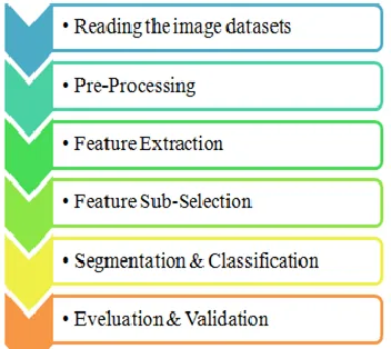
Figure 1: Medical Image Processing Steps
The segmentation and classification techniques have been automated their accuracy and reproducibility of sufficient, and it is also used to improve their diagnostic capabilities. Segmentation refers to pixel by pixel labeling of different regions such as gray matter, white matter, cerebrospinal fluid and abnormal tumor region (if present). The motivation of this research work is to propose and implement a computer aided software system for segmenting the tumor region accurately and classifying the tumors efficiently in benign and malignant tumor CT images. Using CT images, an attempt has been made to improve the quantitative and qualitative similarity measures of accuracy for segmentation and classification of abnormal tumor regions in brain. The different types of abnormalities of the CT images caused by the tumors are categorized as benign and malignant tumors. This study focuses on both benign and malignant tumor brain CT images.
2. LITERATURE SURVEY
[1]ZarNawa Khan Swat, Qinghua Zhao, Muhammad Kabir, Farman Ali, Zakir Ali, Saeed Ahmed &Jianfeng Lu (2019) proposed another methodology for brain tumor image classification dependent on move learning and tweaking. The proposed procedure of move learning with block-wise tweaking recommends an elective methodology, which is unique in relation to utilizing pre-prepared CNN as an off-the-rack include extractor (without preparing) that train the different technique for classification, (for example, k-nearest-neighbors, Support Vector Machines, Boosted Trees, Decision Trees, and Random Forest). It likewise exhibits the transferability of gaining from common images to restorative brain MR images. The proposed methodology might be utilized to create a classification framework for other body organ MRI images and other restorative imaging areas, for example, X-beams, PET, and CT. The strategy is progressively conventional in light of the fact that it requires least preprocessing for 2D MR images and doesn't utilize handcrafted highlights.
[2]QilingTanga, Yangyang Liu, Haihua Liu (2017) proposes a multiscale portrayal learning technique by means of inadequate autoencoder networks to catch the inherent scales in therapeutic images for the classification task. To acquire the multiscale highlight locators by the
scanty auto encoders with various responsive field sizes, and then create the component maps by the convolution activity. This system can more readily describe different size structures in therapeutic imaging than a single-scale variant. Along these lines, the Fisher vector strategy is utilized to encode the extricated highlights to actualize a fixed-length image portrayal that gives increasingly plentiful data of high-request insights and upgrades the graphicness and discriminative capacity of highlight portrayal. To do probes the IRMA-2009 restorative assortment and the mammographic fix dataset. The broad test results exhibit that the proposed technique has prevalent execution. [3]Mamta Mittal, Lalit Mohan Goyal, Sumit Kaur, Iqbaldeep Kaur, Amit Verma, D. Jude Hemanth (2019) proposed a Deep learning-based improved tumor division approach for MR brain images proposed technique incorporates the idea of Stationary Wavelet Transform (SWT) and new Growing CNN (GCNN). The basic objective of this work is to improve the precision of the customary system. A comparable assessment with Support Vector Machine (SVM) and Convolution Neural Network (CNN) is finished right now. A rough 2% improvement in PSNR and SSIM has been accomplished with the proposed strategy over the ordinary CNN approach. The Mean Square Error is fundamentally diminished in the proposed GCNN approach in contrast with the customary CNN approach. A comparative examination with other traditional techniques has additionally demonstrated the proficiency of the proposed framework. The strategy performed similarly powerful and enormous dataset and complex structures. the approach will play out various activities as division, highlight extraction naturally.
[4]Tanya KoohpayehAraghi, Azizah Abd Manaf (2019) proposed a visually impaired image watermarking plan to upgrade the adequacy of half and half Discrete Wavelet Transform (DWT) and Singular Value Decomposition (SVD) based plans using the second level of SVD. Utilizing methods like SVD alongside Discrete Wavelet Transform (DWT) make a solid half and half plan that repays the deterrents of other crossover plans. Proposed a DWT and Two-dimensional SVD conspire, coming from high fiery pieces of Singular Value Decomposition to make the plan hearty just as impalpable. Maintaining a strategic distance from both bogus positive and negative issues, a 2-level verification framework. A visually impaired plan to guarantee security and ease of use in medicinal and non-restorative images. An improved half and half image watermarking dependent on DWT and 2-D SVD. Proposing the image blocking to make independency of limit and host image size.
information depiction language for portraying both spatial and value imperatives in restorative images, which can be utilized to naturally create parsers for organized data extraction from these images. The sentence structure of ODL can be summed up to the next image spaces. Propose a powerful ODL parser, which fabricates the affiliation. between the content boxes from crude OCR results and the comparing depiction in ODL. During the parsing stage, the parser can endure the commotions and blunders brought by OCR acknowledgment, just as incorrect bounding boxes of information portrayal.
3.
MEDICAL
IMAGE
CLASSIFICATION
APPROACHES
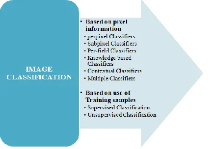
The general goal of image classification systems is to naturally order all pixels in image into land spread classes. Based on pixel data, Images can be delegated Per-pixel, Subpixel, Per-field, Knowledge based, Contextual and numerous classifiers. Per-pixel classifiers might be parametric or non-parametric. Based on the utilization of preparing tests, images can be delegated Supervised and Unsupervised Classification. The unsupervised classification is the distinguishing proof of regular gatherings or structures. The administered classification is the way toward utilizing tests of realized character to arrange (i.e.) to allot unclassified pixels to one of a few enlightening classes. Administered strategy pursues the means, for example, highlight extraction, preparing and naming procedures. The initial step comprises of transforming the image to an element image to lessen the information Dimensionality and improve the information interpretability. This handling stage is discretionary and includes strategies, for example, HIS transformation, head segment investigation and straight blend model. In the preparation stage, a lot of preparing tests in the image is chosen to portray each class. Preparing tests train the classifier to distinguish the classes and are utilized to decide the 'rules' which permit task of a class mark to every pixel in the image. The naming procedure partners name for every pixel or locale. These days, the accessibility of high goals images has expanded the quantity of looks into on urban land use and earth spread classification.
Figure 2: Medical Image Feature Classification
4.
PROPOSED
CLASSIFICATION
FOR
MEDICAL
IMAGE
USING
BASE
PIXEL
TRAINING
Different types of medical images are taken for input and finally using the following enhanced method it is preprocessed.
1. Input the image, identify the denoted pixel as as . 2. The neighborhood of each pixel is a vector and
denoted as , The values in the neighborhood vector is denoted as
3. Each pixel is presented by the vector
4.
5.
6. The labels (classification) of pixels in the neighborhoods are presented as a vector
7.
8. { }
9. A vector presents the labels in the neighborhood without the pixel
10.
11. Calculate the parameters of probability distribution | and
12. Calculate the posterior probabilities | and all labels . Get the image classification result
13. Every pixel is denoted as vector and it Identifies the neighborhood value
14. Calculate the probabilities and identify K centroids (quasi-data points representing the centers of the clusters) are distributed at random among the data points
15. Apply the minimum error classification on a pixel , if the probability of a class being presenting the pixel is the highest among all, then assign as its class.
16. | |
17. The contextual classification rule is described as below, it uses the feature vector rather than
18. | |
19. Calculate the posteriori probability |
20. | |
21. Measure The distance using Euclidean distance,between every image pixel point and each centroid is calculated
22. Each image pixel point is associated with the nearest centroid
23. The centroids move to minimize the distance between them and their associated points, so moving to the Centre of their points
24. Have the centroids moved a sufficiently small amount check for the convergence.
25. The configuration of clusters is stored and the limit on the number of iterations been reached.
26. The cluster configuration with the smallest distance between points and their associated centroids is output as the clustering solution.
27. Finally, image output is displayed with preprocessed.
Algorithmic Steps Enhanced K-means
2909 neighborhood without the pixel .
Identifying the Size of the neighborhood.
There is no restriction of the size, yet it is viewed as generally little for every pixel . Apply the minimum error classification on a pixel , if the probability of a class being presenting the pixel is the highest among all, then assign as its class.The contextual classification rule is described as below, it uses the feature vector rather than
. Calculate the posteriori probability | . The number of vectors is the same as the number of pixels in the image. For the classifier utilizes a vector comparing to each pixel and the vector is created from the pixel's neighborhood. The above medical image preprocessing was implemented in python which includes KERAS and Tensor flow, The Indian pines data set has been used for the implementation.
5.
EXPERIMENTAL
RESULTS
AND
DISCUSSIONS
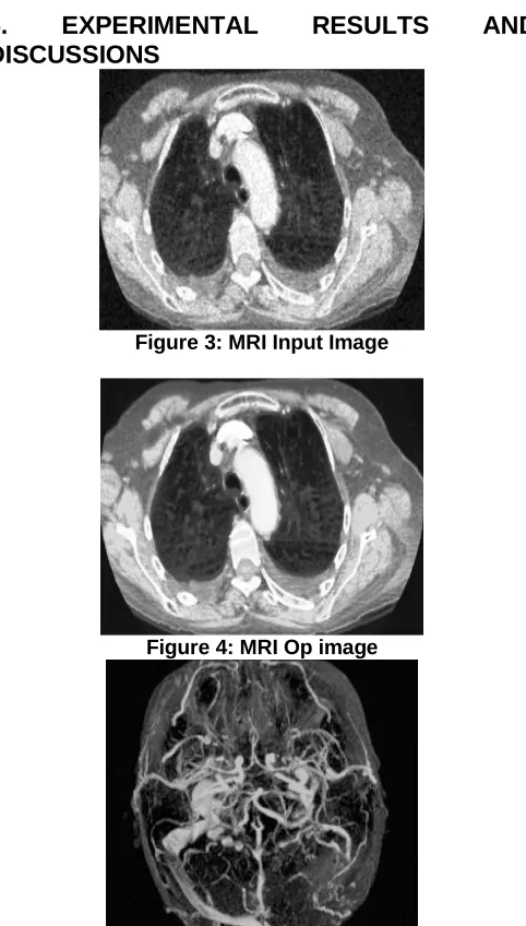
Figure 3: MRI Input Image
Figure 4: MRI Op image
Figure 5: MRI Input Data Before Filtering
Figure 6: MRI Output After Filtering
Figure 7: CT Scan Input Before filtering
Figure 8: CT Scan Output after filtering
Figure 10: Mammography image Output after Filtering
Improve Nearest Pixel-based arrangement algorithms. K-Nearest Neighbors (K-NN) is an administered AI algorithm, for example, it gains from a named preparing set by taking in
the preparation information X alongside its names y and figures out how to outline input X to its ideal yield y. The k-NN algorithm is seemingly the least difficult of the AI algorithms. The model just comprises of the preparation information, that is, the model basically learns the whole preparing set and for expectation gives the yield as the class with the larger part in the 'k' nearest neighbors determined by some distance metric..
The working in a little more detail is as follows:
After the model has put away the preparation set for expectation, it takes a test picture to be anticipated, ascertains the distance to each picture in the preparation set and gets the 'k' preparing pictures nearest to the test picture. It at that point yields the class as indicated by some democratic technique from the marks of these 'k' neighbors,
Test Image Training image Pixel-wise absolute value
difference
58 34 12 20 12 22 26 19 46 12 14 1
92 25 130 135 - 10 12 91 102 = 82 13 39 33 →456
26 28 180 200 14 18 180 172 12 10 0 30
4 2 257 257 6 34 235 114 2 23 22 108
An elective distance metric might be the L2 distance or all the more usually called the Euclidean distance..
√∑( )
L2 distance metric
At the end of the day we would process the pixel-wise distinction as in the past, yet this time we square every one of them, include them up lastly take the square root..
It is fascinating to take note of that because of the squared contrasts in the L2 distance, it is a lot stricter when the pixel distinction is excessively enormous..
Presently, we move onto the down to earth contemplations: Hyperparameters in k-NN and how they influence the presentation..
As k-NN is an extremely straightforward algorithm it doesn't generally have a lot of hyperparameters to tweak, only the two: the distance metric and the estimation of 'k'. So what we can do is, run our model for different estimations of 'k' and get the model with the best approval precision, which will be utilized as our last model on the test set..
Explanation:
Utilizing k-NN from the sklearn library, a circle is run more than 100 estimations of k and the worth that gives most extreme approval precision is utilized. This is acquired as k=43 giving approval precision as 61.01%
Output:
Test Accuracy: 0.5917874396135265 Using our own k-NN
With k = 43 Got 245 / 414 correct => accuracy: 0.591787
CONCLUSION
2911 classification exactness, time utilization and registering
resources.
REFERENCES
[1] ZarNawa Khan Swati ,Qinghua Zhao, Muhammad Kabir, Farman Ali, Zakir Ali, Saeed Ahmed &Jianfeng Lu ―Brain tumor classification for MR images using transfer learning and fine-tuning‖ 0895-6111/© 2019 Elsevier Ltd. All rights reserved.
[2] QilingTanga, Yangyang Liu, Haihua Liu ―Medical image classification via multiscale representation learning‖0933-3657/© 2017 Elsevier B.V. All rights reserved.
[3] Mamta Mittal, Lalit Mohan Goyal, Sumit Kaur, Iqbaldeep Kaur, Amit Verma, D. Jude Hemanth ―Deep learning based enhanced tumor segmentation approach for MR brain images‖ 1568-4946/© 2019 Elsevier B.V. All rights reserved.
[4] Tanya KoohpayehAraghi, Azizah Abd Manaf ―An enhanced hybrid image watermarking scheme for security of medical and non-medical images based on DWT and 2-D SVD‖ 0167-739X/© 2019 Elsevier B.V. All rights reserved
[5] KangqiLuoa, Jinyi Lua, Kenny Q. Zhua, WeiguoGaob, Jia Weib, Meizhuo Zhang ―Layout-aware information extraction from semi-structured medical images‖ 0010-4825/ © 2019 Published by Elsevier Ltd.
[6] Manjun Qin, FengyingXie , Wei Li, Zhenwei Shi and Haopeng Zhang, ―Dehazing for Multispectral Remote Sensing Images Based on a Convolutional Neural Network With the Residual Architecture‖, 1939-1404 © 2018 IEEE.
[7] Mohamed Anis Loghmari, Mohamed Saber Naceur, and Mohamed RachedBoussema, ―A Spectral and Spatial Source Separation of Multispectral Images‖, IEEE TRANSACTIONS ON GEOSCIENCE AND REMOTE SENSING, VOL. 44, NO. 12, DECEMBER 2006.
[8] Saroj K. Meher, B. Uma Shankar, and Ashish Ghosh, ―Wavelet-Feature-Based Classifiers for Multispectral Remote-Sensing Images‖, IEEE TRANSACTIONS ON GEOSCIENCE AND REMOTE SENSING, VOL. 45, NO. 6, JUNE 2007.
[9] Y. Altmann, N. Dobigeon, S. McLaughlin, and J.-Y. Tourneret. Nonlinear spectral unmixing of medical images using Gaussian processes. IEEE Transactions on Signal Processing, 61(10):2442–2453, May 2013. [10] Y. Altmann, N. Dobigeon, and J.-Y. Tourneret.
Unsupervised post-nonlinear unmixing of hyperspectral images using a hamiltonian monte carlo algorithm. IEEE Transactions on Image Processing, 23(6):2663– 2675, June 2014.
[11] Y. Altmann, A. Halimi, N. Dobigeon, and J.-Y. Tourneret. Supervised nonlinear spectral unmixing using a postnonlinear mixing model for hyperspectral imagery. IEEE Transactions on Image Processing, 21(6):3017–3025, June 2012.
[12] Y. Altmann, M. Pereyra, and S. McLaughlin. Bayesian nonlinear hyperspectral unmixing with spatial residual
component analysis. IEEE Transactions on
Computational Imaging, 1(3):174–185, Sept 2015. [13] Yoann Altmann, Nicolas Dobigeon, Steve McLaughlin,
and Jean-Yves Tourneret. Residual component
analysis of hyperspectral imagesapplication to joint nonlinear unmixing and nonlinearity detection. IEEE Transactions on Image Processing, 23(5):2148–2158, 2014.
[14] Christophe Andrieu, Nando De Freitas, Arnaud Doucet, and Michael I Jordan. An introduction to MCMC for machine learning. Machine learning, 50(1-2):5–43, 2003.