Structural Studies of factor B of the Alternative Pathway of
the Complement System
Thesis Presented for the Degree of
Doctor of Philosophy
By
Justin Hinshelwood
Department of Biochemistry and Molecular Biology
Royal Free Campus
Royal Free and University College Medical School
University College London
ProQuest Number: U643925
All rights reserved
INFORMATION TO ALL USERS
The quality of this reproduction is dependent upon the quality of the copy submitted. In the unlikely event that the author did not send a complete manuscript and there are missing pages, these will be noted. Also, if material had to be removed,
a note will indicate the deletion.
uest.
ProQuest U643925
Published by ProQuest LLC(2016). Copyright of the Dissertation is held by the Author. All rights reserved.
This work is protected against unauthorized copying under Title 17, United States Code. Microform Edition © ProQuest LLC.
ProQuest LLC
789 East Eisenhower Parkway P.O. Box 1346
ABSTRACT
Factor B of the complement system is a five-domain, 90 kD serine protease
proenzyme which is activated by the protease factor D in the context of the Mg^^-dependent
complex C3bB during complement activation. The cleavage of factor B into the Ba and Bb
fragments during activation facilitates the assembly of the alternative pathway C3 convertase,
C3bBb. The Ba fragment contains three short consensus repeat (SCR) domains, while the Bb
fragment contains a von Willebrand factor type A (vWF-A) domain and a serine protease
(SP) domain. All the domain types of factor B have been implicated in interactions with
activated C3 and the assembly of the alternative pathway C3 convertase.
This thesis describes a number of structural and functional studies using six different
preparations of factor B and its fragments and domains. Surface-enhanced laser desorption-
ionization affinity mass spectrometry (SELDIAMS) was used to investigate the reaction of
factor B with immobilised activated C3(NH3) in the presence ofMg^^. From this it was
determined that (i) a major function of the vWF-A domain is to bind to activated C3 during
the formation of the C3 convertase, which it does at its active site cleft, and that (ii)
SELDIAMS provides an efficient means of identifying residues involved in protein-protein
interactions. Next, 'H NMR, FTIR and circular dichroism spectroscopy studies were
performed to provide unequivocal evidence to confirm that the factor B vWF-A domain has
allosteric properties and the conformation is dependent upon the presence of bound metal.
This implies that the vWF-A interaction with C3b may alter its Mg^'^-bound coordination.
Further NMR spectroscopic evidence is presented to confirm that the solution structure
of factor B was not in an extended arrangement, but its five domains were involved in close
interactions. It is proposed that the conformation of the Ba fragment is affected by its
incorporation into factor B. The Ba fragment is involved in decoupling an interaction between
the vWF-A and SP domains and the proximity of the vWF-A and SP domains within the Bb
fragment leads to a conformational change in which conserved charged residues may be
important. Allosteric stmctural rearrangements in the SP domain as the result of its interactions
with the vWF-A domain or the Ba fragment provide an explanation of the regulation of the
ACKNOWLEDGEMENTS
I wish to acknowledge the following people for their assistance and support during
the course of my studies at the Royal Free and University College Medical School
(RFUCMS). I would like to thank my supervisor Professor S. J. Perkins for providing me
with the opportunity to study for this thesis and to the members of the protein structure group,
past and present, for their help in all aspects of my work. In particular I would like to thank
Dr C. Ullman and Dr M. K. Boehm for their careful guidance and patience and Mr J. Gor for
his continual assistance. I would like to thank Dr K. Srai and Dr B. Ramesh for allowing me
access to the Wolfson laboratory and the department of Oncology for accommodating us
within their work space. Many thanks are due to Dr R. B. Sim (MRC Immunochemistry Unit,
Oxford) for his collaboration and many useful discussions; Mr A. C. Willis for the protein
sequence analysis (MRC Immunochemistry Unit, Oxford); Professor J. E. Volanakis (Dept,
of Medicine, University of Alabama at Birmingham, U.S.A.) for the provision of factor D;
Professor R. H. J. Begent and Dr D. I. R. Spencer (Dept, of Oncology, Royal Free Hospital)
for access to the PBS-1 mass analyser; Dr J. Feeney, Dr T. Frenkiel, Dr M. Gradwell and
the Medical Research Council for the provision of ’ H NMR facilities at the National Institute
of Medical Research, Mill Hill, London; Dr A. J. Beavil (Kings College, London) for access
to the CD spectrometer; Dr K. J. Welham for access to MALDI and electrospray equipment
(School of Pharmacy, London); Dr. P. I. Haris (De Montfort University, Leicester) for useful
discussions and Dr S. V. L. Narayana for the factor B SP coordinates (PDB code Idle).
Many thanks are also due to Dr. G. Brooker (Wadham College, Oxford) for his assistance
with this document. I offer my apologies for the many people whom I have inevitably forgotten
to mention, but may I offer solace by confirming that you are all wonderful people and that
your influences were undoubtedly crucial for helping me to get this far. I would like to thank
the Wellcome Trust for project grant support.
Finally I would like to thank my family and friends for all their support during these
studies. I would especially like to thank Caroline for her remarkable strength and
encouragement; during this time she has given birth to Lois and Oona and has in the most part
Contents Page
Chapter 1
The Complement System: The First Line of Defence of the Immune System is
The Major Effector of the Humoral Immune System 1
1.1 The biological immune system 2
1.2 The complement system 3
1.3 Classical pathway 7
1.3.1 The structure of Clq, C lr and C ls 8
1.3.2 Activation 8
1.3.3 Mannose binding protein (MB?) 10
1.3.4 C-reactive protein (CRP) 11
1.3.5 Control proteins 11
1.4 Alternative pathway 12
1.4.1 Activation 12
1.4.2 Control proteins 15
1.4.2 Protection mechanisms 16
1.5 Terminal pathway 16
1.5.1 C5 convertase 16
1.5.2 Biological activity 17
1.5.3 Control proteins 18
1.5.4 Protection mechanisms 18
1.5.5 Complement receptors 18
1.6 The physiological effects of complement 21
1.6.1 The inflammatory response 21
1.6.2 Defence against infection 21
1.6.3 Involvement in other systems 22
1.6.4 The role of complement in disease 22
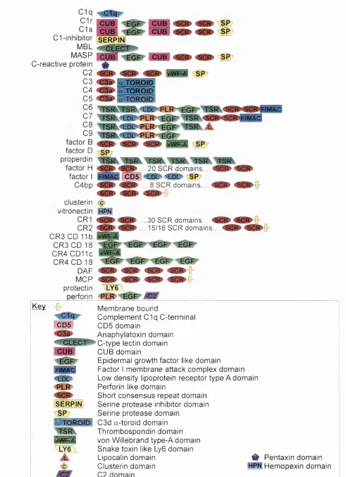
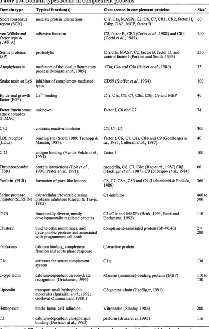
1.7 Domain structures and protein families in complement 24
1.12 Short consensus repeat domain 28
1.7.3 von Willebrand factor type A (vWF-A) domains 28
1.7.4 Serine protease domains 29
1.7.5 LDL receptor (LDLr) domain 30
1.7.6 Factor I module membrane attack complex (FIMAC) domain 30
1.7.7 The CD5 domain 31
1.7.8 Serine protease inhibitor (SERPIN) 32
1.7.9 C3d domain 32
1.7.10 Anaphylatoxin domain 32
Chapter 2
Methods for Protein Expression. Purification. Characterisation and
Crystallisation 33
2.1 Expression methods 34
2.1.1 E. coli as a host 35
2.1.2 E. coli plasmids and promoters 36
2.1.3 trp/lac Hybrid promoters 3 6
2.1.4 Expression systems for fusion proteins 37
2.1.5 The pGEX expression system 3 7
2.1.6 pGEX expression plasmids 37
2.1.7 Purification and isolation of protein products 39
2.1.8 Suitability of pGEX for protein expression 42
2.2 Purification of protein preparations 42
2.3 Characterisation of the preparation 43
2.3.1 Mass spectrometry 43
2.3.2 Instrumentation 44
2.3.3 Electron ionization(EI) and chemical ionization (Cl) 44
2.3.4 Electrospray ionization (ESI) 44
2.3.5 Laser ionization (LIMS) 45
2.3.7 Time-of-flight mass spectrometry (TOP-MS) 46
2.3.8 Surface enhanced laser desorption ionization spectrometry (SELDI) 49
2.4 Crystallisation 49
2.4.1 Purity 49
2.4.2 Solubilities and supersaturation 49
2.4.3 Nucléation and cessation of growth 52
2.4.4 Typical trial arrays 52
Chapter 3
Methods for Protein Structure Determination 54
3.1 Importance of protein structure information 55
3.2 Overview of protein structure 55
3.2.1 Primary structure 55
3.2.2.Secondary and tertiary structure 55
3.2.3 Post translational modification 56
3.2.4 Motifs and domain structures 57
3.3 Protein structure determination 58
3.3.1 Circular dichroism 58
3.3.2 Fourier transform infrared spectroscopy 63
3.4 X-ray crystallography 67
3.5 Nuclear magnetic resonance spectroscopy 68
3.5.1 Signal intensity 74
3.5.2 Chemical shift 76
3.5.3 Spin-spin coupling and multiplet structure 76
3.5.4 Spin-lattice or longitudinal relaxation time T1 76
3.5.5 Transverse or spin-spin relaxation time T2 and line width 76
3.5.6 Protein NMR 77
3.5.7 Structural information 77
3.6 Structure prediction 7 8
3.6.2 The Chou-Fasman method 82
3.6.3 The GOR method 82
3.6.4 The PHD method 83
3.6.5 The SAPIENS method 84
3.6.6 Solvent accessibility 84
3.6.7 Tertiary structure predictions 85
3.7 Analysis of known crystal structures 86
3.7.1 DSSP (Dictionary of Secondary Structure of Proteins) 86
3.7.2 COMPARER 86
3.8 Model building 8 7
3.8.1 Model refinement 88
3.8.2 Structure validation 89
3.8.3 Surface electrostatic potentials 88
Chapter 4
Experimental: Preparation of Factor B Derived Proteins From Blood and bv
Recombinant Expression 91
4.1 Introduction 92
4.2 Materials 97
4.3 Methods 98
4.3.1 GST-vWF-A-218 expression system 98
4.3.2 Preparation of the vWF-A-218 domain 98
4.3.3 Mutagenesis of vWF-A-218 with the “QuickChange” method 99
4.3.4 Mutagenic primer design 99
4.3.5 Preparation of the vWF-A-218 bacterial culture 102
4.3.6 Isolation and purification of vWF-A-218 plasmid DNA 102
4.3.7 Mutagenesis reactions 102
4.3.8 Transformation of the mutant plasmid into competent cells 103
4.3.9 Mini prep of mutant plasmids 104
4.3.12 Screening of the MCI 061 mutants for expression 105
4.3.13 Western blot analysis of the bacterial cell lysates 105
4.3.14 Preparation of the vWF-A-222 domain 107
4.3.15 Preparation of factor B, Bb, Ba, the SP domain and C3 107
4.3.16 Characterisation of protein preparations 108
4.3.17 Mass spectroscopy data collection 109
4.3.18 Crystallisation of the factor B proteins 111
4.4 Results 115
4.4.1 Purification and cleavage of the GST-vWF-A-218 protein 115
4.4.2 Development of a vWF-A-222 expression system 116
4.4.3 Characterisation of the first mutant (vWFCysmut) cDNA plasmids 118
4.4.4 Characterisation of the second mutant (vWFCysext) cDNA plasmids 118
4.4.5 Western blot analysis of the MCI 061 cultures 121
4.4.6 Purification and cleavage of the GST-vWF-A-222 protein 121
4.4.7 Characterisation of the protein preparations 121
4.4.8 Crystallisation of the protein preparations 132
4.5 Discussion 134
Chapter 5
Experimental: Hvdrodvnamic properties and functional activity of the recombinant
vWF-A domain preparations 136
5.1 Introduction 137
5.2 Methods 139
5.2.1 Hydrodynamic analyses of vWF-A-218 and vWF-222 139
5.2.2 Assay of functional (C3b-binding) activity of vWF-A-218 141
5.3 Results and Discussion 143
5.3.1 Hydrodynamic properties of the recombinant vWF-A-218 and vWF-A- 143
222 domains
5.3.2 Functional binding of the vWF-A-218 domain to C3b 147
5.4 Conclusions 151
Chapter 6
Experimental: Identification of the C3b binding site in factor B bv surface enhanced
laser desorption-ionisation affinity mass spectrometry and homology modelling:
Implications for the activity of factor B 154
6.1 Introduction 155
6.2 Materials 158
6.3 Methods 159
6.3.1 SELDI analyses 159
6.3.2 Sequence alignment of vWF-A domains 161
6.3.3 Homology modelling of vWF-A domain 163
6.4 Results and Discussion 165
6.4.1 Preparation of the vWF-A domain 165
6.4.2 Mass spectrometry of immobilised C3(NH3) with factor B and its 165
components
6.4.3 Mass spectrometry of proteolysed vWF-A domain after interaction 167
with C3
6.4.4 Homology model for the vWF-A domain in factor B 172
6.4.5 Location of Peptides 1 and 2 in the vWF-A homology model 180
6.5 Conclusions 183
Chapter 7
Experimental: Metal dependent conformational changes in the recombinant vWF-A
domains and factor B 186
7.1 Introduction 187
7.2 Materials and Methods 193
7.2.1 Molecular graphics analyses of vWF-A domains 193
7.2.2 Preparations of factor B, its Bb fragment and the recombinant vWF-A
7.2.3 Circular dichroism spectroscopy 195
7.2.4 Fourier transform infrared (FT-IR) spectroscopy 195
7.2.5 ’H NMR spectroscopy 196
7.2.6 Calculation of ring current shifts 196
7.3 Results and Discussion 197
7.3.1 The metal binding site in the vWF-A superfamily 197
7.3.2 Circular dichroism studies of factor B and its fragments 198
7.3.3 Mg^^-dependence of the vWF-A structure by FTIR spectroscopy 202
7.3.4 Mg^^-dependence of the vWF-A structure by ’H NMR spectroscopy 207
7.3.5 Temperature and pH stability of the vWF-A domain by ‘H NMR
spectroscopy 211
7.3.6 Ring current calculations of the vWF-A homology model 214
7.4 Conclusions 218
Chapter 8
Experimental: Conformational changes during the assembly of factor B from its
domains bv *H NMR spectroscopy and molecular modelling 221
8.1 Introduction 222
8.2 Materials and Methods 227
8.2.1 Preparation of factor B, the Ba and Bb fragments, C3, factor D and the
SP domain 227
8.2.2 ’H NMR spectroscopy 227
8.2.3 Homology modelling of the domains of factor B 227
8.2.4 Calculation of ring current shifts 229
8.3 Results and Discussion 232
8.3.1 Purification and activity of factor B and its fragments 232
8.3.2 pH stability of factor B by ’H NMR spectroscopy 234
8.3.3 Interactions between the domains of factor B by *H NMR 238
8.3.4 Temperature stability of factor B by 'H NMR 243
8.3.6 Ring current calculations based on homology models or crystal 248
structures
8.3.7 Association of the vWF-A and SP domains 255
8.4 Conclusions 259
Chapter 9
Epilogue 262
9.1 Assessment of the structural studies of factor B 263
9.2 Future work 266
References 268
Abstracts 298
List of Abbreviations
2D-NMR two dimensional nuclear magnetic resonance
a2m a2macroglobulin
APS ammonium persulphate
AMC 7-amino-4-isocoumarin
B„ applied magnetic field
Boc t-butyloxycarbonyl
BSA bovine serum albumin
C8bp C8 binding protein (synonym of homologous restriction factor)
cDNA complementary DNA
Cl/2/3/4/5/6/7/8/9 complement components 1/2/3/4/5/6/7/8/9
Cl-inh complement component 1 inhibitor
Clq component of complement component 1
C lr protease component of complement component 1
Cls protease component of complement component 1
C3a/4a/5a anaphylatoxins
C3b proteolytically activated form of C3
C3i hydrolytically activated form of C3 (synonym of C3(H20))
C3(H20) hydrolytically activated form of C3 (synonym of C3i)
C3(NH3) amidated C3 (activated form of C3)
C4bp C4 binding protein
C8bp C8 binding protein (synonym of HRF)
CCP complement control protein (synonym of SCR)
CD circular dichroism
CDxx cluster of differentiation or determinant (xx is a number)
CDlla/b/c/d a subunit of the p2 integrins
CD18 P subunit of the P2 integrins
CD21 complement receptor 2
CD55 complement receptor 1
CD59 protectin
CI chemical ionization
CRl/2/3/4/5 complement receptor 1/2/3/4/5
CR3 complement receptor 3 (composed of GDI 11
CRP C reactive protein
DAF decay accelerating factor
DEAE diethylaminoethyl
DNA deoxyribonucleic acid
DSS 2,2-dimethyl-2-silapentane-5-sulphonate
DSSP dictionary of secondary structure of proteins
DTP dithiothreitol
EBV Epstein Barr virus
EDTA ethylenediaminetetraacetic acid
EOF epidermal growth factor
El electron ionization
ELISA enzyme linked immunosorbent assay
ESI electrospray ionization
Fab antigen binding region of an immunoglobuli:
FB factor B
Fc C-terminal halves of two heavy chains of an
FI factor I
FID free induction decay
FIM factor I module
FIMAC factor I module/membrane attack complex
FPLC fast performance liquid chromatography
FT-IR Fourier transform infrared
GAL galactose
GST glutathione S-transferase
HIV human immunodeficiency virus
HTH HRF HRF20 lAM Ig IPTG LB LDLr LIMS LFA-1 MAC MALDI MASP-1/-2 MBL MBP MCP MIDAS MHC MIRL MPD mRNA MTPBS NMR nOE OD ORF P P-18 PBS helix-tum-helix
homologous restriction factor (synonym of C8bp)
protectin (synonym of CD59, P-18 and MIRL)
iodoacetamide
immunoglobulin
isopropyl-P-D-galactoside
Luria Bertani
low density lipoprotein receptor
laser ionization mass spectrometry
cartilage matrix protein (composed of GDI la and CD 18)
membrane attack complex
matrix-assisted laser desorption ionisation
mannose binding protein associated serine protease-1/-2
mannose binding lectin
mannose binding protein
membrane cofactor protein
metal ion dependent adhesion site
major histocompatibility complex
membrane inhibitor of reactive lysis (synonym of protectin,
CD59 and HRF20)
2-methyl-2,4-pentanediol
messenger RNA
hypertonic phosphate buffered saline
nuclear magnetic resonance
nuclear Overhauser effect
optical density
open reading frame
properdin
protectin (synonym of CD59, HRF20 and MIRL)
PC PGR PDB PEG PHD PER PMSF PNH PVDF RMS rpm PPM RaRF RCA rFI RMS RNA SCR SDS-PAGE SDW SELDI SERPIN SEE SP40,40 SPA S-protein sFI SP TEMED phosphocholine
polymerase chain reaction
protein databank
polyethylene glycol
profile network system from Heidelberg
perforin-like region
phenylmethylsulphonylfluoride
paroxysmal nocturnal haemoglobinuria
polyvinyl difluoride membrane
root mean squared
revolutions per minute
parts per million
Ra reactive factor (synonym of MBE and MBP)
regulation of complement activation
recombinant factor I
root mean-squared
ribonucleic acid
short consensus (complement) repeat (synonym of CCP) or the
structurally conserved regions (relating to homology modelling)
sodium dodecyl sulphate polyacrylamide gel electrophoresis
sterile deionized and distilled water
surface enhanced laser desorption ionisation
serine protease inhibitor protein
systemic lupus erythematosus
clusterin
surfactant protein A
vitronectin
serum factor I
serine protease
Tm
TOF-MS
TSR
uv
vWF-Amelting temperature
time-of-flight mass spectrometry
thrombospondin repeat
ultraviolet
von Willebrand factor type-A domain
AMINO ACID ABBREVIATIONS
Amino acid 3 letter format 1 1
alanine Ala A
arginine Arg R
aspartic acid Asp D
asparagine Asn N
cysteine Cys C
glutamic acid Glu E
glutamine Gin Q
glycine Gly G
histidine His H
isoleucine lie I
leucine Leu L
lysine Lys K
methionine Met M
phenylalanine Phe F
proline Pro P
serine Ser S
threonine Thr T
tryptophan Trp W
tyrosine Tyr Y
valine Val V
NUCLEOTIDE ABBREVIATIONS
adenine A
guanine G
thymidine T
Figures Legend Page
Chapter 1
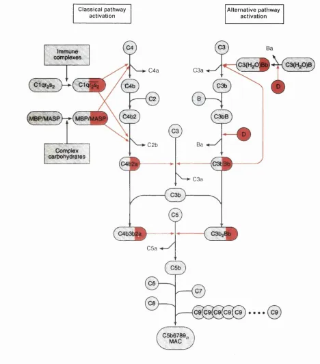
Figure 1.1 The activation steps of the complement system. 6
Figure 1.2 Summary of the domain types that occur in complement protein 26
structures.
Chapter 2
Figure 2.1 Map of the glutathione-S-transferase fusion vectors pGEX-2T and 40
pGEX-3X showing the reading frames and main features.
Figure 2.2 This schematic shows ablation of ions from a solid sample with a pulsed 48
laser in a reflectron time-of-flight mass spectrometer (TOF-MS).
Figure 2.3 Phase diagram representing the solubility of a protein as a function of the 51
single parameter, salt concentration.
F igure 2.4 Flow diagram showing the succession of variables and procedure in the 53
investigation of crystallisation conditions.
Chapter 3
Figure 3.1 (a) Linear polarized light can be viewed as a superposition of opposite 59
circular polarized light of equal amplitude and phase.
Figure 3.2 A representation of the optical activity induced by the chiral centres that 61
occur in amino acids and peptides.
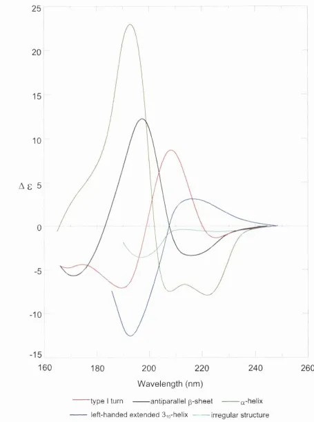
Figure 3.3 The CD spectra associated with various protein secondary structures 64
(Drake 1994).
Figure 3.4 Nuclear spin energy levels of a spin 1/2 nucleus (eg or *^C) placed in 71
an increasing external magnetic field, Bq.
F igure 3.5 At thermal equilibrium there will be a surplus of nuclei in the lower energy 7 2
state (Boltzmann surplus).
Figure 3.6 Schematic representation ofa proton placed in an external magnetic field, 73
Figure 3.7 (a) The application of a radio frequency electromagnetic pulse to an 75
ensemble of spins induces resonances between the different spin states.
Figure 3.8 Flow chart of the procedures used to generate an atomic coordinate 79
model for a target sequence based on a known structure that has the same
fold.
F igure 3.9 V enn diagram shows the relationship of the 20 naturally occurring amino 81
acids to a selection of physio-chemical properties which are important in
the determination of protein structure.
Chapter 4
Figure 4.1 Schematic outline of the domain structure of Factor B; SCR (short 93
consensus/ complement repeat) domains, vWF-A (type A von Willebrand
Factor) domain and SP (serine protease) domain.
Figure 4.2 Schematic representation ofthe “QuickChange” mutagenesis method 96
(Stratagene, La Jolla, USA).
Figure 4.3 The sequence of the cDNA insert (lowercase) of the vWF-A-218 in the 101
pGEX-2T expression vector and the translated protein sequence
(uppercase).
F igure 4.4 Standard curve for the change of absorbance of DTNB at 412 nm against 110
free thiol (glutathione) concentration.
Figure 4.5 A list of précipitants in Hampton Research crystal screen 1. 113
Figure 4.6 A list of précipitants in Hampton Research crystal screen 2. 114
F igure 4.7 1 % agarose gels of the restriction digests of the first mutation of the vWF - 119
A plasmid.
Figure 4.8 1 % agarose gels of the Bfal restriction digests of the mutation of the 120
vWFCysmut 1 plasmid (vWFCysext).
Figure 4.9 (a) 10 % SDS PAGE gel of the bacterial cell lysates from MCI 061 122
cultures of mutants 1 to 8.
Figure 4.10 Mass spectra of the vWF-A domain preparations. 123
and lane 2 contains rainbow protein molecular weight markers
(Amersham RPN756).
Figure 4.12 (a) 10% SDS-PAGE gel; lane 1 contains purified SP domain (non- 126
reduced), lane 2 contains purified SP domain (reduced) and lane 3
contains rainbow protein molecular weight markers (Amersham
RPN756).
Figure 4.13 Theoretical electrophoretic titration curves as determined by the 127
ISOELECTRIC software on GCG at the HGMP computing resource.
Figure 4.14 Theoretical electrophoretic titration curves as determined by the 128
ISOELECTRIC software on GCG at the HGMP computing resource.
Figure 4.15 8% SDS-PAGE gel of the incubation of factor B, C3 and factor D. 130
Samples were incubated at 37°C for 20 minutes across a pH range from
pH 4.0 to 9.0.
Figure 4.16 Assay of the dependence of the SP domain activity on pH. (a) 16 131
%Tricine gel of a 2 hour incubation at 37°C of C3 with the SP domain
stained with Coomassie blue.
Figure4.17 Hexagonal crystal of vWF-A-222. 133
Chapter 5
Figure 5.1 Sedimentation equilibrium data for the vWF-A-222 domain. 144
Figure 5.2 Sedimentation velocity data for the vWF-A-218 domain for a loading 145
concentration of 0.8 mg/ml at 20°C and a rotor speed of 42,000 r.p.m.
F igure 5.3 Plot of data obtained from a boundary sedimentation experiment for the 146
vWF-A-218 domain.
Figure 5.4 Representation of the factor D cleavage site in the recombinant vWF-A 149
domain sequence.
Figure 5.5 The eftect of factor D on vWF-A-218 and vWF-A-222 by mass analysis. 150
Chapter 6
spectrometry technique (SELDIAMS).
F igure 6.2 Mass analysis of the interaction of the recombinant vWF - A-222 domain, 166
the Ba and Bb fragment, factor B and bovine serum albumin with
immobilised C3(NH3).
Figure 6.3 Mass analysis of the peptides derived from the trypsin digest of the vWF- 169
A-222 domain bound to immobilised C3(NH3) on the activated chip.
Figure 6.4 Output from the PAWS program (http://www.proteometrics.com) 170
calculated from the vWF-A-222 amino acid sequence using standard
atomic weights.
Figure 6.5 Sequence alignments of vWF-A domains. 173
F igure 6.6 Sequence alignments of vWF-A domains from human and mouse factor 17 6
B and C2 sequences.
Figure 6.7 A Ramachandran plot of the mainchain torsion angles j and y of the 178
vWF-A domain homology model.
Figure 6.8 Two views rotated by 180° that depict the surface of the factor B vWF-A 179
homology model.
Figure 6.9 Schematic view of the v WF -A structure in relation to C3b and the other 184
domains of factor B.
Chapter 7
F igure 7.1 (a) The active site in open twisted ot/p domains is in a crevice outside the 188
carboxy ends of the P-strands.
Figure 7.2 Supersecondary structure topology the for the vWF-A protein fold. 189
F igure 7.3 Conformational change seen between two superimposed crystal structures 191
for complement receptor type 3 (CR3) in the presence of Mg^^.
Figure 7.4 CD spectra of the factor B, Bb fragment, vWF-A-218 and vWF-A-222 199
domain preparations in 5 mM Tris-HCl, 0.5 mM MgCl2, pH 7.5 at 20°C.
Figure 7.5 CD spectra of the factor B, Bb fragment, vWF-A-218 and vWF-A-222 200
domain preparations in 5 mM Tris-HCl, 0.5 mM MgCl2, pH 7.5 at 20°C
Figure 7.6 Sequence and numbering of the vWF-A domain in factor B. 203
Figure 7.7 FT-IR spectroscopy of the amide I band of vWF -A-222 domain studied 204
in PBS in with either 5 mM MgCl2 (red) or 5 mM EDTA (black).
Figure 7.8 Dependence of the intensity of the second derivative band at 163 5 cm’’ 206
for the vWF-A-222 domain as a function of temperature in the presence
of Mg^^ (•) and EDTA (o).
Figure 7.9 Comparison ofthe vWF-A-218 and vWF-A-222 domains by’H NMR 208
spectroscopy.
Figure7.10 The effect of Mg^^ on the vWF-A-218 and vWF-A-222 domains b y ’H 210
NMR spectroscopy.
Figure7.11 The dependence of the upfield spectral regions of the 500 MHz ’H NMR 212
spectra of the Bb fragment on the presence of Mg^^.
Figure 7.12 Conformational properties of the upheld region of the vWF-A-218 NMR 213
spectrum.
Figure7.13 Methyl-aromatic ring interactions in the homology model for the vWF-A 217
domain of factor B.
Chapter 8
Figure 8.1 Schematic diagram of the structure or chymotrypsin, which is folded into 223
two antiparallel p domains.
Figure 8.2 A diagram of the active site of chymotiypsin with a bound inhibitor, Ac- 224
Pro-Ala-Pro-Tyr-COOH.
Figure 8.3 The pH dependence of the activity of the isolated SP domain from factor 233
B.
Figure 8.4 Plots of the chemical shift of the His proton resonances of the NMR 235
spectra of (a) the Ba fragment and (b) the SP domain of factor B in the
pH titration experiments as a function of pH.
Figure 8.5 pH dependence ofthe upfield’H NMR spectra ofthe Ba fragment and 237
the vWF-A-218 and SP domains.
domains.
Figures.7 Comparisons of the do wnfield^ H NMR spectra the vWF-A-218 and SP 242
domains with their summation and the spectrum of the Bb fragment.
F igure 8.8 Temperature dependence of the upfield * F[ NMR spectra at pH 7.5 of (a) 244
the Ba fragment, (b) the vWF-A-218 domain and (c) the SP domain of
factor B.
Figure 8.9 Temperature dependence ofthe upfield’H NMR spectra at pH 7.5 of (a) 245
the Bb fragment and (b) factor B.
Figures. 10 pH and temperature dependence of the upfield *H NMR spectra at pH 247
7.5 of factor D.
Figure 8.11 Alignment of the human factor B sequence with those of the structures 249
used in homology modelling.
Figures.12 Methyl-aromatic ring interactions in homology models or crystal structures 253
of the domains in factor B and factor D.
Figures. 13 Electrostatic views of the C-terminus of the vWF-A domain and the N- 254
terminus of the SP domain.
Figures. 14 Electrostatic views of the C-terminus of the vWF-A domain and the N- 256
List of Tables Description Page
Chapter 1
Table 1.1
Table 1.2
Table 1.3
Table 1.4
Properties of the water soluble complement proteins
Membrane bound regulators of complement
Receptors for complement proteins and fragments
Domain types found in complement proteins
4 13 20 27 Chapter 2 Table 2.1 Table 2.2 Table 2.3
Examples of recombinant proteins expressed in various E.coli 38
expression systems
Examples of the uses of GST Fusion Proteins 41
Properties of MALDI matrices (from Beavis and Chait, 1996) 47
Chapter 3
Table 3.1
Table 3.2
Characteristic infrared bands of the peptide linkage
Properties of some nuclei
65
70
Chapter 4
Table 4.1
Table 4.2
Primers used for the mutation of the vWF-A-218 cDNA to vWF-A- 100
222 by the “Quick Change” method
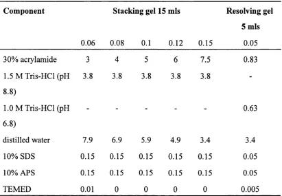
Sodium dodecyl sulphate-polyacrylamide gel electrophoresis (SDS- 106
PAGE) system for the resolution of proteins by their molecular mass
Chapter 6
Table 6.1
Table 6.2
The PDB codes of the v WF-A or 1-domain structures used for the 162
sequence alignment.
Observed masses of the peptides that remain associated with 171
Chapter 7
Table 7.1
Table 7.2
Survey of distances between the metal ion and its coordinating atoms 194
in homologous vWF-A crystal structures
Four ring-current interactions predicted to lead to the signal 2v in the 215
factor B vWF-A ’H NMR spectrum
Chapter 8
Table 8.1
Table 8.2
Table 8.3
Ring-current interactions predicted to lead to upfield-shifted signals 230
in the ’H NMR spectrum of factor B and factor D
The calculated pK^s of the Ba fragment and SP domain histidine^36
Comparison between the predicted and observed upfreld shifted 252
Chapter 1
The Complement System:
The Major Effector
of the Humoral Immune System
is The First Line of Defence of
1.1 The biological immune system
Immunity describes a complex process within the body, rather than a specific group
of organs. Certain organs and tissue types do have a predominantly immunological function,
including reticulo-endothelial tissue, bone marrow, lymph glands, the thymus, spleen and
Peyer’s patches (Kuby, 1997). The classic biological definition of immunity includes all of the
physiological mechanisms that give an organism the ability to recognize foreign substances and
neutralize or degrade them, with or without injury to the organism’s own tissue. The roles of
the humoral and cell-mediated responses of the immune system are interrelated and so provide
the activities necessary for the immune response. It is important to see immunity in its broad
biological context and the different aspects of immunity have been grouped as described
below.
Innate immunity is immunity present from before birth and consists of many
non-specific factors, together with blood-based immunity inherited from the mother, which
operate against almost any substance that threatens the body. Four types of defensive barriers
are involved: (a) Anatomical barriers which include the skin and mucous membranes, (b)
Physiological barriers involve body temperature which inhibits the growth of some pathogens,
acidic pH of the stomach and chemical mediators such as complement, interferon and
lysozyme. Other barriers are provided by (c) endocytosis and phagocytosis by specialised
cells and (d) the inflammatory response where vascular fluid and phagocytic cells leak into
tissues.
Acquired immunity is a more specialised form of immunity found only in the
vertebrates. It is a result of an encounter with a new substance, which triggers events that
induce an immune response specifically against that particular substance. This involves B
lymphocytes, T lymphocytes and macrophages and thus highlights the importance of lymphatic
tissue, the site of lymphocyte maturation and differentiation. The primary functions of acquired
immunity can be summarised in three categories : (a) the production of antibodies; (b) the
stimulation of specialised cells, which destroy invading cells or organisms and neutralize their
immune responses are adaptive and display four characteristic attributes: antigenic specificity;
diversity; immunologic memory and self/nonself recognition.
1.2 The complement system
The complement system is an important component of immune defence against
infection. It is a multi-component protein system found in plasma, that is responsible for
defence and clearance in the blood stream, and it was named as such because it helps the
antibody response. It is part of the innate (non-adaptive) immune system and can respond to
challenges by micro-organisms before an adaptive response has developed. Four major
fimctions of complement are: (a) the recognition of target surfaces; (b) the opsonization and
removal of complement-coated antigens via complement receptors on phagocytic cells; (c)
the recruitment of phagocytic cells into the site of complement activation; (d) the destruction
of membrane integrity of target organisms (reviewed in Law and Reid, 1995). Complement
comprises over 30 proteins which fimction as enzymes, binding proteins, regulators or
membrane bound receptors. The complement cascade proteins are a major constituent in
plasma with a total concentration of around 4 g/1. C3 and C4 are the most abundant
complement proteins at 1.3 and 0.6 g/1, respectively, which is consistent with their central role
in the cascade, whereas factor D is the least abundant ( 1 pg/1). The properties of the major
soluble proteins are summarised in Table 1.1. These proteins acquire the ability to interact with
one another, with antibody and with cell membranes after activation of the system. These
interactions directly generate the various biological interactions that are mediated by
complement and which range from lysis of different kinds of cells, bacteria and viruses to
direct involvement in inflammatory processes. In addition complement is able to enlist the
participation of other humoral and cellular effector systems and to induce histamine release
from mast cells, to direct migration of leukocytes and phagocytes, and to release lysosomal
constituents from phagocytes. Complement is involved in the regulation of B lymphocyte
activity, clearance of host cell breakdown products and the presentation of some antigens to
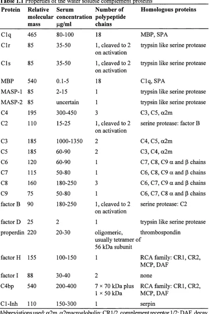
Table 1.1 Properties of the water soluble complement proteins
Protein Relative Serum Number of
molecular concentration polypeptide
mass pg/ml chains
Homologous proteins
Clq 465 80-100 18 MBP, SPA
C lr 85 35-50 1, cleaved to 2
on activation
trypsin like serine protease
C ls 85 35-50 1, cleaved to 2
on activation
trypsin like serine protease
MBP 540 0.1-5 18 Clq, SPA
MASP-1 85 2-15 1 trypsin like serine protease
MASP-2 85 uncertain 1 trypsin like serine protease
C4 195 300-450 3 C3, C5, a2m
C2 110 15-25 1, cleaved to 2
on activation
serine protease: factor B
C3 185 1000-1350 2 C4, C5, a2m
C5 185 60-90 2 C3, C4, a2m
C6 120 60-90 1 C l, C8, C9 a and p chains
C l 115 50-80 1 C6, C8, C9 a and p chains
C8 160 180-250 3 C6, C l, C9 a and p chains
C9 75 50-80 1 C6, C l, C8 a and p chains
factor B 90 180-250 1, cleaved to 2
on activation
serine protease: C2
factor D 25 2 1 trypsin like serine protease
properdin 220 20-30 oligomeric,
usually tetramer 56 kDa subunit
thrombospondin of
factor H 155 100-150 1 RCA family: CR1,CR2,
MCP, DAF
factor I 88 30-40 2 none
C4bp 540 200-400 7 X 70 kDa plus
1 X 50 kDa
RCA family: CRl, CR2, MCP, DAF
Cl-Inh 110 150-300 1 serpin
Various proteins of the complement system detect “targets” and bind to them, usually
by mechanisms that involve the recognition of charge distribution patterns or of carbohydrate
on the surface of the target. Recognition and activation occur by three maj or routes (F igure
1.1): (i) the classical pathway, primarily by immunoglobulin and immune complexes (IgG and
IgM); (ii) a branch of the classical pathway known as the lectin pathway, activated by
complex carbohydrates; and (iii) the alternative pathway, by the molecular structures on the
target cell surface that disrupt the delicate balance of proteins involved. The binding of
complement proteins to the target results in activation of the complement system and
formation, on the target, of unstable complex proteases, the C3 convertases. The C3
convertases (designated C4b2a and C3bBb in Figure 1.1) are each made up of two protein
components. One component (C3b or C4b) is covalently bound to the surface of the
complement activator and the other (C2a or Bb) is a serine protease that is able to cleave and
activate C3. The component C3 is a major plasma glycoprotein and plays a central role in the
system, being common to all three pathways. The major fiagment of activated C3, C3b, binds
covalently to complement activating surfaces such as cells, viruses and immune complexes.
When large amounts of C3b have been deposited, phagocytosis of the substance is greatly
enhanced. This occurs partly through the interaction of surface-bound C3 fragments with C3
receptors located on phagocytic cells. If the complement activator has a lipid bilayer, lysis can
occur through interaction with the membrane of components C5, C6, C7, C8 and C9. The
formation of a C5 convertase enzyme (C4b2a3b classical/lectin and C3bBb3b alternative)
can initiate the terminal or lytic pathway. The end result of the cascade is the formation of the
membrane attack complex (MAC) with an of the order of 1,000,000. The MAC forms
transmembrane channels which displace hpid molecules and other constituents, thus disrupting
the phospholipid bilayer of target cells and leading to osmotic cell lysis.
Other biologically important functions mediated by the complement system involve the
low molecular mass anaphylatoxins C3a, C4a and C5a. While these have effects on a wide
range of cells their major effect is to increase vascular permeability. They can also promote
C la ssic a l pathw ay A ltern ativ e pathw ay
activ atio n a c tiv atio n
C3 C4
Immune complexes
C3a C4a
C3b C4b
C2
C4b2 C3bB
C3
Ba C2b
Complex carbohydrates
031 iBb
C3a
C5a
0 5 b
06
07
08
09(09(09(09{ 09
05b6789, MAO
Figure 1.1 The activation steps of the complement system. The classical pathway (left) is
triggered by immune complexes (via C1 ) or by complex carbohydrates in the lectin pathway
(via MBP/MASP), while the alternative pathway (right) is triggered by a wide variety of
compounds and cell surfaces (via factor B). The number of C9 molecules {n) within the C5b6789„ complex can vary between 1 and 18. Enzymatic cleavage is indicated as solid red
lines and the enzymatically active components are shaded red. Adapted from Law and Reid,
The large C3b and C4b fragments, and degradation fragments of C3 bound to the
complement activator, are important for clearing immune aggregates and so have an influence
on the regulation of immune complex size.
The complement system in humans is well characterised at the biochemical level. The
primary structures of the proteins in the system are mostly known, and knowledge of the
three-dimensional structures is progressing. The activities of the proteins in vitro are generally known in considerable detail, but many uncertainties remain about their activities in vivo. Similar complement systems occur in all mammals and variants of the system have been found
in all other vertebrate classes. A few proteins have been described in invertebrates that are
similar to complement proteins, and these represent stages in the development of the activities
of the mammalian complement system (Dodds and Day, 1993).
Since the system is involved in the removal of materials from the circulation and
tissues, it has the potential to opsonise or lyse host cells. In addition to the beneficial effects
of complement, undesirable complement-mediated tissue damage may occur in many
situations. These situations include mechanical injury, viral infection, tissue damage initiated by
autoantibodies in chronic inflammatory conditions and rheumatoid arthritis. Complement is
responsible for the acute rejection of xenotransplants. Diminished complement activity that
arises through consumption of complement or from genetic deficiencies is associated with
susceptibility to infection and inadequate removal of immune complexes from the circulation,
which may lead to damage of the small blood vessels, particularly of the skin and kidneys.
1.3 Classical pathway
The classical pathway of complement consists of the glycoproteins Cl q. Cl r and Cls
and C2-C9 (Figure 1.1). One molecule of Clq and two of C ls and C lr associate in the
presence of Ca^'^ ions to form a large protein complex known as C1. C1 q is the molecule that
interacts with most potential targets. Classical pathway activation has been mostly studied with
1.3.1 The structure of Clq, C lr and Cls
The complement classical pathway C1 enzyme complex is composed of the subunits
Cl q, Cl r and Cl s. C1 q is made from 18 polypeptide chains of three types, 6 A, 6B and 6C
arranged in a complex structure often described as a “bunch of tulips”. Three of these chains
( 1 A, 1B and 1C) form a collagen-like triple helix at the N-terminal end of the molecule (Reid
and Porter, 1976). The six triple helices are aligned in parallel for half the collagen-like length
and then diverge to terminate at the C-terminus in six globular heads (Brodsky-Doyle et al., 1976). The subcomponents Clr and Cls have homologous amino acid sequences and similar
tertiary structures (Kishumoto et al., 1989). Clr and C ls interact to form a tetrameric chain, C1 s-C 1 r-C 1 r-C 1 s in which the two catalytic domains of Cl r are orientated in a head to tail
manner (Arlaud et al, 1986). Cls interacts with C Ir via the Clr/Cls specific domain. The activated CTr2CTs2 complex has been found to have an asymmetric X structure (Weiss et al,
1986; Perkins and Nealis, 1989). A variety of models have been proposed for the interaction
of the CTr2CTs2 tetramer with C lqto form the C1 complex (Perkins, 1989). These models
can be divided into two groups: those which propose that the CTr2CTs2 tetramer is positioned
on the outside ofClq, summarized in the “ W-model” (Perkins and Nealis, 1989) and the
“02-model” (Cooper, 1985); and those proposing that the CTr2CTs2 subunits are interwoven
between the arms ofClq, summarized in the “S-model” (Schumaker et al., 1986) and the “8-model” (Colomb et al., 1989).
1.3.2 Activation
Activation of the classical pathway is initiated by the binding of C 1 q to a variety of
substances (Sim and Reid, 1991). The most familiar is the formation of immune complexes
of either IgG or IgM with antigens. Many other substances can activate the classical pathway,
without a requirement for antibody (Sim and Malhorta, 1994). These include:
1. nucleic acid and chromatin
2. cytoplasmic intermediate filaments
3. mitochondrial membranes possibly via cardiolipin or mitochondrial proteins
4. some viruses, e.g. murine leukaemia virus (MuLV)
polysaccharide
6. gram negative bacteria via the lipid A component of the lipopolysaccharide of the
cell wall.
The initial step of activating the classical pathway is the binding of Clq via the globular
heads to the activator. The interaction between Clq and the Fc region of immunoglobulin
(IgG or IgM) is facilitated by the close proximity of many IgG molecules (Borsos, 1989) or
a conformational change in the Fab arms of IgM (Perkins et a/., 1991) to expose the Clq binding sites. It is thought that the domain types of the globular heads are likely to have
differing specificities for charge groupings on the surface of the activator. Activation of
complement requires multiple interactions between a single molecule ofClq and the activator,
and therefore the activator is usually of high molecular mass and has a repetitive structure such
as exposed lipid A on bacterial surfaces or multiple antibody molecules bound to a particulate
antigen.
When two or more of the globular heads bind the activator, a conformational change
in the collagenous stalks of C1 q is induced (Heinz, 1989) which increases the affinity ofClq
for C lr2CTs2. C lr2CTs2 will interact with C lq only in the presence of Ca^^ ions, once C1 q
has bound to an efficient activator. The catalytic domain of one C lr autoactivates and cleaves
the corresponding domain in the neighbouring C1 r molecule (Dodds et al, 1978). This in turn activates Cl shy a proteolytic cleavage (Ziccardi, 1976), The activated CTs is then able to
cleave the classical pathway component C4 into C4a and C4b (Thielens et al., 1984). C4 has sequence homology to the complement components C3 and C5 and the non-complement
proteins a2-macroglobulin and pregnancy zone protein. The proenzyme form of C4 is
composed of three chains: a, p and y. CTs cleaves a single peptide bond in the a-chain of
C4 to produce a 9 kDa fragment C4a and a metastable fragment C4b. The C-terminal
portion of the a-chain (a'-chain) of C4b remains disulphide linked to the P-chain. The larger
fragment, C4b, has an exposed thiol ester in the a' chain that is able to react non-specifrcally
amino groups located on a variety of surfaces, which form an amide bond. A proportion,
usually less than 10%, of the C4b ends up covalently bound to the complement activator. The
remainder reacts with water and diffuses away from the site of complement activation.
Proenzyme C2 binds to the surface-bound C4b and, if it is appropriately positioned close to
activated Cls, it is cleaved to form a dependent complex, C4b2a (Horiuchi et al., 1991). The C4b2a complex is the classical pathway C3 convertase enzyme which is able to
cleave and activate C3, a homologue of C4.
1.3.3 Mannose binding protein (MBP)
Molecules other than Clq can also participate in the activation of the classical
pathway of complement (Sim and Malhotra, 1994; Holmskov 1994; Malhotrae^a/.,
1994,1995). The activation of the classical pathway via complex carbohydrates is often
referred to as the lectin pathway. Mannose binding protein (MBP), also known as Ra reactive
factor (RaRF) and mannose- or mannan binding lectin (MBL), is able to substitute for C1 q
after interaction with mannose-rich structures on yeasts, bacteria and viruses (Figure 1.1). It
does not bind to normal IgG, but can activate complement on interaction with the
carbohydrate groups of a glycosylation variant of IgG, which is present at elevated levels in
rheumatoid arthritis (Malhotra et a l, 1995). The structure of MBP resembles that of C1 q in that it has collagenous segments and 6 globular heads. In MBP, each globular head is made
up of three identical C-type lectin domains which interact with carbohydrate in a Ca^"^
dependent manner. MBP can activate C lr and Cls and molecules that are structurally similar
to C lr and C ls that are termed MBP associated proteases (MASPs) (Malhotra et a l, 1994). This antibody-independent pathway may be important for immunodeficient iudividuals
1.3.4 C-reactive protein (CRP)
C-reactive protein (CRP) is a protein which, in mammals, is expressed during the
acute phase response to tissue injury or inflammation. CRP displays several functions
associated with host defense: it promotes agglutination, bacterial capsular swelling,
phagocytosis and complement fixation. CRPs have also been sequenced in an invertebrate,
the Atlantic horseshoe crab, where they are a normal constituent of the hemolymph. CRP can
form complexes with charged groups, including the phosphate groups in choline phosphate
of pneumococcal C-type polysaccharide and microbial polysaccharides (both those containing
phosphocholine (PC) and not containing PC) and lipids, polyanion/polycation complexes and
chromatin. Bound CRP can interact with Clq to activate the classical pathway.
The classical pathway is therefore activated by a wide range of stimuli. Clq and CRP
are principally involved in recognising charge clusters including carbohydrates and lipids. In
contrast MBP recognises neutral sugars.
1.3.5 Control proteins
Regulation of the classical pathway is controlled at two stages: formation of the C l
enzyme complex; and control of the C3 convertase, C4b2a. Many serine proteases in the
blood have relatively specific natural inhibitors that belong to the “serpin” family. Of the
complement proteases, only C1 r, C1 s and possibly both MASPs are controlled by a serpin,
named CT-inhibitor (CT-inh). C 1 inhibitor controls Clr2 activation through non-covalent
interactions (Arlaud et al., 1989; Ziccardi and Cooper, 1979). CT inhibitor can also covalently interact with the four serine protease active sites of the CT complex, resulting in the
dissociation of the complex to form two molecules of CT inhibitor, CTr-CTs-CT inhibitor
(Ziccardi and Cooper, 1979). The free collagen-like stalks of C lq are then free to bind to
cell-surface Clq receptors which may then initiate phagocytosis (Malhotra et al., 1990; Malhotra and Sim, 1989; Ghebrehiwet, 1989). Hereditary or acquired lack of CT-inhibitor
causes angiooedema. Factor J also controls the formation of the CT complex by inhibiting the
Control of the C3 convertase is governed by its inherently unstable nature and
cleavage by factor I. C2a spontaneously dissociates from the complex. This dissociation is
enhanced by C4 binding protein (C4bp) (Gigli et a l, 1979) and decay accelerating factor (DAF) (Nicholson-Weller et al., 1982). Factor I cleaves C4b in two positions to give the fragments C4c and C4d. C4bp (Fujitae/a/., 1978; Gigli etal., 1979), membrane cofactor protein (MCP) (Seya et al., 1986) (Table 1.2) or complement receptor 1 (CRl ) (Table 1.3) act as cofactors for this factor-I mediated cleavage. In most physiological circumstances, the
concentration of C4bp exceeds that of C4b generated by activation, and therefore further
activation of the complement components is prevented.
1.4 Alternative pathway
The proteins of the alternative pathway of complement are factor D, factor B, C3 and
C5 (Figure 1.1). Components C5-C9 are common to both pathways and will be discussed
in the terminal components section. C3 has a central role in both pathways. The regulatory
proteins factor H, factor I and properdin have major roles in controlling activation of the
alternative pathway. Activation of the alternative pathway can be both antibody dependent
and antibody independent. Antibody dependent activation can occur via IgG, IgA and (rarely)
IgE immune complexes. Antibody independent activation can be effected by a whole
spectrum of substances located on the surfaces of bacteria, fungi, viruses, multicellular
parasites and tumour cells.
1.4.1 Activation
The mechanism by which the targets are recognised by the alternative pathway is less
well understood than for the classical pathway. In the alternative pathway, several proteins are
involved simultaneously in recognition. Perturbations in the interaction between C3b deposited
on the activating surface and regulatory molecules is important for determining whether
Table 1.2 Membrane bound regulators of complement.
Regulator Action Characteristics
DAF Di s s o c i a t e s C3 70 kDa glycoprotein present on the
convertases. membranes of peripheral blood cells,
vascular endothelial cells, placenta and
epithelial cells. Extracellular portion
contains 4 SCR domains. Soluble forms
occur.
MCP Binds C3b and acts as 50-60 kDa integral membrane
(CD 46) a cofactor to factor I glycoprotein present on all circulating
m e d i a t i n g C 3 cells except erythrocytes and most other
cleavage. Cellular
receptor for measles
virus.
cell types. Contains 4 SCR domains.
Protectin Binds C5d-8 complex 20 kDa glycoprotein present on the
(CD 59) and prevents formation membranes of all circulating blood cells.
(pl8) of the polymeric C9 endothelial cells, epithelial cells and
(MACIF)
(HRF20)
(MIRL)
complex. spermatozoa.
Homologous HRF/C8bp prevents Poorly characterised to date.
restriction factor the binding of C9 to
(HRF) C8ay and its insertion
(C8 binding protein)
(C8bp)
into membranes.
Abbreviations used: DAF, decay accelerating factor; MCP, membrane cofactor protein;
MIRL, membrane inhibitor of reactive lysis; HRF, homologous restriction factor; C8bp, C8
Native C3, like C4, contains an internal thiolester in the a-chain which can be
hydrolysed to form C3i (also known as C3(H20)) and is capable of attaching to membranes
(Pangbum and Müller-Eberhard, 1980; Pangbum et al, 1981). Although C3i is structurally different from C3 b in that it has an intact a-chain, both C3 i and C3b are functionally similar.
C3 is continually activated at a slow rate in the fluid phase by three mechanisms: (i) C3 may
be cleaved to C3b by serum proteases; (ii) small nucleophiles, or water, may gain access to
C3 and react with the thiolester; (iii) C3 may be subj ect to non-specific perturbation leading
to exposure and hydrolysis of the thiolester. Thus the alternative pathway is in a permanently
activated state. If activated C3 is deposited on an activating surface of the alternative
pathway, it can serve as a seed for the positive amplification loop which operates explosively.
Activated C3 is able to form a C3 convertase with factor B in the presence of factor D.
Removal of C3a by proteolytic cleavage by the C3 convertase induces a conformational
change in C3b which leads to the exposure of the internal thiolester, which is normally buried
in native C3. The exposed thiolester is extremely reactive with nucleophiles, including water
and molecules bearing hydroxyl or amino groups. C3b, activated at the surface of a foreign
cell, is largely restricted to binding to the surface of the same cell or to being inactivated by
water. This puts a limit on the spatial range of the activated C3b. The deposition of C3b is
minimised on host cells because of their inability to activate the host’s own complement
pathways. C3 activation is kept to a low level by the control proteins, factor H and factor I
in the blood. The C3 and C5 convertases decay quite rapidly by the dissociation of the
enzymatic components in the absence of control proteins. The survival of the first C3b
molecule deposited on the surface of a substance determines whether it will be an activator
or non-activator of the alternative pathway
Activation of the alternative pathway does not depend upon antibodies recognising
specific molecules on the target cell surface: rather it relies on molecular structures on the
target cell to upset the delicate balance of proteins involved to focus their activation and
deposition on its surface. Activators include polysaccharides, fungi, bacteria, viruses, parasites
and certain mammalian cells. It is not known what structure all the activators have in common
enabling the formation of the C3bBb complex (Fearon, 1978; Pangbum etal, 1980; Fearon and Austen, 1975). C4b bound to a surface can also activate the alternative pathway by
binding to C3bBbP, which may be important in cases of C2 deficiency (Parries et al, 1990).
The most obvious similarity between the classical and alternative pathway is the C3
convertase enzyme. The C3 convertase of the classical pathway is C4b2a which is very
similar to the alternative pathway C3 convertase, C3bBb. C4b is a homologue of C3b and
C2 a homologue of factor B. Factor D is a serine protease that has a role similar to the
classical pathway Cls. The alternative pathway does not have proteins similar to Cl q or Cl r.
A paradoxical situation arises in which C3b is required in order to assemble the enzymes
responsible for C3 cleavage. In vivo the classical pathway is also present and the amplification of the C3b deposition via the alternative pathway occurs if the classical pathway
C3 convertase (C4b2a) is the source of the initial C3b molecule. Multiple C3b molecules are
deposited in clusters on the complement activator. This cluster formation is important in
mediating multiple interactions with C3 receptors on phagocytic cells.
1.4.2 Control proteins
Control of the alternative pathway is comparable to that of the classical pathway in
that homologous proteins fulfill similar roles. Both positive and negative control mechanisms
exist. The C3bBb convertase is stabilized by the binding of the control protein properdin (P)
to form a C3bBbP complex (Fearon and Austen, 1975; Medicus eta l, 1976; Farries etal, 1987), which facilitates the binding of factor B and prevents cleavage of the convertase by
factor I (Farries et al, 1988a). The versatility of complement to recognise all types of foreign material is desirable, but its binding to host cells must be minimised. Negative control of
activation is governed by factor H which acts in a similar way to C4bp in the classical pathway
by acting as a cofactor for factor I-mediated cleavage of C3b and C3i to form the fi*agments
iC3b + C3f and iC3i + C3f respectively (Pangbum and Müller-Eberhard, 1983). The factor
H molecule can interact with C3b via three binding sites and so prevent the formation of the
C3bBb convertase and it can dissociate the complex (decay accelerating activity). Factor H
activators, which include host cells, carry polyanionic substances like sialic acid or
glycosaminoglycans on the cell surface. The high affinity of factor H for non-activator bound
C3b seems to be a joint recognition of both C3b and surface structures. Various microbes
have capsules rich in sialic acid (group B streptococcus, E. coli capsule type K1 and group B meningococcus) which allows them to escape alternative pathway activation, opsonisation
and phagocytosis.
1.4.3 Protection mechanisms
The majority of C3b and C4b molecules (70 to 90%) formed as a result of
complement activation do not become covalently linked to surfaces but remain in the fluid
phase (Hourcade et al., 1989). Of those that do bind, some will do so to host cells. These are protected against complement-mediated cytolysis by the membrane proteins DAF and
MCP (Atkinson and Farries, 1987). DAF prevents assembly of C3 convertase on
membranes and will dissociate enzymes already bound. MCP has cofactor activity for factor
I mediated cleavage (Liszewski et al., 1991; Fearon, 1979).
1.5 Terminal pathway
1.5.1 C5 convertase
The binding of one or more C3b molecules to either the classical or the alternative C3
convertase (C4b2a or C3bBb) changes the specificity of the complex to a C5 convertase,
C4bC2aC3b or C3bBbC3b respectively (Medicus et al., 1976; Daha et al., 1976). The alternative pathway C5 convertase is regulated by MCP and factor I by the conversion of one
C3b dimer to C3bi (Seya et al., 1991), whereas the classical C5 convertase is controlled by factor H (Ito and Tamura, 1983). C5 binds to the C3b fragment in the C5 convertase
complex. This convertase cleaves a single peptide bond in the a-chain of C5 to yield the
fragments C5a and metastable C5b (Nilsson et al., 1975). Although C5 is very similar in structure to C3, it does not have an internal thiolester bond and therefore does not bind
1.5.2 Biological activity
The C5b fragment contains a labile binding site with specificity for component C6.
The complex C5bC6 remains loosely bound to C3b on the target cell surface until interaction
with Cl. The C5b67 complex undergoes a conformational change in which it dissociates from C3b, and a metastable membrane binding site is formed on the C7 subunit (Preissner et al, 1985). C5b67 binds to a membrane to become an integral membrane protein. C8 is
composed of three chains (a, p and y) and binds to C5b67 via the p-chain (Monahan and
Sodetz, 1981; Stewart et al., 1987). C8 is then able to undergo a conformational change which allows penetration into the membrane of the disulphide linked a- and y-chains. C9
binds to the C5b678 complex via the a-subunit on C8 (Stewart et al, 1987). This catalyses the polymerisation of up to 18 molecules of C9. C5b678 forms small functional channels of
approximately 3 nm in diameter which can enlarge to 10 nm upon incorporation of C9
molecules (Ramm et al., 1985). The composition of the membrane attack complex (MAC) depends on the availability of monomeric C9. If sufficient C9 is present, binding will continue
until it forms a typical cylindrical membrane lesion as seen in electron micrographs.
The assembly of a functional MAC on a cell membrane leads to cytolysis. Two major
hypotheses exist to explain cell death, the “doughnut” hypothesis where the MAC components
form a protein-lined channel or the “leaky patch” hypothesis where there is a destabilisation
of the lipid bilayer due to the insertion of the MAC (reviewed in Bhakdi and Tranum-Jensen,
1991 and in Esser, 1991). The MAC can destroy the membrane of an enveloped virus and
can kill many types of bacteria. The formation of an MAC on the surface of a nucleated cell
will stimulate a rapid increase in the concentration of intracellular Ca^^ which in turn stimulates
the recovery processes of the cell. This allows a cell to survive a mild complement attack and
also initiates an inflammatory response. If a much stronger attack occurs, the recovery
1.5.3 Control proteins
Control of the terminal components occurs at two points. The complement associated
protein SP40,40 (clusterin) binds to C5b6 to prevent the formation of the complete MAC
(Choi etal, 1989). The S-protein (vitronectin) and serum low density lipoprotein (LDL) will compete with membrane lipid for the binding site on C5b-7 and hence can prevent attachment
to the surface of bystander cells (Podack et a l, 1978). Attack on host cells by the MAC is inhibited mainly by the cell surface regulatory protein CD59, which binds to the MAC and
alters its mode of interaction with the membrane.
1.5.4 Protection mechanisms
Complement-mediated cell lysis is dependent on homologous restriction where
complement from one species is inefficient at lysing cells of the same species. The interaction
of specific membrane proteins with the terminal complement components ensures that the self-
inflicted complement damage is kept to a minimum (Lachmann, 1990). Table 1.2 summarises
the actions and characteristics of the membrane-bound regulators. Homologous restriction
factor (HRF) or C8 binding protein (C8bp) (Zalman et al, 1989) prevents the binding of C9 to C8ay and its insertion into membranes (Schonermark et al., 1988). Another of these protective proteins is protectin which is known by many other names; P-18 (Sugita et al.,
1988), HRF20 (Okadae/a/., 1989), membrane inhibitor of reactive lysis (MIRL) (Holguin
etal., 1989) or CD59 (Davies era/., 1989) (Table 1.2). CD59 bound to the C5b-8 complex is thought to prevent the binding of the first molecule of C9 which is required to initiate
polymerization and MAC formation (Lachmann, 1991; Rollins and Sims, 1990).
1.5.5 Complement receptors
In addition to the membrane-bound regulatory proteins of the complement system,
there are several receptors for the complement proteins or their activation fragments. These
receptors are involved in a wide range of biological activities. There are one or more distinct
complement receptors on the surface of most types of cells and on many tissue cells.
Interaction of the complement ligands with these specific cellular receptors triggers various
or other activities depending on the receptor engaged or cell type. A number of these are well
characterised at the biochemical level, but others are identified only by their binding function.
The receptors for the C3 fragments C Rl, CR2, CR3 and CR4 have a number of regulatory
and receptor functions (Table 1.3) (Sim et al., 1987; Ross, 1989). CRl and CR2 are anchored to the membrane via hydrophobic transmembrane segments (Klickstein et a/.,1988). CRl will inhibit complement activation either by accelerating the decay or
dissociation of the C3 convertases or by acting as a cofactor for factor I-mediated cleavage
of C3 b. It is involved in processing of immune complexes and in the mediation of binding and
phagocytosis of C3b-coated particles by phagocytic cells. CR2 bound to C3b-bearing
particles enhances B-cell proliferation in the presence of T-cell derived factors (Lambris,
1988). CR3 adheres to and promotes phagocytic uptake of iC3b-coated particles into
circulating phagocytic cells. CR4 mediates binding of iC3b and C3dg (Gaither et al, 1987).
A C 1 q receptor (also called the collectin receptor) may be principally involved in
phagocytosis of C1 q-bearing particles. It also serves as a receptor for MBP and for a small
family of proteins, the collectins, which are related in structure to Clq and MBP (Malhorta
et a l, 1990,1992). Other receptors of interest in the complement system are the receptors through which the small anaphylotoxins C4a, C3a and C5a exert their effects (see next