MEDICAL IMAGE TEXTURE FEATURE
EXTRACTION USING WAVELET TRANSFORM
1Shahid Eqbal, 2R.L.Yadava
1Research Scholar, 2Professor & Visiting Faculty 1Department of Electronics & Communication Engineering
1Mewar University, Chittorgarh, Rajasthan, India
Abstract: There are several approaches and various methods developed by researchers for computer aided diagnosis of brain tumour problems. Depending on the type of brain tumour, each of the brain pathologies requires a particular approach to follow in order to characterise the disease. Since the focus of this paper is on brain tumour diagnosis & classification, one way is by measuring the characteristics of these tumour masses, we can predict their aggressive behaviour (i.e. how high is their metabolic activity). There could be early signs of cancer so one should try to investigate whether they are benign or malignant. Major approaches in brain tumour detection and classification are discussed in this paper. MRI is the most important technique, in detecting the brain tumor. A new algorithm is purposed which is a combination of support vector machine (SVM) and fuzzy c-means, a hybrid technique for prediction of brain tumors, which gives accurate and more effective result for classification of brain MRI images.
Index Terms - MIP, Image Acquisition, Pre-processing, Segmentation, Feature Extraction, Computer Aided Diagnosis, MRI Image, ROI, Artificial Neural Network (ANN), Malignant, Benign, Grey level run length matrix (GLRLM), Support Vector Machine (SVM) and Fuzzy c-means
I. INTRODUCTION
Medical image processing is the technique which is used to create images of the human body for clinical purposes. It is often perceived to designate the set of techniques that noninvasively produce images of the internal aspect of the body. This implies that cause is inferred from effect. It is very important in improving the diagnosis, prevention and treatment of the diseases. Medical imaging is nothing but a part of biological imaging and incorporates radiology, nuclear medicine, investigative radiological sciences, endoscopy, thermography, medical photography and microscopy [10]. When an image is processed for visual interpretation, the human eye is the judge for the working of a particular method. For medical diagnosis, Computed Tomography (CT) gives the best information on denser tissue with less distortion. Magnetic Resonance Image (MRI) gives better information on soft tissue with more distortion. With more available multimodality medical images in clinical applications, the idea of combining images from different modalities has become very important and so medical image fusion is emerging as a new promising research field [21].
Medical image processing (MIP) is undergoing a revolution in past decades with the advent of faster, more accurate mass invasive devices [3]. This has given rise to the need for corresponding software development which has provided impetus for new algorithms in signal and image processing [4]. Over the recent years, analysis of images such as segmentation, Edge Detection, Boundary detection, classification, clustering and texture property extraction attracts the attention of many researchers in the image processing and pattern recognition area. As compared to ordinary images the medical images, has so many information, in which the feature extraction is not so easy. CT & MRI type of medical images show the information inside the patient body by non-invasive method, so that it is very helpful for diagnoses by doctors and not much painful for patients. However the raw data can only give the material to doctor, he has to decide by himself which is important & which is not.
All the standard computer vision actually aims to duplicate the effect of human vision by perceiving electronically and understanding the image. It is not an easy task by giving computers the ability to see. The specific implementation of a computer vision system also depends on its functionality if it is pre-specified or if some part of it can be learned or modified during operation. There are, however, many typical functions which are found in many computer vision systems. The computer-aided diagnoses (CAD) uses computer for processing the medical images to extract the useful information so that the doctor can make a diagnoses decision easily and quickly. But it is not very easy to locate the problems in medical images if it is noisy or if it is not in a proper format because of the irregular structure of human body. The CAD system consists of five stages such as acquisition of TRUS image of prostate, preprocessing, segmentation, feature extraction and classification. The five stages of the CAD system are described as below:
A. Image Acquisition
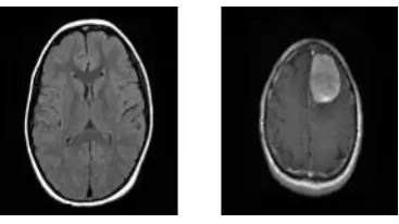
Figure 2: (a) Non-tumor MRI image (b) Tumor MRI image
B. Pre-Processing
Before the application of computer vision method to extract some specific piece of information, it is required to process the data to assure that it satisfies certain assumptions which is implied by the method. Examples are:
1. Noise reduction for assuring that sensor noise does not introduce false information. 2. Re-sampling for assuring that the image coordinate system is correct.
3. Contrast enhancement for assuring that relevant information can be detected.
4. Scale-space representation for enhancing image structures at locally appropriate scales.
Figure 3: (a) Enhanced Non-tumor image (b) Enhanced Tumor image
C. Detection/Segmentation
Segmentation as the name suggest refers to the process of partitioning a digital image into many segments (which is sets of pixels
a
lso known as super pixels). The aim of segmentation is to simplify and/or change the representation of image into something that is more meaningful and easier to analyze. Image segmentation which is typically used to locate objects and boundaries (lines, curves, etc.) in images. More precisely, image segmentation is the process of assigning a label to every pixel in an image such that pixels with the same label share certain visual characteristics. The image segmentation results into a set of segments that collectively cover the whole image, or a set of contours extracted from the image. Each pixel in a region is similar with respect to some characteristic or computed property, such as color, intensity, or texture. Adjacent regions are significantly different with respect to the same characteristic.D. Feature Extraction
When the input data available to an algorithm is very large to be processed and it is suspected that it is notoriously redundant (data with not much information) then the input data will be transformed to a reduced representation of set of features (also named features vector). This transformation of the input data into the set of features is called features extraction. If the features extracted are chosen carefully then it is expected that the features set will extract the relevant & useful information from the input data to perform the required task using this reduced representation in place of the full size input (example, in medical imaging, extract anatomical boundaries before comparison with normal template and diagnosis). Typical examples of such features are: Lines, edges and ridges. Localized interest points such as corners, blobs or points. More complex features may be related to texture, shape or motion. Different methods of feature extraction are being used to detect and classify anomalies in medical images such as wavelets [5,6], statistical methods and most of them used feature extracted using image processing techniques [7].Some other methods are based on fuzzy theory [8] and neural networks [9].
E. Classification
At this step we have the input as typically a small set of data which is a set of points or an image region and is assumed to contain a specific object. The remaining processing deals with for example:
1. Estimating the application specific parameters, such as object poses or objects size.
II.DIFFERENTMETHODSBEINGUSED&CURRENTTREND
Because features are used as part of a classification procedure so it will increase the cost and running time of a recognition system, there is trend in image processing community to design and implement such systems which have small feature sets. On the other hand there is a strong urge to include a sufficient set of features to achieve high recognition rates under complex conditions. This lead to a variety of techniques within the image processing community to find an "optimal" subset of features from a larger set of possible features. Sérgio et al., in his paper gives the advantage of single-valued functions which evaluate rankings to develop a family of feature selection methods. This is based on the genetic algorithm & it improves the accuracy of content-based image retrieval systems and it also evaluates the quality of ranking which in turn improves retrieval performance [14]. Jaba and Shanthi, reviewed previously on continuous feature discretization and they identified defining characteristics of the methods. On that basis defines a new supervised approach which combines discretization and feature selection for selecting the most relevant features useful for classification purpose. Classification technique which is used is Associative Classifiers [15]. Yong Fan, et al., presented a brain classification framework which is based on multi-parametric medical images, and he described the advantage of the method for multi-parametric imaging which provide a set of discriminative features for classifier construction by using a regional feature extraction method to takes into account joint correlations between different image parameters [16]. Ling-Chen et al.,A feature selection algorithm is discussed by him which is based on ant colony optimization (ACO), and said Image feature selection (FS) is an important task & it can affect the performance of image classification and recognition [17]. Ant colony optimization (ACO) is an evolution simulation algorithm which is proposed by M. Dorigo et al., & been successfully used for detecting system fault, job-shop scheduling, network load balancing, graph coloring, robotics and other combinational optimization problems.
Support vector machines were applied in several researches which & is given in [11-13]. H. B. Nandpuru, Dr. S. S. Salankar and academic. V. R. Bora, worked on MRI image brain cancer classification using support vector machine. Support Vector Machines (SVM) was implemented to brain picture classification. In this paper feature extraction from brain MRI pictures were administrated by grey scale, symmetrical and texture feature. They achieved sensibly good result [11]. A. Padma and R. Sukanesh, studies on SVM depend classification of soft Tissues in Brain CT pictures using Wavelet Based Dominant grey Level Run Length Texture feature. They emphasized on the technique of medical CT imaging as one of the widely applied and reliable technique which is used for the detection and site of pathological changes efficiently by using SVM [12]. S.H.S.A. Ubaidillah, R. Sallehuddin and N.A. Ali, worked on cancer found exploitation artificial neural network and support vector machine: A Comparative study. In this paper, they compared the performance on four completely different cancer datasets exploitation of SVM and ANN classifiers. In this study, the ANN classifier gated sensible classification performance on the datasets that have larger quantity of input options (prostate and Ovarian cancer datasets) SVM conjointly given sensible performance as compared to ANN on datasets with smaller quantity of input feature (breast cancer and liver cancer), and finally SVM classifier gives higher result for growth [33]. Guo-Zheng et al., discussed the feature selection methods by using support vector machines which gives satisfactory results, and propose a prediction risk which is based on feature selection method by using multiple classification support vector machines. The performance of this proposed method is compared with the previous methods of optimal brain damage based feature selection methods by using binary support vector machines [21]. Yong and Ding-gang described that feature extraction and selection in neuro image classification are of much importance for identification of informative features and reducing feature dimensionality. This is generally implemented in two separate steps and presented as an integrated feature extraction and selection algorithm having two iterative steps: constrained subspace learning based feature extraction and support vector machine (SVM) based feature selection [19]. Kanazawa et al has detected lung cancer from helical CT images by delineating lung and blood vessels regions and he uses fuzzy clustering algorithm [1]. Then features are extracted related to shape, grey value and position from each region and diagnostic rules were applied to detect lung cancer nodule candidates. Sasikala et al (2006) gives results for magnetic resonance images (MRI’s) for finding malignant tumour by automatic segmentation of brain using optimal texture features. These texture features are extracted from normal and tumor regions (ROI) in the brain images which is under study by using spatial gray level dependence method and wavelet transform [20].
III.WAVELET TRANSFORM
IV.PROPOSEDMETHOD
On the basis of different methods being used by different authors and current trend, I am proposing a new algorithm that is more effective and accurate in the detection of tumor which is given as below:
1. The proposed methodology consists of a group of stages starting from grouping brain MRI images. This is a hybrid technique which involves the steps as follows like enhancement, Skull striping, segmentation through fuzzy c-means clustering, feature extraction and coaching or training the SVM classifier using MRI pictures with wavelet based GLRLM feature using 2D DWT, by storing the information and testing. Obtain the sub-image blocks, starting from the top left corner.
2. Decomposing sub-image blocks using 2 level 2-D discrete wavelet transform (DWT).
3. Deriving the gray level run length matrix (GLRLM) for two level high frequency sub bands of the discrete wavelet decomposed image with 1 for distance and 0,45,90 and 135 degrees for è and averaged.
4. The dominant run length texture features called wavelet dominant run length texture features (WDRLT) are extracted from these gray level run length matrices
5. Then these feature values thus obtained are normalized. This is done by subtracting minimum value and dividing by maximum value minus minimum value. In the data set, if the feature value is less than the minimum value, it is set to minimum value and if the feature value is greater than the maximum value it is set to maximum value
In this method, the image is enhanced using enhancement techniques such as contrast improvement, and mid-range stretch. Skull striping is done through double thresholding and morphological operations. Then to detect the suspicious region in brain MRI image fuzzy c-means (FCM) clustering is used for the segmentation of the image.Wavelet based dominant gray level run length feature extraction method is used for the analysis and characterization of textures present in the medical images. In this we obtain the sub-image blocks, starting from the top left corner and then decomposing sub-image blocks by using 2 level 2-D DWT(Discrete wavelet transform). After which a new hybrid technique which is based on the support vector machine (SVM) and fuzzy c-means for brain tumor classification. This algorithm is a combination of support vector machine (SVM) and fuzzy c-means, a hybrid technique for predicting brain tumors, which gives accurate and more effective result for classification of brain MRI images. The image is processed through hybrid technique. In a nutshell it is reading & enhancement of images, skull striping, segmentation through Fuzzy c-means, feature extraction through GLRLM and finally SVM classifier (Hybrid).
V.CONCLUSION
Different available medical image feature extraction techniques had been studied in this paper and the proposed method is also studied. A need for development of medical image processing method is required that will be less time consuming and effective. These results demonstrate that the developed systems could help the radiologists for a true diagnosis and decrease the number of the missing cancerous regions or unnecessary biopsies. Computer Aided Diagnosis System has to be developed, which acts as a secondary tool for the radiologists for diagnosing the cancer. Also, public medical database should be developed where categorized medical images can be made available to test system being developed by researchers.
In the system proposed brain MRI images proved to be an important way for detecting the brain tumor. In future work, different data mining techniques can be used to train by using different kernel functions for improving the performance of the classifiers and the data sets can also be increased.
REFERENCES
[1] K. Kanazawa, Y. Kawata, N. Niki, H. Satoh, H. Ohmatsu, R. Kakinuma, M. Kaneko, N. Moriyama, and K. Eguchi, "Computer-aided diagnosis for pulmonary nodules based on helical CT images," Computerized Medical Imaging and Graphics, vol. 22, pp. 157-167, Mar-Apr 1998.
[2] Sathish kumar.M, Dinesh.E, MohanRaj.T, “Feature Extraction Method for the Discovery of Breast Cancer Lacerations by Using Mixture Model and EM Algorithm”, International Journal of Scientific & Engineering Research Volume 3, Issue 12, December-2012 1 ISSN 2229-5518
[3] Hashemi, R.H., and Bradley, W.G., (1996), MRI: the Basic, Williams & Williams Baltimore.
[4] Signal Angenent, Eric Pichon, and Allen Tannenbaum, (2005). Mathematical methods in medical image processing, Bulletin (New Series) of the American Mathematical Society Volume 10. Pages 719-725.
[5] C.Chen and G.Lee, “Image segmentation using multitiresolution wavelet analysis and Expectation Maximum (EM) algorithm for mammography”, International Journal of Imaging System and Technology, 8(5):491-504, 1997.
[6] T.Wang and N.Karayaiannis, “Detection of microcalcification in digital mammograms using wavelets”, IEEE Trans. Medical Imaging, 17(4):498-509, 1998.
[7] S.Lai, X.Li and W.Bischof “On techniques for detecting circumscribed masses in mammograms”, IEEE Trans on Medical Imaging, 8(4):377-386, 1989.
[8] D.Brazokovic and M.Nescovic, “Mammogram screening using multisolution based image segmentation”, International journal of pattern recognition and Artificial Intelligence, 7(6):1437-1460, 1993.
[9] I.Christiyanni et al., “Fast detection of masses in computer aided mammography”, IEEE Signal processing Magazine, Pages: 54-64, 2000.
[10] Manavalan Radhakrishnan1 and Thangavel Kuttiannan, “Comparative Analysis of Feature Extraction Methods for the Classification of Prostate Cancer from TRUS Medical Images, IJCSI International Journal of Computer Science Issues, Vol. 9, Issue 1, No 2, January 2012 ISSN (Online): 1694-0814
[11] H. B. Nandpuru, Dr. S. S. Salankar, Prof. V. R. Bora, MRI brain cancer classification using support vector machine. 2014 IEEE Students'Conference on Electrical, Electronics and Computer Science, 978-1- 4799-2526-1/14/$31.00 ©2014 IEEE
[14] Sérgio Francisco da Silva , Marcela Xavier Ribeiro , João do E.S. Batista Neto, Caetano Traina-Jr. ,Agma J.M. Traina, (2011). Improving the ranking quality of medical image retrieval using a genetic feature selection method. Medical Imaging, 1012-1017.
[15]Jaba S. L. and Shanthi V., (2009), International Journal of Computer Theory and Engineering, 1(2), 154-158.
[16] Yong F., Susan M., Resnick, Christos Davatzikos, (2008). Section on Biomedical Image Analysis, Department of Radiology, University of Pennsylvania, Philadelphia, PA Lab of Personality and Cognition, Intramural Research Program, National Institute on Aging, Baltimore, MD
[17] Ling C., Bolun C., and Yixin C., (2011). Image Feature Selection Based on Ant Colony Optimization.
[18] Thiemjarus .S, B. P. L. Lo, Laerhoven K.V and G. Z. Yang, (2005). Feature Selection for Wireless Sensor Networks. In Proc of 1st International Workshop on wearable and Implatable Body Sensors Networks.
[19] Yong F. and Dinggang S., (2009). Integrated Feature Extraction and Selection for Neuroimage classification. Proc of SPIE of Medical Imaging, 7259, 1-8.
[20] Sasikala, M.; Kumaravel, N.; Subhashini, L., "Automatic Tumor Segmentation using Optimal Texture Features,”. IET 3rd International Conference On Advances in Medical, Signal and Information Processing, MEDSIP, pp.1-4, 2006.