Original Research Article.
A Comparative Study of Anterior Cervical Discectomy (ACD) with
Anterior Cervical Discectomy with Fixation (ACDF)
Nilay Adhvaryu
1*, Mukesh Patel
21Senior Resident, Department of Neurosurgery, B.J. Medical College and Civil Hospital, Ahmedabad, Gujarat, India. 2Professor and Head, Department of Neurosurgery, SVP Hospital, Ellisbridge, Ahmedabad, Gujarat, India.
ABSTRACT
Introduction: This study was conducted on 40 patients with symptomatic cervical disc disease with one or two level disc pathology. Clinical and radiological outcome was compared to determine which technique was advantageous for treatment of patients with disc disease. Complications related to graft fusion failures; subsidence, infection etc and donor site chronic pain have stimulated neurosurgeons to revert back to surgeries to avoid fusion.
Methods: Patients were allocated to either ACD (n=21) or ACDF (n=19) procedures. The standard Smith Robinson technique was performed in all patients in this study. Patients were followed up clinically and radiologically according to the study protocol.
Results: The clinical long term outcome was comparable in both groups. Kyphosis and slow rate of fusion was noted with ACD, but on long term follow up, patients were overall satisfied. Donor site pain and graft related problems were major issues with ACDF. The issue of whether to fuse or not to fuse has not come to an end yet. This will require further refinements in surgical technique, graft harvesting, and further
studies. Till then, both of these methods will be used for specific indications.
Keywords: Cervical Spondylosis, Graft Fusion, Anterior Cervical Discectomy, Anterior Cervical Discectomy with Fusion, Donor Site Pain.
*Correspondence to:
Dr. Nilay Adhvaryu, Senior Resident,
Department of Neurosurgery,
B.J. Medical College and Civil Hospital, Ahmedabad, Gujarat, India.
Article History:
Received: 09-08-2019, Revised: 05-09-2019, Accepted: 22-09-2019 Access this article online
Website: www.ijmrp.com
Quick Response code
DOI:
10.21276/ijmrp.2019.5.5.035
INTRODUCTION
The incidence of degenerative spine disease increases with age. Cervical spondylosis is one of the oldest known afflictions of humankind and its surgical correction is one of the most common procedures performed. Basic fundamental idea for treatment of cervical spondylotic myelopathy is arthrodesis or fusion. Many modalities of treatment are available, from age old orthotic immobilization to time tested Anterior cervical discectomy (ACD) alone or with fusion (ACDF) and to relatively recent developments of implants like spacers, cages, arthroplasty Total disc replacement (TDR) devices and Rh –BMP. Only few studies have compared ACD alone with ACDF and they all do not show any superiority of ACDF as compared to ACD alone.1-4 On short term follow up, ACD leads to satisfied outcome but over the long term follow up, patients report increasing complaints with this procedure. This may well be due to the result of ongoing natural degenerative disease and may not be related to surgery alone.5 A similar study was conducted at Bombay hospital Mumbai in 2010, where authors compared results of 80 patients who underwent ACD or ACDF and concluded that “ACD and ACDF groups had similar results in term of hospital stay, mean time for improvement
and patient satisfaction.6 A review of 13 class 2 and class 3 studies comparing the outcome of anterior cervical discectomy with and without fusion was performed by Matz and associates, demonstrated that there was no clinically significant advantage of including fusion.7
So it is not surprising that clinicians are changing their views about surgical management of cervical spondylosis “From No fusion to Fusion and again back to No fusion.”8 Even with this rapid evolution in management, time tested ACD and ACDF are most commonly performed treatments. And overall clinical outcome with them has been excellent.
AIM AND OBJECTIVES
The aim of this study was to ascertain,
▪ Comparison of anterior cervical discectomy (ACD) and anterior cervical discectomy with fusion (ACDF) in view of, - Patient selection
- Operative time - Complication rate
- Return to work - Hospital stay - Cost effectiveness
▪ Is additional burden of cost and complications of fusion justifiable as compared to discectomy alone for improving symptoms?
MATERIALS AND METHODS
This is a prospective non randomized single centre study of patients presenting with cervical spondylosis to a tertiary care centre. A total of 40 patients were included in the study between time periods of 2013 to 2016. Patients presenting with symptoms and signs of cervical spondylosis were evaluated for elective surgery on OPD basis initially. Selected patients were given trial of conservative non operative management with analgesics, cervical collar immobilization, lifestyle modification in the form of avoiding bending, heavy weight lifting, squatting and physiotherapy. Those who were operative candidates and fit for surgery were admitted a day prior to surgery. The goal of surgery for primary radicular symptoms should be decompression of affected nerve root. The goal of surgery for myelopathy is decompression of spinal cord, restoration of sagital alignment and stabilization.
All patients were operated under general anesthesia with use of operative adjuncts like Gardner Wells cervical tong traction, intraoperative fluoroscopy and power drill as and when required. All patients were operated by senior consultant neurosurgeon of the department. Operative procedure has remained uniform. Left cervical skin incision was made and standard dissection done to reach vertebral body. Self-retaining retractor applied below longus colli muscle lip, casper pin retractor is used to open disc space. Annulotomy and discectomy done, PLL is cleared to visualize dura, extruded disc fragments are removed with nerve hook and foraminal part is cleared. In cases of ACDF, we have uniformly used Smith Robinson’s Tricortical iliac bone graft. Tricortical graft
is put in intervertebral disc space with cancellous part facing vertebral end plates. Fusion occurs under Wolff’s law which states that “Bone under stress induces new bone formation”. Anterior cervical plating supplements interbody fusion. Post operatively patients are allowed liquids orally on same day and mobilized with collar on second day, discharged on 3-4 post-operative days. During their stay at hospital, they are assessed for early post-operative complications, if any and improvements in pre-post-operative symptoms. On follow up examination, they are assessed for relief of symptoms, donor site problems, quality of life and return to work parameters. These are graded as per Nurick’s Grades and Odom’s criteria.9
NURICK’S GRADES:
0: Root signs and symptoms. No evidence of cord involvement 1: Signs of cord involvement. Normal gait.
2: Mild gait involved. Able to be employed
3: Gait abnormality prevents employment but ambulant without support.
4: Able to ambulate with assistance 5: Chair bound or bedridden.
Results are also marked in accordance to Odom’s criteria9 after studying post-operative status at follow up visits. They are noted as follows:
Excellent: complete relief of symptoms with full recovery Good: partial relief of symptoms with full activity
Fair: improved after surgery with some persistent limitation of activity
Poor: results denote either no improvement or deterioration after surgery.
X ray cervical spine lateral views are obtained to look for fusion at specified intervals. Fusion is defined as “bony trabeculae of vertebral body immediately above and below level of fusion in line with trabeculae of graft.” Few of the patients were admitted in emergency for post Road Traffic Accident cervical fracture.
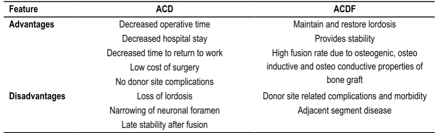
Table 1: Comparison between ACD and ACDF
Feature ACD ACDF
Advantages Decreased operative time Maintain and restore lordosis
Decreased hospital stay Provides stability
Decreased time to return to work High fusion rate due to osteogenic, osteo
inductive and osteo conductive properties of bone graft
Low cost of surgery No donor site complications
Disadvantages Loss of lordosis Donor site related complications and morbidity
Narrowing of neuronal foramen Adjacent segment disease
Late stability after fusion
RESULTS AND DISCUSSION
ACD and ACDF have been extensively studied since many decades now. In the nutshell, comparison between both of these techniques is presented in Table 1.
With ground breaking clinical works by Smith and Robinson, Cloward, Bailey, Simmons, Bhalla etc; ACDF was rapidly accepted by neurosurgical fraternity and became Gold Standard for management of cervical spondylosis. Many studies have shown that ACDF for cervical spondylosis is a cost effective method which avoids complications associated with
instrumentation usage.10 Later on Plating, cages, spacers and other implants were added.
However, donor site complications are one of the most commonly thought for problems associated with this operation. Many of the studies mainly focusing on donor site complications in ACDF have been conducted. Autologous bone graft provides excellent substrate for arthrodesis after ACD and is inexpensive. However, the use of tricortical bone can increase patient discomfort. In1981, Bloom and Raney modified Smith and Robinson technique and inserted the horse shoe type graft in “Reversed Manner” so that the cortical portion headed the disc space, so it could be stronger to resist compressive force.15,16 Since then numerous modifications were introduced and excellent results were reported.
A prospective study was done for evaluation of donor site pain after anterior iliac bone harvesting for cervical fusion on 50 patients.17,18 The researchers found that, main sources of donor site pain were;
▪ Nociceptors adjacent to nerve injury site ▪ Bone micro and macro fractures.
▪ Hematoma
▪ Infection
So the researchers recommended,
• Careful dissection to preserve nerves
• Using oscillating bone saw instead of chisel and hammer to avoid micro fractures.
• Staying away from iliac spine prevents major fractures, which if occurs, may cause pain and instability while walking.
They concluded that after standard approach and anatomical principles, most patients do not experience persistent pain at donor site.17, 18 A similar study was conducted to identify complications of iliac crest graft harvest and concluded that “Harvesting of iliac crest bone graft can be associated with significant morbidity. However, with adequate preoperative planning and proper surgical technique, the incidence of these complications can be reduced.19
In the quest to decrease donor site morbidity several efforts were made. Minimally invasive bicortical autografts are inexpensive technique to harvest iliac graft that may produce reduced amount of general and local donor site complications without affecting outcomes as compared to with prosthetic cages. Misimiliano Vissocchi and Spallone et al described minimally invasive technique to obtain bicortical graft. It offered comparable results to published series in which cage or implants were used.20 so problems associated with donor site complications can be reduced with following measures.
- Meticulous dissection and sufficient orientation to cutaneous
nerves around donor site to avoid injuries. Like lateral femoral cutaneous nerve of thigh, iliohypogastric, ilioinguinal nerves are few of the important ones.
- Perfect and patient hemostasis and again confirming it
before closure.
- Adequate use of thrombin soaked gel and bone wax. - Meticulous closure and vigilant post-operative care to wound
and expedious management of wound related complications.
- Minimally invasive techniques for graft harvest.
One more problem sited with ACDF is of “Adjacent Segment Disease (ASD)”. Definition of symptomatic ASD is “development of radicular or myelopathic signs and or symptoms referable to a motion segment adjacent to prior cervical arthrodesis” Several
studies have looked into this problem and its incidence and prevalence .Symptomatic ASD occurred at a relatively constant incidence of 2.9% per year (range 0 to 4.8% per year) during ten years after operation.21 Xiao Peng Xia et al did extensive research to identify prevalence of ASD. They studied 94 studies with 34,716 patients from 19 countries and found prevalence of clinical cervical ASD at 6.3% (95% confidence limit, range of 4.8 to 7.8%).22
Somewhat less incidence of ASD was noted, when ACDF involved arthrodesis of C5-6 and C6-7 levels as compared to ACDF done in whom these levels were not fused, by Shingo Komura et al.23 As we know these are the most commonly affected level in cervical spine so they are most commonly treated and fused, consequently, possibility of ASD goes down to very less. Thus the “Burdon of the disease ASD” is expected to be very low and TDR are less likely to become gold standard for management of cervical spondylotic myelopathy (CSM). Also there has been no clear evidence that the increased stress or strain of adjacent segment from the fusion, which is cited as the cause of ASD, increases the incidence of reoperation. Even in short-term studies comparing ACDF and TDR have failed to show any significant difference in the prevalence of adjacent segment pathology following surgery.24 So further studies are needed to ascertain that whether ASD is a fusion related phenomenon or due to spondylotic disease progression.25
Fig 1: Age and Sex distribution of disease
Fig 2: Level of diseases
5
8
5
8
1
0 2
4
6
0 0
1
0 1 2 3 4 5 6 7 8 9
21-30 31-40 41-50 51-60 61-70 71-80
Male Female
1
6
12 15 11
0 5 10 15 20
C2-C3 C3-C4 C4-C5 C5-C6 C6-C7
No. of Patients
Le
ve
Results of the present study
Patients in their third and fourth decade of life are most commonly affected.Male to female ratio is approximately 2:1.(Fig 1) Cervical pain and paresthesia of limbs are most common symptom. While dorsal column sensory loss and lower motor neuron symptoms are least common symptom. (Table 2)
Total levels affected are >40 because 5 patients were treated at 2 levels. As we known most commonly involved cervical spine levels in cervical spondylosis are C5-C6 and C6-C7. Maximal mobility is at these levels. Biomechanical basis of these levels affections are “Greater Flexion Extension mobility” at these levels. 26 This is reflected in our study. (Fig 2, Table 3) All over the world, these are most commonly treated levels. Biomechanics of spine make these levels most vulnerable.
Table 2: Clinical presentation of patients
Symptoms and Signs Patients
Cervical Pain 38/40
Limb weakness and motor deficit 18/40 Paraesthesia and sensory symptoms 31/40 Pain and temperature loss (spinothalamic tract) 11/40
Dorsal column sensory loss 4/40
Bowel involvement + Urinary Bladder Involvement 6/40
LMN Features 4/40
Table 3: Comparison of Disc Involvement Disc
Involvement
Present Study
Lunsford Study27
Rishi D.S. Nandoe Tewarie et al5
C6-7 24.44% 37% 47.8%
C5-6 33.33% 48% 40.1%
C4-5 26.66% 10% 5.9%
C3-4 13.33% - 2.0%
C2-3 2.22% - -
Table 4: Clinical syndrome of presentation
Clinical Syndrome No. of Patients
Myelopathy 18
Myelo-radiculopathy 22
Total 40
Table 5: Comparison of Symptomatic Features Symptoms Present Study Lunsford Study27
Myelo Radiculopathy 55% 41%
Myelopathy 45% 40%
Radiculopathy - 19%
Table 6: Modality of treatment given
Modality of Treatment No. of Patients
ACD 21
ACDF 19
Total 40
Patients with mainly radicular symptoms are treated with non-operative conservative management as outlined previously so that is what is reflected in this table. All patients with myelopathy or its component are always considered for surgical management. Patient stratification is done on case by case bases. Patients are assessed clinically and radiologically and treatment required is ascertained pre operatively.
Various levels of treatments given reflect that “different indications exist for each of the treatment modalities.”(Table 4-6)
INDICATIONS ACD
▪ Soft disc herniation. ▪ Single level disease ▪ Young patients.
▪ No evidence of instability, sub luxation ▪ Preserved lordosis.
ACDF WITH BONE GRAFT OR IMPLANT
▪ Hard disc
▪ Multi segmental disease ▪ Osteophytic bars ▪ Instability ▪ Loss of lordosis
▪ Fractures, Tumours, Trauma, Tuberculosis So, specific indications exist for specific operative procedures.
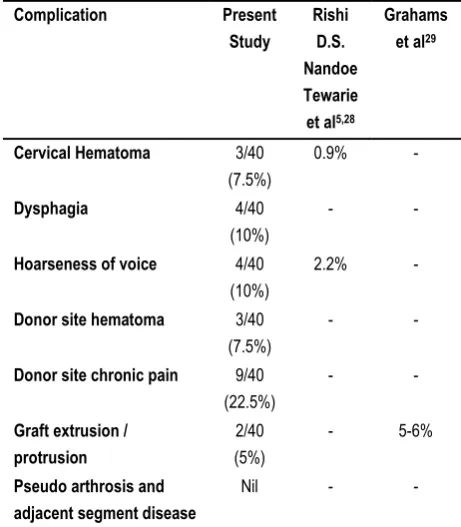
Table 7: Incidence of various complications
Complication Present
Study
Rishi D.S. Nandoe Tewarie et al5,28
Grahams et al29
Cervical Hematoma 3/40 (7.5%)
0.9% -
Dysphagia 4/40
(10%)
- -
Hoarseness of voice 4/40 (10%)
2.2% -
Donor site hematoma 3/40 (7.5%)
- -
Donor site chronic pain 9/40 (22.5%)
- -
Graft extrusion / protrusion
2/40 (5%)
- 5-6%
Pseudo arthrosis and adjacent segment disease
Nil - -
Table 8: Outcome of treatment analysis
Results Present Study Aronson et al30
Excellent 20(50%) 87%
Good 13(32.5%)
Fair 6(15%) 10%
Poor 1(2.5%) 3%
Interpretation: (Table 7)
• Dysphagia and hoarseness of voice were transient and improved on follow up visits, suggestive of neuropraxic injury of which most common mechanism is compression of recurrent laryngeal nerve trachea esophageal segment between Endotracheal Tube and self-retaining retractor.
• Donor site hematoma was managed conservatively with compressive dressing and follow up. Patients showed good recovery.
• Donor site pain was managed initially by analgesics and then reassurance.
• Graft protrusion occurred in 2 patients. But they were asymptomatic and only partial. Lordosis and height were maintain so conservative management and follow up was advised, both patients have shown evidence of fusion on 6 months cervical X-ray.
• No incidence of pseudoarthrosis, adjacent segment disease or implant construct failure have been noted. This is probably the result of strict adherence to management protocols and uniform operative techniques executed by senior operating surgeon.
Results or outcome of treatment was analyzed on basis of symptomatic relief to the patient according to Odom’s criteria. (Table 8) 33 out of 40 (82.5% of patients) have shown excellent to good results after these operations in total.
13 out of 18 patients with myelopathy and 20 out of 22 patients with myeloradiculopathy have shown excellent to good results, so patients with both the clinical syndromes have improved significantly. Of the total patients with excellent to good outcome 20 out of 33 were having myeloradiculopathy and 13 out of 33 were of myelopathy suggesting that patients with myeloradiculopathy are more likely to improve. (Table 9)
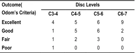
Greater numbers of patients (total 26 out of 45) were operated for C5-6 and C6-7 level and 23 of them showed excellent to good results. So these levels of disease are associated with better outcome. A patient operated at C2-3 level was incorporated in C 3-4 level in this table with fair outcome. (Table 10)
Table 9: Outcome Analysis According To Clinical Syndrome Outcome
(Odom’s
criteria)
Clinical Syndromes Total Myelopathy Myeloradiculopathy
Excellent 9 11 20
Good 4 9 13
Fair 4 2 6
Poor 1 0 1
Total 18 22 40
Table 10: Outcome Analysis According to Level of Disease Outcome(
Odom’s Criteria)
Disc Levels
C3-4 C4-5 C5-6 C6-7
Excellent 4 5 6 9
Good 1 5 6 2
Fair 1 2 3 0
Poor 1 0 0 0
Total is >40 because 5 patients were operated on double level.
Table 11: Outcome Analysis According to Operative Procedure in present study Outcome
(Odom’s criteria)
Operative Procedure Total
ACD ACDF
Excellent 5(23.81%) 15(78.95%) 20
Good 10(47.61%) 3(15.79%) 13
Fair 6(28.57%) 0 6
Poor 0 1(5.26%) 1
Total 21(100%) 19(100%) 40
Table 12: Comparison with Study by C.E. Deopujari et al.6 Outcome
(Odom’s criteria)
Operative Procedure
ACD ACDF
Excellent 13(23.21%) 2(8.33%)
Good 35(62.5%) 16(66.66%)
Fair 5(8.92%) 5(20.83%)
Poor 3(5.35%) 1(4.16%)
Table 13: Return to work
Procedure Means days for return to work Present Study C.E. Deopujari et al6
ACD 7 days 6.76
ACDF 9.67 days 7.75
Most of the patients who have undergone ACD or ACDF have shown excellent to good results in both studies. (Table 13) Due to more morbid neurological involvement in ACDF patients like traumatic cervical spine injuries and Tuberculosis, time taken to return to work were longer for few of the ACDF patients. Else between ACD and ACDF, it is nearly same range. 3 patients could not return to work while on follow up.
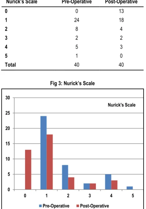
NURICK’S SCALE (Table 14, Fig 3)
Pre-operative mean Nurick’s Scale
= Total of (sum of) individual scales of patients pre operatively. Total no. of patients studied.
= 71/40 = 1.775
Post-operative mean Nurick’s Scale
= Sum of individual scales of patients post operatively. Total no. of patients studied.
= 44/40 = 1.1
This indicates mean improvement of 0.665 in Nurick’s scale and functional improvement.
Graft Fusion at 3 months
Criteria of fusion were taken as described in materials and methods. 18 out of 19 grafts were fused so fusion rate is 94.73%. 21 Patients of ACD did not undergo grafting.
Table 14: Nurick’s Scale
Nurick’s Scale Pre-Operative Post-Operative
0 0 13
1 24 18
2 8 4
3 2 2
4 5 3
5 1 0
Total 40 40
Fig 3: Nurick’s Scale
CONCLUSION
ACD is still a viable operative procedure and it is still being practiced in European and Asian countries. On long term follow up of ACD patients, one may notice that patient may complaint of cervical pain and sensory symptoms again. But fusion at operative site was still sufficient and symptoms were more probably due to progression of natural spondylotic disease. No study was able to show superiority of other fusion techniques over ACD alone, statistically and it holds true today is well.
On the other hand, enthusiasm to operate for ACDF has still persisted since its inception from the era of Robinson and Smith, with time to time additions and modifications of techniques and technologies with plates, cages and spacers. The problems of Donor site morbidities and adjacent segment disease have been studied in deep and their solutions made available.
So, as per the aim of the study, patient selection criteria are noted. Complication rate with both procedures (ACD and ACDF) were acceptable. Hospital stay, symptomatic relief with outcome analysis and time to return to work were comparable between ACD and ACDF. Cost did not hinder selection of ACDF over ACD. So, we conclude that both these procedures are viable in present day clinical practice is well with proper selection of patients. Key to achieve desirable clinical results is proper patient selection and adequate surgical decompression. No single gold standard procedure exists for treatment of patients with cervical spondylosis and the treatment strategies need to be tailored to match the specific changes present in a given patient. The incidence of degenerative cervical spondylosis increases with age and ability to perform its surgery and treat this ailment is very important and
satisfying asset in the armamentarium of a neurosurgeon. The need for prospective, randomized, multicenter studies with long term follow up in this area is becoming more imperative than ever.
REFERENCES
1. Abd - Alrahman N, Dokmak AS, Abou – Madawi A. Anterior cervical discectomy (ACD) versus Anterior cervical fusion (ACF), Clinical and radiological outcome study. In Acta Neurochirurgica (1999) October, 141(10): 1089-92.
2. De La Torre M, Martinez – Quinonez JV, Isla A et al. Anterior cervical discectomy with and without bone grafting. Multicentric comparative study. (1995). In J Bone Joint Surgery Britain B; 77, 290-95.
3. Dowd GC, Wirth FP. Anterior cervical discectomy: is fusion necessary? In J Neurosurg. 1999 Jan; 90 (1 Suppl): 8-12. 4. Martins AN. Anterior cervical discectomy with and without interbody bone graft. In J Neurosurg. 1976 Mar; 44(3): 290-95. 5. Nandoe Tewarie, Rishi DS; Bartels, Ronald HMA; Peul, Wilco C. Long term outcome after anterior cervical discectomy without fusion. In European Spine Journal October 2007, 16 (9): 1411-16. 6. Kumar, Rajiv; Deopujari, CE; Shah, Rajan; Kumar, Ashish. A study of anterior cervical discectomy with or without fusion. In Bombay hospital Journal, 52(1): 2010.
7. Ellenbogen RG; Abdulrauf SI; Sekhar LN. Principles of Neurosurgery. Degenerative Spine Disease. 3rd edition, Vol. 2, Chapter 30, Elsevier Saunders USA, 2012.
8. Feiz – Erfan I, Klopfenstein JD, Bambakidis NC, Sonntag VKH. Chapter 43. Surgical management of cervical disc disease: From No Fusion to Fusion and Back Again. In The Congress of Neurological Surgeons.
9. Son S, Lee SG, Yoo CJ, Park CW, Kim WK. Single stage circumferential cervical surgery (selective anterior cervical corpectomy with fusion and laminoplasty) for multilevel ossification of posterior longitudinal ligament with spinal cord ischemia on MRI. In J Korean Neurosurg Soc. 2010 Oct ; 48(4): 335-41. 10. Jagadesh Babu K, Sidda Reddy, Murali GV, Ponraj S, Mahendra Kumar K, Hanuman DS. Anterior cervical discectomy and bone graft fusion – clinical presentation, post operative outcome and complications. In International Journal of Research and Development of Health. March 2014; 2(1): 1-6.
11. Wright IP, Eisenstein SM. Anterior cervical discectomy and fusion without instrumentation. In Spine (Phila Pa 1976) 2007 Apr 1; 32(7): 772-5.
12. Epstein NE. Anterior cervical discectomy and fusion without plate instrumentation in 178 patients. In J Spinal Disorders. 2000 Feb; 13(1); 1-8.
13. Bolesta MJ, Rechtine GR 2nd, Chrin AM. One and two level anterior cervical discectomy and fusion: The effect of plate fixation. In Spine J. 2002 May – June; 2(3): 197-203.
14. Jacobs WCH, Anderson PG, Van Limbeek J, Williams PC, Paulau P. Single or double level anterior interbody fusion techniques for cervical degenerative disc disease. Cochrane Database Systematic Review 4: CD 004958.
15. Bloom MH, Raney FL Jr. Anterior intervertebral fusion of cervical spine. A technical note. In J Bone Joint Surg Am. 1981 Jun; 63(5): 842.
16. Emery SE, Bolesta MJ, Banks MA, Jones PK. Robinson anterior cervical fusion. Comparison of standard and modified techniques. In Spine (Phila Pa 1976). 1994 Mar 15; 19(6): 660-3.
0 5 10 15 20 25 30
0 1 2 3 4 5
Nurick's Scale
17. Shamsaldin M, Mouchaty H, Desogus N, Costaglioba C, Di Lorenzo N. Evaluation of donor site pain after anterior iliac bone harvesting for cervical fusion. A prospective study on 50 patients. In Acta Neurochir (Wien) (2006) 148: 1071-74.
18. Cioni, Beatrice. Evaluation of donor site pain after anterior iliac crest harvesting for cervical fusion: A prospective study on 50 patients – Comment. In Acta Neurochir (Wien) 2006 Oct; 148(10): 1074-74.
19. Arrington ED, Smith WJ, Chambers HG, Bucknell AL, Davino NA. Complications of iliac crest bone graft harvesting. In Clin Orthop Relat Res. 1996 Aug; (329): 300-9.
20. Spallone A, Marchione P et al. Anterior cervical discectomy and fusion with minimally invasive harvesting of iliac crest graft versus polyetheretherketone (PEEK) cages: a retrospective outcome analysis. In Int J Surg. 2014 Dec; 12(12): 1328-32. 21. Hillbrand AS, Carlson GD et al. Radiculopathy and myelopathy at segments adjacent to site of previous anterior cervical arthrodesis. In J Bone Joint Surg Am. 1999 Apr; 81(4): 519-28. 22. Xia XP, Chen HL, Cheng HB. Prevalence of adjacent segment degeneration after spine surgery: a systemic review and meta-analysis. In Spine. April 2013. 38(7): 597-608.
23. Komura S, Miyamoto K et al. Lower incidence of adjacent segment degeneration after anterior cervical fusion found with those fusing C5-6 and C6-7 than those leaving C5-6 or C6-7 as an adjacent level. In J Spinal Disorders Tech. 2012 Feb; 25(1):23-9. 24. Maldonado CV, Paz RD, Martin CB. Adjacent level degeneration after cervical disc arthroplasty versus fusion. In Eu Spine J, 2011 Aug; 20 suppl 3: 403-7.
25. Song KJ, Choi BW, Jeon TS, Lee KB, Chang H. Adjacent segment degenerative disease: is it due to disease progression or a fusion associated phenomenon? Comparison between segments adjacent to the fused and non fused segments. In Eur Spine J. 2011 Nov; 20(11): 1940-5.
26. Richard Winn H. Youmans Neurological Surgery. 6th edition Volume 3. Chapter 278, p 2859; Chapter 280, p 2876; Chapter 291, p 2980-81; Chapter 297, p 3028. Elsevier Saunders, USA. 2011.
27. Lunsford LD, Bissonette DJ, Jannetta PJ, Sheptak PE, Zorub DS. Anterior surgery for cervical disc disease. Part 1: Treatment of lateral cervical disc herniation in 253 cases. In J Neurosurg. 1980 Jul; 53 (1): 1-11.
28. Flynn TB. Neurogenic complications of anterior cervical interbody fusion. Spine (Phila Pa 1976) 1982 Nov-Dec;7(6):536-9. 29. Graham JJ. Complications of cervical spine surgery. In Shark HH, et al (eds) The Cervical Spine, 2nd edition. Lippincott, Philadelphia, pp 831-837, 1989.
30. Aronson NI, Filtzer DL, Bagan M. Anterior cervical fusion by the Smith Robinson Approach. In J Neurosurg 1968 Oct: 29.
[
Source of Support: Nil.
Conflict of Interest: None Declared.
Copyright: © the author(s) and publisher. IJMRP is an official publication of Ibn Sina Academy of Medieval Medicine & Sciences, registered in 2001 under Indian Trusts Act, 1882. This is an open access article distributed under the terms of the Creative Commons Attribution Non-commercial License, which permits unrestricted non-commercial use, distribution, and reproduction in any medium, provided the original work is properly cited.